Design of New Polyaspartamide Copolymers for siRNA Delivery in Antiasthmatic Therapy


Abstract
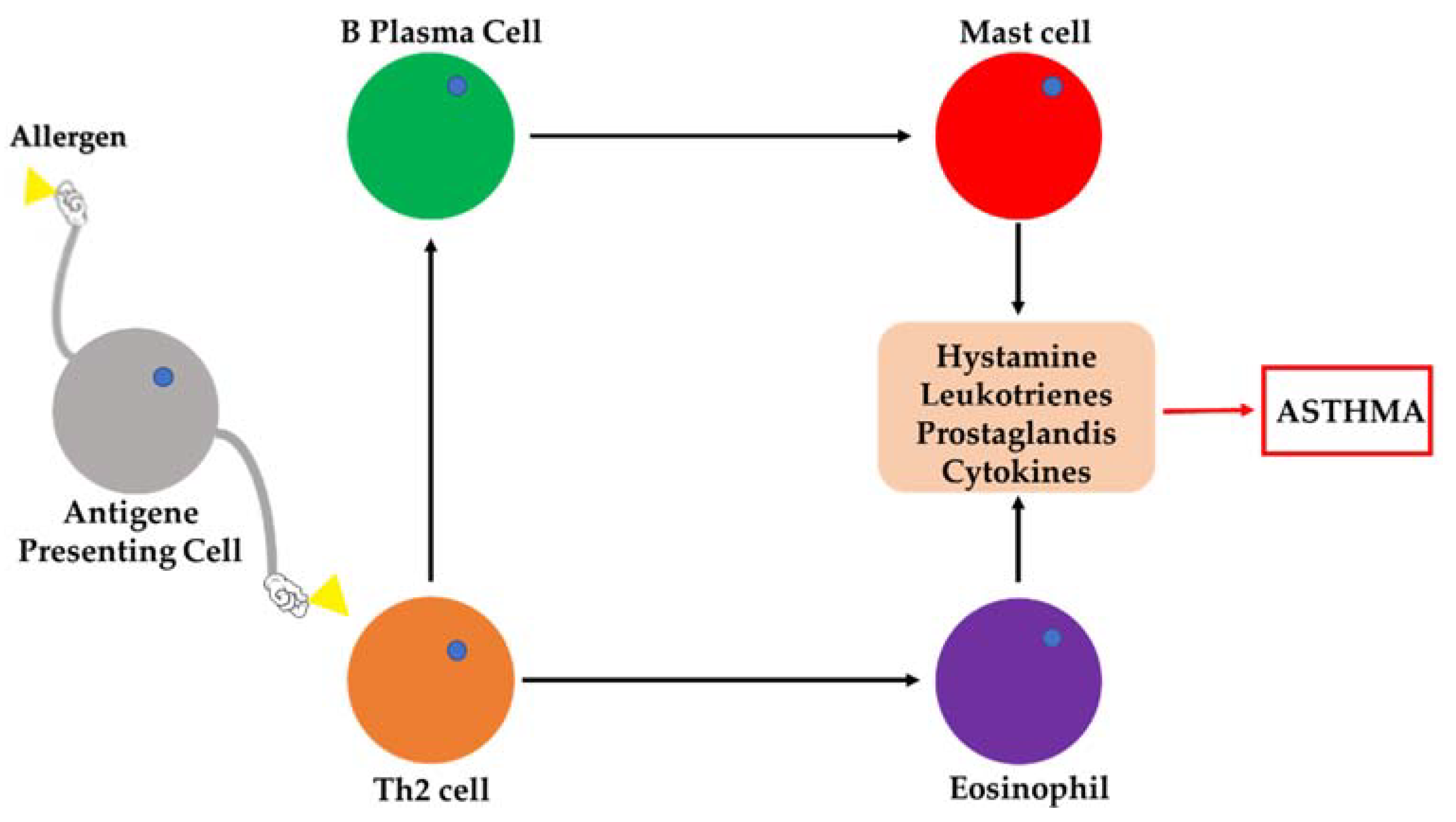
:1. Introduction
2. Materials and Methods
2.1. Materials
2.2. Copolymer Synthesis
2.2.1. General Procedure for the Derivatization and Characterization of α,β-poly(N-2-hydroxyethyl)d,l-aspartamide with 1,2-Bis(3-aminopropylamino)ethane (PHEA-g-bAPAE)
2.2.2. General Procedure for the Derivatization and Characterization of PHEA with methoxy polyethylene glycol amine (H3CO-PEG-NH2)
2.2.3. General Procedure for the Derivatization and Characterization of PHEA-g-PEG with bAPAE
2.3. Determination of the Amine Content
2.4. Size Exclusion Chromatography
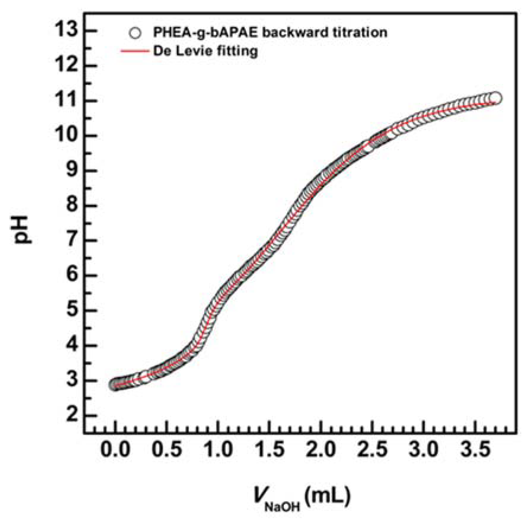
2.5. Potentiometric Titration of PHEA-g-bAPAE Graft Copolymer
2.5.1. Qualitative Titration of PHEA-g-bAPAE Copolymer
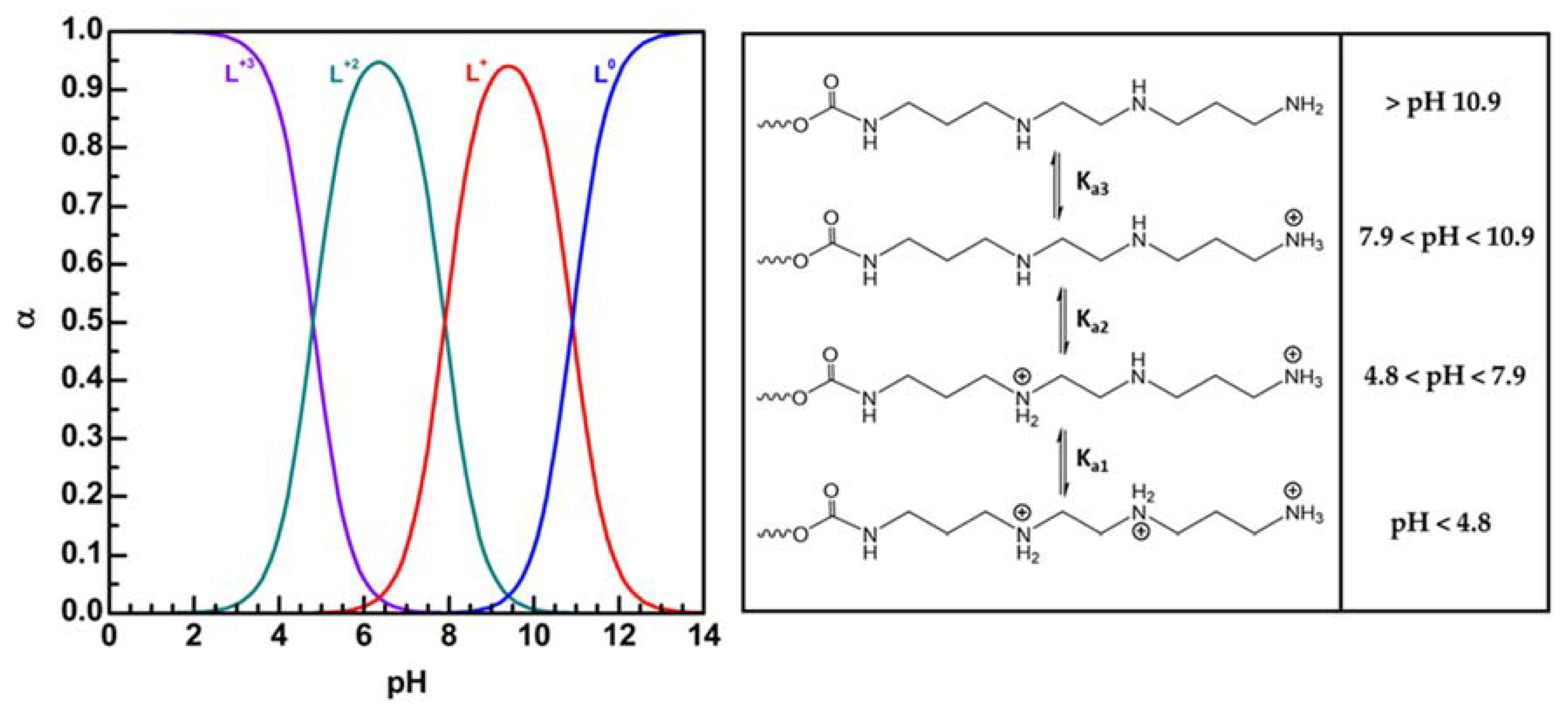
2.5.2. Determination of the pKa Values of PHEA-g-bAPAE Copolymer by Potentiometric Titration
2.6. Biological Studies
2.6.1. Cell Culture
2.6.2. Membrane Destabilization Study
2.6.3. Cell Viability Assay
2.7. Complexation Study
2.8. Polyplex Stability in Presence of Mucin
2.8.1. Polyanionic Exchange
2.8.2. Turbidimetric Assay
2.9. Gene Silencing Assay
3. Results and Discussion
3.1. Polymer Synthesis and Characterization
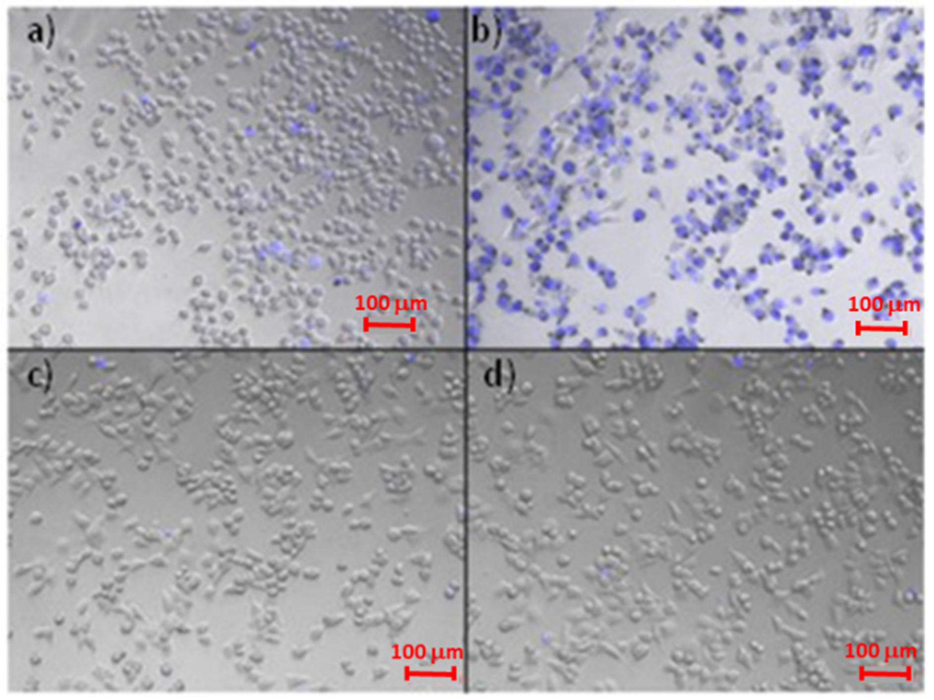
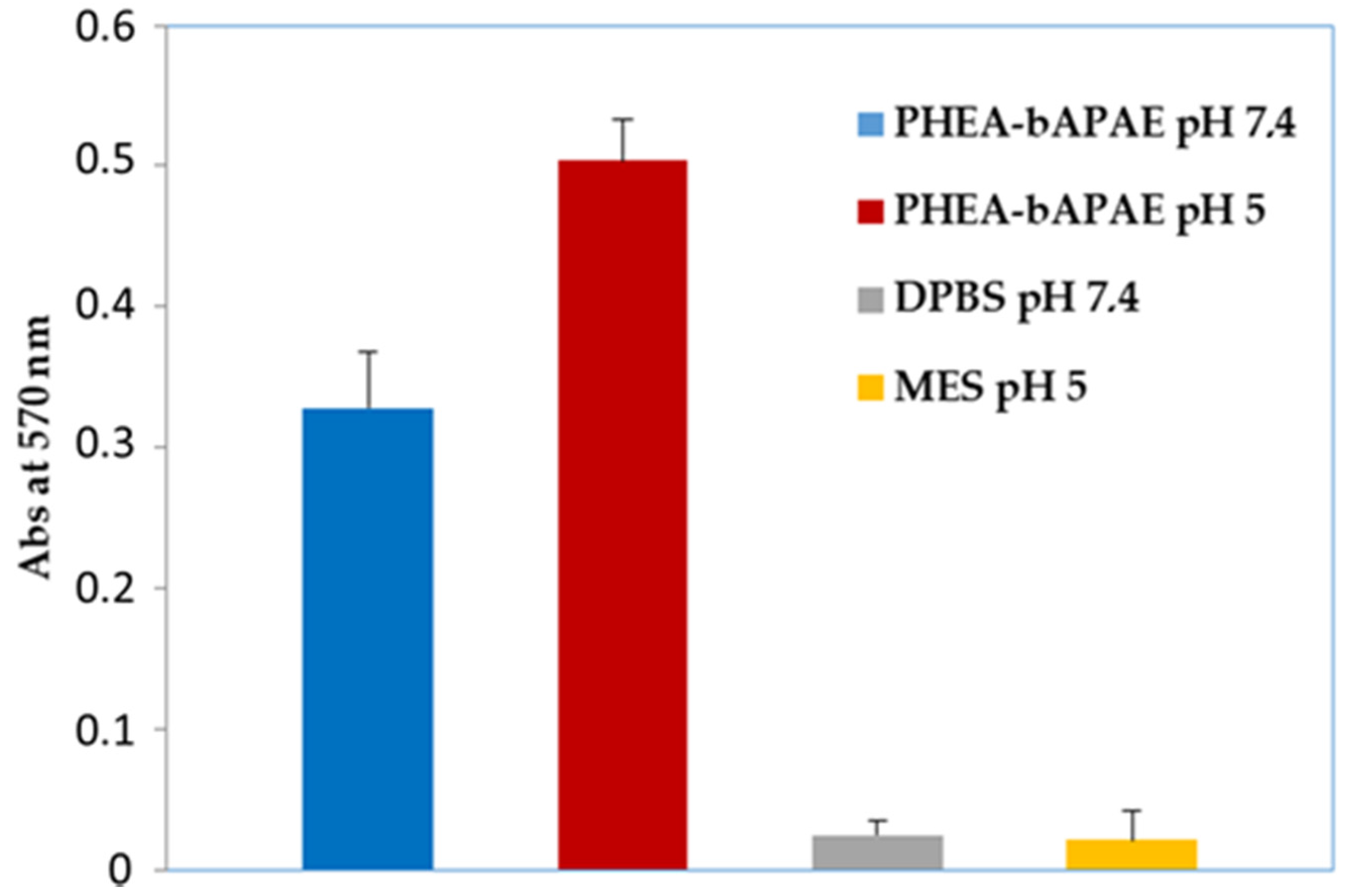
3.1.1. Membrane Destabilization Study
3.1.2. Pegylation
3.1.3. Cell Viability Assay
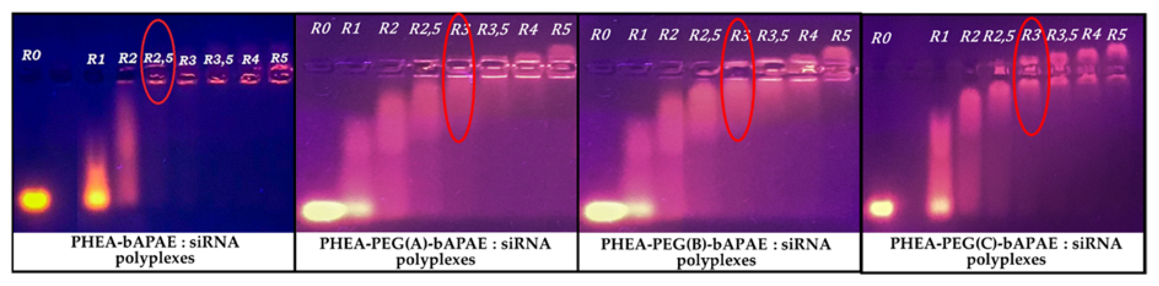
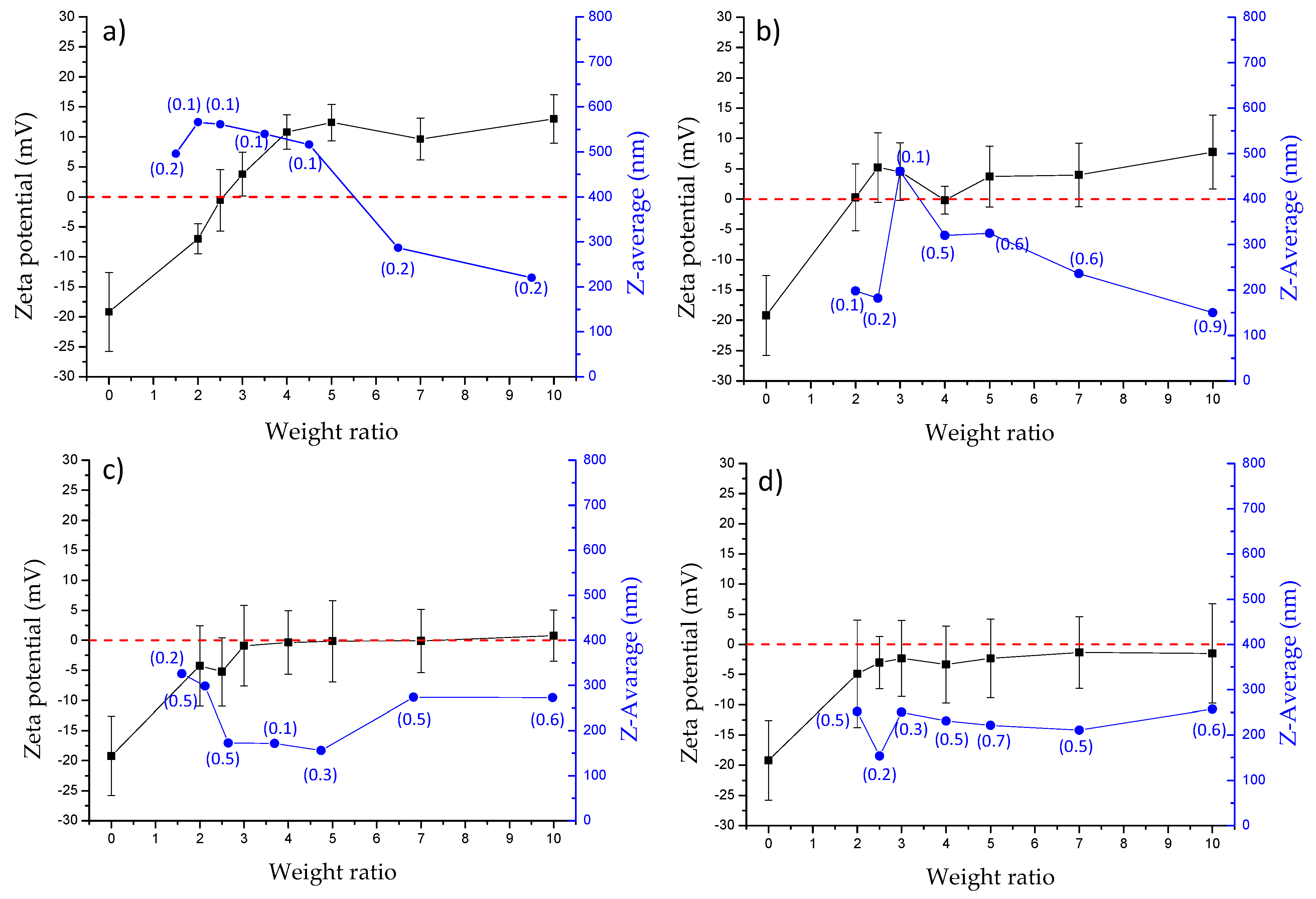
3.2. Complexation Studies and Characterization of Obtained Polyplexes
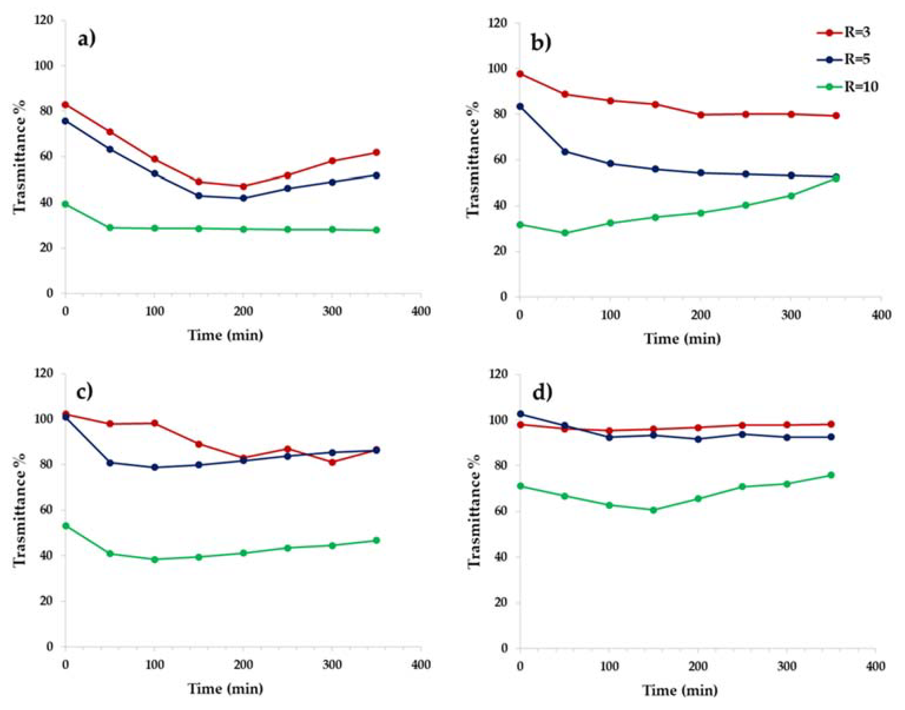
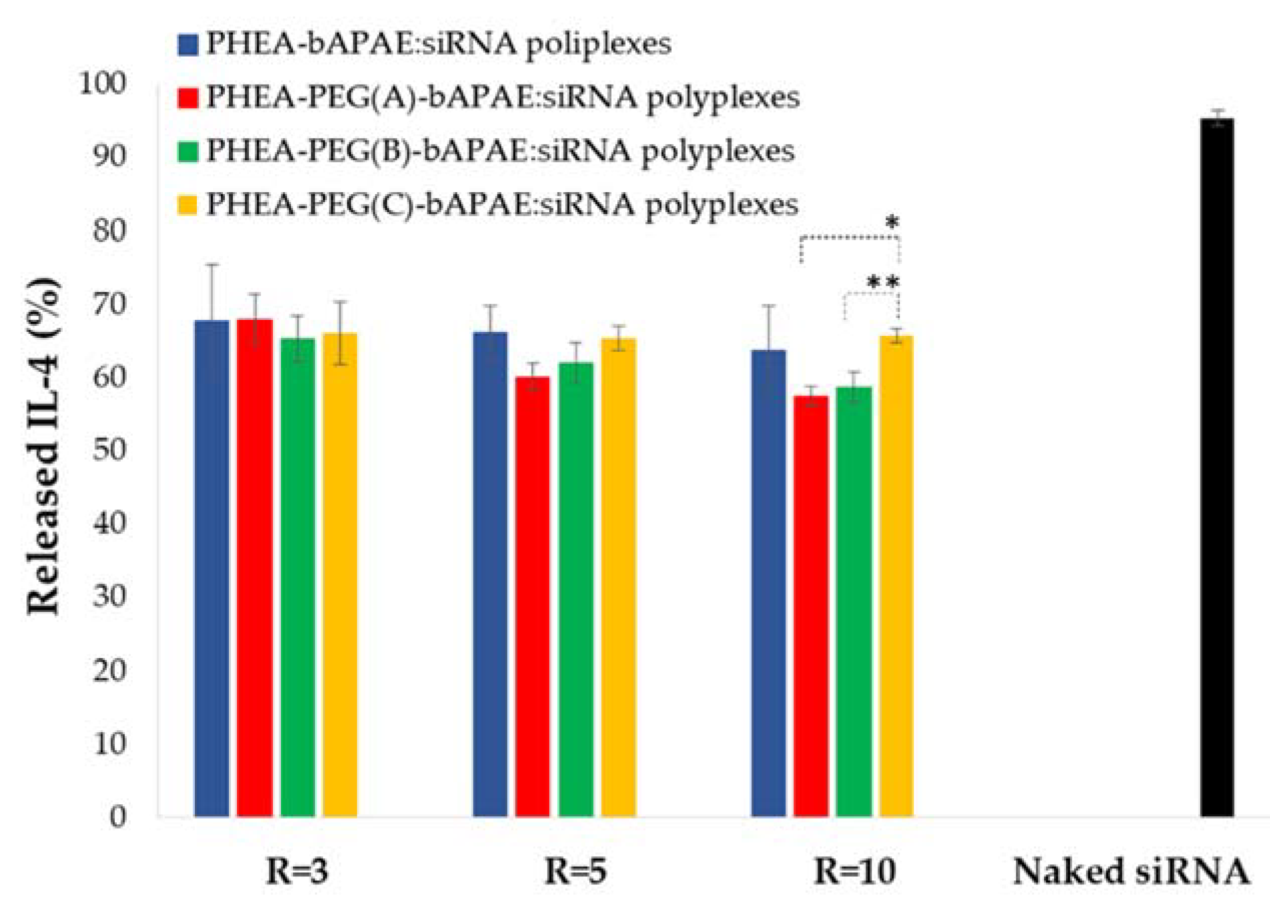
3.3. Interaction Studies of Polyplexes with Mucin
3.4. Gene Silencing Assay
4. Conclusions
Supplementary Materials
Author Contributions
Funding
Acknowledgments
Conflicts of Interest
References
- Shakshuki, A.; Agu, R.U. Improving the Efficiency of Respiratory Drug Delivery: A Review of Current Treatment Trends and Future Strategies for Asthma and Chronic Obstructive Pulmonary Disease. Pulm. Ther. 2017, 3, 267–281. [Google Scholar] [CrossRef] [PubMed]
- Anderson, S.D. Repurposing drugs as inhaled therapies in asthma. Adv. Drug Deliv. Rev. 2018, 133, 19–33. [Google Scholar] [CrossRef] [PubMed]
- Kanemitsu, Y.; Matsumoto, H.; Izuhara, K.; Tohda, Y.; Kita, H.; Horiguchi, T.; Kuwabara, K.; Tomii, K.; Otsuka, K.; Fujimura, M.; et al. Increased periostin associates with greater airflow limitation in patients receiving inhaled corticosteroids. J. Allergy Clin. Immunol. 2013, 132, 305–312. [Google Scholar] [CrossRef] [PubMed] [Green Version]
- Miyake, T.; Miyake, T.; Sakaguchi, M.; Nankai, H.; Nakazawa, T.; Morishita, R. Prevention of Asthma Exacerbation in a Mouse Model by Simultaneous Inhibition of NF-κB and STAT6 Activation Using a Chimeric Decoy Strategy. Mol. Ther. Nucleic Acids 2018, 10, 159–169. [Google Scholar] [CrossRef] [Green Version]
- Cho, S.H.; Jo, A.; Casale, T.; Jeong, S.J.; Hong, S.J.; Cho, J.K.; Holbrook, J.T.; Kumar, R.; Smith, L.J. Soy isoflavones reduce asthma exacerbation in asthmatic patients with high PAI-1–producing genotypes. J. Allergy Clin. Immunol. 2019, 144, 109–117. [Google Scholar] [CrossRef] [Green Version]
- Tabeling, C.; Herbert, J.; Hocke, A.C.; Lamb, D.J.; Wollin, S.L.; Erb, K.J.; Boiarina, E.; Movassagh, H.; Scheffel, J.; Doehn, J.M.; et al. Spleen tyrosine kinase inhibition blocks airway constriction and protects from Th2-induced airway inflammation and remodeling. Allergy Eur. J. Allergy Clin. Immunol. 2017, 72, 1061–1072. [Google Scholar] [CrossRef]
- Cavallaro, G.; Sardo, C.; Craparo, E.F.; Porsio, B.; Giammona, G. Polymeric nanoparticles for siRNA delivery: Production and applications. Int. J. Pharm. 2017, 525, 313–333. [Google Scholar] [CrossRef]
- Park, J.; Park, J.; Pei, Y.; Xu, J.; Yeo, Y. Pharmacokinetics and biodistribution of recently-developed siRNA nanomedicines. Adv. Drug Deliv. Rev. 2016, 104, 93–109. [Google Scholar] [CrossRef] [Green Version]
- David, S.; Pitard, B.; Benoît, J.P.; Passirani, C. Non-viral nanosystems for systemic siRNA delivery. Pharmacol. Res. 2010, 62, 100–114. [Google Scholar] [CrossRef] [Green Version]
- Cooper, B.M.; Putnam, D. Polymers for siRNA delivery: A Critical Assessment of Current Technology Prospects for Clinical Application. ACS Biomater. Sci. Eng. 2016, 2, 1837–1850. [Google Scholar] [CrossRef]
- Giammona, G.; Carlisi, B.; Palazzo, S. Reaction of α,β-poly(N-hydroxyethyl)-DL-aspartamide with derivatives of carboxylic acids. J. Polym. Sci. Part A Polym. Chem. 1987, 25, 2813–2818. [Google Scholar] [CrossRef]
- Cavallaro, G.; Farra, R.; Craparo, E.F.; Sardo, C.; Porsio, B.; Giammona, G.; Perrone, F.; Grassi, M.; Pozzato, G.; Grassi, G.; et al. Galactosylated polyaspartamide copolymers for siRNA targeted delivery to hepatocellular carcinoma cells. Int. J. Pharm. 2017, 525, 397–406. [Google Scholar] [CrossRef] [PubMed]
- Gioia, S.D.; Sardo, C.; Belgiovine, G.; Triolo, D.; d’Apolito, M.; Castellani, S.; Carbone, A.; Giardino, I.; Giammona, G.; Cavallaro, G.; et al. Cationic polyaspartamide-based nanocomplexes mediate siRNA entry and down-regulation of the pro-inflammatory mediator high mobility group box 1 in airway epithelial cells. Int. J. Pharm. 2015, 491, 359–366. [Google Scholar] [CrossRef] [PubMed]
- Craparo, E.F.; Cavallaro, G.; Bondì, M.L.; Giammona, G. Preparation of polymeric nanoparticles by photo-crosslinking of an acryloylated polyaspartamide in w/o microemulsion. Macromol. Chem. Phys. 2004, 205, 1955–1964. [Google Scholar] [CrossRef]
- Pitarresi, G.; Palumbo, F.S.; Calabrese, R.; Craparo, E.F.; Giammona, G. Crosslinked hyaluronan with a protein-like polymer: Novel bioresorbable films for biomedical applications. J. Biomed. Mater. Res.-Part A 2008, 84, 413–424. [Google Scholar] [CrossRef]
- Craparo, E.F.; Teresi, G.; Licciardi, M.; Bondí, M.L.; Cavallaro, G. Novel composed galactosylated nanodevices containing a ribavirin prodrug as hepatic cell-targeted carriers for HCV treatment. J. Biomed. Nanotechnol. 2013, 9, 1107–1122. [Google Scholar] [CrossRef]
- Licciardi, M.; Di Stefano, M.; Craparo, E.F.; Amato, G.; Fontana, G.; Cavallaro, G.; Giammona, G. PHEA-graft-polybutylmethacrylate copolymer microparticles for delivery of hydrophobic drugs. Int. J. Pharm. 2012, 433. [Google Scholar] [CrossRef]
- Suma, T.; Miyata, K.; Ishii, T.; Uchida, S.; Uchida, H.; Itaka, K.; Nishiyama, N.; Kataoka, K. Enhanced stability and gene silencing ability of siRNA-loaded polyion complexes formulated from polyaspartamide derivatives with a repetitive array of amino groups in the side chain. Biomaterials 2012, 33, 2770–2779. [Google Scholar] [CrossRef]
- Darcan-Nicolaisen, Y.; Meinicke, H.; Fels, G.; Hegend, O.; Haberland, A.; Kühl, A.; Loddenkemper, C.; Witzenrath, M.; Kube, S.; Henke, W.; et al. Small Interfering RNA against Transcription Factor STAT6 Inhibits Allergic Airway Inflammation and Hyperreactivity in Mice. J. Immunol. 2009, 182, 7501–7508. [Google Scholar] [CrossRef]
- Rippmann, J.F.; Schnapp, A.; Weith, A.; Hobbie, S. Gene silencing with STAT6 specific siRNAs blocks eotaxin release in IL-4/TNFα stimulated human epithelial cells. FEBS Lett. 2005, 579, 173–178. [Google Scholar] [CrossRef] [Green Version]
- Lomonossoff, G. Modified Plant Virus Particles and Uses Therefor. U.S. Patent US20120015899A1, 19 January 2012. [Google Scholar]
- Wu, Z.; Liu, J.; Hu, S.; Zhu, Y.; Li, S. Serine/threonine kinase 35, a target gene of stat3, regulates the proliferation and apoptosis of osteosarcoma cells. Cell. Physiol. Biochem. 2018, 45, 808–818. [Google Scholar] [CrossRef] [PubMed]
- Craparo, E.F.; Porsio, B.; Sardo, C.; Giammona, G.; Cavallaro, G. Pegylated Polyaspartamide–Polylactide-Based Nanoparticles Penetrating Cystic Fibrosis Artificial Mucus. Biomacromolecules 2016, 17, 767–777. [Google Scholar] [CrossRef]
- Craparo, E.F.; Porsio, B.; Mauro, N.; Giammona, G.; Cavallaro, G. Polyaspartamide-Polylactide Graft Copolymers with Tunable Properties for the Realization of Fluorescent Nanoparticles for Imaging. Macromol. Rapid Commun. 2015, 36, 1409–1415. [Google Scholar] [CrossRef]
- Montana, G.; Bondì, M.L.; Carrotta, R.; Picone, P.; Craparo, E.F.; San Biagio, P.L.; Giammona, G.; Di Carlo, M. Employment of cationic solid-lipid nanoparticles as RNA carriers. Bioconjug. Chem. 2007, 18, 302–308. [Google Scholar] [CrossRef] [PubMed]
- Craparo, E.F.; Triolo, D.; Pitarresi, G.; Giammona, G.; Cavallaro, G. Galactosylated micelles for a ribavirin prodrug targeting to hepatocytes. Biomacromolecules 2013, 14, 1838–1849. [Google Scholar] [CrossRef] [PubMed]
- Behr, J.P. The proton sponge: A trick to enter cells the viruses did not exploit. Chimia 1997, 51, 34–36. [Google Scholar]
- Patel, S.; Kim, J.; Herrera, M.; Mukherjee, A.; Kabanov, A.V.; Sahay, G. Brief update on endocytosis of nanomedicines. Adv. Drug Deliv. Rev. 2019, 144, 90–111. [Google Scholar] [CrossRef]
- Mauro, N.; Fiorica, C.; Varvarà, P.; Di Prima, G.; Giammona, G. A facile way to build up branched high functional polyaminoacids with tunable physicochemical and biological properties. Eur. Polym. J. 2016, 77, 124–138. [Google Scholar] [CrossRef]
- Ferruti, P.; Mauro, N.; Falciola, L.; Pifferi, V.; Bartoli, C.; Gazzarri, M.; Chiellini, F.; Ranucci, E. Amphoteric, prevailingly cationic L-arginine polymers of poly(amidoamino acid) structure: Synthesis, acid/base properties and preliminary cytocompatibility and cell-permeating characterizations. Macromol. Biosci. 2014, 14, 390–400. [Google Scholar] [CrossRef]
- De Levie, R. How to Use Excel ® in Analytical Chemistry and in General Scientific Data Analysis; Cambridge University Press: Cambridge, UK, 2001; ISBN 9780511808265. [Google Scholar]
- Scialabba, C.; Sciortino, A.; Messina, F.; Buscarino, G.; Cannas, M.; Roscigno, G.; Condorelli, G.; Cavallaro, G.; Giammona, G.; Mauro, N. Highly Homogeneous Biotinylated Carbon Nanodots: Red-Emitting Nanoheaters as Theranostic Agents toward Precision Cancer Medicine. ACS Appl. Mater. Interfaces 2019, 11, 19854–19866. [Google Scholar] [CrossRef]


| Copolymers | R3 | R4 | R5 | PEG Solution (mL) | PEG Weight Amount (mg) |
|---|---|---|---|---|---|
| PHEA-g-PEG(A) | 0.03 | 0.04 | 1 | 4 | 200 |
| PHEA-g-PEG(B) | 0.075 | 0.1 | 1 | 9.5 | 475 |
| PHEA-g-PEG(C) | 0.12 | 0.16 | 1 | 15 | 750 |
|
R3 = (mmol of aminoPEG/mmol of functionalizable RU on PHEA) R4 = (mmol of DSC/mmol of functionalizable RU on PHEA) R5 = (mmol of TEA/mmol of DSC) | |||||
| Copolymers | Molecular Weight | Degree of Derivatization (DD) | ||
|---|---|---|---|---|
| DDPEG | DDbAPAE | |||
| PHEA | 67500 | 1.24 | --- | --- |
| PHEA-g-PEG(A) | 82,410 | 1.2 | 1.9 | --- |
| PHEA-g-PEG(B) | 95,360 | 1.3 | 2.7 | --- |
| PHEA-g-PEG(C) | 110,800 | 1.3 | 4.4 | --- |
| PHEA-g-bAPAE | 20,921 | 1.41 | --- | 34 |
| PHEA-g-PEG(A)-g-bAPAE | 25,400 | 1.32 | 1.9 | 35 |
| PHEA-g-PEG(B)-g-bAPAE | 32,100 | 1.35 | 2.7 | 36 |
| PHEA-g-PEG(C)-g-bAPAE | 34,700 | 1.41 | 4.4 | 33 |
© 2020 by the authors. Licensee MDPI, Basel, Switzerland. This article is an open access article distributed under the terms and conditions of the Creative Commons Attribution (CC BY) license (http://creativecommons.org/licenses/by/4.0/).
Share and Cite
Craparo, E.F.; Drago, S.E.; Mauro, N.; Giammona, G.; Cavallaro, G. Design of New Polyaspartamide Copolymers for siRNA Delivery in Antiasthmatic Therapy. Pharmaceutics 2020, 12, 89. https://doi.org/10.3390/pharmaceutics12020089
Craparo EF, Drago SE, Mauro N, Giammona G, Cavallaro G. Design of New Polyaspartamide Copolymers for siRNA Delivery in Antiasthmatic Therapy. Pharmaceutics. 2020; 12(2):89. https://doi.org/10.3390/pharmaceutics12020089
Chicago/Turabian StyleCraparo, Emanuela Fabiola, Salvatore Emanuele Drago, Nicolò Mauro, Gaetano Giammona, and Gennara Cavallaro. 2020. "Design of New Polyaspartamide Copolymers for siRNA Delivery in Antiasthmatic Therapy" Pharmaceutics 12, no. 2: 89. https://doi.org/10.3390/pharmaceutics12020089