Injectable Hydrogels for Cancer Therapy over the Last Decade

 , , and
Abstract
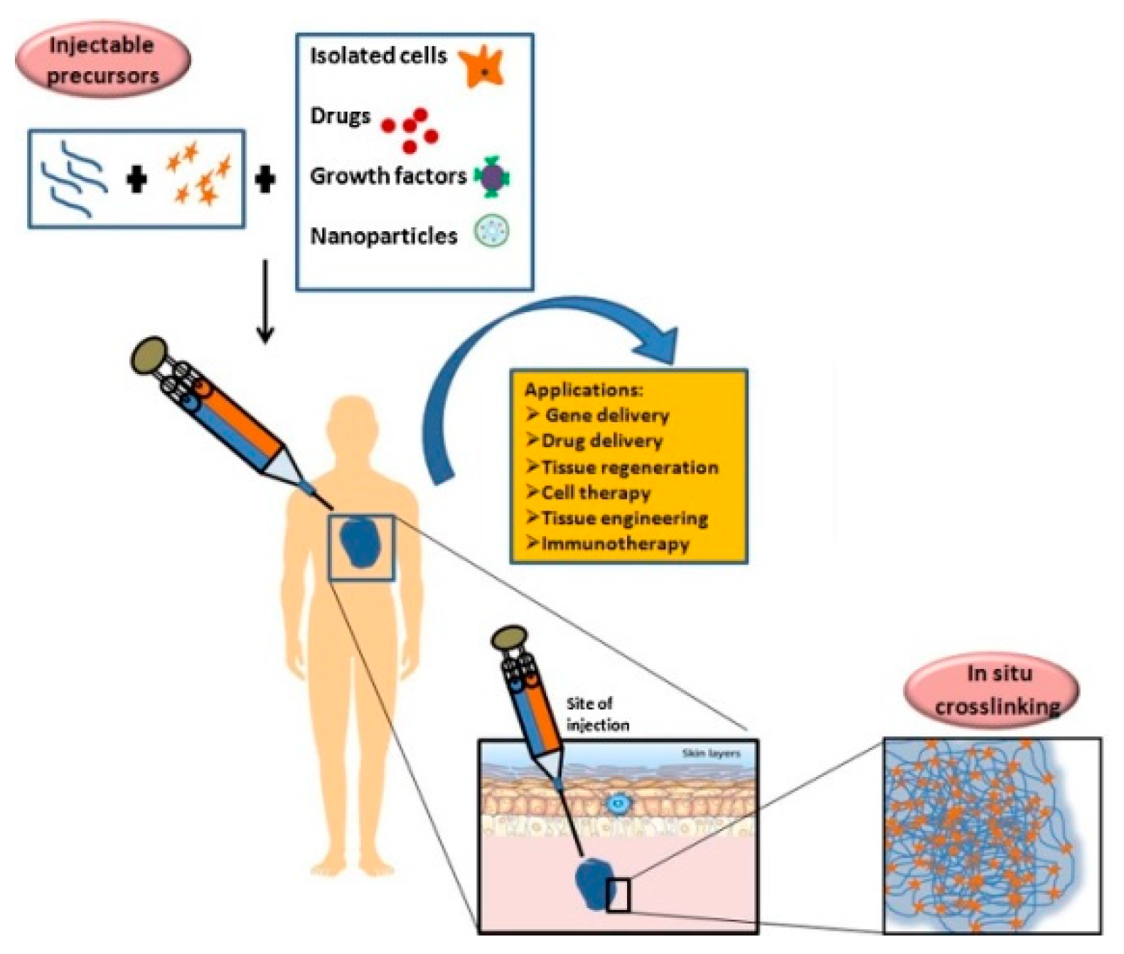
:1. Introduction
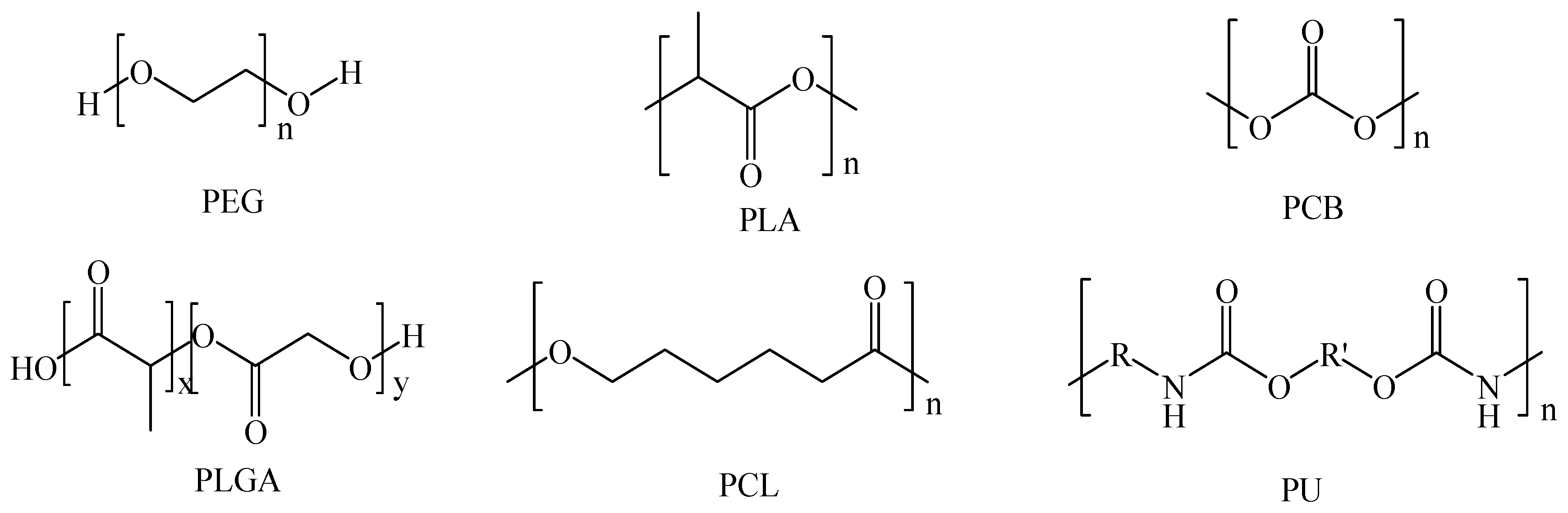
2. Synthetic Injectable Hydrogels
2.1. Polyphosphazenes
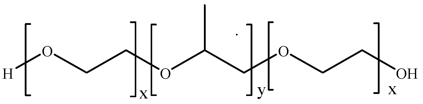
2.2. Polaxamers
2.3. Polyesters
2.4. Polyacrylates
2.5. Synthetic Polypeptide
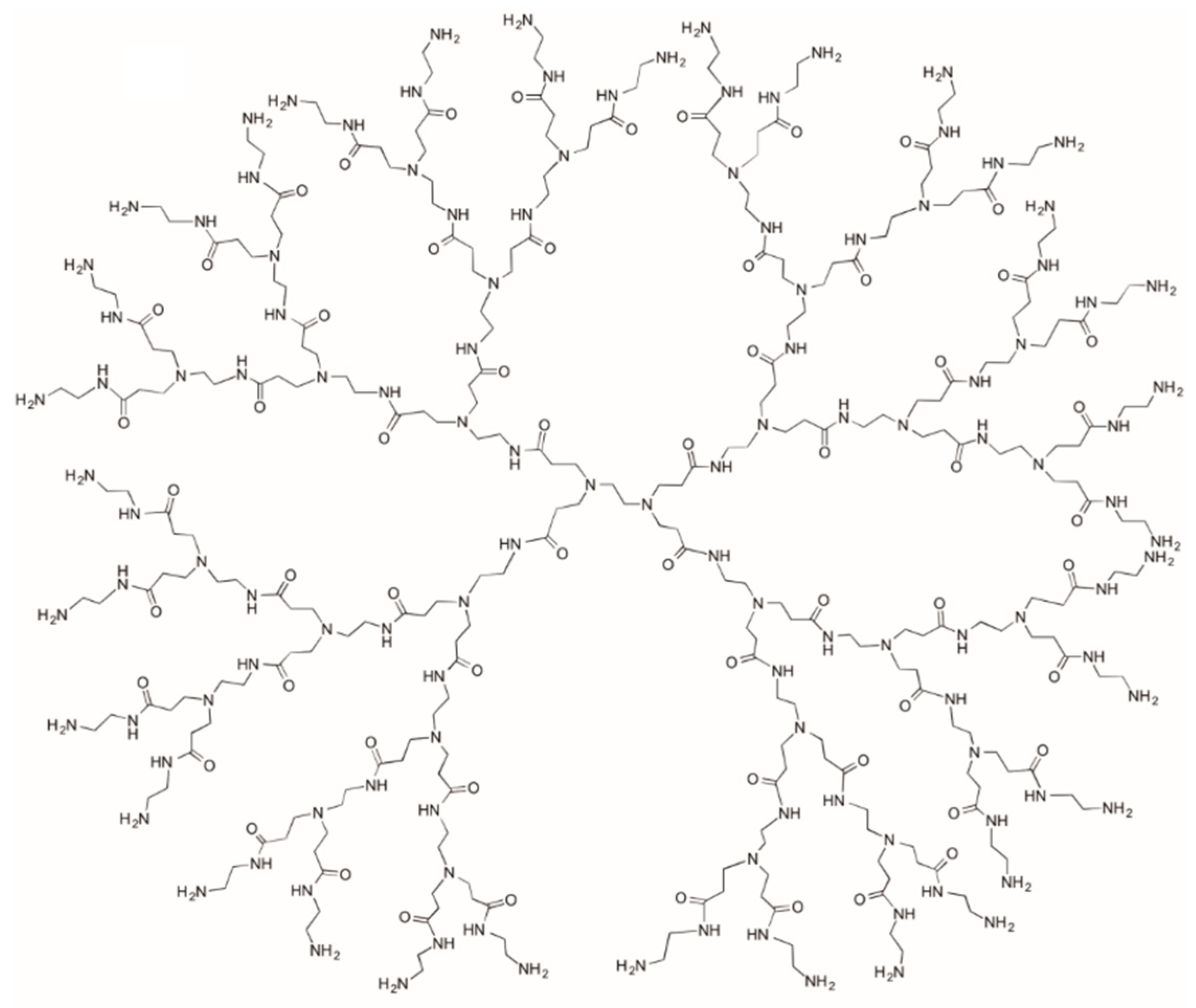
2.6. Dendrimers and Other Systems
3. Natural Polymers
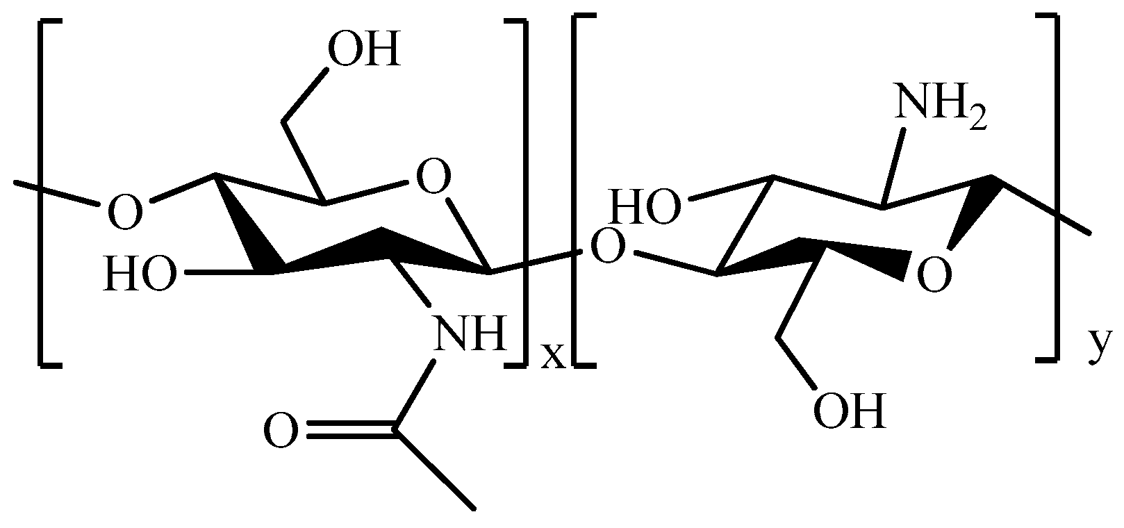
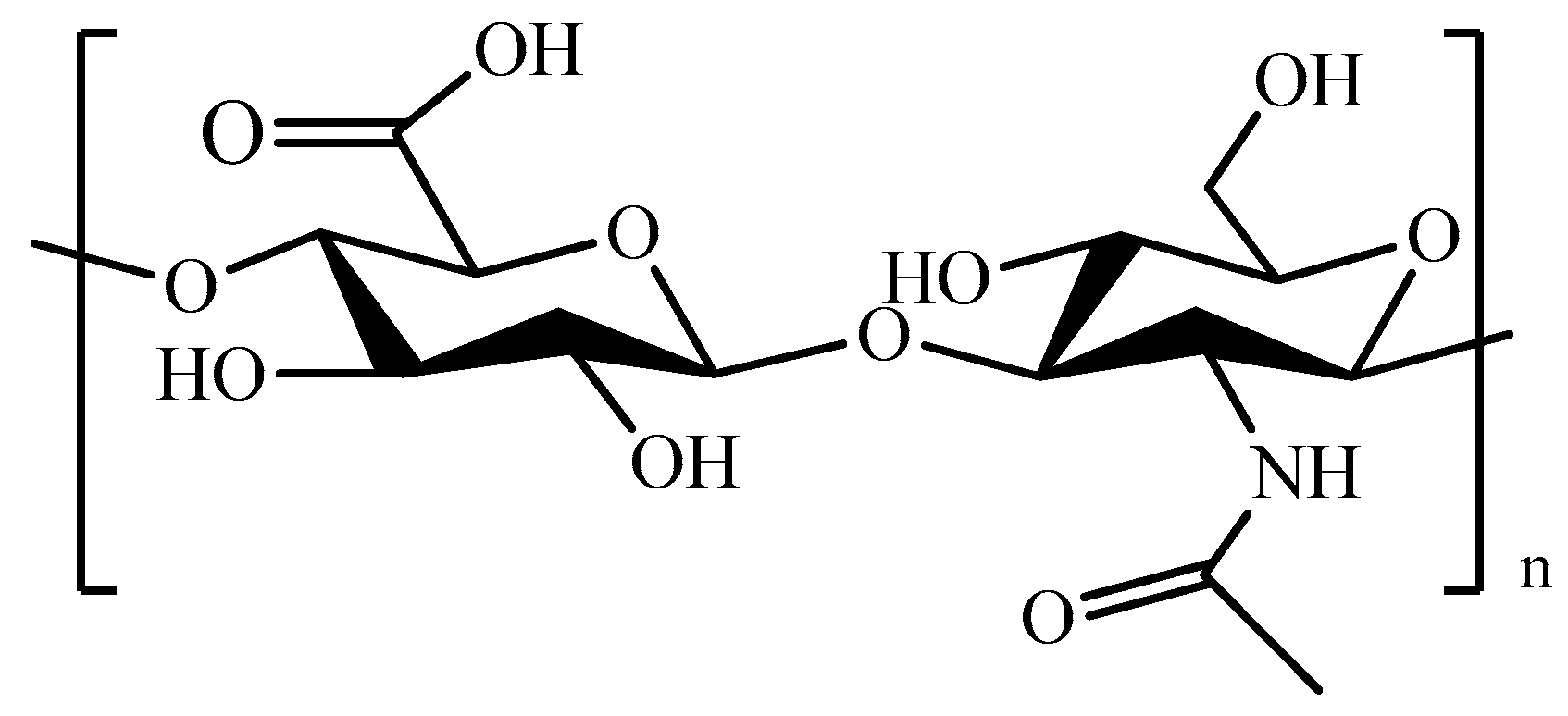
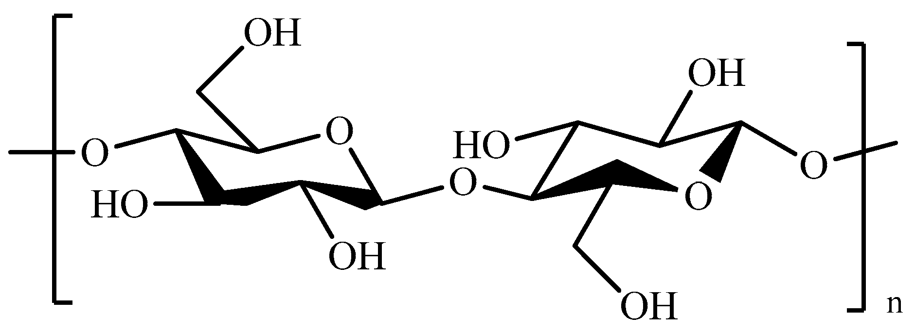
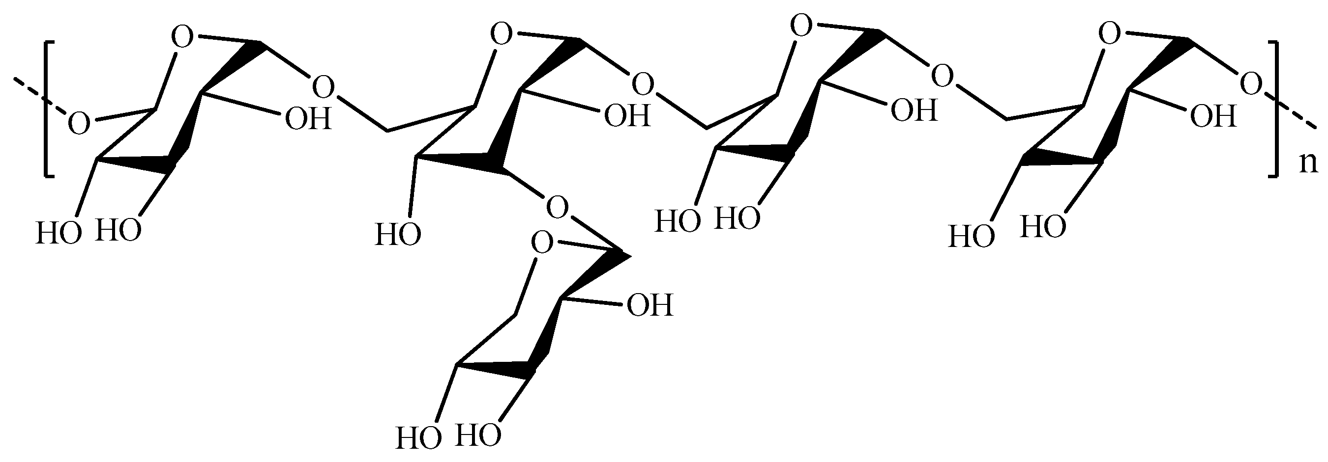
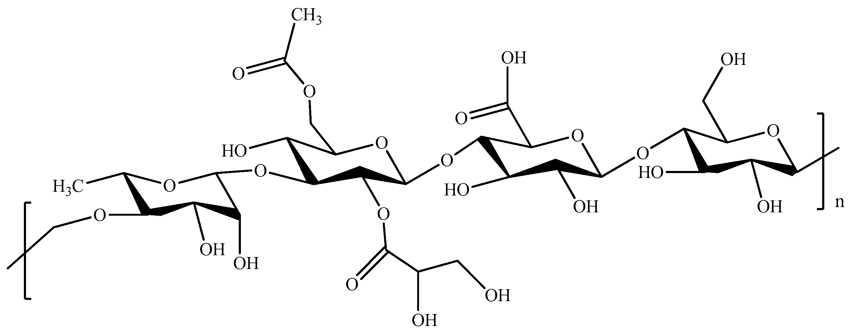
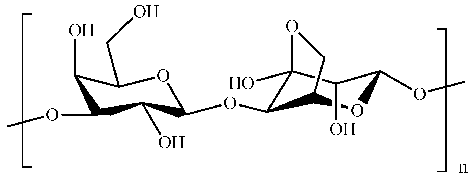
3.1. Polysaccharides
3.2. Proteins
4. Conclusions and Future Perspectives
Funding
Conflicts of Interest
References
- Li, Y.; Rodrigues, J.; Tomás, H. Injectable and biodegradable hydrogels: Gelation, biodegradation and biomedical applications. Chem. Soc. Rev. 2012, 41, 2193–2221. [Google Scholar] [CrossRef] [PubMed]
- Norouzi, M.; Nazari, B.; Miller, D.W. Injectable hydrogel-based drug delivery systems for local cancer therapy. Drug Discov. Today 2016, 21, 1835–1849. [Google Scholar] [CrossRef] [PubMed]
- Mathew, A.P.; Uthaman, S.; Cho, K.H.; Cho, C.S.; Park, I.K. Injectable hydrogels for delivering biotherapeutic molecules. Int. J. Biol. Macromol. 2018, 110, 17–29. [Google Scholar] [CrossRef] [PubMed]
- Yu, S.; He, C.; Chen, X. Injectable Hydrogels as Unique Platforms for Local Chemotherapeutics-Based Combination Antitumor Therapy. Macromol. Biosci. 2018, 18. [Google Scholar] [CrossRef]
- Ko, D.Y.; Shinde, U.P.; Yeon, B.; Jeong, B. Recent progress of in situ formed gels for biomedical applications. Prog. Polym. Sci. 2013, 38, 672–701. [Google Scholar] [CrossRef]
- Qi, C.; Liu, J.; Jin, Y.; Xu, L.; Wang, G.; Wang, Z.; Wang, L. Photo-crosslinkable, injectable sericin hydrogel as 3D biomimetic extracellular matrix for minimally invasive repairing cartilage. Biomaterials 2018, 163, 89–104. [Google Scholar] [CrossRef] [PubMed]
- Wu, X.; He, C.; Wu, Y.; Chen, X.; Cheng, J. Nanogel-Incorporated Physical and Chemical Hybrid Gels for Highly Effective Chemo-Protein Combination Therapy. Adv. Funct. Mater. 2015, 25, 6744–6755. [Google Scholar] [CrossRef]
- Zhang, Z.; He, C.L.; Xu, Q.H.; Zhuang, X.L.; Chen, X.S. Preparation of Poly(L-glutamic acid)-based Hydrogels via Diels-Alder Reaction and Study on Their Biomolecule-responsive Properties. Acta. Polym. Sin. 2018, 1, 99–108. [Google Scholar] [CrossRef]
- Xu, Q.; Guo, L.; Sigen, A.; Gao, Y.; Zhou, D.; Greiser, U.; Creagh-Flynn, J.; Zhang, H.; Dong, Y.; Cutlar, L.; et al. Injectable hyperbranched poly(β-amino ester) hydrogels with on-demand degradation profiles to match wound healing processes. Chem. Sci. 2018, 9, 2179–2187. [Google Scholar] [CrossRef]
- Castro, V.; Rodríguez, H.; Albericio, F. CuAAC: An Efficient Click Chemistry Reaction on Solid Phase. ACS Comb. Sci. 2016, 18, 1–14. [Google Scholar] [CrossRef]
- Dey, P.; Hemmati-Sadeghi, S.; Haag, R. Hydrolytically degradable, dendritic polyglycerol sulfate based injectable hydrogels using strain promoted azide-alkyne cycloaddition reaction. Polym. Chem. 2016, 7, 375–383. [Google Scholar] [CrossRef]
- Yang, Z.; Gao, D.; Cao, Z.; Zhang, C.; Cheng, D.; Liu, J.; Shuai, X. Drug and gene co-delivery systems for cancer treatment. Biomater. Sci. 2015, 3, 1035–1049. [Google Scholar] [CrossRef] [PubMed]
- Nezhad-Mokhtari, P.; Ghorbani, M.; Roshangar, L.; Soleimani Rad, J. Chemical gelling of hydrogels-based biological macromolecules for tissue engineering: Photo- and enzymatic-crosslinking methods. Int. J. Biol. Macromol. 2019, 139, 760–772. [Google Scholar] [CrossRef] [PubMed]
- Xu, Q.; He, C.; Zhang, Z.; Ren, K.; Chen, X. Injectable, Biomolecule-Responsive Polypeptide Hydrogels for Cell Encapsulation and Facile Cell Recovery through Triggered Degradation. ACS Appl. Mater. Interfaces 2016, 8, 30692–30702. [Google Scholar] [CrossRef] [PubMed]
- Yu, S.; Wang, C.; Yu, J.; Wang, J.; Lu, Y.; Zhang, Y.; Zhang, X.; Hu, Q.; Sun, W.; He, C.; et al. Injectable Bioresponsive Gel Depot for Enhanced Immune Checkpoint Blockade. Adv. Mater. 2018, 30. [Google Scholar] [CrossRef]
- Kim, S.H.; Tan, J.P.K.; Nederberg, F.; Fukushima, K.; Colson, J.; Yang, C.; Nelson, A.; Yang, Y.Y.; Hedrick, J.L. Hydrogen bonding-enhanced micelle assemblies for drug delivery. Biomaterials 2010, 31, 8063–8071. [Google Scholar] [CrossRef]
- Huebsch, N.; Kearney, C.J.; Zhao, X.; Kim, J.; Cezar, C.A.; Suo, Z.; Mooney, D.J. Ultrasound-triggered disruption and self-healing of reversibly cross-linked hydrogels for drug delivery and enhanced chemotherapy. Proc. Natl. Acad. Sci. USA 2014, 111, 9762–9767. [Google Scholar] [CrossRef] [Green Version]
- Appel, E.A.; Del Barrio, J.; Loh, X.J.; Scherman, O.A. Supramolecular polymeric hydrogels. Chem. Soc. Rev. 2012, 41, 6195–6214. [Google Scholar] [CrossRef]
- Bai, Y.; Li, S.; Li, X.; Han, X.; Li, Y.; Zhao, J.; Zhang, J.; Hou, X.; Yuan, X. An injectable robust denatured albumin hydrogel formed via double equilibrium reactions. J. Biomater. Sci. Polym. Ed. 2019, 30, 662–678. [Google Scholar] [CrossRef]
- Gačanin, J.; Kovtun, A.; Fischer, S.; Schwager, V.; Quambusch, J.; Kuan, S.L.; Liu, W.; Boldt, F.; Li, C.; Yang, Z.; et al. Spatiotemporally Controlled Release of Rho-Inhibiting C3 Toxin from a Protein–DNA Hybrid Hydrogel for Targeted Inhibition of Osteoclast Formation and Activity. Adv. Healthc. Mater. 2017, 6. [Google Scholar] [CrossRef]
- Yan, C.; Pochan, D.J. Rheological properties of peptide-based hydrogels for biomedical and other applications. Chem. Soc. Rev. 2010, 39, 3528–3540. [Google Scholar] [CrossRef] [PubMed] [Green Version]
- Slaughter, B.V.; Khurshid, S.S.; Fisher, O.Z.; Khademhosseini, A.; Peppas, N.A. Hydrogels in regenerative medicine. Adv. Mater. 2009, 21, 3307–3329. [Google Scholar] [CrossRef] [PubMed]
- Bakaic, E.; Smeets, N.M.B.; Hoare, T. Injectable hydrogels based on poly(ethylene glycol) and derivatives as functional biomaterials. RSC Adv. 2015, 5, 35469–35486. [Google Scholar] [CrossRef]
- Singh, N.K.; Lee, D.S. In situ gelling pH- and temperature-sensitive biodegradable block copolymer hydrogels for drug delivery. J. Control. Release 2014, 193, 214–227. [Google Scholar] [CrossRef] [PubMed]
- Nguyen, M.K.; Lee, D.S. Injectable biodegradable hydrogels. Macromol. Biosci. 2010, 10, 563–579. [Google Scholar] [CrossRef] [PubMed]
- Tran, R.T.; Gyawali, D.; Nair, P.; Yang, J. Biodegradable injectable systems for bone tissue engineering. In A Handbook of Applied Biopolymer Technology: Synthesis, Degradation and Applications; Sharma, S., Mudhoo, A., Eds.; RSC: London, UK, 2011; pp. 419–451. [Google Scholar]
- Srinivasan, C.; Weight, A.K.; Bussemer, T.; Klibanov, A.M. Non-aqueous suspensions of antibodies are much less viscous than equally concentrated aqueous solutions. Pharm. Res. 2013, 30, 1749–1757. [Google Scholar] [CrossRef]
- Sun, S.; Cao, H.; Su, H.; Tan, T. Preparation and characterization of a novel injectable in situ cross-linked hydrogel. Polym. Bull. 2009, 62, 699–711. [Google Scholar] [CrossRef]
- Tu, Y.; Chen, N.; Li, C.; Liu, H.; Zhu, R.; Chen, S.; Xiao, Q.; Liu, J.; Ramakrishna, S.; He, L. Advances in injectable self-healing biomedical hydrogels. Acta. Biomater. 2019, 90, 1–20. [Google Scholar] [CrossRef]
- Kretlow, J.D.; Klouda, L.; Mikos, A.G. Injectable matrices and scaffolds for drug delivery in tissue engineering. Adv. Drug Deliv. Rev. 2007, 59, 263–273. [Google Scholar] [CrossRef]
- Říhová, B. Immunocompatibility and biocompatibility of cell delivery systems. Adv. Drug Deliv. Rev. 2000, 42, 65–80. [Google Scholar] [CrossRef]
- Fu, C.X.; Lin, X.X.; Wang, J.; Zheng, X.Q.; Li, X.Y.; Lin, Z.F.; Lin, G.Y. Injectable micellar supramolecular hydrogel for delivery of hydrophobic anticancer drugs. J. Mater. Sci. 2016, 27, 1–7. [Google Scholar] [CrossRef] [PubMed]
- Brigger, I.; Dubernet, C.; Couvreur, P. Nanoparticles in cancer therapy and diagnosis. Adv. Drug Deliv. Rev. 2012, 64, 24–36. [Google Scholar] [CrossRef]
- Wu, X.; He, C.; Wu, Y.; Chen, X. Synergistic therapeutic effects of Schiff’s base cross-linked injectable hydrogels for local co-delivery of metformin and 5-fluorouracil in a mouse colon carcinoma model. Biomaterials 2016, 75, 148–162. [Google Scholar] [CrossRef] [PubMed]
- Cirillo, G.; Nicoletta, F.P.; Curcio, M.; Spizzirri, U.G.; Picci, N.; Iemma, F. Enzyme immobilization on smart polymers: Catalysis on demand. React. Funct. Polym. 2014, 83, 62–69. [Google Scholar] [CrossRef]
- Allcock, H.R.; Morozowich, N.L. Bioerodible polyphosphazenes and their medical potential. Polym. Chem. 2012, 3, 578–590. [Google Scholar] [CrossRef]
- Baillargeon, A.L.; Mequanint, K. Biodegradable polyphosphazene biomaterials for tissue engineering and delivery of therapeutics. BioMed Res. Int. 2014, 2014. [Google Scholar] [CrossRef] [PubMed]
- Hindenlang, M.D.; Soudakov, A.A.; Imler, G.H.; Laurencin, C.T.; Nair, L.S.; Allcock, H.R. Iodine-containing radio-opaque polyphosphazenes. Polym. Chem. 2010, 1, 1467–1474. [Google Scholar] [CrossRef]
- Singh, A.; Krogman, N.R.; Sethuraman, S.; Nair, L.S.; Sturgeon, J.L.; Brown, P.W.; Laurencin, C.T.; Allcock, H.R. Effect of side group chemistry on the properties of biodegradable l-alanine cosubstituted polyphosphazenes. Biomacromolecules 2006, 7, 914–918. [Google Scholar] [CrossRef]
- Cho, J.K.; Hong, K.Y.; Park, J.W.; Yang, H.K.; Song, S.C. Injectable delivery system of 2-methoxyestradiol for breast cancer therapy using biodegradable thermosensitive poly(organophosphazene) hydrogel. J. Drug Target. 2011, 19, 270–280. [Google Scholar] [CrossRef]
- Allcock, H.R.; Pucher, S.R.; Scopelianos, A.G. Poly[(amino acid ester)phosphazenes] as substrates for the controlled release of small molecules. Biomaterials 1994, 15, 563–569. [Google Scholar] [CrossRef]
- Teasdale, I.; Brüggemann, O. Polyphosphazenes: Multifunctional, biodegradable vehicles for drug and gene delivery. Polymers 2013, 5, 161–187. [Google Scholar] [CrossRef] [PubMed]
- Ogueri, K.S.; Allcock, H.R.; Laurencin, C.T. Polyphosphazene Polymer. Encycl. Polym. Sci. Technol. 2019. [Google Scholar] [CrossRef]
- Kwak, M.K.; Hur, K.; Yu, J.E.; Han, T.S.; Yanagihara, K.; Kim, W.H.; Lee, S.M.; Song, S.C.; Yang, H.K. Suppression of in vivo tumor growth by using a biodegradable thermosensitive hydrogel polymer containing chemotherapeutic agent. Investig. New Drugs 2010, 28, 284–290. [Google Scholar] [CrossRef] [PubMed]
- Al-Abd, A.M.; Hong, K.Y.; Song, S.C.; Kuh, H.J. Pharmacokinetics of doxorubicin after intratumoral injection using a thermosensitive hydrogel in tumor-bearing mice. J. Control. Release 2010, 142, 101–107. [Google Scholar] [CrossRef] [PubMed]
- Wang, J.; Wang, D.; Yan, H.; Tao, L.; Wei, Y.; Li, Y.; Wang, X.; Zhao, W.; Zhang, Y.; Zhao, L.; et al. An injectable ionic hydrogel inducing high temperature hyperthermia for microwave tumor ablation. J. Mater. Chem. B 2017, 5, 4110–4120. [Google Scholar] [CrossRef]
- Cho, J.K.; Hong, J.M.; Han, T.; Yang, H.K.; Song, S.C. Injectable and biodegradable poly(organophosphazene) hydrogel as a delivery system of docetaxel for cancer treatment. J. Drug Target. 2013, 21, 564–573. [Google Scholar] [CrossRef] [PubMed]
- Kim, J.H.; Lee, J.H.; Kim, K.S.; Na, K.; Song, S.C.; Lee, J.; Kuh, H.J. Intratumoral delivery of paclitaxel using a thermosensitive hydrogel in human tumor xenografts. Arch. Pharmacal Res. 2013, 36, 94–101. [Google Scholar] [CrossRef] [PubMed]
- Cho, J.K.; Kuh, H.J.; Song, S.C. Injectable poly(organophosphazene) hydrogel system for effective paclitaxel and doxorubicin combination therapy. J. Drug Target. 2014, 22, 761–767. [Google Scholar] [CrossRef] [PubMed]
- Kim, Y.M.; Park, M.R.; Song, S.C. An injectable cell penetrable nano-polyplex hydrogel for localized siRNA delivery. Biomaterials 2013, 34, 4493–4500. [Google Scholar] [CrossRef] [PubMed]
- Cho, J.K.; Chun, C.; Kuh, H.J.; Song, S.C. Injectable poly(organophosphazene)-camptothecin conjugate hydrogels: Synthesis, characterization, and antitumor activities. Eur. J. Pharm. Biopharm. 2012, 81, 582–590. [Google Scholar] [CrossRef] [PubMed]
- Kim, J.I.; Kim, B.; Chun, C.; Lee, S.H.; Song, S.C. MRI-monitored long-term therapeutic hydrogel system for brain tumors without surgical resection. Biomaterials 2012, 33, 4836–4842. [Google Scholar] [CrossRef] [PubMed]
- Zhang, Z.Q.; Song, S.C. Thermosensitive/superparamagnetic iron oxide nanoparticle-loaded nanocapsule hydrogels for multiple cancer hyperthermia. Biomaterials 2016, 106, 13–23. [Google Scholar] [CrossRef] [PubMed]
- Akash, M.S.H.; Rehman, K. Recent progress in biomedical applications of pluronic (PF127): Pharmaceutical perspectives. J. Control. Release 2015, 209, 120–138. [Google Scholar] [CrossRef] [PubMed]
- Moebus, K.; Siepmann, J.; Bodmeier, R. Alginate-poloxamer microparticles for controlled drug delivery to mucosal tissue. Eur. J. Pharm. Biopharm. 2009, 72, 42–53. [Google Scholar] [CrossRef] [PubMed]
- Klouda, L. Thermoresponsive hydrogels in biomedical applications A seven-year update. Eur. J. Pharm. Biopharm. 2015, 97, 338–349. [Google Scholar] [CrossRef]
- Cabana, A.; Aït-Kadi, A.; Juhász, J. Study of the gelation process of polyethylene oxide(a)-polypropylene oxide(b)-polyethylene oxide, copolymer (poloxamer 407) aqueous solutions. J. Colloid Interface Sci. 1997, 190, 307–312. [Google Scholar] [CrossRef]
- Thimmaraju, M.K.; Bheemanapally, K.; Dharavath, R.; Kakarla, L.; Botlagunta, M. Improved anticancer activity of meloxicam hydrogels in K562 and HL60 cell lines. J. Young Pharm. 2017, 9, 209–213. [Google Scholar] [CrossRef]
- Hu, H.; Lin, Z.; He, B.; Dai, W.; Wang, X.; Wang, J.; Zhang, X.; Zhang, H.; Zhang, Q. A novel localized co-delivery system with lapatinib microparticles and paclitaxel nanoparticles in a peritumorally injectable in situ hydrogel. J. Control. Release 2015, 220, 189–200. [Google Scholar] [CrossRef]
- Xu, G.; Li, B.; Wang, T.; Wan, J.; Zhang, Y.; Huang, J.; Shen, Y. Enhancing the anti-ovarian cancer activity of quercetin using a self-assembling micelle and thermosensitive hydrogel drug delivery system. RSC Adv. 2018, 8, 21229–21242. [Google Scholar] [CrossRef] [Green Version]
- Kim, D.Y.; Kwon, D.Y.; Kwon, J.S.; Park, J.H.; Park, S.H.; Oh, H.J.; Kim, J.H.; Min, B.H.; Park, K.; Kim, M.S. Synergistic anti-tumor activity through combinational intratumoral injection of an in-situ injectable drug depot. Biomaterials 2016, 85, 232–245. [Google Scholar] [CrossRef]
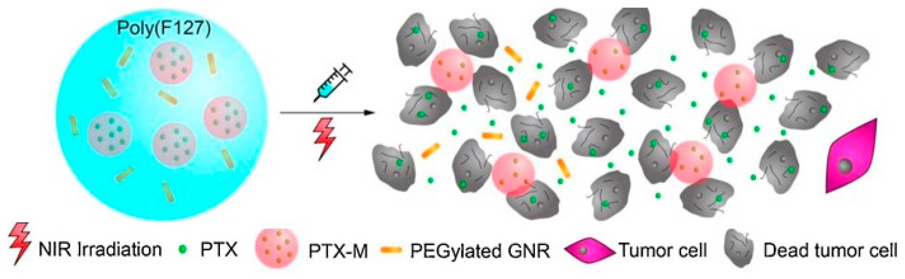
- Zhang, N.; Xu, X.; Zhang, X.; Qu, D.; Xue, L.; Mo, R.; Zhang, C. Nanocomposite hydrogel incorporating gold nanorods and paclitaxel-loaded chitosan micelles for combination photothermal-chemotherapy. Int. J. Pharm. 2016, 497, 210–221. [Google Scholar] [CrossRef] [PubMed]
- Fu, J.J.; Chen, M.Y.; Li, J.X.; Zhou, J.H.; Xie, S.N.; Yuan, P.; Tang, B.; Liu, C.C. Injectable hydrogel encapsulating Cu2MnS2 nanoplates for photothermal therapy against breast cancer. J. Nanobiotechnology 2018, 16. [Google Scholar] [CrossRef] [PubMed]
- Bruschi, M.L.; Borghi-Pangoni, F.B.; Junqueira, M.V.; de Souza Ferreira, S.B.; Ficai, D.; Grumezescu, A.M. Chapter 12—Nanostructured therapeutic systems with bioadhesive and thermoresponsive properties. In Nanostructures for Novel Therapy; Ficai, D., Grumezescu, A., Eds.; Elsevier: Amsterdam, The Netherlands, 2017; pp. 313–342. [Google Scholar]
- Lin, H.R.; Tseng, C.C.; Lin, Y.J.; Ling, M.H. A novel in-situ-gelling liquid suppository for site-targeting delivery of anti-colorectal cancer drugs. J. Biomater. Sci. Polym. Ed. 2012, 23, 807–822. [Google Scholar] [CrossRef] [PubMed]
- Gao, L.; Wang, X.; Ma, J.; Hao, D.; Wei, P.; Zhou, L.; Liu, G. Evaluation of TPGS-modified thermo-sensitive Pluronic PF127 hydrogel as a potential carrier to reverse the resistance of P-gp-overexpressing SMMC-7721 cell lines. Colloids Surf. B Biointerfaces 2016, 140, 307–316. [Google Scholar] [CrossRef] [PubMed]
- Sheu, M.T.; Jhan, H.J.; Su, C.Y.; Chen, L.C.; Chang, C.E.; Liu, D.Z.; Ho, H.O. Codelivery of doxorubicin-containing thermosensitive hydrogels incorporated with docetaxel-loaded mixed micelles enhances local cancer therapy. Colloids Surf. B Biointerfaces 2016, 143, 260–270. [Google Scholar] [CrossRef]
- Jhan, H.J.; Liu, J.J.; Chen, Y.C.; Liu, D.Z.; Sheu, M.T.; Ho, H.O. Novel injectable thermosensitive hydrogels for delivering hyaluronic acid-doxorubicin nanocomplexes to locally treat tumors. Nanomedicine 2015, 10, 1263–1274. [Google Scholar] [CrossRef] [PubMed]
- Khan, S.; Minhas, M.U.; Ahmad, M.; Sohail, M. Self-assembled supramolecular thermoreversible β-cyclodextrin/ethylene glycol injectable hydrogels with difunctional Pluronic®127 as controlled delivery depot of curcumin. Development, characterization and in vitro evaluation. J. Biomater. Sci. Polym. Ed. 2018, 29, 1–34. [Google Scholar] [CrossRef]
- Hu, X.; Li, D.; Tan, H.; Pan, C.; Chen, X. Injectable graphene oxide/graphene composite supramolecular hydrogel for delivery of anti-cancer drugs. J. Macromol. Sci. A Pure Appl. Chem. 2014, 51, 378–384. [Google Scholar] [CrossRef]
- Moon, H.J.; Ko, D.Y.; Park, M.H.; Joo, M.K.; Jeong, B. Temperature-responsive compounds as in situ gelling biomedical materials. Chem. Soc. Rev. 2012, 41, 4860–4883. [Google Scholar] [CrossRef]
- Kang, Y.M.; Kim, G.H.; Kim, J.I.; Kim, D.Y.; Lee, B.N.; Yoon, S.M.; Kim, J.H.; Kim, M.S. In vivo efficacy of an intratumorally injected in situ-forming doxorubicin/poly(ethylene glycol)-b-polycaprolactone diblock copolymer. Biomaterials 2011, 32, 4556–4564. [Google Scholar] [CrossRef]
- Lei, N.; Gong, C.; Qian, Z.; Luo, F.; Wang, C.; Wang, H.; Wei, Y. Therapeutic application of injectable thermosensitive hydrogel in preventing local breast cancer recurrence and improving incision wound healing in a mouse model. Nanoscale 2012, 4, 5686–5693. [Google Scholar] [CrossRef] [PubMed]
- Kondiah, P.J.; Choonara, Y.E.; Kondiah, P.P.D.; Marimuthu, T.; Kumar, P.; Du Toit, L.C.; Pillay, V. A review of injectable polymeric hydrogel systems for application in bone tissue engineering. Molecules 2016, 21. [Google Scholar] [CrossRef] [PubMed]
- Choi, B.; Lee, M. Injectable Hydrogels for Articular Cartilage Regeneration. In Injectable Hydrogels for Regenerative Engineering; Nair, L.S., Ed.; Imperial College Press: London, UK, 2016; pp. 355–376. [Google Scholar]
- Shi, K.; Wang, Y.L.; Qu, Y.; Liao, J.F.; Chu, B.Y.; Zhang, H.P.; Luo, F.; Qian, Z.Y. Synthesis, characterization, and application of reversible PDLLA-PEG-PDLLA copolymer thermogels in vitro and in vivo. Sci. Rep. 2016, 6. [Google Scholar] [CrossRef] [PubMed]
- Shi, K.; Xue, B.; Jia, Y.; Yuan, L.; Han, R.; Yang, F.; Peng, J.; Qian, Z. Sustained co-delivery of gemcitabine and cis-platinum via biodegradable thermo-sensitive hydrogel for synergistic combination therapy of pancreatic cancer. Nano Res. 2019. [Google Scholar] [CrossRef]
- Fan, R.; Tong, A.; Li, X.; Gao, X.; Mei, L.; Zhou, L.; Zhang, X.; You, C.; Guo, G. Enhanced antitumor effects by docetaxel/LL 37-loaded thermosensitive hydrogel nanoparticles in peritoneal carcinomatosis of colorectal cancer. Int. J. Nanomed. 2015, 10, 7291–7305. [Google Scholar] [CrossRef]
- Li, X.; Fan, R.; Wang, Y.; Wu, M.; Tong, A.; Shi, J.; Xiang, M.; Zhou, L.; Guo, G. In situ gel-forming dual drug delivery system for synergistic combination therapy of colorectal peritoneal carcinomatosis. RSC Adv. 2015, 5, 101494–101506. [Google Scholar] [CrossRef]
- Liang, Y.; Dong, C.; Zhang, J.; Deng, L.; Dong, A. A reconstituted thermosensitive hydrogel system based on paclitaxel-loaded amphiphilic copolymer nanoparticles and antitumor efficacy. Drug Dev. Ind. Pharm. 2017, 43, 972–979. [Google Scholar] [CrossRef]
- Park, M.H.; Joo, M.K.; Choi, B.G.; Jeong, B. Biodegradable thermogels. Acc. Chem. Res. 2012, 45, 424–433. [Google Scholar] [CrossRef]
- Qiu, B.; Stefanos, S.; Ma, J.; Lalloo, A.; Perry, B.A.; Leibowitz, M.J.; Sinko, P.J.; Stein, S. A hydrogel prepared by in situ cross-linking of a thiol-containing poly(ethylene glycol)-based copolymer: A new biomaterial for protein drug delivery. Biomaterials 2003, 24, 11–18. [Google Scholar] [CrossRef]
- Ma, H.; He, C.; Cheng, Y.; Yang, Z.; Zang, J.; Liu, J.; Chen, X. Localized Co-delivery of Doxorubicin, Cisplatin, and Methotrexate by Thermosensitive Hydrogels for Enhanced Osteosarcoma Treatment. ACS Appl. Mater. Interfaces 2015, 7, 27040–27048. [Google Scholar] [CrossRef]
- He, C.; Kim, S.W.; Lee, D.S. In situ gelling stimuli-sensitive block copolymer hydrogels for drug delivery. J. Control. Release 2008, 127, 189–207. [Google Scholar] [CrossRef] [PubMed]
- Chang, G.; Ci, T.; Yu, L.; Ding, J. Enhancement of the fraction of the active form of an antitumor drug topotecan via an injectable hydrogel. J. Control. Release 2011, 156, 21–27. [Google Scholar] [CrossRef] [PubMed]
- Yang, Z.; Yu, S.; Li, D.; Gong, Y.; Zang, J.; Liu, J.; Chen, X. The effect of PLGA-based hydrogel scaffold for improving the drug maximum-tolerated dose for in situ osteosarcoma treatment. Colloids Surf. B Biointerfaces 2018, 172, 387–394. [Google Scholar] [CrossRef] [PubMed]
- Gong, C.; Wang, C.; Wang, Y.; Wu, Q.; Zhang, D.; Luo, F.; Qian, Z. Efficient inhibition of colorectal peritoneal carcinomatosis by drug loaded micelles in thermosensitive hydrogel composites. Nanoscale 2012, 4, 3095–3104. [Google Scholar] [CrossRef] [PubMed]
- Liu, J.; Jiang, Y.; Cui, Y.; Xu, C.; Ji, X.; Luan, Y. Cytarabine-AOT catanionic vesicle-loaded biodegradable thermosensitive hydrogel as an efficient cytarabine delivery system. Int. J. Pharm. 2014, 473, 560–571. [Google Scholar] [CrossRef] [PubMed]
- Xing, Y.; Chen, H.; Li, S.; Guo, X. In vitro and in vivo investigation of a novel two-phase delivery system of 2-methoxyestradiol liposomes hydrogel. J. Liposome Res. 2014, 24, 10–16. [Google Scholar] [CrossRef] [PubMed]
- Jiang, L.; Ding, Y.; Xue, X.; Zhou, S.; Li, C.; Zhang, X.; Jiang, X. Entrapping multifunctional dendritic nanoparticles into a hydrogel for local therapeutic delivery and synergetic immunochemotherapy. Nano Res. 2018, 11, 6062–6073. [Google Scholar] [CrossRef]
- Guo, X.; Cui, F.; Xing, Y.; Mei, Q.; Zhang, Z. Investigation of a new injectable thermosensitive hydrogel loading solid lipid nanoparticles. Pharmazie 2011, 66, 948–952. [Google Scholar] [CrossRef]
- Ma, H.; He, C.; Cheng, Y.; Li, D.; Gong, Y.; Liu, J.; Tian, H.; Chen, X. PLK1shRNA and doxorubicin co-loaded thermosensitive PLGA-PEG-PLGA hydrogels for osteosarcoma treatment. Biomaterials 2014, 35, 8723–8734. [Google Scholar] [CrossRef]
- Shen, W.; Chen, X.; Luan, J.; Wang, D.; Yu, L.; Ding, J. Sustained Codelivery of Cisplatin and Paclitaxel via an Injectable Prodrug Hydrogel for Ovarian Cancer Treatment. ACS Appl. Mater. Interfaces 2017, 9, 40031–40046. [Google Scholar] [CrossRef]
- Liu, Y.; Xiao, L.; Joo, K.I.; Hu, B.; Fang, J.; Wang, P. In situ modulation of dendritic cells by injectable thermosensitive hydrogels for cancer vaccines in mice. Biomacromolecules 2014, 15, 3836–3845. [Google Scholar] [CrossRef] [PubMed]
- Wang, Y.; Gong, C.; Yang, L.; Wu, Q.; Shi, S.; Shi, H.; Qian, Z.; Wei, Y. 5-FU-hydrogel inhibits colorectal peritoneal carcinomatosis and tumor growth in mice. BMC Cancer 2010, 10. [Google Scholar] [CrossRef] [PubMed]
- Lin, X.; Deng, L.; Xu, Y.; Dong, A. Thermosensitive in situ hydrogel of paclitaxel conjugated poly(ε-caprolactone)-poly(ethylene glycol)-poly(ε-caprolactone). Soft Matter 2012, 8, 3470–3477. [Google Scholar] [CrossRef]
- Liu, L.; Wu, Q.; Ma, X.; Xiong, D.; Gong, C.; Qian, Z.; Zhao, X.; Wei, Y. Camptothecine encapsulated composite drug delivery system for colorectal peritoneal carcinomatosis therapy: Biodegradable microsphere in thermosensitive hydrogel. Colloids Surf. B Biointerfaces 2013, 106, 93–101. [Google Scholar] [CrossRef] [PubMed]
- Liu, M.; Huang, P.; Wang, W.; Feng, Z.; Zhang, J.; Deng, L.; Dong, A. An injectable nanocomposite hydrogel co-constructed with gold nanorods and paclitaxel-loaded nanoparticles for local chemo-photothermal synergetic cancer therapy. J. Mater. Chem. B 2019, 7, 2667–2677. [Google Scholar] [CrossRef]
- Peng, M.; Xu, S.; Zhang, Y.; Zhang, L.; Huang, B.; Fu, S.; Xue, Z.; Da, Y.; Dai, Y.; Qiao, L.; et al. Thermosensitive injectable hydrogel enhances the antitumor effect of embelin in mouse hepatocellular carcinoma. J. Pharm. Sci. 2014, 103, 965–973. [Google Scholar] [CrossRef] [PubMed]
- Wang, W.; Deng, L.; Xu, S.; Zhao, X.; Lv, N.; Zhang, G.; Gu, N.; Hu, R.; Zhang, J.; Liu, J.; et al. A reconstituted “two into one” thermosensitive hydrogel system assembled by drug-loaded amphiphilic copolymer nanoparticles for the local delivery of paclitaxel. J. Mater. Chem. B 2013, 1, 552–563. [Google Scholar] [CrossRef]
- Huang, P.; Song, H.; Zhang, Y.; Liu, J.; Zhang, J.; Wang, W.; Li, C.; Kong, D. Bridging the Gap between Macroscale Drug Delivery Systems and Nanomedicines: A Nanoparticle-Assembled Thermosensitive Hydrogel for Peritumoral Chemotherapy. ACS Appl. Mater. Interfaces 2016, 8, 29323–29333. [Google Scholar] [CrossRef]
- Huang, P.; Zhang, Y.; Wang, W.; Zhou, J.; Sun, Y.; Liu, J.; Kong, D.; Dong, A. Co-delivery of doxorubicin and 131 I by thermosensitive micellar-hydrogel for enhanced in situ synergetic chemoradiotherapy. J. Control. Release 2015, 220, 456–464. [Google Scholar] [CrossRef]
- Zhu, W.; Li, Y.; Liu, L.; Chen, Y.; Xi, F. Supramolecular hydrogels as a universal scaffold for stepwise delivering Dox and Dox/cisplatin loaded block copolymer micelles. International, J. Pharm. 2012, 437, 11–19. [Google Scholar] [CrossRef]
- Ren, L.; He, L.; Sun, T.; Dong, X.; Chen, Y.; Huang, J.; Wang, C. Dual-responsive supramolecular hydrogels from water-soluble PEG-grafted copolymers and cyclodextrin. Macromol. Biosci. 2009, 9, 902–910. [Google Scholar] [CrossRef] [PubMed]
- Xu, S.; Wang, W.; Li, X.; Liu, J.; Dong, A.; Deng, L. Sustained release of PTX-incorporated nanoparticles synergized by burst release of DOX·HCl from thermosensitive modified PEG/PCL hydrogel to improve anti-tumor efficiency. European, J. Pharm. Sci. 2014, 62, 267–273. [Google Scholar] [CrossRef] [PubMed]
- Zhu, W.; Li, Y.; Liu, L.; Chen, Y.; Wang, C.; Xi, F. Supramolecular hydrogels from cisplatin-loaded block copolymer nanoparticles and α-cyclodextrins with a stepwise delivery property. Biomacromolecules 2010, 11, 3086–3092. [Google Scholar] [CrossRef] [PubMed]
- Kuang, H.; He, H.; Zhang, Z.; Qi, Y.; Xie, Z.; Jing, X.; Huang, Y. Injectable and biodegradable supramolecular hydrogels formed by nucleobase-terminated poly(ethylene oxide)s and α-cyclodextrin. J. Mater. Chem. B 2014, 2, 659–667. [Google Scholar] [CrossRef]
- Liu, X.; Li, Z.; Loh, X.J.; Chen, K.; Wu, Y.L. Targeted and Sustained Corelease of Chemotherapeutics and Gene by Injectable Supramolecular Hydrogel for Drug-Resistant Cancer Therapy. Macromol. Rapid Commun. 2019, 40. [Google Scholar] [CrossRef] [PubMed]
- Shahin, M.; Lavasanifar, A. Novel self-associating poly(ethylene oxide)-b-poly(ε-caprolactone) based drug conjugates and nano-containers for paclitaxel delivery. Int. J. Pharm. 2010, 389, 213–222. [Google Scholar] [CrossRef]
- Ma, G.; Miao, B.; Song, C. Thermosensitive PCL-PEG-PCL hydrogels: Synthesis, characterization, and delivery of proteins. J. Appl. Polym. Sci. 2010, 116, 1985–1993. [Google Scholar] [CrossRef]
- Wang, W.; Deng, L.; Liu, S.; Li, X.; Zhao, X.; Hu, R.; Zhang, J.; Han, H.; Dong, A. Adjustable degradation and drug release of a thermosensitive hydrogel based on a pendant cyclic ether modified poly(e-caprolactone) and poly(ethylene glycol)co-polymer. Acta. Biomater. 2012, 8, 3963–3973. [Google Scholar] [CrossRef]
- Wang, W.; Song, H.; Zhang, J.; Li, P.; Li, C.; Wang, C.; Kong, D.; Zhao, Q. An injectable, thermosensitive and multicompartment hydrogel for simultaneous encapsulation and independent release of a drug cocktail as an effective combination therapy platform. J. Control. Release 2015, 203, 57–66. [Google Scholar] [CrossRef]
- Yin, L.; Xu, S.; Feng, Z.; Deng, H.; Zhang, J.; Gao, H.; Deng, L.; Tang, H.; Dong, A. Supramolecular hydrogel based on high-solid-content mPECT nanoparticles and cyclodextrins for local and sustained drug delivery. Biomater. Sci. 2017, 5, 698–706. [Google Scholar] [CrossRef]
- Kunz-Schughart, L.A.; Dubrovska, A.; Peitzsch, C.; Ewe, A.; Aigner, A.; Schellenburg, S.; Muders, M.H.; Hampel, S.; Cirillo, G.; Iemma, F.; et al. Nanoparticles for radiooncology: Mission, vision, challenges. Biomaterials 2017, 120, 155–184. [Google Scholar] [CrossRef] [PubMed] [Green Version]
- Peng, C.L.; Shih, Y.H.; Liang, K.S.; Chiang, P.F.; Yeh, C.H.; Tang, I.C.; Yao, C.J.; Lee, S.Y.; Luo, T.Y.; Shieh, M.J. Development of in situ forming thermosensitive hydrogel for radiotherapy combined with chemotherapy in a mouse model of hepatocellular carcinoma. Mol. Pharm. 2013, 10, 1854–1864. [Google Scholar] [CrossRef] [PubMed]
- Biolato, M.; Marrone, G.; Racco, S.; Di Stasi, C.; Miele, L.; Gasbarrini, G.; Landolfi, R.; Grieco, A. Transarterial chemoembolization (TACE) for unresectable HCC: A new life begins? Eur. Rev. Med. Pharmacol. Sci. 2010, 14, 356–362. [Google Scholar]
- Lym, J.S.; Nguyen, Q.V.; Ahn, D.W.; Huynh, C.T.; Jae, H.J.; Kim, Y.I.; Lee, D.S. Sulfamethazine-based pH-sensitive hydrogels with potential application for transcatheter arterial chemoembolization therapy. Acta. Biomater. 2016, 41, 253–263. [Google Scholar] [CrossRef] [PubMed]
- Huynh, C.T.; Nguyen, Q.V.; Lym, J.S.; Kim, B.S.; Huynh, D.P.; Jae, H.J.; Kim, Y.I.; Lee, D.S. Intraarterial gelation of injectable cationic pH/temperature-sensitive radiopaque embolic hydrogels in a rabbit hepatic tumor model and their potential application for liver cancer treatment. RSC Adv. 2016, 6, 47687–47697. [Google Scholar] [CrossRef]
- Gil, M.S.; Thambi, T.; Phan, V.H.G.; Kim, S.H.; Lee, D.S. Injectable hydrogel-incorporated cancer cell-specific cisplatin releasing nanogels for targeted drug delivery. J. Mater. Chem. B 2017, 5, 7140–7152. [Google Scholar] [CrossRef]
- Varghese, O.P.; Liu, J.; Sundaram, K.; Hilborn, J.; Oommen, O.P. Chondroitin sulfate derived theranostic nanoparticles for targeted drug delivery. Biomater. Sci. 2016, 4, 1310–1313. [Google Scholar] [CrossRef] [PubMed]
- Andrgie, A.T.; Mekuria, S.L.; Addisu, K.D.; Hailemeskel, B.Z.; Hsu, W.H.; Tsai, H.C.; Lai, J.Y. Non-Anticoagulant Heparin Prodrug Loaded Biodegradable and Injectable Thermoresponsive Hydrogels for Enhanced Anti-Metastasis Therapy. Macromol. Biosci. 2019, 19. [Google Scholar] [CrossRef]
- Phan, V.H.G.; Lee, E.; Maeng, J.H.; Thambi, T.; Kim, B.S.; Lee, D.; Lee, D.S. Pancreatic cancer therapy using an injectable nanobiohybrid hydrogel. RSC Adv. 2016, 6, 41644–41655. [Google Scholar] [CrossRef]
- Nguyen, Q.V.; Lym, J.S.; Huynh, C.T.; Kim, B.S.; Jae, H.J.; Kim, Y.I.; Lee, D.S. A novel sulfamethazine-based pH-sensitive copolymer for injectable radiopaque embolic hydrogels with potential application in hepatocellular carcinoma therapy. Polym. Chem. 2016, 7, 5805–5818. [Google Scholar] [CrossRef]
- Bobbala, S.; Tamboli, V.; McDowell, A.; Mitra, A.K.; Hook, S. Novel Injectable Pentablock Copolymer Based Thermoresponsive Hydrogels for Sustained Release Vaccines. AAPS J. 2016, 18, 261–269. [Google Scholar] [CrossRef]
- Kim, S.H.; Tan, J.P.K.; Fukushima, K.; Nederberg, F.; Yang, Y.Y.; Waymouth, R.M.; Hedrick, J.L. Thermoresponsive nanostructured polycarbonate block copolymers as biodegradable therapeutic delivery carriers. Biomaterials 2011, 32, 5505–5514. [Google Scholar] [CrossRef] [PubMed]
- Zawaneh, P.N.; Singh, S.P.; Padera, R.F.; Henderson, P.W.; Spector, J.A.; Putnam, D. Design of an injectable synthetic and biodegradable surgical biomaterial. Proc. Natl. Acad. Sci. USA 2010, 107, 11014–11019. [Google Scholar] [CrossRef] [PubMed] [Green Version]
- Lee, A.L.Z.; Ng, V.W.L.; Gao, S.; Hedrick, J.L.; Yang, Y.Y. Injectable hydrogels from triblock copolymers of vitamin E-functionalized polycarbonate and poly(ethylene glycol) for subcutaneous delivery of antibodies for cancer therapy. Adv. Funct. Mater. 2014, 24, 1538–1550. [Google Scholar] [CrossRef]
- Yang, C.; Lee, A.; Gao, S.; Liu, S.; Hedrick, J.L.; Yang, Y.Y. Hydrogels with prolonged release of therapeutic antibody: Block junction chemistry modification of ‘ABA’ copolymers provides superior anticancer efficacy. J. Control. Release 2019, 293, 193–200. [Google Scholar] [CrossRef] [PubMed]
- Leprince, J.G.; Palin, W.M.; Hadis, M.A.; Devaux, J.; Leloup, G. Progress in dimethacrylate-based dental composite technology and curing efficiency. Dent. Mater. 2013, 29, 139–156. [Google Scholar] [CrossRef]
- Cirillo, G.; Spataro, T.; Curcio, M.; Spizzirri, U.G.; Nicoletta, F.P.; Picci, N.; Iemma, F. Tunable thermo-responsive hydrogels: Synthesis, structural analysis and drug release studies. Mater. Sci. Eng. C 2015, 48, 499–510. [Google Scholar] [CrossRef]
- Pal, A.; Vernon, B.L.; Nikkhah, M. Therapeutic neovascularization promoted by injectable hydrogels. Bioact. Mater. 2018, 3, 389–400. [Google Scholar] [CrossRef]
- Fourniols, T.; Randolph, L.D.; Staub, A.; Vanvarenberg, K.; Leprince, J.G.; Préat, V.; Des Rieux, A.; Danhier, F. Temozolomide-loaded photopolymerizable PEG-DMA-based hydrogel for the treatment of glioblastoma. J. Control. Release 2015, 210, 95–104. [Google Scholar] [CrossRef]
- Zhao, M.; Danhier, F.; Bastiancich, C.; Joudiou, N.; Ganipineni, L.P.; Tsakiris, N.; Gallez, B.; Rieux, A.D.; Jankovski, A.; Bianco, J.; et al. Post-resection treatment of glioblastoma with an injectable nanomedicine-loaded photopolymerizable hydrogel induces long-term survival. Int. J. Pharm. 2018, 548, 522–529. [Google Scholar] [CrossRef]
- Zhang, H.; Zhu, X.; Ji, Y.; Jiao, X.; Chen, Q.; Hou, L.; Zhang, Z. Near-infrared-triggered in situ hybrid hydrogel system for synergistic cancer therapy. J. Mater. Chem. B 2015, 3, 6310–6326. [Google Scholar] [CrossRef]
- Wu, H.; Song, L.; Chen, L.; Huang, Y.; Wu, Y.; Zang, F.; An, Y.; Lyu, H.; Ma, M.; Chen, J.; et al. Injectable thermosensitive magnetic nanoemulsion hydrogel for multimodal-imaging-guided accurate thermoablative cancer therapy. Nanoscale 2017, 9, 16175–16182. [Google Scholar] [CrossRef] [PubMed]
- Khang, M.K.; Zhou, J.; Huang, Y.; Hakamivala, A.; Tang, L. Preparation of a novel injectable in situ-gelling nanoparticle with applications in controlled protein release and cancer cell entrapment. RSC Adv. 2018, 8, 34625–34633. [Google Scholar] [CrossRef] [Green Version]
- Ishii, S.; Kaneko, J.; Nagasaki, Y. Development of a long-acting, protein-loaded, redox-active, injectable gel formed by a polyion complex for local protein therapeutics. Biomaterials 2016, 84, 210–218. [Google Scholar] [CrossRef]
- Xu, X.; Huang, Z.; Zhang, X.; He, S.; Sun, X.; Shen, Y.; Yan, M.; Zhao, C. Injectable, NIR/pH-Responsive Nanocomposite Hydrogel as Long-Acting Implant for Chemophotothermal Synergistic Cancer Therapy. ACS Appl. Mater. Interfaces 2017, 9, 20361–20375. [Google Scholar] [CrossRef]
- Wu, Y.; Wang, H.; Gao, F.; Xu, Z.; Dai, F.; Liu, W. An Injectable Supramolecular Polymer Nanocomposite Hydrogel for Prevention of Breast Cancer Recurrence with Theranostic and Mammoplastic Functions. Adv. Funct. Mater. 2018, 28. [Google Scholar] [CrossRef]
- Song, Z.; Han, Z.; Lv, S.; Chen, C.; Chen, L.; Yin, L.; Cheng, J. Synthetic polypeptides: From polymer design to supramolecular assembly and biomedical application. Chem. Soc. Rev. 2017, 46, 6570–6599. [Google Scholar] [CrossRef]
- Shen, Y.; Fu, X.; Fu, W.; Li, Z. Biodegradable stimuli-responsive polypeptide materials prepared by ring opening polymerization. Chem. Soc. Rev. 2015, 44, 612–622. [Google Scholar] [CrossRef]
- Deming, T.J. Synthesis of Side-Chain Modified Polypeptides. Chem. Rev. 2016, 116, 786–808. [Google Scholar] [CrossRef]
- Maude, S.; Ingham, E.; Aggeli, A. Biomimetic self-assembling peptides as scaffolds for soft tissue engineering. Nanomedicine 2013, 8, 823–847. [Google Scholar] [CrossRef]
- Szkolar, L.; Guilbaud, J.B.; Miller, A.F.; Gough, J.E.; Saiani, A. Enzymatically triggered peptide hydrogels for 3D cell encapsulation and culture. J. Pept. Sci. 2014, 20, 578–584. [Google Scholar] [CrossRef] [PubMed]
- Rodriguez, A.L.; Wang, T.Y.; Bruggeman, K.F.; Li, R.; Williams, R.J.; Parish, C.L.; Nisbet, D.R. Tailoring minimalist self-assembling peptides for localized viral vector gene delivery. Nano Res. 2016, 9, 674–684. [Google Scholar] [CrossRef]
- Abbas, M.; Xing, R.; Zhang, N.; Zou, Q.; Yan, X. Antitumor Photodynamic Therapy Based on Dipeptide Fibrous Hydrogels with Incorporation of Photosensitive Drugs. ACS Biomater. Sci. Eng. 2018, 4, 2046–2052. [Google Scholar] [CrossRef]
- Xing, R.; Li, S.; Zhang, N.; Shen, G.; Möhwald, H.; Yan, X. Self-Assembled Injectable Peptide Hydrogels Capable of Triggering Antitumor Immune Response. Biomacromolecules 2017, 18, 3514–3523. [Google Scholar] [CrossRef] [PubMed]
- Weiden, J.; Voerman, D.; Dölen, Y.; Das, R.K.; Van Duffelen, A.; Hammink, R.; Eggermont, L.J.; Rowan, A.E.; Tel, J.; Figdor, C.G. Injectable biomimetic hydrogels as tools for efficient T Cell expansion and delivery. Front. Immunol. 2018, 9. [Google Scholar] [CrossRef] [PubMed]
- Yamada, Y.; Chowdhury, A.; Schneider, J.P.; Stetler-Stevenson, W.G. Macromolecule-Network Electrostatics Controlling Delivery of the Biotherapeutic Cell Modulator TIMP-2. Biomacromolecules 2018, 19, 1285–1293. [Google Scholar] [CrossRef]
- Qi, Y.; Min, H.; Mujeeb, A.; Zhang, Y.; Han, X.; Zhao, X.; Anderson, G.J.; Zhao, Y.; Nie, G. Injectable Hexapeptide Hydrogel for Localized Chemotherapy Prevents Breast Cancer Recurrence. ACS Appl. Mater. Interfaces 2018, 10, 6972–6981. [Google Scholar] [CrossRef]
- Mei, L.; Xu, K.; Zhai, Z.; He, S.; Zhu, T.; Zhong, W. Doxorubicin-reinforced supramolecular hydrogels of RGD-derived peptide conjugates for pH-responsive drug delivery. Org. Biomol. Chem. 2019, 17, 3853–3860. [Google Scholar] [CrossRef]
- Leach, D.G.; Dharmaraj, N.; Piotrowski, S.L.; Lopez-Silva, T.L.; Lei, Y.L.; Sikora, A.G.; Young, S.; Hartgerink, J.D. STINGel: Controlled release of a cyclic dinucleotide for enhanced cancer immunotherapy. Biomaterials 2018, 163, 67–75. [Google Scholar] [CrossRef]
- Jin, H.; Wan, C.; Zou, Z.; Zhao, G.; Zhang, L.; Geng, Y.; Chen, T.; Huang, A.; Jiang, F.; Feng, J.P.; et al. Tumor Ablation and Therapeutic Immunity Induction by an Injectable Peptide Hydrogel. ACS Nano 2018, 12, 3295–3310. [Google Scholar] [CrossRef]
- Hu, C.; Liu, X.; Ran, W.; Meng, J.; Zhai, Y.; Zhang, P.; Yin, Q.; Yu, H.; Zhang, Z.; Li, Y. Regulating cancer associated fibroblasts with losartan-loaded injectable peptide hydrogel to potentiate chemotherapy in inhibiting growth and lung metastasis of triple negative breast cancer. Biomaterials 2017, 144, 60–72. [Google Scholar] [CrossRef] [PubMed]
- Li, L.; Gu, J.; Zhang, J.; Xie, Z.; Lu, Y.; Shen, L.; Dong, Q.; Wang, Y. Injectable and biodegradable pH-responsive hydrogels for localized and sustained treatment of human fibrosarcoma. ACS Appl. Mater. Interfaces 2015, 7, 8033–8040. [Google Scholar] [CrossRef] [PubMed]
- Yu, S.; Zhang, D.; He, C.; Sun, W.; Cao, R.; Cui, S.; Deng, M.; Gu, Z.; Chen, X. Injectable Thermosensitive Polypeptide-Based CDDP-Complexed Hydrogel for Improving Localized Antitumor Efficacy. Biomacromolecules 2017, 18, 4341–4348. [Google Scholar] [CrossRef] [PubMed]
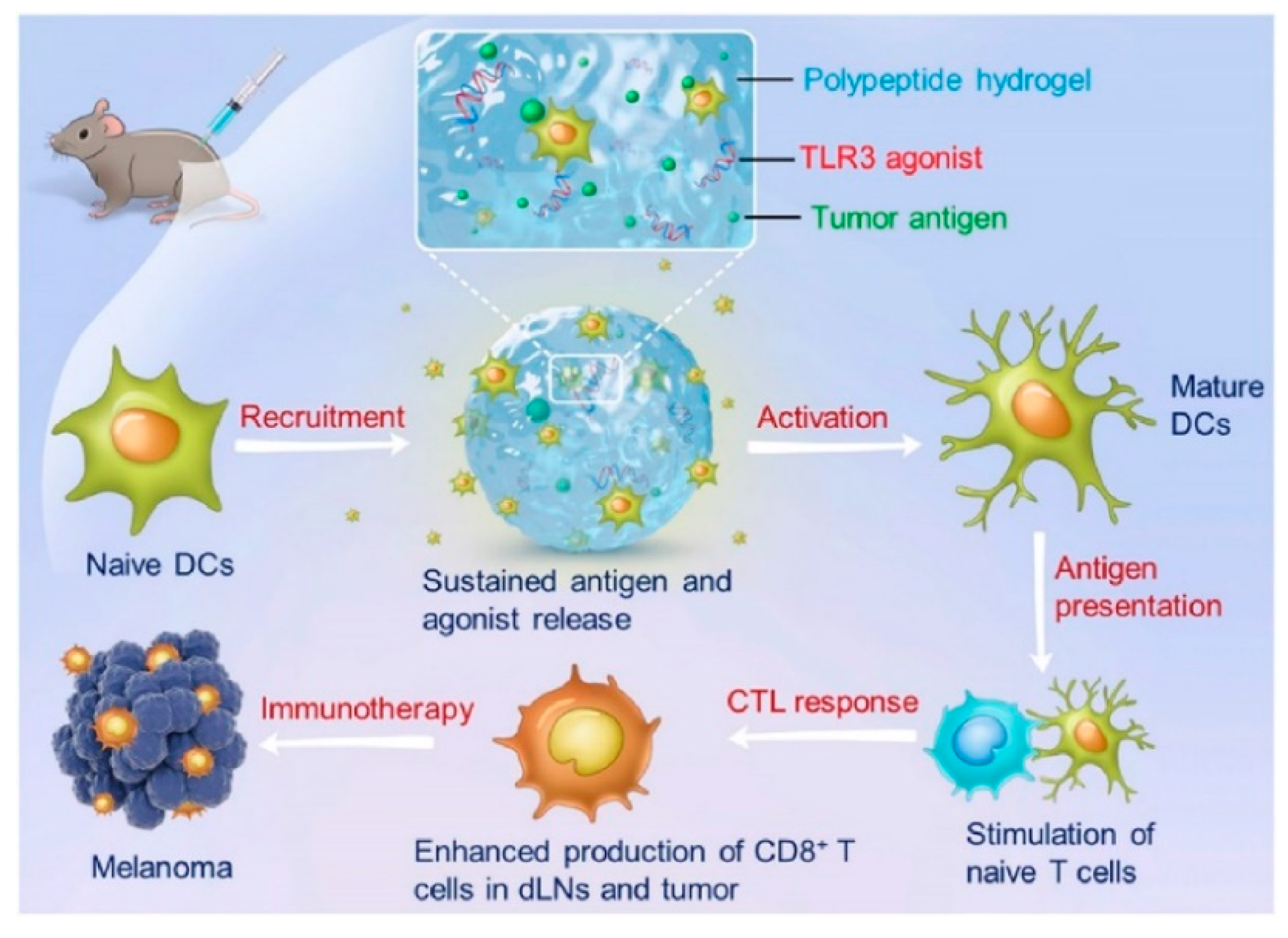
- Song, H.; Huang, P.; Niu, J.; Shi, G.; Zhang, C.; Kong, D.; Wang, W. Injectable polypeptide hydrogel for dual-delivery of antigen and TLR3 agonist to modulate dendritic cells in vivo and enhance potent cytotoxic T-lymphocyte response against melanoma. Biomaterials 2018, 159, 119–129. [Google Scholar] [CrossRef]
- Wei, L.; Chen, J.; Zhao, S.; Ding, J.; Chen, X. Thermo-sensitive polypeptide hydrogel for locally sequential delivery of two-pronged antitumor drugs. Acta. Biomater. 2017, 58, 44–53. [Google Scholar] [CrossRef]
- Cheng, Y.; He, C.; Ding, J.; Xiao, C.; Zhuang, X.; Chen, X. Thermosensitive hydrogels based on polypeptides for localized and sustained delivery of anticancer drugs. Biomaterials 2013, 34, 10338–10347. [Google Scholar] [CrossRef]
- Wu, C.; Li, R.; Yin, Y.; Wang, J.; Zhang, L.; Zhong, W. Redox-responsive supramolecular hydrogel based on 10-hydroxy camptothecin-peptide covalent conjugates with high loading capacity for drug delivery. Mater. Sci. Eng. C 2017, 76, 196–202. [Google Scholar] [CrossRef]
- Wang, H.; Lv, L.; Xu, G.; Yang, C.; Sun, J.; Yang, Z. Molecular hydrogelators consist of Taxol and short peptides/amino acids. J. Mater. Chem. 2012, 22, 16933–16938. [Google Scholar] [CrossRef]
- Singh, M.; Kundu, S.; Reddy, M.A.; Sreekanth, V.; Motiani, R.K.; Sengupta, S.; Srivastava, A.; Bajaj, A. Injectable small molecule hydrogel as a potential nanocarrier for localized and sustained in vivo delivery of doxorubicin. Nanoscale 2014, 6, 12849–12855. [Google Scholar] [CrossRef]
- Lin, Q.; Yang, Y.; Hu, Q.; Guo, Z.; Liu, T.; Xu, J.; Wu, J.; Kirk, T.B.; Ma, D.; Xue, W. Injectable supramolecular hydrogel formed from α-cyclodextrin and PEGylated arginine-functionalized poly(L-lysine) dendron for sustained MMP-9 shRNA plasmid delivery. Acta. Biomater. 2017, 49, 456–471. [Google Scholar] [CrossRef]
- Ma, Y.; Fu, X.; Shen, Y.; Fu, W.; Li, Z. Irreversible low critical solution temperature behaviors of thermal-responsive OEGylated poly(L-cysteine) containing disulfide bonds. Macromolecules 2014, 47, 4684–4689. [Google Scholar] [CrossRef]
- Zhang, S.; Fu, W.; Li, Z. Supramolecular hydrogels assembled from nonionic poly(ethylene glycol)-b-polypeptide diblocks containing OEGylated poly-l-glutamate. Polym. Chem. 2014, 5, 3346–3351. [Google Scholar] [CrossRef]
- Singh, S.K.; Singh, S.; Wlillard, J.; Singh, R. Drug delivery approaches for breast cancer. Int. J. Nanomed. 2017, 12, 6205–6218. [Google Scholar] [CrossRef] [PubMed]
- Kesharwani, P.; Gothwal, A.; Iyer, A.K.; Jain, K.; Chourasia, M.K.; Gupta, U. Dendrimer nanohybrid carrier systems: An expanding horizon for targeted drug and gene delivery. Drug Discov. Today 2018, 23, 300–314. [Google Scholar] [CrossRef] [PubMed]
- Kitchens, K.M.; El-Sayed, M.E.H.; Ghandehari, H. Transepithelial and endothelial transport of poly (amidoamine) dendrimers. Adv. Drug Deliv. Rev. 2005, 57, 2163–2176. [Google Scholar] [CrossRef] [PubMed]
- Northfelt, D.W.; Dezube, B.J.; Thommes, J.A.; Miller, B.J.; Fischl, M.A.; Friedman-Kien, A.; Kaplan, L.D.; Du Mond, C.; Mamelok, R.D.; Henry, D.H. Pegylated-liposomal doxorubicin versus doxorubicin, bleomycin, and vincristine in the treatment of AIDS-related Kaposi’s sarcoma: Results of a randomized phase III clinical trial. J. Clin. Oncol. 1998, 16, 2445–2451. [Google Scholar] [CrossRef] [PubMed]
- Lo, Y.W.; Sheu, M.T.; Chiang, W.H.; Chiu, Y.L.; Tu, C.M.; Wang, W.Y.; Wu, M.H.; Wang, Y.C.; Lu, M.; Ho, H.O. In situ chemically crosslinked injectable hydrogels for the subcutaneous delivery of trastuzumab to treat breast cancer. Acta. Biomater. 2019, 86, 280–290. [Google Scholar] [CrossRef]
- Zhang, H.; Zhao, C.; Cao, H.; Wang, G.; Song, L.; Niu, G.; Yang, H.; Ma, J.; Zhu, S. Hyperbranched poly(amine-ester) based hydrogels for controlled multi-drug release in combination chemotherapy. Biomaterials 2010, 31, 5445–5454. [Google Scholar] [CrossRef]
- Xu, L.; Cooper, R.C.; Wang, J.; Yeudall, W.A.; Yang, H. Synthesis and Application of Injectable Bioorthogonal Dendrimer Hydrogels for Local Drug Delivery. ACS Biomater. Sci. Eng. 2017, 3, 1641–1653. [Google Scholar] [CrossRef]
- Patil, S.S.; Shinde, V.S.; Misra, R.D.K. pH and reduction dual-stimuli-responsive PEGDA/PAMAM injectable network hydrogels via aza-michael addition for anticancer drug delivery. J. Polym. Sci. A Polym. Chem. 2018, 56, 2080–2095. [Google Scholar] [CrossRef] [Green Version]
- Yang, W.J.; Zhou, P.; Liang, L.; Cao, Y.; Qiao, J.; Li, X.; Teng, Z.; Wang, L. Nanogel-Incorporated Injectable Hydrogel for Synergistic Therapy Based on Sequential Local Delivery of Combretastatin-A4 Phosphate (CA4P) and Doxorubicin (DOX). ACS Appl. Mater. Interfaces 2018, 10, 18560–18573. [Google Scholar] [CrossRef] [PubMed]
- Kharkar, P.M.; Kloxin, A.M.; Kiick, K.L. Dually degradable click hydrogels for controlled degradation and protein release. J. Mater. Chem. B 2014, 2, 5511–5521. [Google Scholar] [CrossRef] [PubMed]
- Huang, Z.; Delparastan, P.; Burch, P.; Cheng, J.; Cao, Y.; Messersmith, P.B. Injectable dynamic covalent hydrogels of boronic acid polymers cross-linked by bioactive plant-derived polyphenols. Biomater. Sci. 2018, 6, 2487–2495. [Google Scholar] [CrossRef] [PubMed] [Green Version]
- Gao, W.; Liang, Y.; Peng, X.; Hu, Y.; Zhang, L.; Wu, H.; He, B. In situ injection of phenylboronic acid based low molecular weight gels for efficient chemotherapy. Biomaterials 2016, 105, 1–11. [Google Scholar] [CrossRef] [PubMed] [Green Version]
- Seib, F.P.; Tsurkan, M.; Freudenberg, U.; Kaplan, D.L.; Werner, C. Heparin-Modified Polyethylene Glycol Microparticle Aggregates for Focal Cancer Chemotherapy. ACS Biomater. Sci. Eng. 2016, 2, 2287–2293. [Google Scholar] [CrossRef] [Green Version]
- Fang, Y.; Xue, J.; Gao, S.; Lu, A.; Yang, D.; Jiang, H.; He, Y.; Shi, K. Cleavable PEGylation: A strategy for overcoming the “PEG dilemma” in efficient drug delivery. Drug Deliv. 2017, 24, 22–32. [Google Scholar] [CrossRef] [PubMed]
- Bastiancich, C.; Bianco, J.; Vanvarenberg, K.; Ucakar, B.; Joudiou, N.; Gallez, B.; Bastiat, G.; Lagarce, F.; Préat, V.; Danhier, F. Injectable nanomedicine hydrogel for local chemotherapy of glioblastoma after surgical resection. J. Control. Release 2017, 264, 45–54. [Google Scholar] [CrossRef] [PubMed]
- Ye, Y.; Hu, X. A pH-sensitive injectable nanoparticle composite hydrogel for anticancer drug delivery. J. Nanomater. 2016, 2016. [Google Scholar] [CrossRef]
- Makharza, S.; Cirillo, G.; Bachmatiuk, A.; Ibrahim, I.; Ioannides, N.; Trzebicka, B.; Hampel, S.; Ruemmeli, M.H. Graphene oxide-based drug delivery vehicles: Functionalization, characterization, and cytotoxicity evaluation. J. Nanoparticle Res. 2013, 15. [Google Scholar] [CrossRef]
- Vittorio, O.; Le Grand, M.; Makharza, S.A.; Curcio, M.; Tucci, P.; Iemma, F.; Nicoletta, F.P.; Hampel, S.; Cirillo, G. Doxorubicin synergism and resistance reversal in human neuroblastoma BE(2)C cell lines: An in vitro study with dextran-catechin nanohybrids. Eur. J. Pharm. Biopharm. 2018, 122, 176–185. [Google Scholar] [CrossRef]
- Lerra, L.; Farfalla, A.; Sanz, B.; Cirillo, G.; Vittorio, O.; Voli, F.; Grand, M.L.; Curcio, M.; Nicoletta, F.P.; Dubrovska, A.; et al. Graphene oxide functional nanohybrids with magnetic nanoparticles for improved vectorization of doxorubicin to neuroblastoma cells. Pharmaceutics 2019, 11. [Google Scholar] [CrossRef] [PubMed]
- Thambi, T.; Phan, V.H.G.; Lee, D.S. Stimuli-Sensitive Injectable Hydrogels Based on Polysaccharides and Their Biomedical Applications. Macromol. Rapid Commun. 2016, 37, 1881–1896. [Google Scholar] [CrossRef] [PubMed]
- Spizzirri, U.G.; Altimari, I.; Puoci, F.; Parisi, O.I.; Iemma, F.; Picci, N. Innovative antioxidant thermo-responsive hydrogels by radical grafting of catechin on inulin chain. Carbohydr. Polym. 2011, 84, 517–523. [Google Scholar] [CrossRef]
- Ahsan, S.M.; Thomas, M.; Reddy, K.K.; Sooraparaju, S.G.; Asthana, A.; Bhatnagar, I. Chitosan as biomaterial in drug delivery and tissue engineering. Int. J. Biol. Macromol. 2018, 110, 97–109. [Google Scholar] [CrossRef] [PubMed]
- Kozen, B.G.; Kircher, S.J.; Henao, J.; Godinez, F.S.; Johnson, A.S. An alternative hemostatic dressing: Comparison of CELOX, HemCon, and QuikClot. Acad. Emerg. Med. 2008, 15, 74–81. [Google Scholar] [CrossRef] [PubMed]
- Ueno, H.; Mori, T.; Fujinaga, T. Topical formulations and wound healing applications of chitosan. Adv. Drug Deliv. Rev. 2001, 52, 105–115. [Google Scholar] [CrossRef]
- Karimi, A.R.; Khodadadi, A.; Hadizadeh, M. A nanoporous photosensitizing hydrogel based on chitosan cross-linked by zinc phthalocyanine: An injectable and pH-stimuli responsive system for effective cancer therapy. RSC Adv. 2016, 6, 91445–91452. [Google Scholar] [CrossRef]
- Abdel-Bar, H.M.; Abdel-Reheem, A.Y.; Osman, R.; Awad, G.A.S.; Mortada, N. Defining cisplatin incorporation properties in thermosensitive injectable biodegradable hydrogel for sustained delivery and enhanced cytotoxicity. Int. J. Pharm. 2014, 477, 623–630. [Google Scholar] [CrossRef]
- Fathi, M.; Alami-Milani, M.; Geranmayeh, M.H.; Barar, J.; Erfan-Niya, H.; Omidi, Y. Dual thermo-and pH-sensitive injectable hydrogels of chitosan/(poly(N-isopropylacrylamide-co-itaconic acid)) for doxorubicin delivery in breast cancer. Int. J. Biol. Macromol. 2019, 128, 957–964. [Google Scholar] [CrossRef]
- Zhang, W.; Jin, X.; Li, H.; Zhang, R.R.; Wu, C.W. Injectable and body temperature sensitive hydrogels based on chitosan and hyaluronic acid for pH sensitive drug release. Carbohydr. Polym. 2018, 186, 82–90. [Google Scholar] [CrossRef]
- Saeednia, L.; Yao, L.; Cluff, K.; Asmatulu, R. Sustained Releasing of Methotrexate from Injectable and Thermosensitive Chitosan-Carbon Nanotube Hybrid Hydrogels Effectively Controls Tumor Cell Growth. ACS Omega 2019, 4, 4040–4048. [Google Scholar] [CrossRef] [PubMed]
- Huang, F.Y.J.; Hung, C.C.; Chang, C.W.; Chao, J.H.; Hsieh, B.T. Evaluation of injectable chitosan-based co-crosslinking hydrogel for local delivery of 188Re-LIPO-DOX to breast-tumor-bearing mouse model. Anticancer. Res. 2018, 38, 4651–4659. [Google Scholar] [CrossRef] [PubMed]
- Alexander, A.; Ajazuddin, A.; Khan, J.; Saraf, S. Formulation and evaluation of chitosan-based long-acting injectable hydrogel for PEGylated melphalan conjugate. J. Pharm. Pharmacol. 2014, 66, 1240–1250. [Google Scholar] [CrossRef] [PubMed]
- López-Noriega, A.; Hastings, C.L.; Ozbakir, B.; O’Donnell, K.E.; O’Brien, F.J.; Storm, G.; Hennink, W.E.; Duffy, G.P.; Ruiz-Hernández, E. Hyperthermia-Induced Drug Delivery from Thermosensitive Liposomes Encapsulated in an Injectable Hydrogel for Local Chemotherapy. Adv. Healthc. Mater. 2014, 3, 854–859. [Google Scholar] [CrossRef] [PubMed]
- Xing, J.; Qi, X.; Jiang, Y.; Zhu, X.; Zhang, Z.; Qin, X.; Wu, Z. Topotecan hydrochloride liposomes incorporated into thermosensitive hydrogel for sustained and efficient in situ therapy of H22 tumor in Kunming mice. Pharm. Dev. Technol. 2015, 20, 812–819. [Google Scholar] [CrossRef] [PubMed]
- Huang, F.Y.J.; Gan, G.Y.; Lin, W.Y.; Huang, L.K.; Luo, T.Y.; Hong, J.J.; Hsieh, B.T. Investigation of the local delivery of an intelligent chitosan-based 188Re thermosensitive in situ-forming hydrogel in an orthotopic hepatoma-bearing rat model. J. Radioanal. Nucl. Chem. 2014, 299, 31–40. [Google Scholar] [CrossRef]
- Zhang, D.; Sun, P.; Li, P.; Xue, A.; Zhang, X.; Zhang, H.; Jin, X. A magnetic chitosan hydrogel for sustained and prolonged delivery of Bacillus Calmette-Guérin in the treatment of bladder cancer. Biomaterials 2013, 34, 10258–10266. [Google Scholar] [CrossRef] [PubMed]
- Le Renard, P.E.; Jordan, O.; Faes, A.; Petri-Fink, A.; Hofmann, H.; Rüfenacht, D.; Bosman, F.; Buchegger, F.; Doelker, E. The in vivo performance of magnetic particle-loaded injectable, in situ gelling, carriers for the delivery of local hyperthermia. Biomaterials 2010, 31, 691–705. [Google Scholar] [CrossRef]
- Zhu, X.; Zhang, H.; Huang, H.; Zhang, Y.; Hou, L.; Zhang, Z. Functionalized graphene oxide-based thermosensitive hydrogel for magnetic hyperthermia therapy on tumors. Nanotechnology 2015, 26. [Google Scholar] [CrossRef] [PubMed]
- Saeednia, L.; Yao, L.; Berndt, M.; Cluff, K.; Asmatulu, R. Structural and biological properties of thermosensitive chitosan–graphene hybrid hydrogels for sustained drug delivery applications. J. Biomed. Mater. Res. A 2017, 105, 2381–2390. [Google Scholar] [CrossRef]
- Fletcher, N.A.; Krebs, M.D. Sustained delivery of anti-VEGF from injectable hydrogel systems provides a prolonged decrease of endothelial cell proliferation and angiogenesis: In vitro. RSC Adv. 2018, 8, 8999–9005. [Google Scholar] [CrossRef]
- Chen, C.H.; Kuo, C.Y.; Chen, S.H.; Mao, S.H.; Chang, C.Y.; Shalumon, K.T.; Chen, J.P. Thermosensitive injectable hydrogel for simultaneous intraperitoneal delivery of doxorubicin and prevention of peritoneal adhesion. Int. J. Mol. Sci. 2018, 19. [Google Scholar] [CrossRef] [PubMed]
- Fong, Y.T.; Chen, C.H.; Chen, J.P. Intratumoral delivery of doxorubicin on folate-conjugated graphene oxide by in-situ forming thermo-sensitive hydrogel for breast cancer therapy. Nanomaterials 2017, 7. [Google Scholar] [CrossRef] [PubMed]
- Hyun, H.; Park, M.H.; Lim, W.; Kim, S.Y.; Jo, D.; Jung, J.S.; Jo, G.; Um, S.; Lee, D.W.; Yang, D.H. Injectable visible light-cured glycol chitosan hydrogels with controlled release of anticancer drugs for local cancer therapy in vivo: A feasible study. Artif. Cells Nanomed. Biotechnol. 2018, 46, 874–882. [Google Scholar] [CrossRef] [PubMed]
- Zhou, X.; Li, Y.; Chen, S.; Fu, Y.N.; Wang, S.; Li, G.; Tao, L.; Wei, Y.; Wang, X.; Liang, J.F. Dynamic agent of an injectable and self-healing drug-loaded hydrogel for embolization therapy. Colloids Surf. B Biointerfaces 2018, 172, 601–607. [Google Scholar] [CrossRef] [PubMed]
- Xia, L.Y.; Zhang, X.; Cao, M.; Chen, Z.; Wu, F.G. Enhanced Fluorescence Emission and Singlet Oxygen Generation of Photosensitizers Embedded in Injectable Hydrogels for Imaging-Guided Photodynamic Cancer Therapy. Biomacromolecules 2017, 18, 3073–3081. [Google Scholar] [CrossRef]
- Xie, W.; Gao, Q.; Guo, Z.; Wang, D.; Gao, F.; Wang, X.; Wei, Y.; Zhao, L. Injectable and self-healing thermosensitive magnetic hydrogel for asynchronous control release of doxorubicin and docetaxel to treat triple-negative breast cancer. ACS Appl. Mater. Interfaces 2017, 9, 33660–33673. [Google Scholar] [CrossRef] [PubMed]
- Liu, Z.; Xu, G.; Wang, C.; Li, C.; Yao, P. Shear-responsive injectable supramolecular hydrogel releasing doxorubicin loaded micelles with pH-sensitivity for local tumor chemotherapy. Int. J. Pharm. 2017, 530, 53–62. [Google Scholar] [CrossRef]
- Wang, Q.Q.; Kong, M.; An, Y.; Liu, Y.; Li, J.J.; Zhou, X.; Feng, C.; Li, J.; Jiang, S.Y.; Cheng, X.J.; et al. Hydroxybutyl chitosan thermo-sensitive hydrogel: A potential drug delivery system. J. Mater. Sci. 2013, 48, 5614–5623. [Google Scholar] [CrossRef]
- Khan, S.; Akhtar, N.; Minhas, M.U.; Badshah, S.F. pH/Thermo-Dual Responsive Tunable In Situ Cross-Linkable Depot Injectable Hydrogels Based on Poly(N-Isopropylacrylamide)/Carboxymethyl Chitosan with Potential of Controlled Localized and Systemic Drug Delivery. AAPS Pharm. Sci. Tech. 2019, 20. [Google Scholar] [CrossRef]
- Wang, H.; Song, F.; Chen, Q.; Hu, R.; Jiang, Z.; Yang, Y.; Han, B. Antitumor and antimetastasis effects of macerating solutions from an injectable chitosan-based hydrogel on hepatocarcinoma. J. Biomed. Mater. Res.-A 2015, 103, 3879–3885. [Google Scholar] [CrossRef] [PubMed]
- Qu, J.; Zhao, X.; Ma, P.X.; Guo, B. pH-responsive self-healing injectable hydrogel based on N-carboxyethyl chitosan for hepatocellular carcinoma therapy. Acta. Biomater. 2017, 58, 168–180. [Google Scholar] [CrossRef] [PubMed]
- Chen, X.; Fan, M.; Tan, H.; Ren, B.; Yuan, G.; Jia, Y.; Li, J.; Xiong, D.; Xing, X.; Niu, X.; et al. Magnetic and self-healing chitosan-alginate hydrogel encapsulated gelatin microspheres via covalent cross-linking for drug delivery. Mater. Sci. Eng. C 2019, 101, 619–629. [Google Scholar] [CrossRef] [PubMed]
- Qian, C.; Zhang, T.; Gravesande, J.; Baysah, C.; Song, X.; Xing, J. Injectable and self-healing polysaccharide-based hydrogel for pH-responsive drug release. Int. J. Biol. Macromol. 2019, 123, 140–148. [Google Scholar] [CrossRef] [PubMed]
- Gao, N.; Lü, S.; Gao, C.; Wang, X.; Xu, X.; Bai, X.; Feng, C.; Liu, M. Injectable shell-crosslinked F127 micelle/hydrogel composites with pH and redox sensitivity for combined release of anticancer drugs. Chem. Eng. J. 2016, 287, 20–29. [Google Scholar] [CrossRef]
- Li, J.; Hu, W.; Zhang, Y.; Tan, H.; Yan, X.; Zhao, L.; Liang, H. PH and glucose dually responsive injectable hydrogel prepared by in situ crosslinking of phenylboronic modified chitosan and oxidized dextran. J. Polym. Sci. A Polym. Chem. 2015, 53, 1235–1244. [Google Scholar] [CrossRef]
- Liang, Y.; Zhao, X.; Ma, P.X.; Guo, B.; Du, Y.; Han, X. pH-responsive injectable hydrogels with mucosal adhesiveness based on chitosan-grafted-dihydrocaffeic acid and oxidized pullulan for localized drug delivery. J. Colloid Interface Sci. 2019, 536, 224–234. [Google Scholar] [CrossRef] [PubMed]
- Jalalvandi, E.; Shavandi, A. In situ-forming and pH-responsive hydrogel based on chitosan for vaginal delivery of therapeutic agents. J. Mater. Sci. Mater. Med. 2018, 29. [Google Scholar] [CrossRef] [PubMed]
- Shi, J.; Guobao, W.; Chen, H.; Zhong, W.; Qiu, X.; Xing, M.M.Q. Schiff based injectable hydrogel for in situ pH-triggered delivery of doxorubicin for breast tumor treatment. Polym. Chem. 2014, 5, 6180–6189. [Google Scholar] [CrossRef]
- Zahedi, P.; De Souza, R.; Piquette-Miller, M.; Allen, C. Docetaxel distribution following intraperitoneal administration in mice. Journal of pharmacy & pharmaceutical sciences: A publication of the Canadian Society for Pharmaceutical Sciences. J. Pharm. Pharm. Sci. 2011, 14, 90–99. [Google Scholar]
- Yavvari, P.S.; Pal, S.; Kumar, S.; Kar, A.; Awasthi, A.K.; Naaz, A.; Srivastava, A.; Bajaj, A. Injectable, Self-Healing Chimeric Catechol-Fe(III) Hydrogel for Localized Combination Cancer Therapy. ACS Biomater. Sci. Eng. 2017, 3, 3404–3413. [Google Scholar] [CrossRef]
- Belali, S.; Karimi, A.R.; Hadizadeh, M. Cell-specific and pH-sensitive nanostructure hydrogel based on chitosan as a photosensitizer carrier for selective photodynamic therapy. Int. J. Biol. Macromol. 2018, 110, 437–448. [Google Scholar] [CrossRef] [PubMed]
- Ning, P.; Lü, S.; Bai, X.; Wu, X.; Gao, C.; Wen, N.; Liu, M. High encapsulation and localized delivery of curcumin from an injectable hydrogel. Mater. Sci. Eng. C 2018, 83, 121–129. [Google Scholar] [CrossRef] [PubMed]
- Burdick, J.A. Injectable gels for tissue/organ repair. Biomed. Mater. 2012, 7. [Google Scholar] [CrossRef] [PubMed]
- Seliktar, D. Designing cell-compatible hydrogels for biomedical applications. Science 2012, 336, 1124–1128. [Google Scholar] [CrossRef] [PubMed]
- Mitragotri, S.; Burke, P.A.; Langer, R. Overcoming the challenges in administering biopharmaceuticals: Formulation and delivery strategies. Nat. Rev. Drug Discov. 2014, 13, 655–672. [Google Scholar] [CrossRef] [PubMed]
- Burdick, J.A.; Prestwich, G.D. Hyaluronic acid hydrogels for biomedical applications. Adv. Mater. 2011, 23, H41–H56. [Google Scholar] [CrossRef]
- Chen, Y.Y.; Wu, H.C.; Sun, J.S.; Dong, G.C.; Wang, T.W. Injectable and thermoresponsive self-assembled nanocomposite hydrogel for long-term anticancer drug delivery. Langmuir 2013, 29, 3721–3729. [Google Scholar] [CrossRef]
- Zhao, Y.; Yan, H.; Qiao, S.; Zhang, L.; Wang, T.; Meng, Q.; Chen, X.; Lin, F.H.; Guo, K.; Li, C.; et al. Hydrogels bearing bioengineered mimetic embryonic microenvironments for tumor reversion. J. Mater. Chem. B 2016, 4, 6183–6191. [Google Scholar] [CrossRef]
- Ohta, S.; Hiramoto, S.; Amano, Y.; Emoto, S.; Yamaguchi, H.; Ishigami, H.; Kitayama, J.; Ito, T. Intraperitoneal Delivery of Cisplatin via a Hyaluronan-Based Nanogel/in Situ Cross-Linkable Hydrogel Hybrid System for Peritoneal Dissemination of Gastric Cancer. Mol. Pharm. 2017, 14, 3105–3113. [Google Scholar] [CrossRef]
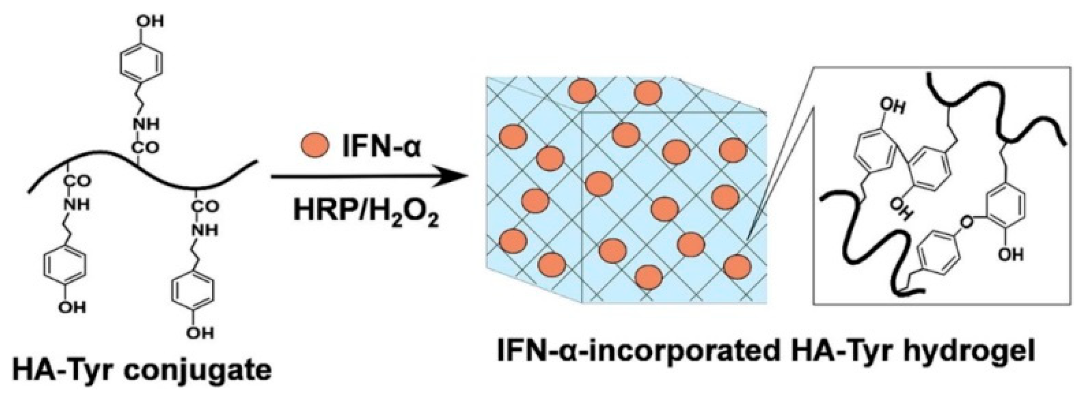
- Xu, K.; Lee, F.; Gao, S.J.; Chung, J.E.; Yano, H.; Kurisawa, M. Injectable hyaluronic acid-tyramine hydrogels incorporating interferon-α2a for liver cancer therapy. J. Control. Release 2013, 166, 203–210. [Google Scholar] [CrossRef] [PubMed]
- Xu, K.; Lee, F.; Gao, S.; Tan, M.H.; Kurisawa, M. Hyaluronidase-incorporated hyaluronic acid-tyramine hydrogels for the sustained release of trastuzumab. J. Control. Release 2015, 216, 47–55. [Google Scholar] [CrossRef] [PubMed]
- Ueda, K.; Akiba, J.; Ogasawara, S.; Todoroki, K.; Nakayama, M.; Sumi, A.; Kusano, H.; Sanada, S.; Suekane, S.; Xu, K.; et al. Growth inhibitory effect of an injectable hyaluronic acid-tyramine hydrogels incorporating human natural interferon-α and sorafenib on renal cell carcinoma cells. Acta. Biomater. 2016, 29, 103–111. [Google Scholar] [CrossRef] [PubMed]
- He, M.; Sui, J.; Chen, Y.; Bian, S.; Cui, Y.; Zhou, C.; Sun, Y.; Liang, J.; Fan, Y.; Zhang, X. Localized multidrug co-delivery by injectable self-crosslinking hydrogel for synergistic combinational chemotherapy. J. Mater. Chem. B 2017, 5, 4852–4862. [Google Scholar] [CrossRef]
- Chen, X.; Liu, Z. A pH-Responsive Hydrogel Based on a Tumor-Targeting Mesoporous Silica Nanocomposite for Sustained Cancer Labeling and Therapy. Macromol. Rapid Commun. 2016, 37, 1533–1539. [Google Scholar] [CrossRef] [PubMed]
- Chen, X.; Liu, Z.; Parker, S.G.; Zhang, X.; Gooding, J.J.; Ru, Y.; Liu, Y.; Zhou, Y. Light-Induced Hydrogel Based on Tumor-Targeting Mesoporous Silica Nanoparticles as a Theranostic Platform for Sustained Cancer Treatment. ACS Appl. Mater. Interfaces 2016, 8, 15857–15863. [Google Scholar] [CrossRef] [PubMed]
- Ranga, A.; Lutolf, M.P.; Hilborn, J.; Ossipov, D.A. Hyaluronic Acid Hydrogels Formed in Situ by Transglutaminase-Catalyzed Reaction. Biomacromolecules 2016, 17, 1553–1560. [Google Scholar] [CrossRef]
- Moon, R.J.; Martini, A.; Nairn, J.; Simonsen, J.; Youngblood, J. Cellulose nanomaterials review: Structure, properties and nanocomposites. Chem. Soc. Rev. 2011, 40, 3941–3994. [Google Scholar] [CrossRef]
- Ngwabebhoh, F.A.; Yildiz, U. Nature-derived fibrous nanomaterial toward biomedicine and environmental remediation: Today’s state and future prospects. J. Appl. Polym. Sci. 2019, 136. [Google Scholar] [CrossRef]
- You, J.; Cao, J.; Zhao, Y.; Zhang, L.; Zhou, J.; Chen, Y. Improved Mechanical Properties and Sustained Release Behavior of Cationic Cellulose Nanocrystals Reinforeced Cationic Cellulose Injectable Hydrogels. Biomacromolecules 2016, 17, 2839–2848. [Google Scholar] [CrossRef]
- Ding, L.; Wang, Q.; Shen, M.; Sun, Y.; Zhang, X.; Huang, C.; Chen, J.; Li, R.; Duan, Y. Thermoresponsive nanocomposite gel for local drug delivery to suppress the growth of glioma by inducing autophagy. Autophagy 2017, 13, 1176–1190. [Google Scholar] [CrossRef] [PubMed]
- Xing, C.; Chen, S.; Qiu, M.; Liang, X.; Liu, Q.; Zou, Q.; Li, Z.; Xie, Z.; Wang, D.; Dong, B.; et al. Conceptually Novel Black Phosphorus/Cellulose Hydrogels as Promising Photothermal Agents for Effective Cancer Therapy. Adv. Healthc. Mater. 2018, 7. [Google Scholar] [CrossRef] [PubMed]
- Yang, J.S.; Xie, Y.J.; He, W. Research progress on chemical modification of alginate: A review. Carbohydr. Polym. 2011, 84, 33–39. [Google Scholar] [CrossRef]
- Wróblewska-Krepsztul, J.; Rydzkowski, T.; Michalska-Pożoga, I.; Thakur, V.K. Biopolymers for biomedical and pharmaceutical applications: Recent advances and overview of alginate electrospinning. Nanomaterials 2019, 9. [Google Scholar] [CrossRef] [PubMed]
- Wang, C.; Wang, X.; Dong, K.; Luo, J.; Zhang, Q.; Cheng, Y. Injectable and responsively degradable hydrogel for personalized photothermal therapy. Biomaterials 2016, 104, 129–137. [Google Scholar] [CrossRef] [PubMed]
- Chalanqui, M.J.; Pentlavalli, S.; McCrudden, C.; Chambers, P.; Ziminska, M.; Dunne, N.; McCarthy, H.O. Influence of alginate backbone on efficacy of thermo-responsive alginate-g-P(NIPAAm) hydrogel as a vehicle for sustained and controlled gene delivery. Mater. Sci. Eng. C 2019, 95, 409–421. [Google Scholar] [CrossRef] [PubMed]
- Liu, M.; Song, X.; Wen, Y.; Zhu, J.L.; Li, J. Injectable Thermoresponsive Hydrogel Formed by Alginate-g-Poly(N-isopropylacrylamide) That Releases Doxorubicin-Encapsulated Micelles as a Smart Drug Delivery System. ACS Appl. Mater. Interfaces 2017, 9, 35673–35682. [Google Scholar] [CrossRef] [PubMed]
- Davoodi, P.; Ng, W.C.; Srinivasan, M.P.; Wang, C.H. Codelivery of anti-cancer agents via double-walled polymeric microparticles/injectable hydrogel: A promising approach for treatment of triple negative breast cancer. Biotechnol. Bioeng. 2017, 114, 2931–2946. [Google Scholar] [CrossRef]
- Davoodi, P.; Ng, W.C.; Yan, W.C.; Srinivasan, M.P.; Wang, C.H. Double-walled microparticles-embedded self-cross-linked, injectable, and antibacterial hydrogel for controlled and sustained release of chemotherapeutic agents. ACS Appl. Mater. Interfaces 2016, 8, 22785–22800. [Google Scholar] [CrossRef]
- Liu, J.; Qi, C.; Tao, K.; Zhang, J.; Xu, L.; Jiang, X.; Zhang, Y.; Huang, L.; Li, Q.; Xie, H.; et al. Sericin/Dextran Injectable Hydrogel as an Optically Trackable Drug Delivery System for Malignant Melanoma Treatment. ACS Appl. Mater. Interfaces 2016, 8, 6411–6422. [Google Scholar] [CrossRef]
- Li, L.; Wang, C.; Huang, Q.; Xiao, J.; Zhang, Q.; Cheng, Y. A degradable hydrogel formed by dendrimer-encapsulated platinum nanoparticles and oxidized dextran for repeated photothermal cancer therapy. J. Mater. Chem. B 2018, 6, 2474–2480. [Google Scholar] [CrossRef]
- Deng, J.; Xun, X.; Zheng, W.; Su, Y.; Zheng, L.; Wang, C.; Su, M. Sequential delivery of bismuth nanoparticles and doxorubicin by injectable macroporous hydrogels for combined anticancer kilovoltage X-ray radio- and chemo-therapy. J. Mater. Chem. B 2018, 6, 7966–7973. [Google Scholar] [CrossRef]
- GuhaSarkar, S.; More, P.; Banerjee, R. Urothelium-adherent, ion-triggered liposome-in-gel system as a platform for intravesical drug delivery. J. Control. Release 2017, 245, 147–156. [Google Scholar] [CrossRef] [PubMed]
- Zheng, Y.; Liang, Y.; Zhang, D.; Zhou, Z.; Li, J.; Sun, X.; Liu, Y.N. Fabrication of injectable CuS nanocomposite hydrogels based on UCST-type polysaccharides for NIR-triggered chemo-photothermal therapy. Chem. Commun. 2018, 54, 13805–13808. [Google Scholar] [CrossRef] [PubMed]
- Hou, M.; Yang, R.; Zhang, L.; Liu, G.; Xu, Z.; Kang, Y.; Xue, P. Injectable and Natural Humic Acid/Agarose Hybrid Hydrogel for Localized Light-Driven Photothermal Ablation and Chemotherapy of Cancer. ACS Biomater. Sci. Eng. 2018, 4, 4266–4277. [Google Scholar] [CrossRef]
- Niu, X.; Zhang, Z.; Zhong, Y. Hydrogel loaded with self-assembled dextran sulfate-doxorubicin complexes as a delivery system for chemotherapy. Mater. Sci. Eng. C 2017, 77, 888–894. [Google Scholar] [CrossRef]
- Nguyen, K.; Dang, P.N.; Alsberg, E. Functionalized, biodegradable hydrogels for control over sustained and localized siRNA delivery to incorporated and surrounding cells. Acta. Biomater. 2013, 9, 4487–4495. [Google Scholar] [CrossRef] [PubMed]
- Vittorio, O.; Cirillo, G.; Iemma, F.; Di Turi, G.; Jacchetti, E.; Curcio, M.; Barbuti, S.; Funel, N.; Parisi, O.I.; Puoci, F.; et al. Dextran-catechin conjugate: A potential treatment against the pancreatic ductal adenocarcinoma. Pharm. Res. 2012, 29, 2601–2614. [Google Scholar] [CrossRef]
- Vittorio, O.; Brandl, M.; Cirillo, G.; Kimpton, K.; Hinde, E.; Gaus, K.; Yee, E.; Kumar, N.; Duong, H.; Fleming, C.; et al. Dextran-Catechin: An anticancer chemically-modified natural compound targeting copper that attenuates neuroblastoma growth. Oncotarget 2016, 7, 47479–47493. [Google Scholar] [CrossRef] [Green Version]
- Agarwal, A.; Gupta, U.; Asthana, A.; Jain, N.K. Dextran conjugated dendritic nanoconstructs as potential vectors for anti-cancer agent. Biomaterials 2009, 30, 3588–3596. [Google Scholar] [CrossRef]
- Abdo Qasem, A.A.; Alamri, M.S.; Mohamed, A.A.; Hussain, S.; Mahmood, K.; Ibraheem, M.A. High Soluble-Fiber Pudding: Formulation, Processing, Texture and Sensory Properties. J. Food Process. Preserv. 2017, 41. [Google Scholar] [CrossRef]
- Carlini, A.S.; Gaetani, R.; Braden, R.L.; Luo, C.; Christman, K.L.; Gianneschi, N.C. Enzyme-responsive progelator cyclic peptides for minimally invasive delivery to the heart post-myocardial infarction. Nat. Commun. 2019, 10. [Google Scholar] [CrossRef] [PubMed]
- Haines-Butterick, L.; Rajagopal, K.; Branco, M.; Salick, D.; Rughani, R.; Pilarz, M.; Lamm, M.S.; Pochan, D.J.; Schneider, J.P. Controlling hydrogelation kinetics by peptide design for three-dimensional encapsulation and injectable delivery of cells. Proc. Natl. Acad. Sci. USA 2007, 104, 7791–7796. [Google Scholar] [CrossRef] [PubMed] [Green Version]
- Mano, J.F. Stimuli-responsive polymeric systems for biomedical applications. Adv. Eng. Mater. 2008, 10, 515–527. [Google Scholar] [CrossRef]
- Xing, R.; Liu, K.; Jiao, T.; Zhang, N.; Ma, K.; Zhang, R.; Zou, Q.; Ma, G.; Yan, X. An Injectable Self-Assembling Collagen-Gold Hybrid Hydrogel for Combinatorial Antitumor Photothermal/Photodynamic Therapy. Adv. Mater. 2016, 28, 3669–3676. [Google Scholar] [CrossRef] [PubMed]
- Upadhyay, A.; Kandi, R.; Rao, C.P. Injectable, Self-Healing, and Stress Sustainable Hydrogel of BSA as a Functional Biocompatible Material for Controlled Drug Delivery in Cancer Cells. ACS Sustain. Chem. Eng. 2018, 6, 3321–3330. [Google Scholar] [CrossRef]
- Kim, I.; Choi, J.S.; Lee, S.; Byeon, H.J.; Lee, E.S.; Shin, B.S.; Choi, H.G.; Lee, K.C.; Youn, Y.S. In situ facile-forming PEG cross-linked albumin hydrogels loaded with an apoptotic TRAIL protein. J. Control. Release 2015, 214, 30–39. [Google Scholar] [CrossRef]
- Qian, H.Q.; Qian, K.Y.; Cai, J.; Yang, Y.; Zhu, L.J.; Liu, B.R. Therapy for Gastric Cancer with Peritoneal Metastasis Using Injectable Albumin Hydrogel Hybridized with Paclitaxel-Loaded Red Blood Cell Membrane Nanoparticles. ACS Biomater. Sci. Eng. 2019, 5, 1100–1112. [Google Scholar] [CrossRef]
- Curcio, M.; Altimari, I.; Spizzirri, U.G.; Cirillo, G.; Vittorio, O.; Puoci, F.; Picci, N.; Iemma, F. Biodegradable gelatin-based nanospheres as pH-responsive drug delivery systems. J. Nanoparticle Res. 2013, 15. [Google Scholar] [CrossRef]
- Curcio, M.; Spizzirri, U.G.; Iemma, F.; Puoci, F.; Cirillo, G.; Parisi, O.I.; Picci, N. Grafted thermo-responsive gelatin microspheres as delivery systems in triggered drug release. Eur. J. Pharm. Biopharm. 2010, 76, 48–55. [Google Scholar] [CrossRef]
- Oh, E.; Oh, J.E.; Hong, J.; Chung, Y.; Lee, Y.; Park, K.D.; Kim, S.; Yun, C.O. Optimized biodegradable polymeric reservoir-mediated local and sustained co-delivery of dendritic cells and oncolytic adenovirus co-expressing IL-12 and GM-CSF for cancer immunotherapy. J. Control. Release 2017, 259, 115–127. [Google Scholar] [CrossRef] [PubMed]
- Takei, T.; Sugihara, K.; Yoshida, M.; Kawakami, K. Injectable and biodegradable sugar beet pectin/gelatin hydrogels for biomedical applications. J. Biomater. Sci. Polym. Ed. 2013, 24, 1333–1342. [Google Scholar] [CrossRef] [PubMed]
- Ciobanu, B.C.; Cadinoiu, A.N.; Popa, M.; Desbrières, J.; Peptu, C.A. Modulated release from liposomes entrapped in chitosan/gelatin hydrogels. Mater. Sci. Eng. C 2014, 43, 383–391. [Google Scholar] [CrossRef] [PubMed]
- Franke, K.; Baur, M.; Daum, L.; Vaegler, M.; Sievert, K.D.; Schlosshauer, B. Prostate carcinoma cell growth-inhibiting hydrogel supports axonal regeneration in vitro. Neurosci. Lett. 2013, 541, 248–252. [Google Scholar] [CrossRef] [PubMed]
- Cirillo, G.; Vittorio, O.; Hampel, S.; Spizzirri, U.G.; Picci, N.; Iemma, F. Incorporation of carbon nanotubes into a gelatin-catechin conjugate: Innovative approach for the preparation of anticancer materials. Int. J. Pharm. 2013, 446, 176–182. [Google Scholar] [CrossRef] [PubMed]
- Zhou, M.; Liu, S.; Jiang, Y.; Ma, H.; Shi, M.; Wang, Q.; Zhong, W.; Liao, W.; Xing, M.M.Q. Doxorubicin-Loaded Single Wall Nanotube Thermo-Sensitive Hydrogel for Gastric Cancer Chemo-Photothermal Therapy. Adv. Funct. Mater. 2015, 25, 4730–4739. [Google Scholar] [CrossRef]
- Cirillo, G.; Hampel, S.; Spizzirri, U.G.; Parisi, O.I.; Picci, N.; Iemma, F. Carbon Nanotubes Hybrid Hydrogels in Drug Delivery: A Perspective Review. Biomed. Res. Int. 2014. [Google Scholar] [CrossRef]
- Cirillo, G.; Caruso, T.; Hampel, S.; Haase, D.; Puoci, F.; Ritschel, M.; Leonhardt, A.; Curcio, M.; Iemma, F.; Khavrus, V.; et al. Novel carbon nanotube composites by grafting reaction with water-compatible redox initiator system. Colloid Polym. Sci. 2013, 291, 699–708. [Google Scholar] [CrossRef]
- He, G.; Chen, S.; Xu, Y.J.; Miao, Z.H.; Ma, Y.; Qian, H.S.; Lu, Y.; Zha, Z.B. Charge reversal induced colloidal hydrogel acts as a multi-stimuli responsive drug delivery platform for synergistic cancer therapy. Mater. Horiz. 2019, 6, 711–716. [Google Scholar] [CrossRef]
- Maitz, M.F.; Sperling, C.; Wongpinyochit, T.; Herklotz, M.; Werner, C.; Seib, F.P. Biocompatibility assessment of silk nanoparticles: Hemocompatibility and internalization by human blood cells. Nanomedicine 2017, 13, 2633–2642. [Google Scholar] [CrossRef]
- Omenetto, F.G.; Kaplan, D.L. New opportunities for an ancient material. Science 2010, 329, 528–531. [Google Scholar] [CrossRef] [PubMed]
- Seib, F.P.; Pritchard, E.M.; Kaplan, D.L. Self-assembling doxorubicin silk hydrogels for the focal treatment of primary breast cancer. Adv. Funct. Mater. 2013, 23, 58–65. [Google Scholar] [CrossRef] [PubMed]
- Wu, P.; Liu, Q.; Wang, Q.; Qian, H.; Yu, L.; Liu, B.; Li, R. Novel silk fibroin nanoparticles incorporated silk fibroin hydrogel for inhibition of cancer stem cells and tumor growth. Int. J. Nanomed. 2018, 13, 5405–5418. [Google Scholar] [CrossRef] [PubMed]
- Wu, H.; Liu, S.; Xiao, L.; Dong, X.; Lu, Q.; Kaplan, D.L. Injectable and pH-Responsive Silk Nanofiber Hydrogels for Sustained Anticancer Drug Delivery. ACS Appl. Mater. Interfaces 2016, 8, 17118–17126. [Google Scholar] [CrossRef] [PubMed]
- He, W.; Li, P.; Zhu, Y.; Liu, M.; Huang, X.; Qi, H. An injectable silk fibroin nanofiber hydrogel hybrid system for tumor upconversion luminescence imaging and photothermal therapy. New J. Chem. 2019, 43, 2213–2219. [Google Scholar] [CrossRef]
- Ribeiro, V.P.; Silva-Correia, J.; Goncalves, C.; Pina, S.; Radhouani, H.; Montonen, T.; Hyttinen, J.; Roy, A.; Oliveira, A.L.; Reis, R.L.; et al. Rapidly responsive silk fibroin hydrogels as an artificial matrix for the programmed tumor cells death. PLoS ONE 2018, 13. [Google Scholar] [CrossRef] [PubMed]
- Schaal, J.L.; Li, X.; Mastria, E.; Bhattacharyya, J.; Zalutsky, M.R.; Chilkoti, A.; Liu, W. Injectable polypeptide micelles that form radiation crosslinked hydrogels in situ for intratumoral radiotherapy. J. Control. Release 2016, 228, 58–66. [Google Scholar] [CrossRef] [Green Version]
- Poursaid, A.; Jensen, M.M.; Nourbakhsh, I.; Weisenberger, M.; Hellgeth, J.W.; Sampath, S.; Cappello, J.; Ghandehari, H. Silk-Elastinlike Protein Polymer Liquid Chemoembolic for Localized Release of Doxorubicin and Sorafenib. Mol. Pharm. 2016, 13, 2736–2748. [Google Scholar] [CrossRef]
- Gustafson, J.A.; Price, R.A.; Greish, K.; Cappello, J.; Ghandehari, H. Silk-elastin-like hydrogel improves the safety of adenovirus-mediated gene-directed enzyme-’prodrug therapy. Mol. Pharm. 2010, 7, 1050–1056. [Google Scholar] [CrossRef]
- Hoffman, A.S. Hydrogels for biomedical applications. Adv. Drug Deliv. Rev. 2012, 64, 18–23. [Google Scholar] [CrossRef]
- Naahidi, S.; Jafari, M.; Logan, M.; Wang, Y.; Yuan, Y.; Bae, H.; Dixon, B.; Chen, P. Biocompatibility of hydrogel-based scaffolds for tissue engineering applications. Biotechnol. Adv. 2017, 35, 530–544. [Google Scholar] [CrossRef] [PubMed]
- Steinwachs, M.; Cavalcanti, N.; Mauuva Venkatesh Reddy, S.; Werner, C.; Tschopp, D.; Choudur, H.N. Arthroscopic and open treatment of cartilage lesions with BST-CARGEL scaffold and microfracture: A cohort study consecutive patients. Knee 2019, 26, 174–184. [Google Scholar] [CrossRef] [PubMed]
- Elstad, N.L.; Fowers, K.D. OncoGel (ReGel/paclitaxel) - Clinical applications for a novel paclitaxel delivery system. Adv. Drug Deliv. Rev. 2009, 61, 785–794. [Google Scholar] [CrossRef] [PubMed]
- Shalhoub, J.; Hinchliffe, R.J.; Powell, J.T. The world of legoo assessed: A short systematic and critical review. Eur. J. Vasc. Endovasc. Surg. 2013, 45, 44–45. [Google Scholar] [CrossRef] [PubMed]
- Moreno, E.; Schwartz, J.; Larrañeta, E.; Nguewa, P.A.; Sanmartín, C.; Agüeros, M.; Irache, J.M.; Espuelas, S. Thermosensitive hydrogels of poly(methyl vinyl ether-co-maleic anhydride) - Pluronic® F127 copolymers for controlled protein release. Int. J. Pharm. 2014, 459, 1–9. [Google Scholar] [CrossRef]
- Hwang, M.E.; Black, P.J.; Elliston, C.D.; Wolthuis, B.A.; Smith, D.R.; Wu, C.C.; Wenske, S.; Deutsch, I. A novel model to correlate hydrogel spacer placement, perirectal space creation, and rectum dosimetry in prostate stereotactic body radiotherapy. Radiat. Oncol. 2018, 13. [Google Scholar] [CrossRef] [PubMed]
- Rao, A.D.; Feng, Z.; Shin, E.J.; He, J.; Waters, K.M.; Coquia, S.; DeJong, R.; Rosati, L.M.; Su, L.; Li, D.; et al. A Novel Absorbable Radiopaque Hydrogel Spacer to Separate the Head of the Pancreas and Duodenum in Radiation Therapy for Pancreatic Cancer. Int. J. Radiat. Oncol. Biol. Phys. 2017, 99, 1111–1120. [Google Scholar] [CrossRef] [Green Version]


| Ref | Composition | Carrier Properties | Delivery Properties | Cancer Model | Health Model | ||||||
|---|---|---|---|---|---|---|---|---|---|---|---|
| Hydrogel (Gelation Process) | Composite Component | Degradation Time (Days) | Smart Responsivity | Bioactive Agent (DL% w/w) | Release Time (Days) | Type | In Vitro | In Vivo | In Vitro | In Vivo | |
| [40] | PPZ (Physical – T) | - - - | 30 | - - - | ME (0.15) | 35 | Breast | MDA-MB-231 | MDA-MB-231 | - - - | - - - |
| [44] | PPZ (Physical – T) | - - - | - - - | - - - | DOX (10) | 30 | Stomach | HSC44Luc | HSC44Luc | - - - | - - - |
| [45] | PPZ (Physical – T) | - - - | >50 | - - - | DOX (0.3–0.6) | 35 | Stomach | SNU-601 | SNU-601 | - - - | - - - |
| [46] | PPZ (Physical – T) | - - - | - - - | - - - | DTX (10) | 28# | Stomach | 44As3Luc | 44As3Luc | - - - | Mice |
| [47] | PPZ (Physical – T) | - - - | 10–20 | - - - | DTX (1–3) | 10–20 | Stomach | SNU-601 | SNU-601 | - - - | Mice |
| Pancreas | AsPC-1 | - - - | |||||||||
| Liver | SNU-398 | - - - | |||||||||
| [48] | PPZ (Physical – T) | - - - | - - - | - - - | PTX (0.6–0.9) | 60 | Colon | DLD-1 | - - - | - - - | - - - |
| Stomach | SNU-601 | SNU-601 | |||||||||
| [49] | PPZ (Physical – T) | - - - | - - - | - - - | PTX-DOX (0.6) | 60–100# | Stomach | SNU-601 | SNU-601 | - - - | - - - |
| [50] | PPZ (Physical – T) | - - - | - - - | - - - | sRNA | 30# | Prostate | PC3 | PC3 | - - - | - - - |
| [51] | PPZ (Physical – T) | - - - | 12–25 | - - - | CPT* (10) | 60 | Lung | A549 | - - - | - - - | - - - |
| Colon | DLD-1 | HT-29 | |||||||||
| HCT-116 | |||||||||||
| HT-29 | |||||||||||
| [52] | PPZ (Physical – T) | CoFe2O4 | 30 | Magnetic | SN-38 (0.8–0.12) | 60 | Glioblastoma | U-87 | U87 | 3T3 | - - - |
| [53] | PPZ (Physical – T) | Zn0.47Mn0.53Fe2O4 | 25 | Magnetic | - - - | - - - | Glioblastoma | U-87 | U87 | 3T3 | Mice |
| Ref | Composition | Carrier Properties | Delivery Properties | Cancer Model | Health Model | ||||||
|---|---|---|---|---|---|---|---|---|---|---|---|
| Hydrogel (Gelation Process) | Composite Component | Degradation Time (Days) | Smart Responsivity | Bioactive Agent (DL% w/w) | Release Time (Days) | Type | In Vitro | In Vivo | In Vitro | In Vivo | |
| [58] | PF127 (Physical – T) | - - - | - - - | pH | MLX (7.5) | 1 | Leukemia | K562 | - - - | - - - | - - - |
| HL60 | |||||||||||
| [59] | PF127 (Physical – T) | - - - | - - - | - - - | PTX (2.0) LAP (4.0) | 4 | Breast | MCF7 | - - - | BT 474 | Mice |
| MCF7-ADR | |||||||||||
| [60] | PF127 (Physical – T) | MPEG-PCL | - - - | - - - | Q* (7.0) | 9 | Ovary | SK-OV-3 | SK-OV-3 | - - - | - - - |
| [61] | PF127 (Physical – T) | PLGA | - - - | - - - | 5-FU (2.0) DOX* (2.0) | 35 | Melanoma | B16F10 | B16F10 | - - - | - - - |
| [62] | PF127 (Physical – T) | OCS MPEG-AuNRs | - - - | PTX* (34.8) | 18# | Liver | HepG2 | HepG2 | - - - | - - - | |
| [63] | PF127 (Physical – T) | PVP | - - - | - - - | Cu2MnS2* | - - - | Murine breast | 4T1 | 4T1 | - - - | - - - |
| [65] | PF127/PAA (Physical – I,T) | - - - | - - - | - - - | OXA (2.3) | 1 | Colon | SW480 | - - - | IEC-6 | - - - |
| [66] | PF127/TPGS (Physical – T) | - - - | - - - | Temperature | DTX (5.0) | 3 | Liver | SMMC- 7721/RT | SMMC- 7721/RT | - - - | - - - |
| [69] | PF127/β-CD (Physical – T) | - - - | - - - | Temperature pH | CUR (10) | 2 | Cervix | HeLa | - - - | L929 | - - - |
| Breast | MCF-7 | ||||||||||
| [70] | PF127/α-CD (Physical – T) | GO | 8 | - - - | DOX (6.0) CPT (14) | 8 6 | - - - | - - - | - - - | - - - | - - - |
| PF127/α-CD (Physical – T) | rGO | ||||||||||
| Ref | Composition | Carrier Properties | Delivery Properties | Cancer Model | Health Model | ||||||
|---|---|---|---|---|---|---|---|---|---|---|---|
| Hydrogel (Gelation Process) | Composite Component | Degradation Time (Days) | Smart Responsivity | Bioactive Agent (DL% w/w) | Release Time (Days) | Type | In Vitro | In Vitro | In Vitro | In Vitro | |
| [77] | PLA-PEG-PLA (Physical – T) | PLA-PEG-PLA | - - - | - - - | GEM* (10) CisPt* (0.2) | 10 | Pancreas | Bxpc-3 | Bxpc-3 | - - - | Mice |
| [78] | PLA-PL64-PLA (Physical – T) | PLA-PL35-PLA | - - - | - - - | DTX (4.5) LL37* (7.5) | 24 | Colon | HCT 116 | - - - | HEK293 | Mice |
| [79] | PLA-PL35-PLA (Physical – T) | PLA-PL35-PLA | - - - | - - - | OXA DTX* (4.4) | 14 | Murine colon | CT26 | CT26 | 3T3 | - - - |
| HEK293 | |||||||||||
| [80] | MPEG-POA-LAO (Physical – T) | MPEG-POA-LAO | - - - | - - - | PTX* (0.9) | 17# | Breast | MCF7 | MCF7 | - - - | - - - |
| Cervix | HeLa | - - - | |||||||||
| [83] | PLGA-PEG-PLGA (Physical – T) | - - - | - - - | - - - | DOX (1.0) | 12 | Osteosarcoma | Saos-2 | Saos-2 | L929 | - - - |
| CisPt (1.0) | MG-63 | - - - | |||||||||
| MTX (1.0) | |||||||||||
| [85] | PLGA-PEG-PLGA (Physical – T) | - - - | - - - | - - - | TPT (1.0) | 5 | Sarcoma | - - - | S180 | - - - | - - - |
| [86] | PLGA-PEG-PLGA (Physical – T) | - - - | 44 | - - - | DOX (1.0) | 15 | Osteosarcoma | Saos-2 | - - - | - - - | - - - |
| Murine osteosarcoma | K7 | K7 | |||||||||
| [88] | PLGA-PEG-PLGA (Physical – T) | Vesicles | - - - | - - - | CYT* (25.4) | 12 | Leukemia | K562 | - - - | - - - | Rabbit |
| HL-60 | |||||||||||
| [89] | PLGA-PEG-PLGA (Physical – T) | Liposome | - - - | - - - | ME* (5.6) | 70 | Murine breast | - - - | 4T1 mice | - - - | - - - |
| [90] | PLGA-PEG-PLGA (Physical – T) | Arg Dendrimers | - - - | pH | DOX* (13.6) | 20 | Murine breast | 4T1 | 4T1 mice | 3T3 | - - - |
| Lys Dendrimers | DOX* (14.3) | RAW267 | |||||||||
| [91] | PLGA-PEG-PLGA (Physical – T) | SLN | - - - | pH | ME* (2.05–2.23) | 45 | - - - | - - - | - - - | - - - | - - - |
| [92] | PLGA-PEG-PLGA (Physical – T) | PEI-Lys | 40 | - - - | DOX (1.0) sRNA* | 16# | Osteosarcoma | Saos2 | Saos2 | - - - | - - - |
| MG-63 | - - - | ||||||||||
| [93] | MPEG-PLGA (Physical – T) | MPEG-PLGA | - - - | - - - | PTX (4.0) Pt* (0.8) | 80 | Ovarian | SKOV-3 | SKOV-3 | - - - | - - - |
| [94] | MPEG-PLGA (Physical – T) | - - - | - - - | - - - | GM-CSF (15–25) | 14 | Murine Melanoma | B16 | - - - | - - - | |
| B16-F10 | B16-F10 | ||||||||||
| [95] | PCL-PEG (Physical – T) | - - - | - - - | - - - | 5-FU (1.0) | 7 | Colon | CT26 | CT26 | - - - | - - - |
| [73] | PEG-PCL-PEG (Physical – T) | MPEG-PCL | - - - | - - - | PTX* (8.3) | 20 | Breast | 4T1 | 4T1 | - - - | - - - |
| [96] | PCL-PEG-PCL (Physical – T) | - - - | - - - | pH | PTX** (20) | 30 | Liver | HepG2 | - - - | L929 | Mice |
| Breast | MCF7 | ||||||||||
| [97] | PCL-PEG-PCL (Physical – T) | PCL-PEG-PCL | - - - | CPT* (4.1–13.5) | 14 | Colon | - - - | CT26 | L929 | - - - | |
| [98] | MPEG-PCL- α-CD (Physical – T) | MPEG-PCL AuNRs | 14 3§ | - - - | PTX* (3.0) | 14 | Murine Breast | 4T1 | 4T1 | - - - | - - - |
| [100] | PCL-PEG-PCL (Physical – T) | PCL-PEG-PCL | - - - | - - - | PTX* (1.25, 2.5) | 45 | Erlich ascites | EAC | EAC | - - - | - - - |
| Ovarian | - - - | OVCAR-3 | |||||||||
| [101] | PCL-PEG-PCL (Physical – T) | PCL-PEG-PCL | - - - | pH | DOX* (1.0) | 35 | Liver | HepG2 | - - - | - - - | - - - |
| Breast | - - - | Bcap-37 | |||||||||
| [102] | PCL-PEG-PCL (Physical – T) | HA | - - - | pH | DOX 131I* (0.5–2) | 35 | Liver | HepG2 | HepG2 | - - - | - - - |
| [32] | MPEG-PCL-α-CD (Physical) | MPEG-PCL | - - - | - - - | PTX* (18.8) | 3 | Lung | A549 | - - - | - - - | - - - |
| [103] | MPEG-PCL-α-CD (Physical) | MPEG-PCL | - - - | pH | DOX* (15) CisPt* (20) | 8 | Bladder | EJ | - - - | HEK293 | - - - |
| [106] | MPEG-PAA-α-CD (Physical) | MPEG-PAA | - - - | - - - | CisPt* (0.5–1.0) | 4 | Bladder | EJ | - - - | HEK293 | - - - |
| [107] | APEGA/TPEGT-α-CD (Physical) | - - - | - - - | - - - | DOX (0.2–0.6) | 11 | Buccal | - - - | U14 | L929 | - - - |
| [108] | MPEG-PCL-PEI-α-CD (Physical – SE) | - - - | - - - | - - - | PTX (10) pDNA | 7# | Liver | HepG2 | - - - | HEK293 | - - - |
| Lymphoma | Bcl-2 | LO2 | |||||||||
| [112] | PCL-PEG-PCL/ MPEG-PPFEMA (Physical – T) | - - - | - - - | - - - | PTX (4.2) DOX (4.2) | 42 | Breast | MCF7 | Bcap-37 | - - - | - - - |
| [113] | MPEG-PCL-α-CD (Physical) | MPEG-PCL | - - - | - - - | PTX* (1–3) | 20 | Murine breast | - - - | 4T1 | - - - | - - - |
| [115] | PCL-PEG-PCL (Physical – T) | Liposome | - - - | - - - | 188Re DOX* (2.0) | 10 | Murine liver | BNL-Luc | BNL-Luc | - - - | - - - |
| [117] | PCLMA-PEGMA-SMA (Chemical – RP) | - - - | - - - | pH | DOX (0.2–0.4) | 27 | Liver | HepG2 | VX2 | L929 | - - - |
| [118] | PAEU (Physical – T, pH) | - - - | - - - | - - - | DOX (10) | 14 | Liver | HepG2 | VX2 | L929 | - - - |
| [119] | PEG–PAEU (Physical – T) | CHS-Nanogel | 28 | pH | CisPt* (0.2) | 14 | Lung | A549 | - - - | 3T3 | Mice |
| [121] | PCLA-PEG-PCLA (Physical – T) | PCLA-PEG-PCLA | 35 | - - - | Hep* | 10 | Cervix | HeLa | HeLa | HaCaT | - - - |
| [122] | PCLA-PEG-PCLA (Physical – T) | MMT | 56 | - - - | GEM* (10) | 7 | Pancreas | - - - | Panc-1 | 293T | - - - |
| [123] | PCLA-PEG-PUSSM (Physical – T) | - - - | 28 | pH | DOX (5.0) | 28 | Liver | HepG2 | VX2 | 293T | - - - |
| [124] | PEG-PCL-PLA-PCL-PEG (Physical – T) | PLGA | 30 | - - - | OVA (0.8) MPL (0.6) QA (1.1) | 30 | Melanoma | - - - | B16 OT-I B16 OT-II | - - - | - - - |
| [125] | Bis-MPA-PEG (Chemical – ROP) | - - - | - - - | Temperature | PTX (3.9) | 7 | Liver | HepG2 | - - - | - - - | - - - |
| [126] | PEG-pDHA (Physical) | - - - | 1 | - - - | - - - | - - - | Breast | - - - | Rat | - - - | - - - |
| [127] | VitE-PCB-VitE (Physical) | - - - | 42 | - - - | TZB (4.0) | 16 | MCF-7 | - - - | HDF | - - - | |
| BT474 | BT474 | ||||||||||
| [128] | VitD-PCB-VitD VitE-PCB-VitE (Physical) | - - - | - - - | - - - | TZB (4.0) | 42 | Breast | BT474 | BT474 | - - - | Mice |
| Ref | Composition | Carrier Properties | Delivery Properties | Cancer Model | Health Model | ||||||
|---|---|---|---|---|---|---|---|---|---|---|---|
| Hydrogel (Gelation Process) | Composite Component | Degradation Time (Days) | Smart Responsivity | Bioactive Agent (DL% w/w) | Release Time (Days) | Type | In Vitro | In Vivo | In Vitro | In Vivo | |
| [132] | PEGDMA (Chemical – RP) | PEG–PCL | - - - | - - - | TMZ* (0.9–1.3) | 12 | Glioblastoma | - - - | U87 | - - - | - - - |
| [133] | PEGDMA (Chemical – RP) | PLGA | - - - | - - - | PTX* (4.0) | 7 | Glioblastoma | U87 | U87 | - - - | Mice |
| [134] | PEGDMA (Chemical – RP) | TiO2MWCNT | - - - | NIR | DOX* (10) | 3 | Breast | MCF7 | MCF7 | - - - | - - - |
| [135] | PEGDMA (Physical – T) | ZnFe2O4 | - - - | Magnetic | - - - | - - - | Breast | - - - | 4T1 | - - - | - - - |
| [136] | p(MEO2MA-OEGMA-AA) (Chemical – RP) | - - - | - - - | - - - | BSA (1.6) Epo (1.6) | 3 1.5 | Prostate | - - - | PC3 | 3T3 | - - - |
| [137] | PMNT–PEG–PMNT/PAA (Physical) | - - - | - - - | - - - | IL-12 (0.07) | 15 | Murine Colon | - - - | C26 | - - - | - - - |
| [138] | p(NIPAAm-MPCD) (Chemical – RP) | AuNRs | - - - | pH NIR | DOX (6.6) | 30 | Breast | MCF7 | - - - | - - - | - - - |
| Cervix | HeLa | - - - | |||||||||
| Murine sarcoma | - - - | S180 | |||||||||
| [139] | PNAm (Chemical – RP) | PDA–AuRNs | - - - | NIR | DOX | 2§ | Murine breast | - - - | 4T1 | - - - | - - - |
| Ref | Composition | Carrier Properties | Delivery Properties | Cancer Model | Health Model | ||||||
|---|---|---|---|---|---|---|---|---|---|---|---|
| Hydrogel (Gelation Process) | Composite Component | Degradation Time (Days) | Smart Responsivity | Bioactive Agent (DL% w/w) | Release Time (Days) | Type | In Vitro | In Vivo | In Vitro | In Vivo | |
| [146] | Fmoc-FF/PLL (Physical – I) | - - - | - - - | - - - | Ce6 (0.4) | 14# | Breast | - - - | MCF7 | - - - | - - - |
| [147] | Fmoc-FF/PLL (Physical – I) | - - - | - - - | - - - | - - - | - - - | - - - | - - - | - - - | - - - | Mice |
| [148] | RGD–PIC (Physical – T) | - - - | - - - | - - - | - - - | - - - | - - - | - - - | - - - | - - - | Mice |
| [149] | AcVES3 (Physical – T) | - - - | - - - | - - - | TIMP-2 (4.0) | 28 | Lung | A549 | - - - | - - - | - - - |
| [150] | FEFFFK (Physical – T) | - - - | - - - | - - - | DOX (0.5) | 20# | Breast | MDA-MB 231 | MDA-MB 231 | - - - | - - - |
| Murine breast | 4T1 | 4T1 | |||||||||
| [151] | Nap-GFFYGRGDHn (n = 0–2) (Physical – T) | - - - | - - - | pH | DOX** (3.9–12.4) | 7 | Lung | A549 | - - - | - - - | Mice |
| Cervix | HeLa | ||||||||||
| Breast | MCF-7 | ||||||||||
| [152] | K2(SL)6K2 (Physical – T) | - - - | - - - | - - - | CDN (40) | 1 | Murine Oral | MOC2-E6E7 | MOC2-E6E7 | - - - | - - - |
| [153] | (RADA)8 (Physical – T) | - - - | 10 | - - - | MEL** DOX (16) | 7 | Murine Melanoma | B16F10 | B16F10 | - - - | - - - |
| [154] | C16-GNNQQNYKD-OH (Physical – T) | Liposome | - - - | - - - | LST DOX* | 9 | Breast | - - - | 4T1 | - - - | - - - |
| [155] | PEG–PAH (Physical – T) | - - - | - - - | pH | DOX (1.7) | 2 | Fibrosarcoma | HT1080 | HT1080 | 3T3 | - - - |
| [156] | MPEG–(PELG–LG) (Physical – T) | - - - | 28 | - - - | CisPt (1.0) | 7 | Murine colon | C26 | C26 | - - - | - - - |
| Cervix | HeLa | - - - | |||||||||
| Breast | MCF-7 | - - - | |||||||||
| [157] | MPEG–PV (Physical – T) | - - - | - - - | - - - | TCL (50) | 21# | Murine Melanoma | - - - | B16 | - - - | - - - |
| [158] | MPEG–PAF (Physical – T) | - - - | 28 | Temperature | DOX (6.0) CA4 (6.0) | 28 | Murine cervix | - - - | U14 | - - - | - - - |
| [163] | MPEG–PLD–Arg-α–CD (Physical) | - - - | - - - | - - - | sRNA | 6 | Epithelium | HNE-1 | HNE-1 | 3T3 | - - - |
| [159] | PELG–PEG–PELG (Physical – T) | - - - | 14 | - - - | PTX (6.0) | 21 | Liver | HEPG2 | HEPG2 | - - - | Mice |
| [15] | (Me-D-1MT)–PEG–(Me-D-1MT) (Physical – T) | - - - | 21 | ROS | PD-1/PD-L1 CTLA-4 | 16# | Murine Melanoma | B16F10 | B16F10 | - - - | - - - |
| [160] | FFE–EE (Physical – Red) | - - - | - - - | Redox | SN-38** | 3 | Breast | MD-MBA-231 | - - - | - - - | - - - |
| [161] | KE–EE/AcKE–EE/E–EE/R–EE/S–EE (Physical – Red) | - - - | - - - | Redox | DXM** TX** | 1 | Liver | HepG2 | - - - | - - - | - - - |
| Ref | Composition | Carrier Properties | Delivery Properties | Cancer Model | Health Model | ||||||
|---|---|---|---|---|---|---|---|---|---|---|---|
| Hydrogel (Gelation Process) | Composite Component | Degradation Time (Days) | Smart Responsivity | Bioactive Agent (DL% w/w) | Release Time (Days) | Type | In Vitro | In Vivo | In Vitro | In Vivo | |
| Dendrimers | |||||||||||
| [170] | PGA–MA/PEG-4–SH (Chemical – MR) | - - - | - - - | - - - | TZB (1.0–13) | 42 | Breast | BT474 | BT474 | - - - | - - - |
| [171] | HPAE (Chemical – RP) | - - - | 7 | - - - | DOX (0.05) | 5 | Breast | MCF7 | - - - | L929 | - - - |
| 5-FU (0.5) | |||||||||||
| LC (0.2) | |||||||||||
| [172] | PAMAM(G4)/PEG–PDBCO (Physical) | - - - | - - - | - - - | 5-FU (4) | 0.5 | Head/Neck | HN12 | HN12 | 3T3 | - - - |
| [173] | PEGDA–PAMAM (Chemical – MR) | - - - | 14-22 1.4-2.0 | pH Redox | DOX (4) | 2 | Cervix | HeLa | - - - | - - - | - - - |
| [174] | PEG-4–SH/PDMA–PEGMA (Physical) | p(AA-co-4-VPBA) | 21 | pH Redox | CA4P (0.05) DOX* (14.3–96.3) | 3 14 | Breast | MCF-7 | - - - | 3T3 | - - - |
| Liver | - - - | HepG2 | |||||||||
| [175] | PEG-4–SH/PEG-2–MI (Physical – pH) | - - - | 2.7 | Redox | BSA (1.2) | 7 | - - - | - - - | - - - | - - - | - - - |
| [176] | PEGBA-4/PEGBA-8 (Chemical – RP) | PEGBA-4/PEGBA-8 | - - - | pH | PLP* | 20 | Oral | CAL-27 | - - - | - - - | - - - |
| [177] | PBA–PEG (Physical) | - - - | - - - | pH | DOX (1.2) | 5 | Murine Breast | 4T1 | 4T1 | 3T3 | - - - |
| [34] | PEG-4–SH/PLL (Chemical – C) | - - - | 14 | pH | MTF (5.0) 5-FU (0.5) | 14 | Colon | C26 | C26 | - - - | - - - |
| [178] | Hep–PEG-4–SH (Physical) | - - - | - - - | - - - | DOX (0.004–0.08) | 4 | Breast | MCF-7 | MDA-MB-231 | - - - | - - - |
| MDA-MB-231 | |||||||||||
| Other systems | |||||||||||
| [181] | PVA/GO (Physical) | β-CD | - - - | pH | CPT (5.0) | 5 | - - - | - - - | - - - | - - - | - - - |
| [180] | GemC12LNC (Physical – PI) | - - - | - - - | - - - | - - - | - - - | Glioblastoma | U251 | - - - | - - - | - - - |
| T98-G | - - - | ||||||||||
| 9L-LacZ | - - - | ||||||||||
| U-87 | U87 | ||||||||||
| Ref | Composition | Carrier Properties | Delivery Properties | Cancer Model | Health Model | ||||||
|---|---|---|---|---|---|---|---|---|---|---|---|
| Hydrogel (Gelation Process) | Composite Component | Degradation Time (Days) | Smart Responsivity | Bioactive Agent (DL% w/w) | Release Time (Days) | Type | In Vitro | In Vivo | In Vitro | In Vivo | |
| Naturals | |||||||||||
| [190] | CS (Chemical – C) | TA-ZnPc | 1 | Light pH | TA-ZnPc (6.0) | 8 | Breast | MDA-MB-231 | - - - | - - - | - - - |
| Melanoma | A435 | ||||||||||
| [191] | CS/β-GP (Physical – T) | - - - | - - - | - - - | CisPt (1.0) | 15 | Colon | HCT-116 | - - - | - - - | - - - |
| Breast | MCF7 | ||||||||||
| [193] | CS/β-GP/HA (Physical – I) | - - - | - - - | pH | DOX (0.016–0.033) | 5 | Cervix | HeLa | - - - | - - - | - - - |
| [194] | CS/β-GP/CNT (Physical – T) | - - - | 21 | - - - | MTX | 7 | Breast | MCF7 | - - - | 3T3 | - - - |
| [196] | CS/β-GP (Physical – T) | MPEG | - - - | - - - | MEL** (4.6–10.6) | 4 | - - - | - - - | - - - | - - - | - - - |
| [198] | CS/β-GP (Physical – T) | Liposome | - - - | pH | TPT* (0.97) | 2 | Murine Liver | - - - | H22 | - - - | - - - |
| [197] | CS/β-GP (Physical – T) | Liposome | - - - | - - - | DOX* (4.5) | 7 | Ovarian | A2780 | - - - | - - - | - - - |
| [195] | CS/β-GP (Physical – T) | Liposome | - - - | - - - | DOX* (0.2) 188Re* | 21 | Murine Breast | - - - | 4T1 | - - - | - - - |
| [199] | CS/β-GP (Physical – T) | Sn | - - - | - - - | DOX (0.025) 188Re | 2 | Liver | N1-S1 | N1-S1 | - - - | - - - |
| [200] | CS/β-GP (Physical – T) | Fe3O4 | 48 | Magnetic | BCG | - - - | Bladder | - - - | Mice | - - - | - - - |
| [201] | CS/β-GP (Physical – T) | F3O4 | - - - | Magnetic | - - - | - - - | Breast | - - - | SK-BR-3 | - - - | - - - |
| Ovarian | SKOV-3 | ||||||||||
| ALG (Physical – T) | Glioblastoma | LN229 | |||||||||
| Colon | Co112 | ||||||||||
| T380 | |||||||||||
| [202] | CS/β-GP (Physical – T) | GO/PEI–Fe3O4 | - - - | pH Magnetic | DOX* (200) | 0.5 | Breast | MCF7 | - - - | - - - | - - - |
| Murine Sarcoma | - - - | S180 | |||||||||
| [203] | CS/G (Physical – T) | - - - | 21 | - - - | MTX (0.0125) | 7.5 | Breast | MCF7 | - - - | - - - | - - - |
| [204] | CS-ALG (Physical – I) | - - - | 31 | - - - | anti-VEGF (0.018) | - - - | - - - | - - - | - - - | HUVECs | - - - |
| [212] | HBCS (Physical) | - - - | 45 | - - - | DOX (2.5–10) | 3 | Murine Breast | 4-T1 | - - - | HUVEC | - - - |
| [214] | CMCS-oxALG (Chemical – C) | - - - | - - - | - - - | HDBP | - - - | Liver | Bel-7402 | - - - | L02 | - - - |
| Murine Liver | H-22 | H-22 | |||||||||
| [216] | CECS-oxALG (Chemical – C) | MGM | 14 | Magnetic | 5-FU* (4.0) | 35 | - - - | - - - | - - - | - - - | - - - |
| [217] | CECS/HA (Chemical – C) | - - - | 10 | pH | DOX (0.3) | 3.5 | Cervix | HeLa | - - - | - - - | - - - |
| [218] | CS–oxDEX (Chemical – C) | PF127 | 10 | pH Redox | 5-FU (2.5) CUR* (7.6) | 10 | Cervix | HeLa | - - - | - - - | - - - |
| [219] | PBCS–oxDEX (Physical – T) | - - - | - - - | pH Glucose | DOX (1.0) | 0.5 | - - - | - - - | - - - | L929 | - - - |
| [220] | CS–DA/oxPLN (Chemical – C) | - - - | - - - | pH | DOX (0.01–0.32) AMX (0.5) | 2.5 1.5 | Colon | HCT116 | - - - | - - - | - - - |
| [221] | SCS–oxCS (Chemical – C) | - - - | 11 | pH | DOX (3.0) FeG1 (5.0) | 6 2 | - - - | - - - | - - - | MSC | - - - |
| [222] | SCS–oxALG (Physical) | - - - | - - - | pH | DOX** (7.6) | 2 | Breast | MCF7 | - - - | - - - | - - - |
| MDA-MB-231 | |||||||||||
| [223] | GTMACS/ePC/LA (Physical) | - - - | - - - | - - - | DTX | - - - | - - - | - - - | - - - | - - - | mice |
| [224] | CS–CAT (Physical – I) | - - - | - - - | - - - | DOX DTX (2.5) | 18 | Murine Lung | - - - | LLC | C212 | - - - |
| Murine Breast | 4T1 | 4T1 | |||||||||
| [225] | CS–TRIPOD (Chemical – C) | - - - | - - - | Light pH | TPP** | 12 | Breast | MCF7 | - - - | - - - | - - - |
| Liver | HepG2 | ||||||||||
| N/S Hybrids | |||||||||||
| [192] | CS/β-GP/NIPAAm–IA (Physical – T) | pH Thermo | DOX (3.0) | 8 | Breast | MCF7 | - - - | - - - | - - - | ||
| [205] | CS–HA–NIPAAm (Chemical – C) | - - - | 40 | pH | DOX (10) | 12 | Murine Colon | CT-26 | CT-26 | - - - | - - - |
| [206] | CS–HA–NIPAAm (Physical – T) | GO | 60 | pH | DOX* (14.20) | 9 | Breast | MCF7 | MCF7 | - - - | - - - |
| [207] | GCS/GMA (Chemical – IRD) | - - - | - - - | - - - | DOX (1.0) | 7 | Breast | MCF7 | MCF7 | - - - | - - - |
| [208] | GCS–PEG (Physical – T) | - - - | - - - | - - - | CRB (2.0) | 0.25 | - - - | - - - | - - - | - - - | mice |
| [209] | GCS–PEG (Physical – T) | - - - | - - - | - - - | TMPyP (0.05–0.2) | 7# | Cervix | U14 | U14 | - - - | - - - |
| [210] | GCS–PEG (Chemical – C) | PLGA-F3O4 | - - - | Magnetic | DTX* (9.0) | 30 | Breast | MDA-MB-231 | MDA-MB-231 | - - - | Mice |
| [211] | GCS/PF127/α-CD (Physical) | - - - | 11 | pH | DOX (1.0–5.0) | 8 | Liver | HepG2 | - - - | - - - | - - - |
| Murine Liver | - - - | H22 | |||||||||
| [7] | PPLG–HPCS–PPLL (Physical – I) | oxDEX | 21 | - - - | DOX* (22.1) IL-2* (8.3) IFN-γ* (8.7) | 24# | Breast | MCF7 | - - - | - - - | - - - |
| Cervix | HeLa | ||||||||||
| Lung | A549 | A549 | |||||||||
| [213] | CMCS–NIPAAm (Chemical – RP) | - - - | - - - | pH Thermo | 5-FU (6.2–8.9) | 2 | Breast | MCF-7 | - - - | L929 | - - - |
| Cervix | HeLa | ||||||||||
| [215] | CECS–PEG (Chemical – C) | - - - | 8 | pH | DOX | 7.5 | Liver | HepG2 | - - - | L929 | - - - |
| [226] | TCS–PEGDMA (Chemical – MR) | STC | - - - | Enzyme | CUR (3.8) LSZ* | 7 0.5 | Liver | HepG2 | HepG2 | - - - | - - - |
| Ref | Composition | Carrier Properties | Delivery Properties | Cancer Model | Health Model | ||||||
|---|---|---|---|---|---|---|---|---|---|---|---|
| Hydrogel (Gelation Process) | Composite Component | Degradation Time (Days) | Smart Responsivity | Bioactive Agent (DL% w/w) | Release Time (Days) | Type | In Vitro | In Vivo | In Vitro | In Vivo | |
| Naturals | |||||||||||
| [232] | oxHA (Chemical – C) | - - - | 10 | - - - | Anti-2B11 | 24# | Breast | MCF7 | - - - | - - - | - - - |
| MDA-MB-231 | MDA-MB-231 | ||||||||||
| Murine Breast | - - - | BT-474 | |||||||||
| Murine Melanoma | - - - | B16 | |||||||||
| [233] | oxHA (Chemical – C) | HA-IDA HA-MA | - - - | - - - | CisPt* (200) | 7.5 | Stomach | - - - | MKN45P | - - - | - - - |
| [236] | HA–Tyr (Chemical – HRP) | - - - | - - - | IFN-α | 1 | Kidney | ACHN | ACHN | - - - | - - - | |
| [234] | HA–Tyr (Chemical – HRP) | - - - | - - - | Enzyme (HAse) | IFN-α | 1 | Liver | HAK-1B | HAK-1B | - - - | - - - |
| [235] | HA–Tyr (Chemical – HRP) | - - - | 28 | Enzyme (HAse) | TZB (0.3) | 28 | Breast | BT474 | BT474 | - - - | - - - |
| [237] | HA–SH (Chemical – Red) | - - - | - - - | Redox | DOX (1.0) | 21 | Breast | MCF7 | - - - | - - - | - - - |
| DOX/SRB (1.0/1.0) | MDA-MB-231 | ||||||||||
| DOX/SRB/MTF (1.0/1.0/1.0) | Murine Breast | 4T1 | 4T1 | ||||||||
| [238] | HA (Physical – pH) | MSNs | - - - | Enzyme (HAse) | DOX* (27) | 6 | Breast | SKBR3 | - - - | 293T | - - - |
| [239] | HA–αCD (Physical) | AuBNs-MSNs | 7 | Enzyme (HAse) | DOX* (11.1–32.0) | 7 | Squamous Carcinoma | SCC | - - - | HaCaT | - - - |
| N/S Hybrids | |||||||||||
| [231] | HA/PF127 (Physical – T) | - - - | 31 | - - - | DOX (0.5) | 31 | Breast | MCF7 | - - - | - - - | mice |
| [68] | PF127/HA (Physical – T) | - - - | - - - | - - - | DOX (1.0) | 1 | Murine colon | C26 | C26 | - - - | - - - |
| Colon | HT29 | - - - | |||||||||
| Breast | MCF7 | - - - | |||||||||
| [67] | PF127/HA (Physical – T) | PF127_PL121 | - - - | - - - | DOX (1.0) DTX* (1.6) | 3 | Colorectal | - - - | CT26 | - - - | - - - |
| [240] | HA–Gln/PEG-8–SH–Lys (Chemical – E) | - - - | - - - | - - - | - - - | - - - | Breast | MCF7 | - - - | C2C12 | - - - |
| Ref | Composition | Carrier Properties | Delivery Properties | Cancer Model | Health Model | ||||||
|---|---|---|---|---|---|---|---|---|---|---|---|
| Hydrogel (Gelation Process) | Composite Component | Degradation Time (Days) | Smart Responsivity | Bioactive Agent (DL% w/w) | Release Time (Days) | Type | In Vitro | In Vivo | In Vitro | In Vivo | |
| Naturals | |||||||||||
| [243] | QCL–CCNCs (Physical – I) | - - - | 18 | - - - | DOX (0.5) | 21 | Murine Liver | - - - | H22 | COS-7 | - - - |
| [245] | CL (Chemical – C) | BPNs | - - - | - - - | - - - | - - - | Murine Melanoma | B16 | - - - | J774A.1 | - - - |
| Liver | SMMC-7721 | SMMC-7721 | |||||||||
| [248] | ALG (Physical – I) | PAMAM | - - - | - - - | CisPt* (37.0) | 30# | Breast | MFC7 | - - - | 3T3 | - - - |
| MDA-MB-231 | |||||||||||
| Lung | PC9 | PC9 | |||||||||
| [253] | oxDEX–SRC (Chemical – C) | - - - | 70 | - - - | HRP (0.39–1.36) DOX (0.39–1.36) | 50 30 | Melanoma | - - - | B16F10 | C2C12 | mice |
| HL7702 | |||||||||||
| [254] | oxDEX (Chemical – C) | PAMAM | 14 | - - - | Pt* | 9# | Breast | MDA-MB-231 | MDA-MB-231 | - - - | mice |
| [255] | MADEX–SH/MAHA (Chemical – Red) | Bi NPs | - - - | - - - | DOX* (3.1) | 7.5 | Murine Breast | 4T1 | 4T1 | - - - | - - - |
| [256] | GG (Physical) | Liposome | - - - | - - - | PTX (33) | 2 | Bladder | T24 | - - - | - - - | mice |
| Murine Bladder | NBT-II | ||||||||||
| [257] | GG (Physical) | CuS NPs | - - - | NIR | DOX* (0.1) | 0.2 | Murine Breast | 4T1 | 4T1 | - - - | - - - |
| [258] | AGR (Physical) | - - - | 7 | pH NIR | DOX (4.5) | 2 | Murine Breast | 4T1 | 4T1 | L929 | - - - |
| Cervix | HeLa | - - - | HUVEC | ||||||||
| [259] | AGR (Physical) | DEX-SH | - - - | - - - | DOX* (20–50) | 80 | Breast | MDA-MB-231 | - - - | 3T3 | - - - |
| N/S Hybrids | |||||||||||
| [244] | HPMCL/PF127/ALG (Physical) | MPEG–DPPE | - - - | - - - | PTX* (5.1) TMZ* (5.3) | 3 | Murine Glioma | C6 | C6 | - - - | - - - |
| [249] | ALG–NIPAAm (Physical – T) | - - - | 365 | Thermo | DNA | 29 | Prostate | PC3 | - - - | - - - | - - - |
| [250] | ALG–NIPAAm (Physical – T) | - - - | - - - | Thermo | DOX (1.2) | 21 | Prostate | AT3B-1N | - - - | - - - | - - - |
| AT3B-1 | |||||||||||
| [251] | oxALG–PEI (Physical) | PLGA–PLA | - - - | - - - | CisPt* (0.01–2.48) PTX* (1.0–1.7) | 45 | Breast | MDA-MB-231 | - - - | - - - | - - - |
| [252] | oxALG–PEI (Physical) | PLGA–PLA | - - - | - - - | CisPt* (0.01–2.48) PTX* (1.49–1.70) | 45 | Liver | HepG2 | - - - | MRC-5 | - - - |
| [260] | DEX–HEMA/PEI–MA (Chemical – RP) | - - - | 9–17 | - - - | sRNA | 9–17 | - - - | - - - | - - - | HEK 293 | - - - |
| Ref | Composition | Carrier Properties | Delivery Properties | Cancer Model | Health Model | ||||||
|---|---|---|---|---|---|---|---|---|---|---|---|
| Hydrogel (Gelation Process) | Composite Component | Degradation Time (Days) | Smart Responsivity | Bioactive Agent (DL% w/w) | Release Time (Days) | Type | In Vitro | In Vivo | In Vitro | In Vivo | |
| Naturals | |||||||||||
| [269] | BSA (Chemical – CR) | - - - | 3 | - - - | DOX (0.11–0.14) | 5 | Cervix | HeLa | - - - | - - - | - - - |
| Breast | MCF-7 | ||||||||||
| MDA-MB 231 | |||||||||||
| [277] | GEL (Chemical – E) | - - - | - - - | - - - | AraC | - - - | Prostate | DU-145 | - - - | L929 | mice |
| [274] | GEL–HPA (Chemical – HRP) | - - - | - - - | Enzyme (COLase) | DCs Ad | 19# | Murine Lung | - - - | LL | - - - | - - - |
| [275] | GEL–SBP (Chemical – HRP) | - - - | - - - | - - - | DOX (0.9) | 6 | Murine Melanoma | - - - | B16F10 | - - - | - - - |
| [276] | GEL–CS (Chemical - C) | Liposome | - - - | - - - | CAL (0.39–0.47) CAL* | 7 21 | - - - | - - - | - - - | - - - | - - - |
| [282] | GEL (Physical – T) | PDA | 7 | pH Enzyme NIR | DOX* | 7 | Murine Breast | - - - | 4T1 | - - - | - - - |
| [286] | SF (Physical – US) | SF NPs | - - - | - - - | SAL* (12) PTX* (12) | 5 28 | Murine Liver | - - - | H22 | - - - | mice |
| [287] | SF (Physical – CDP) | - - - | - - - | pH | DOX (8–24) | 56 | Breast | MDA-MB-231 | MDA-MB-231 | - - - | - - - |
| [288] | SF (Physical – T) | GO | - - - | - - - | NaLuF4:Er3+,Yb3+ | - - - | Murine Breast | 4T1 | 4T1 | - - - | - - - |
| [290] | SELP (Physical – T) | - - - | - - - | - - - | 131I | - - - | Prostate | - - - | PC3 | - - - | - - - |
| Pancreas | BxPc3 | ||||||||||
| [291] | SELP (Physical – T) | - - - | - - - | - - - | DOX (21–28) SRB (21–28) | 15-30 | - - - | - - - | - - - | - - - | - - - |
| [292] | SELP (Physical – T) | - - - | - - - | - - - | HSVtk/GCV | - - - | - - - | - - - | - - - | - - - | mice |
| N/S Hybrids | |||||||||||
| [270] | HSA–SH/PEG-4–SH (Physical – T) | - - - | 21 | - - - | TRAIL (5.8) | 7 | Pancreas | Mia PAca-2 | Mia PAca-2 | - - - | - - - |
| [271] | PEG–BSA (Physical – T) | PRNP | 50 | - - - | PTX* (22.1) | 6 | Stomach | MKN45 | MKN-45 | - - - | - - - |
| [279] | GEL–SWCNT–pNIPAAm (Chemical – RP) | - - - | - - - | Temperature | DOX (1.11) | 28 | Stomach | BGC-823 | BGC-823 | - - - | - - - |
© 2019 by the authors. Licensee MDPI, Basel, Switzerland. This article is an open access article distributed under the terms and conditions of the Creative Commons Attribution (CC BY) license (http://creativecommons.org/licenses/by/4.0/).
Share and Cite
Cirillo, G.; Spizzirri, U.G.; Curcio, M.; Nicoletta, F.P.; Iemma, F. Injectable Hydrogels for Cancer Therapy over the Last Decade. Pharmaceutics 2019, 11, 486. https://doi.org/10.3390/pharmaceutics11090486
Cirillo G, Spizzirri UG, Curcio M, Nicoletta FP, Iemma F. Injectable Hydrogels for Cancer Therapy over the Last Decade. Pharmaceutics. 2019; 11(9):486. https://doi.org/10.3390/pharmaceutics11090486
Chicago/Turabian StyleCirillo, Giuseppe, Umile Gianfranco Spizzirri, Manuela Curcio, Fiore Pasquale Nicoletta, and Francesca Iemma. 2019. "Injectable Hydrogels for Cancer Therapy over the Last Decade" Pharmaceutics 11, no. 9: 486. https://doi.org/10.3390/pharmaceutics11090486