Design of Soft Nanocarriers Combining Hyaluronic Acid with Another Functional Polymer for Cancer Therapy and Other Biomedical Applications

Abstract
:1. Introduction
2. Types of Polymeric Nanocarrier Systems Based on HA
2.1. Solid Nanoparticles
2.2. Nanogels
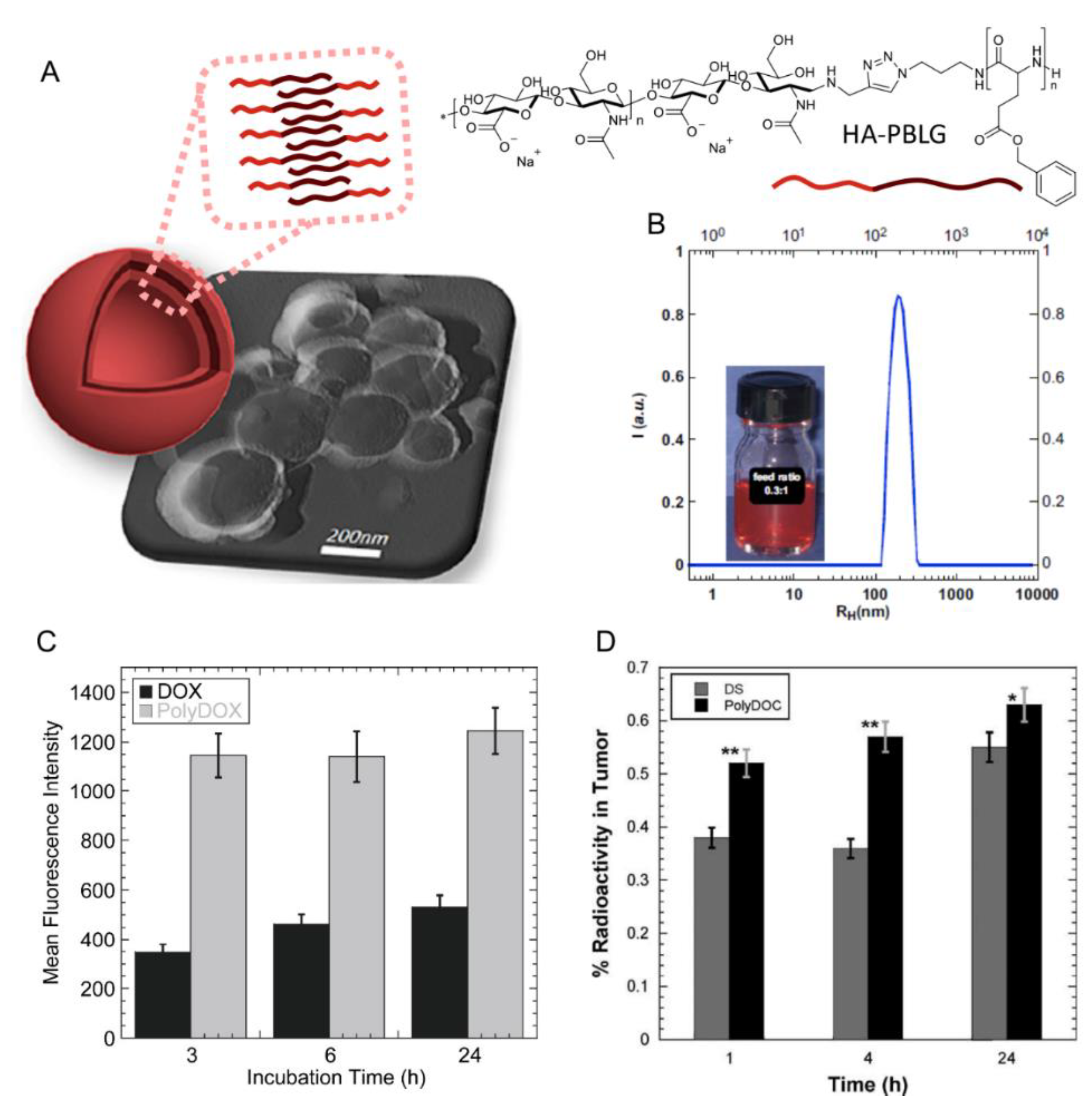
2.3. Polymersomes
2.4. Micelles
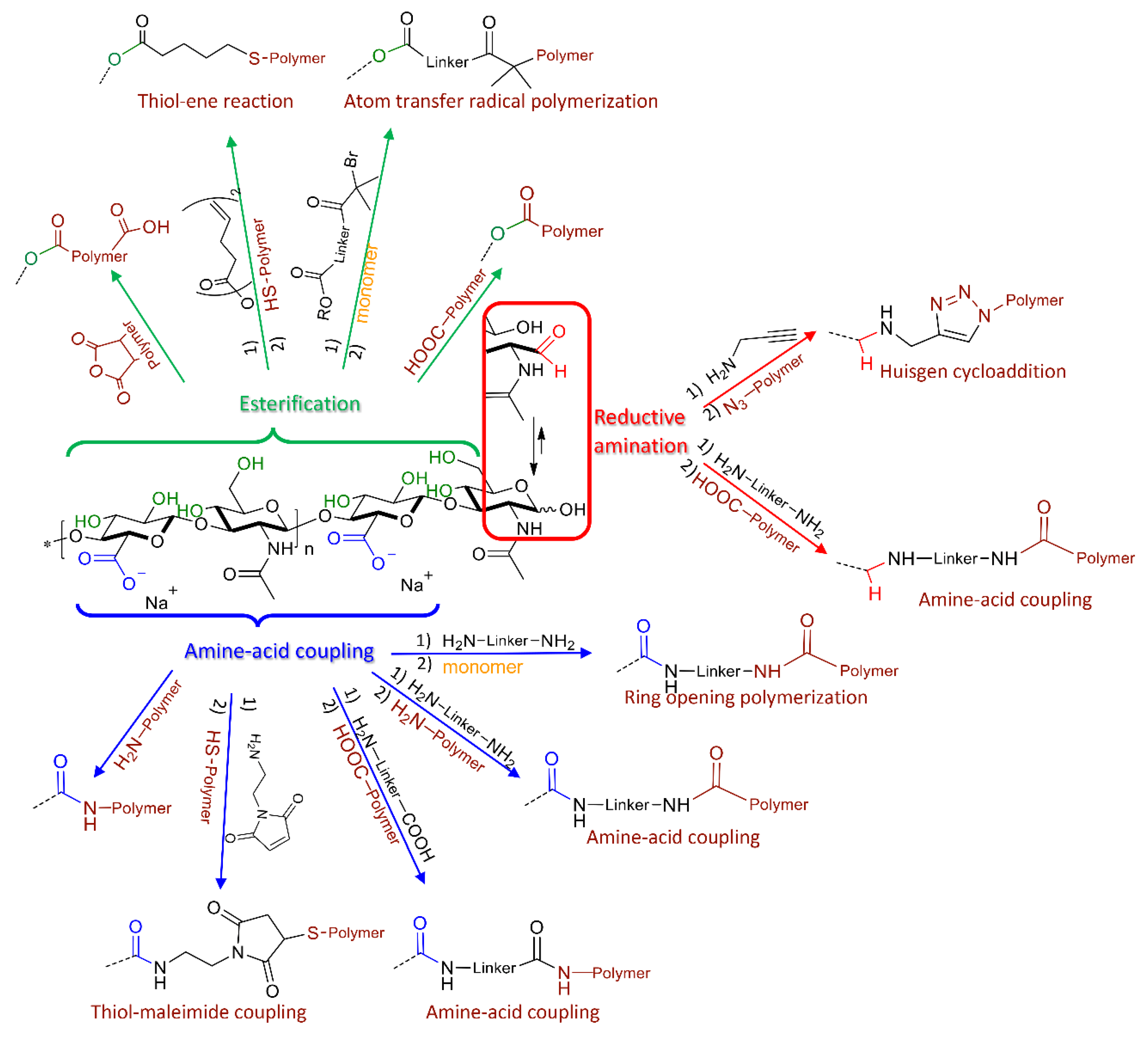
3. Synthetic Strategies for the Design of HA-Based Nanocarriers
3.1. Block Copolymers from HA
3.2. Graft Copolymers via “Grafting onto” or “Grafting from” HA
3.3. HA-Polymer Complexes
4. Nanocarriers Based on HA-Polymers Conjugates: Biomedical Applications
4.1. Biodegradable Nanocarriers
4.2. Polyelectrolyte Nanocarriers
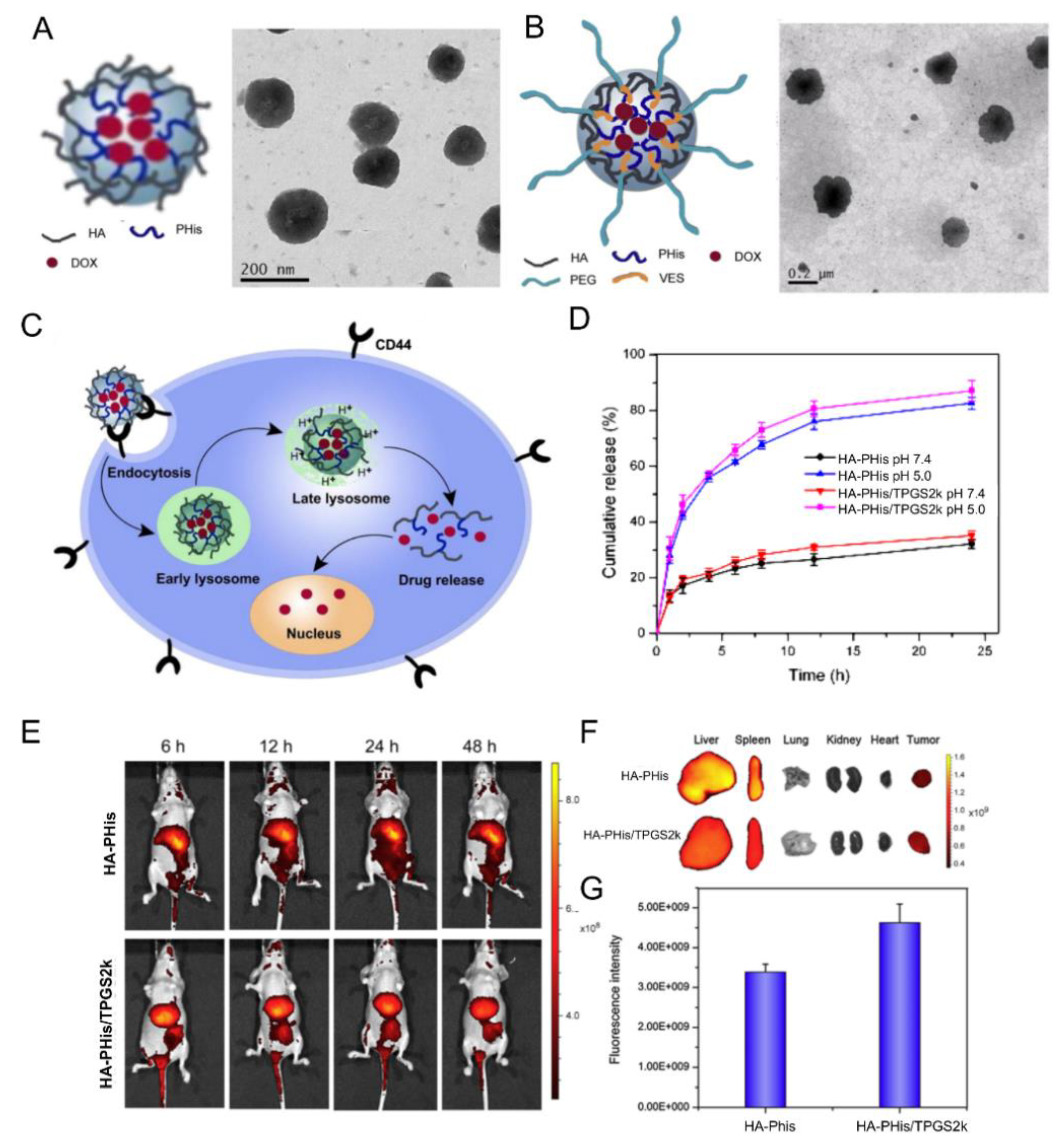
4.3. pH-Responsive Nanocarriers
4.4. Thermoresponsive Nanocarriers
4.5. Redox-Responsive Nanocarriers
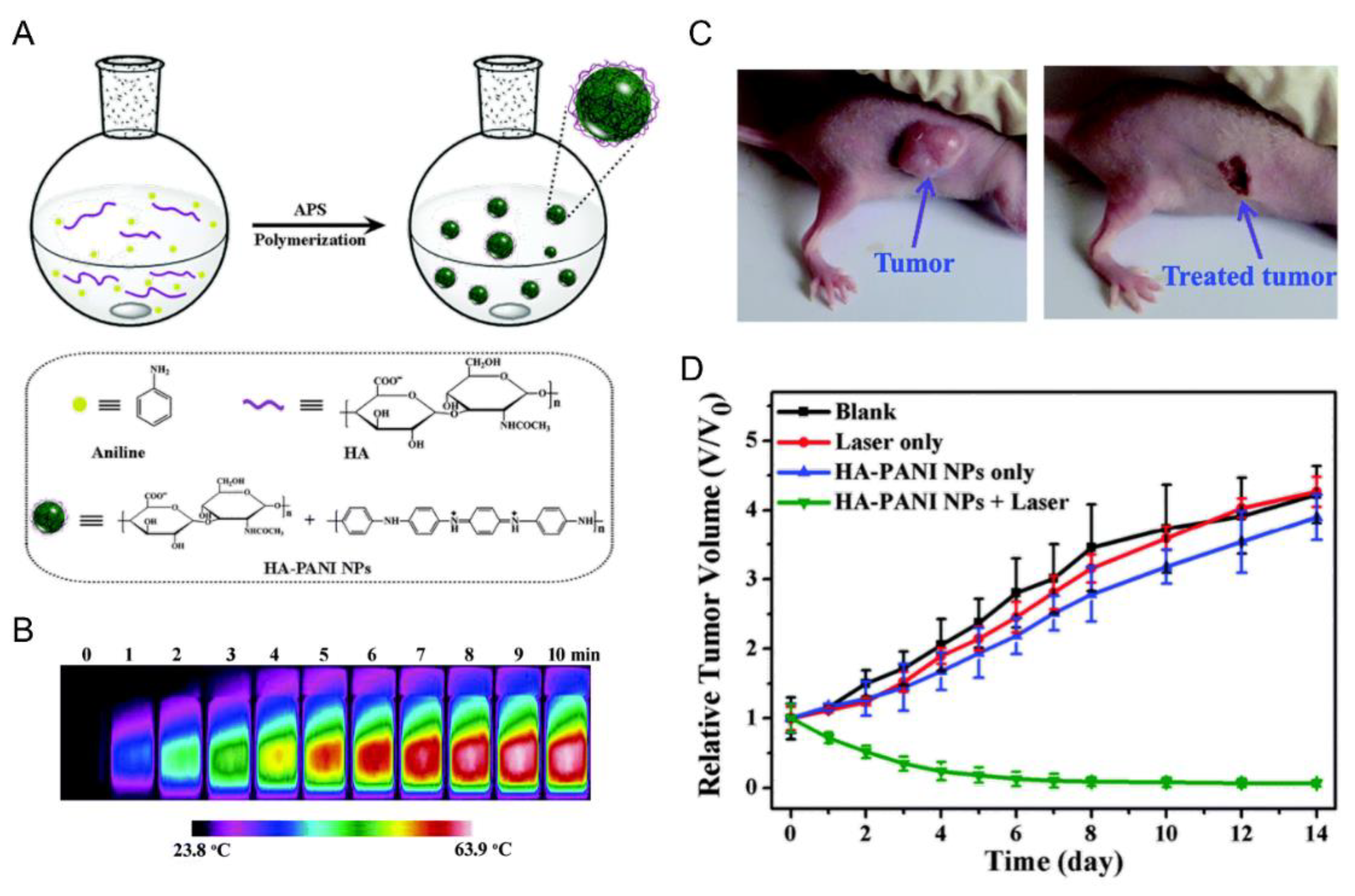
4.6. Light-Responsive Nanocarriers
5. HA-Based Polymeric Nanocarriers: Challenges and Future Prospects
6. Conclusions
Funding
Conflicts of Interest
References
- Husseini, G.A.; Pitt, W.G. Micelles and nanoparticles for ultrasonic drug and gene delivery. Adv. Drug Deliv. Rev. 2008, 60, 1137–1152. [Google Scholar] [CrossRef] [PubMed] [Green Version]
- Rawat, M.; Singh, D.; Saraf, S.; Saraf, S. Nanocarriers: Promising vehicle for bioactive drugs. Biol. Pharm. Bull. 2006, 29, 1790–1798. [Google Scholar] [CrossRef] [PubMed]
- Fang, J.; Nakamura, H.; Maeda, H. The EPR effect: Unique features of tumor blood vessels for drug delivery, factors involved, and limitations and augmentation of the effect. Adv. Drug Deliv. Rev. 2011, 63, 136–151. [Google Scholar] [CrossRef] [PubMed]
- Giner-Casares, J.J.; Henriksen-Lacey, M.; Coronado-Puchau, M.; Liz-Marzan, L.M. Inorganic nanoparticles for biomedicine: Where materials scientists meet medical research. Mater. Today 2016, 19, 19–28. [Google Scholar] [CrossRef]
- Karlsson, J.; Vaughan, H.J.; Green, J.J. Biodegradable Polymeric Nanoparticles for Therapeutic Cancer Treatments. Annu. Rev. Chem. Biomol. Eng. 2018, 9, 105–127. [Google Scholar] [CrossRef]
- Kim, J.H.; Moon, M.J.; Kim, D.Y.; Heo, S.H.; Jeong, Y.Y. Hyaluronic acid-based nanomaterials for cancer therapy. Polymers 2018, 10, 1133. [Google Scholar] [CrossRef]
- Banik Brittany, L.; Fattahi, P.; Brown Justin, L. Polymeric nanoparticles: The future of nanomedicine. Wiley Interdiscip. Rev. Nanomed. Nanobiotechnol. 2016, 8, 271–299. [Google Scholar] [CrossRef]
- Dicker, K.T.; Gurski, L.A.; Pradhan-Bhatt, S.; Witt, R.L.; Farach-Carson, M.C.; Jia, X. Hyaluronan: A simple polysaccharide with diverse biological functions. Acta Biomater. 2014, 10, 1558–1570. [Google Scholar] [CrossRef]
- Nasir, A.; Kausar, A.; Younus, A. A Review on Preparation, Properties and Applications of Polymeric Nanoparticle-Based Materials. Polym. Plast. Technol. Eng. 2015, 54, 325–341. [Google Scholar] [CrossRef]
- Jiang, B.-P.; Zhang, L.; Zhu, Y.; Shen, X.-C.; Ji, S.-C.; Tan, X.-Y.; Cheng, L.; Liang, H. Water-soluble hyaluronic acid-hybridized polyaniline nanoparticles for effectively targeted photothermal therapy. J. Mater. Chem. B Mater. Biol. Med. 2015, 3, 3767–3776. [Google Scholar] [CrossRef]
- Gelmi, A.; Higgins, M.J.; Wallace, G.G. Attractive and Repulsive Interactions Originating from Lateral Nanometer Variations in Surface Charge/Energy of Hyaluronic Acid and Chondroitin Sulfate Doped Polypyrrole Observed Using Atomic Force Microscopy. J. Phys. Chem. B 2012, 116, 13498–13505. [Google Scholar] [CrossRef] [PubMed]
- Vinogradov, S.; Batrakova, E.; Kabanov, A. Poly(ethylene glycol)-polyethylenimine NanoGel particles: Novel drug delivery systems for antisense oligonucleotides. Colloids Surf. B Biointerfaces 1999, 16, 291–304. [Google Scholar] [CrossRef]
- Messager, L.; Portecop, N.; Hachet, E.; Lapeyre, V.; Pignot-Paintrand, I.; Catargi, B.; Auzely-Velty, R.; Ravaine, V. Photochemical crosslinking of hyaluronic acid confined in nanoemulsions: Towards nanogels with a controlled structure. J. Mater. Chem. B Mater. Biol. Med. 2013, 1, 3369–3379. [Google Scholar] [CrossRef]
- Neamtu, I.; Rusu, A.G.; Diaconu, A.; Nita, L.E.; Chiriac, A.P. Basic concepts and recent advances in nanogels as carriers for medical applications. Drug Deliv. 2017, 24, 539–557. [Google Scholar] [CrossRef] [PubMed] [Green Version]
- Soni, G.; Yadav Khushwant, S. Nanogels as potential nanomedicine carrier for treatment of cancer: A mini review of the state of the art. Saudi Pharm. J. Spj Off. Publ. Saudi Pharm. Soc. 2016, 24, 133–139. [Google Scholar] [CrossRef] [PubMed]
- Soni, K.S.; Desale, S.S.; Bronich, T.K. Nanogels: An overview of properties, biomedical applications and obstacles to clinical translation. J. Control. Release 2016, 240, 109–126. [Google Scholar] [CrossRef] [PubMed]
- Dorwal, D. Nanogels as novel and versatile pharmaceuticals. Int. J. Pharm. Pharm. Sci. 2012, 4, 67–74. [Google Scholar]
- Kabanov, A.V.; Vinogradov, S.V. Nanogels as Pharmaceutical Carriers: Finite Networks of Infinite Capabilities. Angew. Chem. Int. Ed. 2009, 48, 5418–5429. [Google Scholar] [CrossRef] [Green Version]
- Yadav, H.; Al Halabi, N.; Alsalloum, G. Nanogels as Novel Drug Delivery Systems—A Review. J. Pharm. Pharm. Res. 2017, 1, 5. [Google Scholar]
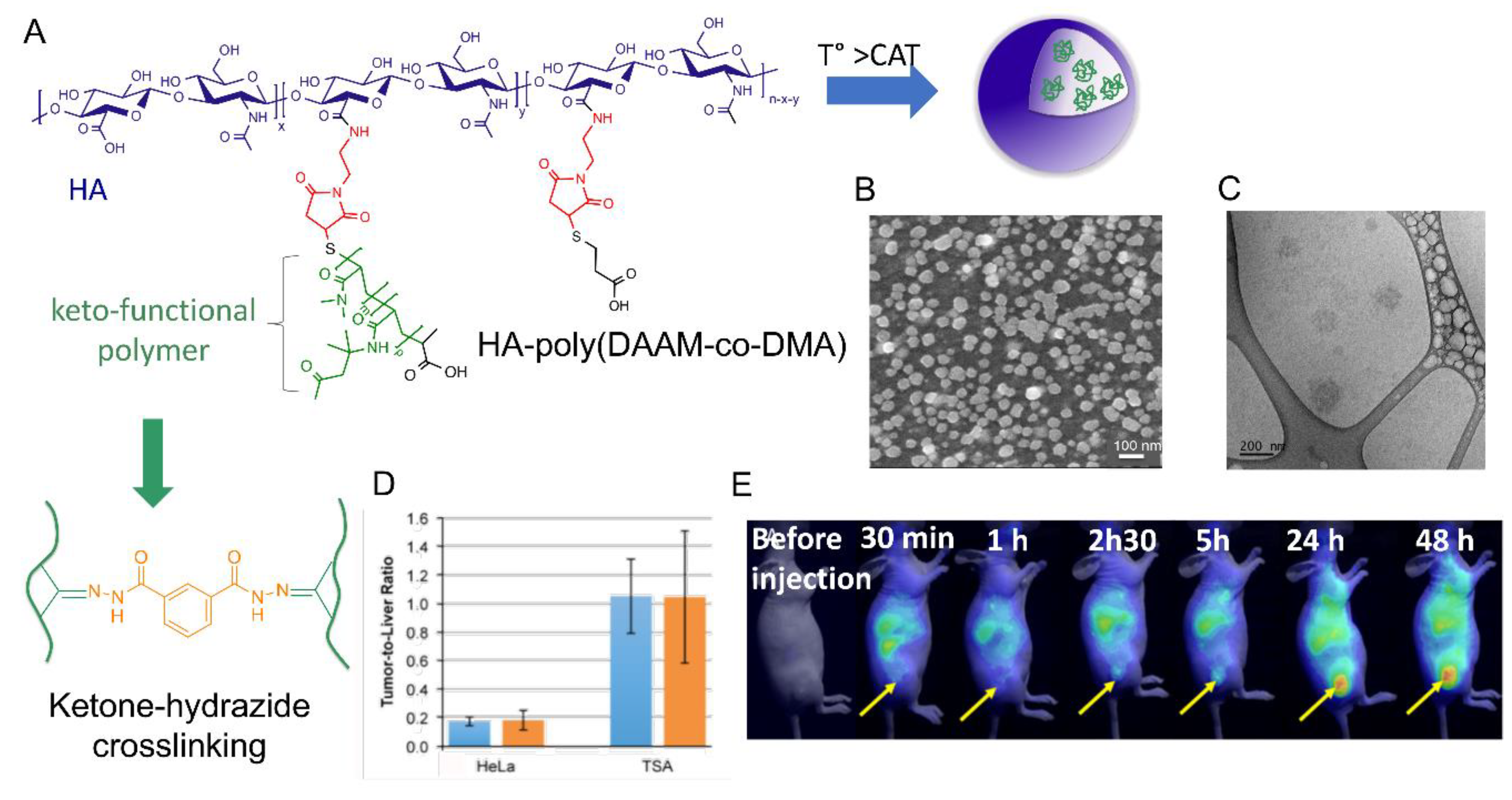
- Garcia, F.P.; Rippe, M.; Companhoni, M.V.P.; Stefanello, T.F.; Louage, B.; Van Herck, S.; Sancey, L.; Coll, J.-L.; De Geest, B.G.; Vataru Nakamura, C.; et al. A versatile method for the selective core-crosslinking of hyaluronic acid nanogels via ketone-hydrazide chemistry: From chemical characterization to in vivo biodistribution. Biomater. Sci. 2018, 6, 1754–1763. [Google Scholar] [CrossRef]
- Jha, A.K.; Hule, R.A.; Jiao, T.; Teller, S.S.; Clifton, R.J.; Duncan, R.L.; Pochan, D.J.; Jia, X. Structural Analysis and Mechanical Characterization of Hyaluronic Acid-Based Doubly Cross-Linked Networks. Macromolecules 2009, 42, 537–546. [Google Scholar] [CrossRef] [PubMed] [Green Version]
- Deng, X.; Cao, M.; Zhang, J.; Hu, K.; Yin, Z.; Zhou, Z.; Xiao, X.; Yang, Y.; Sheng, W.; Wu, Y.; et al. Hyaluronic acid-chitosan nanoparticles for co-delivery of MiR-34a and doxorubicin in therapy against triple negative breast cancer. Biomaterials 2014, 35, 4333–4344. [Google Scholar] [CrossRef] [PubMed]
- Novoa-Carballal, R.; Pergushov, D.V.; Mueller, A.H.E. Interpolyelectrolyte complexes based on hyaluronic acid-block-poly(ethylene glycol) and poly-l-lysine. Soft Matter 2013, 9, 4297–4303. [Google Scholar] [CrossRef]
- Park, H.S.; Lee, J.E.; Cho, M.Y.; Hong, J.H.; Cho, S.H.; Lim, Y.T. Hyaluronic Acid/Poly(β-Amino Ester) Polymer Nanogels for Cancer-Cell-Specific NIR Fluorescence Switch. Macromol. Rapid Commun. 2012, 33, 1549–1555. [Google Scholar] [CrossRef] [PubMed]
- Anajafi, T.; Mallik, S. Polymersome-based drug-delivery strategies for cancer therapeutics. Ther. Deliv. 2015, 6, 521–534. [Google Scholar] [CrossRef] [PubMed] [Green Version]
- Hu, X.; Zhang, Y.; Xie, Z.; Jing, X.; Bellotti, A.; Gu, Z. Stimuli-Responsive Polymersomes for Biomedical Applications. Biomacromolecules 2017, 18, 649–673. [Google Scholar] [CrossRef] [PubMed]
- Schatz, C.; Lecommandoux, S. Polysaccharide-containing block copolymers: Synthesis, properties and applications of an emerging family of glycoconjugates. Macromol. Rapid Commun. 2010, 31, 1664–1684. [Google Scholar] [CrossRef] [PubMed]
- Upadhyay, K.K.; Bhatt, A.N.; Castro, E.; Mishra, A.K.; Chuttani, K.; Dwarakanath, B.S.; Schatz, C.; Le Meins, J.-F.; Misra, A.; Lecommandoux, S. In vitro and In vivo Evaluation of Docetaxel Loaded Biodegradable Polymersomes. Macromol. Biosci. 2010, 10, 503–512. [Google Scholar] [CrossRef]
- Upadhyay, K.K.; Bhatt, A.N.; Mishra, A.K.; Dwarakanath, B.S.; Jain, S.; Schatz, C.; Le Meins, J.-F.; Farooque, A.; Chandraiah, G.; Jain, A.K.; et al. The intracellular drug delivery and anti tumor activity of doxorubicin loaded poly(γ-benzyl -glutamate)-b-hyaluronan polymersomes. Biomaterials 2010, 31, 2882–2892. [Google Scholar] [CrossRef] [PubMed]
- Upadhyay, K.K.; Mishra, A.K.; Chuttani, K.; Kaul, A.; Schatz, C.; Le Meins, J.-F.; Misra, A.; Lecommandoux, S. The in vivo behavior and antitumor activity of doxorubicin-loaded poly(γ-benzyl L-glutamate)-block-hyaluronan polymersomes in Ehrlich ascites tumor-bearing BalB/c mice. Nanomedicine 2012, 8, 71–80. [Google Scholar] [CrossRef]
- Haas, S.; Hain, N.; Raoufi, M.; Handschuh-Wang, S.; Wang, T.; Jiang, X.; Schoenherr, H. Enzyme Degradable Polymersomes from Hyaluronic Acid-block-poly(ε-caprolactone) Copolymers for the Detection of Enzymes of Pathogenic Bacteria. Biomacromolecules 2015, 16, 832–841. [Google Scholar] [CrossRef] [PubMed]
- Thakur, S.; Pramod, K.; Malviya, R. Utilization of Polymeric Nanoparticle in Cancer Treatment: A Review. J. Pharm. Care Health Syst. 2017, 4, 172. [Google Scholar] [CrossRef]
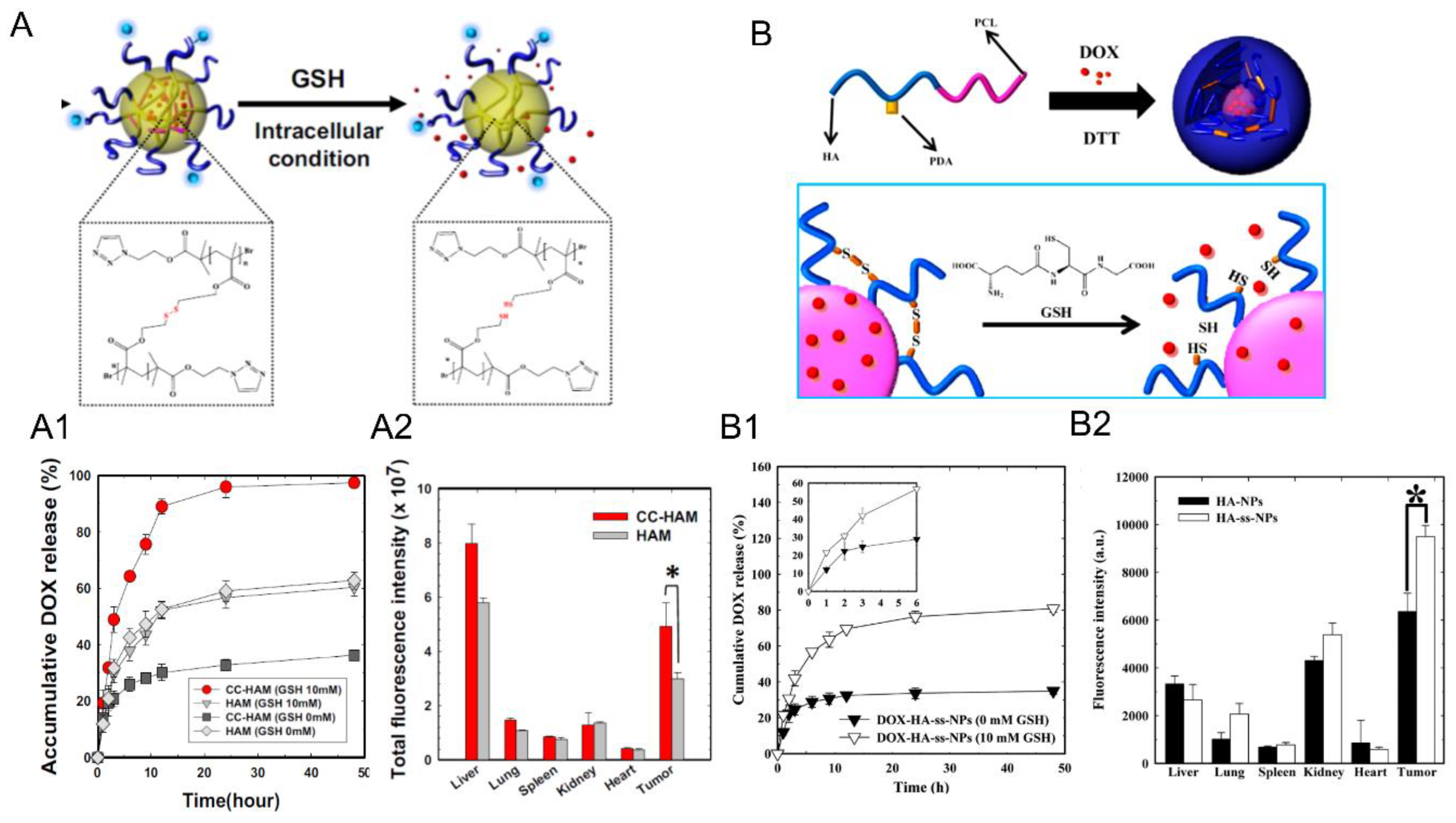
- Park, H.-K.; Lee, S.J.; Oh, J.-S.; Lee, S.-G.; Jeong, Y.-I.; Lee, H.C. Smart Nanoparticles Based on Hyaluronic Acid for Redox-Responsive and CD44 Receptor-Mediated Targeting of Tumor. Nanoscale Res. Lett. 2015, 10, 288. [Google Scholar] [CrossRef]
- Han, H.S.; Thambi, T.; Choi, K.Y.; Son, S.; Ko, H.; Lee, M.C.; Jo, D.-G.; Chae, Y.S.; Kang, Y.M.; Lee, J.Y.; et al. Bioreducible Shell-Cross-Linked Hyaluronic Acid Nanoparticles for Tumor-Targeted Drug Delivery. Biomacromolecules 2015, 16, 447–456. [Google Scholar] [CrossRef] [PubMed]
- Jeannot, V.; Mazzaferro, S.; Lavaud, J.; Vanwonterghem, L.; Henry, M.; Arboleas, M.; Vollaire, J.; Josserand, V.; Coll, J.-L.; Lecommandoux, S.; et al. Targeting CD44 receptor-positive lung tumors using polysaccharide-based nanocarriers: Influence of nanoparticle size and administration route. Nanomedicine 2016, 12, 921–932. [Google Scholar] [CrossRef] [PubMed]
- Zhang, Y.; Wu, K.; Sun, H.; Zhang, J.; Yuan, J.; Zhong, Z. Hyaluronic Acid-Shelled Disulfide-Cross-Linked Nanopolymersomes for Ultrahigh-Efficiency Reactive Encapsulation and CD44-Targeted Delivery of Mertansine Toxin. Acs Appl. Mater. Interfaces 2018, 10, 1597–1604. [Google Scholar] [CrossRef] [PubMed]
- Han, H.S.; Choi, K.Y.; Ko, H.; Jeon, J.; Saravanakumar, G.; Suh, Y.D.; Lee, D.S.; Park, J.H. Bioreducible core-crosslinked hyaluronic acid micelle for targeted cancer therapy. J. Control. Release 2015, 200, 158–166. [Google Scholar] [CrossRef] [PubMed]
- Tizzotti, M.; Charlot, A.; Fleury, E.; Stenzel, M.; Bernard, J. Modification of Polysaccharides Through Controlled/Living Radical Polymerization Grafting-Towards the Generation of High Performance Hybrids. Macromol. Rapid Commun. 2010, 31, 1751–1772. [Google Scholar] [CrossRef] [PubMed]
- Yadav, A.K.; Mishra, P.; Jain, S.; Mishra, P.; Mishra, A.K.; Agrawal, G.P. Preparation and characterization of HA-PEG-PCL intelligent core-corona nanoparticles for delivery of doxorubicin. J. Drug Target. 2008, 16, 464–478. [Google Scholar] [CrossRef] [PubMed]
- Palumbo, F.S.; Bavuso Volpe, A.; Bongiovi, F.; Pitarresi, G.; Giammona, G. A New Hyaluronic Acid Derivative Obtained from Atom Transfer Radical Polymerization as a siRNA Vector for CD44 Receptor Tumor Targeting. Macromol. Biosci. 2015, 15, 1605–1615. [Google Scholar] [CrossRef] [PubMed]
- Oh, E.J.; Park, K.; Kim, K.S.; Kim, J.; Yang, J.-A.; Kong, J.-H.; Lee, M.Y.; Hoffman, A.S.; Hahn, S.K. Target specific and long-acting delivery of protein, peptide, and nucleotide therapeutics using hyaluronic acid derivatives. J. Control. Release 2010, 141, 2–12. [Google Scholar] [CrossRef] [PubMed]
- Jing, J.; Alaimo, D.; De Vlieghere, E.; Jerome, C.; De Wever, O.; De Geest, B.G.; Auzely-Velty, R. Tunable self-assembled nanogels composed of well-defined thermoresponsive hyaluronic acid-polymer conjugates. J. Mater. Chem. B Mater. Biol. Med. 2013, 1, 3883–3887. [Google Scholar] [CrossRef]
- Stefanello, T.F.; Couturaud, B.; Szarpak-Jankowska, A.; Fournier, D.; Louage, B.; Garcia, F.P.; Nakamura, C.V.; De Geest, B.G.; Woisel, P.; van der Sanden, B.; et al. Coumarin-containing thermoresponsive hyaluronic acid-based nanogels as delivery systems for anticancer chemotherapy. Nanoscale 2017, 9, 12150–12162. [Google Scholar] [CrossRef] [PubMed]
- Hu, K.; Zhou, H.; Liu, Y.; Liu, Z.; Liu, J.; Tang, J.; Li, J.; Zhang, J.; Sheng, W.; Zhao, Y.; et al. Hyaluronic acid functional amphipathic and redox-responsive polymer particles for the co-delivery of doxorubicin and cyclopamine to eradicate breast cancer cells and cancer stem cells. Nanoscale 2015, 7, 8607–8618. [Google Scholar] [CrossRef] [PubMed]
- Huang, W.-C.; Chen, S.-H.; Chiang, W.-H.; Huang, C.-W.; Lo, C.-L.; Chern, C.-S.; Chiu, H.-C. Tumor Microenvironment-Responsive Nanoparticle Delivery of Chemotherapy for Enhanced Selective Cellular Uptake and Transportation within Tumor. Biomacromolecules 2016, 17, 3883–3892. [Google Scholar] [CrossRef] [PubMed]
- Yadav, A.K.; Agarwal, A.; Rai, G.; Mishra, P.; Jain, S.; Mishra, A.K.; Agrawal, H.; Agrawal, G.P. Development and characterization of hyaluronic acid decorated PLGA nanoparticles for delivery of 5-fluorouracil. Drug Deliv. 2010, 17, 561–572. [Google Scholar] [CrossRef]
- Schante, C.E.; Zuber, G.; Herlin, C.; Vandamme, T.F. Chemical modifications of hyaluronic acid for the synthesis of derivatives for a broad range of biomedical applications. Carbohydr. Polym. 2011, 85, 469–489. [Google Scholar] [CrossRef]
- Fernandes Stefanello, T.; Szarpak-Jankowska, A.; Appaix, F.; Louage, B.; Hamard, L.; De Geest, B.G.; van der Sanden, B.; Nakamura, C.V.; Auzely-Velty, R. Thermoresponsive hyaluronic acid nanogels as hydrophobic drug carrier to macrophages. Acta Biomater. 2014, 10, 4750–4758. [Google Scholar] [CrossRef]
- Rippe, M.; Stefanello, T.F.; Kaplum, V.; Britta, E.A.; Garcia, F.P.; Poirot, R.; Companhoni, M.V.P.; Nakamura, C.V.; Szarpak-Jankowska, A.; Auzely-Velty, R. Heparosan as a potential alternative to hyaluronic acid for the design of biopolymer-based nanovectors for anticancer therapy. Biomater. Sci. 2019, 7, 2850–2860. [Google Scholar] [CrossRef]
- Pitarresi, G.; Palumbo, F.S.; Albanese, A.; Fiorica, C.; Picone, P.; Giammona, G. Self-assembled amphiphilic hyaluronic acid graft copolymers for targeted release of antitumoral drug. J. Drug Target. 2010, 18, 264–276. [Google Scholar] [CrossRef]
- Son, G.M.; Kim, H.Y.; Ryu, J.H.; Chu, C.W.; Kang, D.H.; Park, S.B.; Jeong, Y.-I. Self-assembled polymeric micelles based on hyaluronic acid-g-poly(d,l-lactide-co-glycolide) copolymer for tumor targeting. Int. J. Mol. Sci. 2014, 15, 16057–16068. [Google Scholar] [CrossRef] [PubMed]
- Yadav, A.K.; Mishra, P.; Mishra, A.K.; Mishra, P.; Jain, S.; Agrawal, G.P. Development and characterization of hyaluronic acid-anchored PLGA nanoparticulate carriers of doxorubicin. Nanomedicine 2007, 3, 246–257. [Google Scholar] [CrossRef] [PubMed]
- Kesharwani, P.; Banerjee, S.; Padhye, S.; Sarkar, F.H.; Iyer, A.K. Hyaluronic Acid Engineered Nanomicelles Loaded with 3,4-Difluorobenzylidene Curcumin for Targeted Killing of CD44+ Stem-Like Pancreatic Cancer Cells. Biomacromolecules 2015, 16, 3042–3053. [Google Scholar] [CrossRef] [PubMed]
- Ganesh, S.; Iyer, A.K.; Morrissey, D.V.; Amiji, M.M. Hyaluronic acid based self-assembling nanosystems for CD44 target mediated siRNA delivery to solid tumors. Biomaterials 2013, 34, 3489–3502. [Google Scholar] [CrossRef] [PubMed] [Green Version]
- Ganesh, S.; Iyer, A.K.; Gattacceca, F.; Morrissey, D.V.; Amiji, M.M. In vivo biodistribution of siRNA and cisplatin administered using CD44-targeted hyaluronic acid nanoparticles. J. Control. Release 2013, 172, 699–706. [Google Scholar] [CrossRef] [PubMed] [Green Version]
- Yin, T.; Wang, L.; Yin, L.; Zhou, J.; Huo, M. Co-delivery of hydrophobic paclitaxel and hydrophilic AURKA specific siRNA by redox-sensitive micelles for effective treatment of breast cancer. Biomaterials 2015, 61, 10–25. [Google Scholar] [CrossRef] [PubMed]
- Qiu, L.; Qiao, M.; Long, M.; Wang, M.; Zhang, X.; Li, Z.; Tian, C.; Chen, D. Self-assembled pH-responsive hyaluronic acid-g-poly((L)-histidine) copolymer micelles for targeted intracellular delivery of doxorubicin. Acta Biomater. 2014, 10, 2024–2035. [Google Scholar] [CrossRef] [PubMed]
- Qiu, L.; Qiao, M.; Chen, Q.; Tian, C.; Long, M.; Wang, M.; Li, Z.; Hu, W.; Li, G.; Cheng, L.; et al. Enhanced effect of pH-sensitive mixed copolymer micelles for overcoming multidrug resistance of doxorubicin. Biomaterials 2014, 35, 9877–9887. [Google Scholar] [CrossRef] [PubMed]
- Chen, Q.; Long, M.; Qiu, L.; Zhu, M.; Li, Z.; Qiao, M.; Hu, H.; Zhao, X.; Chen, D. Decoration of pH-sensitive copolymer micelles with tumor-specific peptide for enhanced cellular uptake of doxorubicin. Int. J. Nanomed. 2016, 11, 5415–5427. [Google Scholar] [CrossRef] [PubMed]
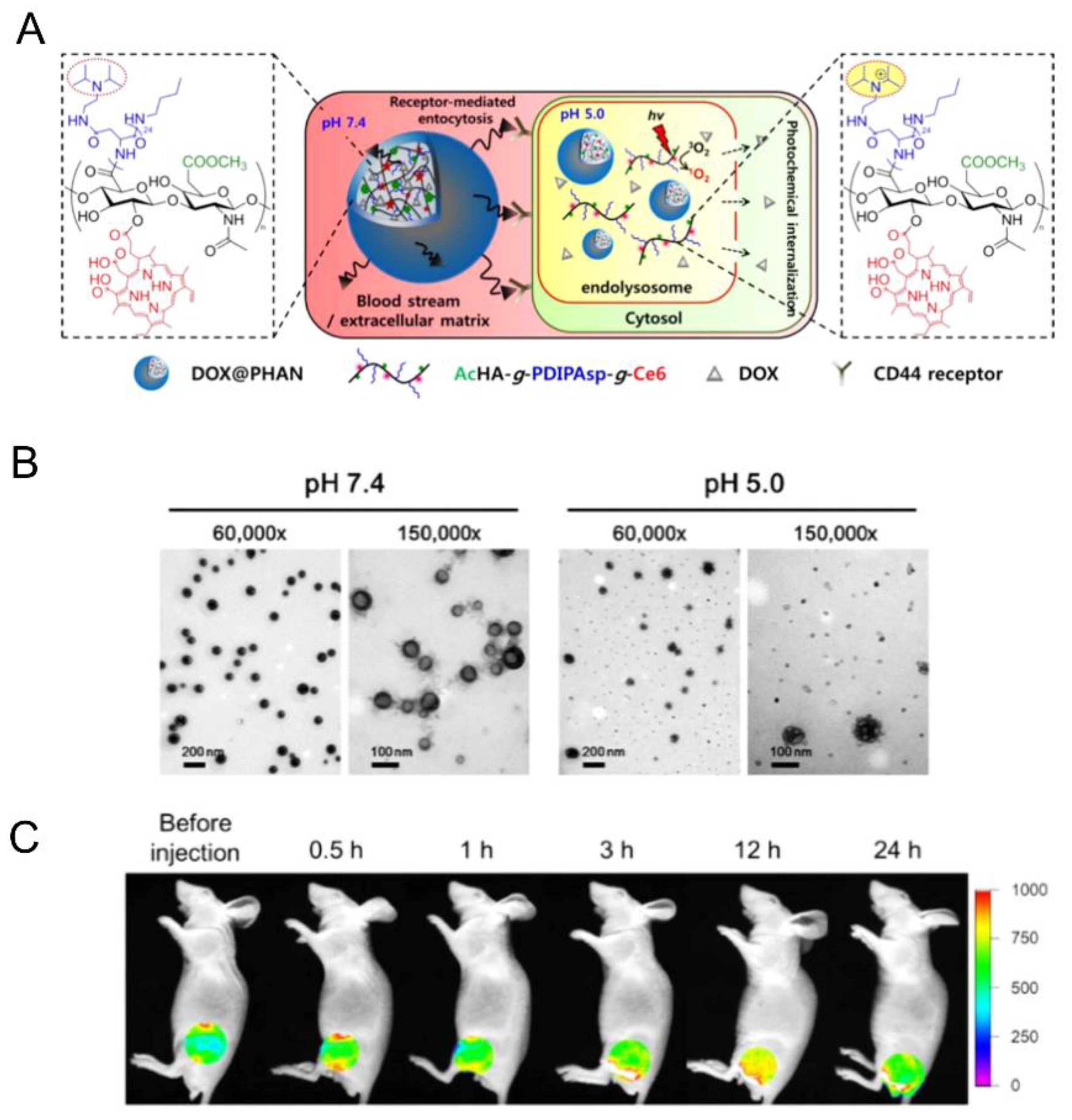
- Lee, C.-S.; Na, K. Photochemically triggered cytosolic drug delivery using pH-responsive hyaluronic acid nanoparticles for light-induced cancer therapy. Biomacromolecules 2014, 15, 4228–4238. [Google Scholar] [CrossRef] [PubMed]
- Ulery, B.D.; Nair, L.S.; Laurencin, C.T. Biomedical applications of biodegradable polymers. J. Polym. Sci. Part B Polym. Phys. 2011, 49, 832–864. [Google Scholar] [CrossRef] [Green Version]
- Fleige, E.; Quadir, M.A.; Haag, R. Stimuli-responsive polymeric nanocarriers for the controlled transport of active compounds: Concepts and applications. Adv. Drug Deliv. Rev. 2012, 64, 866–884. [Google Scholar] [CrossRef] [PubMed]
- Kost, J.; Langer, R. Responsive polymeric delivery systems. Adv. Drug Deliv. Rev. 2001, 46, 125–148. [Google Scholar] [CrossRef]
- Coury, A.J.; Levy, R.J.; Ratner, B.D.; Schoen, F.J.; Williams, D.F.; Williams, R.L. Degradation of materials in the biological environment. In Biomaterial Science An Introduction to Materials in Medicine, 2nd ed.; Ratner, B.D., Hoffman, A.S., Schoen, F.J., Lemons, J.E., Eds.; Elsevier Academic Press: San Diego, CA, USA, 2004; pp. 411–454. [Google Scholar]
- Li, Y.; Gao, G.H.; Lee, D.S. Stimulus-Sensitive Polymeric Nanoparticles and Their Applications as Drug and Gene Carriers. Adv. Healthc. Mater. 2013, 2, 388–417. [Google Scholar] [CrossRef] [PubMed]
- Hong, W.; Chen, D.; Zhang, X.; Zeng, J.; Hu, H.; Zhao, X.; Qiao, M. Reversing multidrug resistance by intracellular delivery of Pluronic® P85 unimers. Biomaterials 2013, 34, 9602–9614. [Google Scholar] [CrossRef] [PubMed]
- Schafer, F.Q.; Buettner, G.R. Redox environment of the cell as viewed through the redox state of the glutathione disulfide/glutathione couple. Free Radic. Biol. Med. 2001, 30, 1191–1212. [Google Scholar] [CrossRef]
- Cao, J.; Huang, S.; Chen, Y.; Li, S.; Li, X.; Deng, D.; Qian, Z.; Tang, L.; Gu, Y. Near-infrared light-triggered micelles for fast controlled drug release in deep tissue. Biomaterials 2013, 34, 6272–6283. [Google Scholar] [CrossRef] [PubMed]
- Kumar, S.; Allard, J.-F.; Morris, D.; Dory, Y.L.; Lepage, M.; Zhao, Y. Near-infrared light sensitive polypeptide block copolymer micelles for drug delivery. J. Mater. Chem. 2012, 22, 7252–7257. [Google Scholar] [CrossRef]
- Zhao, P.; Zheng, M.; Luo, Z.; Gong, P.; Gao, G.; Sheng, Z.; Zheng, C.; Ma, Y.; Cai, L. NIR-driven Smart Theranostic Nanomedicine for On-demand Drug Release and Synergistic Antitumour Therapy. Sci. Rep. 2015, 5, 14258. [Google Scholar] [CrossRef] [Green Version]
- Harris, E.N.; Weigel, P.H. The ligand-binding profile of HARE: Hyaluronan and chondroitin sulfates A, C, and D bind to overlapping sites distinct from the sites for heparin, acetylated low-density lipoprotein, dermatan sulfate, and CS-E. Glycobiology 2008, 18, 638–648. [Google Scholar] [CrossRef]
- Choi, K.Y.; Min, K.H.; Yoon, H.Y.; Kim, K.; Park, J.H.; Kwon, I.C.; Choi, K.; Jeong, S.Y. PEGylation of hyaluronic acid nanoparticles improves tumor targetability in vivo. Biomaterials 2011, 32, 1880–1889. [Google Scholar] [CrossRef] [PubMed]
- Choi, K.Y.; Yoon, H.Y.; Kim, J.-H.; Bae, S.M.; Park, R.-W.; Kang, Y.M.; Kim, I.-S.; Kwon, I.C.; Choi, K.; Jeong, S.Y.; et al. Smart Nanocarrier Based on PEGylated Hyaluronic Acid for Cancer Therapy. Acs Nano 2011, 5, 8591–8599. [Google Scholar] [CrossRef] [PubMed]
- Han, H.S.; Lee, J.; Kim, H.R.; Chae, S.Y.; Kim, M.; Saravanakumar, G.; Yoon, H.Y.; You, D.G.; Ko, H.; Kim, K.; et al. Robust PEGylated hyaluronic acid nanoparticles as the carrier of doxorubicin: Mineralization and its effect on tumor targetability in vivo. J. Control. Release 2013, 168, 105–114. [Google Scholar] [CrossRef] [PubMed]
- Garay, R.P.; El-Gewely, R.; Armstrong, J.K.; Garratty, G.; Richette, P. Antibodies against polyethylene glycol in healthy subjects and in patients treated with PEG-conjugated agents. Expert Opin. Drug Deliv. 2012, 9, 1319–1323. [Google Scholar] [CrossRef] [PubMed]
- Knop, K.; Hoogenboom, R.; Fischer, D.; Schubert, U.S. Poly(ethylene glycol) in Drug Delivery: Pros and Cons as Well as Potential Alternatives. Angew. Chem. Int. Ed. 2010, 49, 6288–6308. [Google Scholar] [CrossRef] [PubMed]
- Verhoef, J.J.F.; Carpenter, J.F.; Anchordoquy, T.J.; Schellekens, H. Potential induction of anti-PEG antibodies and complement activation toward PEGylated therapeutics. Drug Discov. Today 2014, 19, 1945–1952. [Google Scholar] [CrossRef] [PubMed]
- Yang, Q.; Lai, S.K. Anti-PEG immunity: Emergence, characteristics, and unaddressed questions. Wiley Interdiscip. Rev. Nanomed. Nanobiotechnol. 2015, 7, 655–677. [Google Scholar] [CrossRef]
- Choi, K.Y.; Han, H.S.; Lee, E.S.; Shin, J.M.; Almquist, B.D.; Lee, D.S.; Park, J.H. Hyaluronic acid-based activatable nanomaterials for stimuli-responsive imaging and therapeutics: Beyond CD44-mediated drug delivery. Adv. Mater. 2019. Ahead of Print. [Google Scholar] [CrossRef]
- Naor, D.; Nedvetzki, S. CD44 in rheumatoid arthritis. Arthritis Res. Ther. 2003, 5, 105–115. [Google Scholar] [CrossRef]
- Saba, G.-E.; Abdullah, M.A. Polymeric nanoparticle mediated targeted drug delivery to cancer cells. In Biotechnology and Bioinformatics; Apple Academic Press: Waretown, NJ, USA, 2015; pp. 1–34. [Google Scholar]
- Huang, G.; Huang, H. Application of hyaluronic acid as carriers in drug delivery. Drug Deliv. 2018, 25, 766–772. [Google Scholar] [CrossRef]
- Rao, N.V.; Yoon, H.Y.; Han, H.S.; Ko, H.; Son, S.; Lee, M.; Lee, H.; Jo, D.-G.; Kang, Y.M.; Park, J.H. Recent developments in hyaluronic acid-based nanomedicine for targeted cancer treatment. Expert Opin. Drug Deliv. 2016, 13, 239–252. [Google Scholar] [CrossRef] [PubMed]
- Shah, K.; Chawla, S.; Gadeval, A.; Reddy, G.; Maheshwari, R.; Kalia, K.; Tekade, R.K. Nanostructured Hyaluronic Acid-based Materials for the Delivery of siRNA. Curr. Pharm. Des. 2018, 24, 2678–2691. [Google Scholar] [CrossRef] [PubMed]
- Cai, J.; Fu, J.; Li, R.; Zhang, F.; Ling, G.; Zhang, P. A potential carrier for anti-tumor targeted delivery-hyaluronic acid nanoparticles. Carbohydr. Polym. 2019, 208, 356–364. [Google Scholar] [CrossRef] [PubMed]
- Dosio, F.; Arpicco, S.; Stella, B.; Fattal, E. Hyaluronic acid for anticancer drug and nucleic acid delivery. Adv. Drug Deliv. Rev. 2016, 97, 204–236. [Google Scholar] [CrossRef] [PubMed]


| Mw of HA kg/mol | Polymer Structure a | Size (nm) b | DL (EE) % | In Vitro and/or in Vivo Biological Studies a | Ref |
|---|---|---|---|---|---|
| 5 |  PCL, biodegradable | - c | - | Detection of pathogenic bacteria “Staphylococcus aureus” and drug release of after enzymatic degradation. | [31] |
| 2100 | ~200 d | ||||
| 12 | 198 e,f | 7 (74) f | In vitro: cytotoxicity and intracellular drug release of DOX in SCC7 cell line. In vivo: biodistribution and antitumor efficacy in SCC7 cells-bearing mice. | [34] | |
| 162 g,h | 9 (90) g | ||||
| 7.5 |  PLGA, biodegradable | <200 h | 8 | In vitro: cytotoxicity in MDA-MB231 and NIH3T3 cell lines and release behavior of DOX. In vivo: biodistribution and antitumor efficacy in mice bearing MDA-MB231 or NIH3T3 cells grafted. | [33] |
| 5 |  PBLG, biodegradable | 220 d | 12 (40) | In vitro: cytotoxicity, cell uptake of DOX containing nanocarriers in MCF-7 and U87 cells lines. In vivo: antitumor efficacy in mice with DMBA induced tumor. | [29] |
| 135 d | 9.8 (49) | In vitro: cytotoxicity in MCF-7 and U87 cells lines and release behavior of DOC. In vivo: biodistribution in EAT–bearing mice. | [28] | ||
| - i | - i | In vivo: stability, biodistribution, pharmacokinetics and antitumor activity of DOX containing nanocarriers in EAT-bearing mice. | [30] | ||
| 30 and 300 d | - | In vitro and in vivo lung tumor cells targeting effect of the size. | [35] | ||
| 8 |  P(TMC-co-DTC), biodegradable and redox-responsive | 103 d,g | 17 (99) | In vitro: cytotoxicity in MDA-MB-231 cell line and release behavior of DM1. In vivo: pharmacokinetics and anticancer efficacy in MDA-MB-231 cells-bearing mice. | [36] |
| 7.4 |  PDSMA, redox-responsive | 215 f,h 187 g,h | 8 (80) f 9 (87) g | In vitro: drug release of DOX. In vivo: biodistribution, pharmacokinetics, tumor accumulation profiles and antitumor efficacy in SCC7 cells-bearing mice. | [37] |
| Mw of HA kg/mol | Polymer Structure a | Size (nm) b | DL (EE) % a | In Vitro and/or in Vivo Biological Studies a | Ref |
|---|---|---|---|---|---|
| 5.7 |  PCL, biodegradable | 95 c | (96) | In vitro: hemolytic toxicity and stability of DOX containing nanocarriers. In vivo: biodistribution and tumor inhibition in EAT-bearing mice. | [39] |
| 1500 |  PLA, biodegradable | 30 c | 5 (10) | In vitro: cytotoxicity and uptake in HCT-166-cells and release behavior of DOX. | [50] |
| 8.3 |  PLGA, biodegradable | 103 c | 8 | In vitro: cell viability and uptake in Hep G2 cells and CT26 cell lines and release behavior of DOX. | [51] |
| 15 | - d | 16 (87) | In vitro: cytotoxicity and uptake in Raw 264.7-cells and release behavior of SN38 In vivo biodistribution, and tumor inhibition in tramp-C1 cells–bearing mice. | [45] | |
| 5.7 | 152 e | (80) | In vitro: cytotoxicity in EAT-cells and release behavior of 5-FU In vivo: biodistribution, and tumor inhibition in EAT–bearing mice. | [46] | |
| 6.4 | 245 c | (71) DOX (58) CYC | In vitro: cell viability and uptake in MCF-7 and MDA-MB-231 cell lines and release behavior of DOX and CYC. In vivo: synergic antitumor efficacy of DOX and CYC in MDA-MB231 cell-bearing mice. | [44] | |
| 5.7 |  PEG-co-PLGA, biodegradable | - d | (~90) | In vitro: drug release of DOX. In vivo: biodistribution, and tumor inhibition in EAT–bearing mice. | [52] |
| 10 |  SMA, pH-responsive after hydrolysis of anhydrides | - d | (16) | In vitro: cell viability and uptake of CDF containing nanocarriers in MiaPaCa-2 and AsPC-1; activity on CD44+ and CD44- pancreatic cells. | [53] |
| 20 |  Branched PEI, positive charged at pH 7.4 | ~200 c,f | - | In vitro: siRNA release, cell uptake and gene silencing in MDA-MB468 cell line. In vivo: tumor uptake and gene-silencing in A549 cells-bearing mice. | [54] |
| ~100 c,g | - | In vivo: biodistribution and quantification of siRNA in mice bearing A549, A549DDP, H69 or H69Ar cells grafted. | [55] | ||
| ~200 c,f | 33 (86) | In vitro: cytotoxicity, cell uptake and gene silencing of siRNA and PTX-containing nanocarriers in MDA-MB-231 cell line. In vivo: biodistribution and antitumor efficacy in MDA-MB-231 cells-bearing mice. | [56] | ||
| - g |  pDEAEMA, positive charged at pH 7.4 | 155 c | - | In vitro: cytotoxicity in HCT 116 cells and uptake of siRNA. | [40] |
| 11 |  pHis, pH-responsive | ~400 c (pH = 7.4) | 7 (90) | In vitro: cell viability and uptake in MCF-7 cell line; endocytosis inhibition. pH release behavior of DOX containing nanocarriers. | [57] |
| 10 (92) | In vitro: cell viability and uptake in MCF-7 and MCF-7/ADR cell lines; endocytosis inhibition. pH release behavior of DOX and tocopheryl-PEG containing nanocarriers. In vivo: biodistribution in MCF-7/ADR cells–bearing mice. | [58] | |||
| - d | 10 (93) | In vitro: cytotoxicity and cellular uptake in MDA-MB-231 cell lines. pH release behavior of DOX and Her2 peptide-tocopheryl-PEG containing nanocarriers. In vivo: biodistribution and antitumor efficacy in MDA-MB-231 cell-bearing mice. | [59] | ||
| 5.8 |  PDIPASP, pH-responsive | - d | 14 | In vitro: cell viability and uptake in CT-26 cell line; photosensible endosome scape; pH release behavior of DOX. In vivo: biodistribution and antitumor efficacy in CT-26 cell-bearing mice. | [60] |
| 120 |  p(DEGMA-co-OEGMA), thermosensible (CAT of HA-copolymer: 34 °C) | 150 c (40 °C) | ~3 (70) | In vitro: cellular uptake by RAW264.7 macrophages of DSB containing nanocarriers. In vivo: biodistribution, and macrophage uptake in mice. | [48] |
| 300 | 211 c (40 °C) | - | - | ||
| 200 | 95 c (40 °C) | (80) | In vitro: cell viability of PTX containing nanocarriers in HCT-8/E11 and SKOV-3 cell lines. | [42] | |
| 40 |  p(DEGMA-co-BMA), thermosensible | 108 c,i (37 °C) 151 c,j (37 °C) | - | In vitro: vero cells viability. In vivo: biodistribution in EAC cells-bearing mice. | [49] |
| 40 |  p(DEGMA-co-CMA), photo and thermosensible | ~100 c (37 °C) | 1.5 (52) | In vitro: cell viability in Vero cells and uptake in HeLa cells of PTX containing nanocarriers. In vivo: biodistribution in mice bearing HeLa subcutaneous grafts. | [43] |
| 40 |  p(DAAM-co-DMA), thermoresponsive | 144 c,i (40 °C) 150 c,j (40 °C) | - | In vitro: cell viability and uptake in HeLa and TS/A-pc cell lines. In vivo: biodistribution in mice bearing HeLa or TS/A-pc subcutaneous grafts. | [20] |
| Mw of HA kg/mol | Polymer Structure | Size a in nm (T, °C) | DL (EE) % | In Vitro and/or in Vivo Biological Studies a | Ref |
|---|---|---|---|---|---|
| - b |  Polyaniline, esmeraldine salt, positive charged, photothermal | 100 c | - d | In vitro: cytotoxicity and photothermal therapy effect in HFF, HCT-116 and HeLa cell lines. In vivo: photothermal therapy in HeLa tumor-bearing mice. | [10] |
| 170 |  Chitosan hydrochloride salt, biodegradable, positive charged | 189 c,e | (48, DOX) (91, miR-34a) | In vitro: cytotoxicity and uptake of DOX and miR-34a containing nanocarriers in MDA-MB-231 cell line. In vivo tumor inhibition in MDA-MB-231 cells-bearing mice. | [22] |
| 1300-1800 |  Poly(β-amino ester), biodegradable, partially positive charged at pH 7.4 | - f | - b | In vitro: cell viability and uptake in MDA-MB-231 cells. | [24] |
| 54.3 g |  Polyd -lysine, biodegradable positive charged at pH 7.4 | 122 c,h 145 c,i | - d | - | [23] |
© 2019 by the authors. Licensee MDPI, Basel, Switzerland. This article is an open access article distributed under the terms and conditions of the Creative Commons Attribution (CC BY) license (http://creativecommons.org/licenses/by/4.0/).
Share and Cite
Rippe, M.; Cosenza, V.; Auzély-Velty, R. Design of Soft Nanocarriers Combining Hyaluronic Acid with Another Functional Polymer for Cancer Therapy and Other Biomedical Applications. Pharmaceutics 2019, 11, 338. https://doi.org/10.3390/pharmaceutics11070338
Rippe M, Cosenza V, Auzély-Velty R. Design of Soft Nanocarriers Combining Hyaluronic Acid with Another Functional Polymer for Cancer Therapy and Other Biomedical Applications. Pharmaceutics. 2019; 11(7):338. https://doi.org/10.3390/pharmaceutics11070338
Chicago/Turabian StyleRippe, Marlène, Vanina Cosenza, and Rachel Auzély-Velty. 2019. "Design of Soft Nanocarriers Combining Hyaluronic Acid with Another Functional Polymer for Cancer Therapy and Other Biomedical Applications" Pharmaceutics 11, no. 7: 338. https://doi.org/10.3390/pharmaceutics11070338