1. Introduction
The combination of chemotherapy drugs and siRNA is recently emerging as a strategy for cancer therapy [
1,
2,
3]. Rational design for the combination therapy strategy is essential to obtain maximum efficacy with minimum dosage, side effects, and drug tolerance. Methotrexate (MTX) is a dihydrofolate reductase (DHFR) inhibitor and has shown anticancer activity [
4,
5]. However, MTX monotherapy at high dosage is often associated with systemic toxicity, drug resistance, and low efficacy [
6,
7]. Therefore, MTX needs to be combined with other agents to reduce side effects and enhance tumor efficacy [
8,
9]. Small interfering RNA (siRNA) with a length of 20–25 base pairs has drawn much attention from researchers and is regarded as a potential therapeutic modality for cancer [
10,
11]. A siRNA could specifically silence the expression of the targeted gene through RNA interference [
12]. Survivin has recently been found to be a crucial protein to tumor growth and metastasis and is a promising therapeutic target for tumor [
13]. Moreover, studies had indicated that the survivin could facilitate tumor drug resistance. The inhibition of survivin expression may boost the chemotherapeutic efficacy of cancer [
14]. Thus, a co-delivery of siRNA targeting survivin expression and MTX may be a promising approach to overcome cancer drug resistance [
1]. In addition, the distinct mechanisms of MTX and survivin siRNA suggest that their combination may produce synergy [
15].
Nanocarriers such as liposomes, micelles, dendrimers, or supramolecular systems have been evaluated as vehicles for the co-delivery of chemotherapeutic drugs and gene agents [
2,
16]. Micelles for the co-delivery of chemotherapeutic drugs and nucleic acid have been shown to have good stability and to control drug release. They can increase the effectiveness of drug combination therapy and can reduce drug resistance [
17,
18,
19,
20]. Compared to the pristine micelles, mixed micelles self-assembled from two or more amphiphilic polymers provide greater flexibility. They have recently drawn much attention for use in combination cancer therapy [
21,
22]. The mixed micelles are easy to optimize, in terms of kinetic stability, drug loading capacity, size distribution, and the preparation of multifunctional carriers [
21,
22,
23,
24]. The mixed micelles also could be prepared with simplified procedures, could achieve a desirable antitumor efficacy, and could reduce variability when expanding to a large scale for clinical application [
25,
26]. However, the efficacy of the mixed micelles is still restricted due to rapid drug release especially for the co-delivery of two or more different types of therapeutic drugs in blood circulation. Polymer-drug conjugates have been studied as nanomedicine for the improvement of disease treatment efficacy recently [
27]. Drug covalently bound to the polymers could avoid the drug dissociation and rapid clearance and could improve the drug stability during dilution or exposure to components in blood. Therefore, a system for the co-delivery of siRNA and MTX was developed based on mixed micelles consisted of functionalized polymer-drug conjugates [
28].
MTX conjugated to dendrimers or polymers had previously been shown to retain good antitumor activity in vitro and in vivo [
29,
30]. Moreover, MTX has been suggested to act both as a targeting ligand and a therapeutic agent in recent studies [
31]. When conjugated to dendrimers, MTX may directly target the folic acid (FA) receptor, be internalized into the cell, and then act on its target [
32]. Polyethylenimine (PEI) has been used extensively for the delivery of nucleic acids such as siRNA, miRNA, and oligonucleotides because of its superior ability to electrostatically complex with nucleic acids and to facilitate the endosomal escape through its proton sponge effect. However, PEI has had limited clinical use due to its toxicity [
33,
34]. We previously demonstrated that fatty acid modified PEI showed reduced toxicity and enhanced the efficiency for oligonucleotides delivery [
35,
36]. Here, MTX was conjugated to linolenic acid modified branched PEI (MTX-bPEI-LA). The MTX-bPEI-LA has several advantages: (i) MTX conjugated to the polymers by amide bond is stable and the MTX conjugated micelles have a long circulation time; (ii) it reduced systemic toxicity compared to free MTX [
37]; (iii) bPEI-LA with MTX conjugation may be less toxic than bPEI due to a reduction in charged amino groups [
38], and (iv) polymer-MTX conjugates may act in dual roles of a targeting and a therapeutic agent [
31,
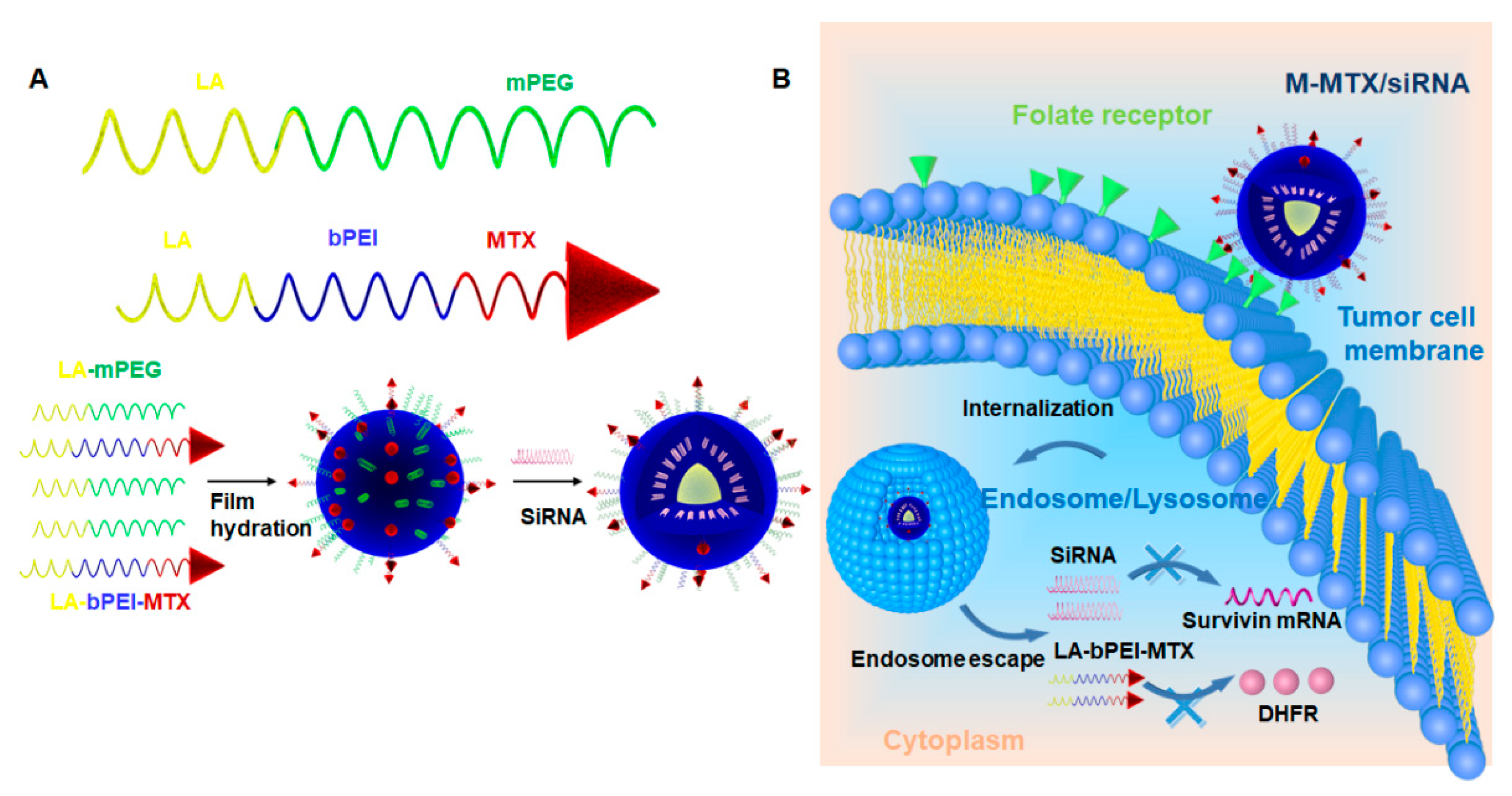
32]. In this study, we also synthesized linolenic acid (LA)-modified methoxy-polyethyleneglycol (mPEG-LA), which was combined with MTX-bPEI-LA to formed mixed micelles (M-MTX) (
Scheme 1). In order to evaluate the co-delivery efficiency of the system, we studied their cellular uptake, cytotoxicity, siRNA target modulation, biodistribution and the therapeutic effect in vitro and in vivo.
2. Materials and Methods
2.1. Materials
Branched polyethylenimine (bPEI, 25 kDa) was purchased from Sigma-Aldrich (St. Louis, MO, USA). Methotrexate (MTX) and 3-(4,5-dimethyl-2-thiazolyl)-2,5-diphenyl-2-H-tetrazolium bromide (MTT) were purchased from Shanghai Yuanye Biological Technology (Shanghai, China). mPEG-NH2 (2000 Da) was purchased from Yarebio (Shanghai, China). Linolyl chloride (LC) was obtained from Tokyo Chemical Industry Co., Ltd. (Shanghai, China). Survivin siRNA: Sense (5′–3′): mGCAGGUUCCUmUAUCUGUCAdTdT; Antisense (5′–3′): UGAmCAGAmUAAGGAACCUGmCdTdT; Survivin siRNA negative control: Sense (5′–3′): mUUCUCCGAACmGUGUCACGUdTdT; Antisense (5′–3′): ACGmUGACmACGUUCGGAGAmAdTdT, and Cy3, 5′-FAM and Cy5-labeled survivin siRNA for cellular uptake and biodistribution studies were synthesized by Ribo Biochemistry (Guangzhou, China). HeLa cells were purchased from ATCC (Rockefeller, MD, USA). 4′,6-Diamidino-2-phenylindole (DAPI) and Lyso Tracker™ Green DND-99 were purchased from Invitrogen Co. (Carlsbad, CA, USA). The Dihydrofolate Reductase Assay Kit was also purchased from BioVision (S. Milpitas Blvd., Milpitas, CA, USA). All chemical reagents used were of analytical grade.
2.2. Synthesis and Characterization of the Amphiphilic Polymers
LA was separately conjugated to the mPEG and b-PEI as shown in
Figure S1 using a previously reported method [
35,
36]. Briefly, linolenic chloride (LC) dissolved in anhydrous dichloromethane (DCM) (Sinopharm Chemical Reagent Co., Ltd., Shanghai, China) was added dropwise to the mPEG2000-NH
2 and bPEI (25 kDa) anhydrous DCM solution, respectively. After 12 h, the reaction mixture was precipitated and washed three times by diethyl ether (Sinopharm Chemical Reagent Co., Ltd., Shanghai, China). The products mPEG-LA and bPEI-LA were obtained by removing organic solvent in a rotary evaporator (Shanghai Yukang Scientific Instrument Co., Ltd., Shanghai, China) and then vacuumed for 2 h. MTX modified bPEI-LA(MTX-bPEI-LA) was prepared through the reaction of the amino groups of bPEI-LA and carboxy groups of MTX (
Figure S1B). The activated reagents of 1-hydroxybenzotriazole (HOBT) (Xiya Chemical Industry Co., Ltd., Linshu, China),
O-benzotriazole-
N,
N,
N′,
N′-tetramethyl-uroniumhexafluorophosphate (HBTU) (Xiya Chemical Industry Co., Ltd., Linshu, China), and
N,
N-diisopropylethylamine (DIEA) (Sinopharm Chemical Reagent Co., Ltd., Shanghai, China) were first added to the MTX solution to activate the carboxyl groups of MTX for 2 h. Then, the bPEI-LA solution dissolved in anhydrous methanol (Sinopharm Chemical Reagent Co., Ltd., Shanghai, China) was added dropwise to the MTX solution. The mixture was incubated at room temperature for 24 h under nitrogen atmosphere. The reaction mixture was placed in a dialysis bag with a molecular weight cutoff (MWCO) of 8000 to 14,000 Da and dialyzed against deionized water. The dialysate was changed every 4 h. After 48 h, MTX-bPEI-LA was freeze-dried on a Christ epsilon 2-6D LSC (Osterode, Germany). The structures of mPEG-LA, bPEI-LA, and MTX-bPEI-LA were confirmed by
1H NMR on a spectrometer from Bruker (Fällanden, Switzerland). mPEG-LA and bPEI-LA were dissolved in deuterated chloroform (CDCl
3, Cambridge Isotope Laboratories, Inc., Tewksbury, MA, USA). MTX-bPEI-LA was dissolved in deuterated water (D
2O, (Cambridge Isotope Laboratories, Inc., Tewksbury, MA, USA). The concentration of MTX in MTX-bPEI-LA was determined based on a calibration curve of MTX, and the drug reaction efficiency was calculated. The reaction efficiency of MTX was defined as the ratio of the weight of MTX which was conjugated to the bPEI-LA to the total weight of MTX added to the reaction. The drug loading efficiency was obtained by calculating the ratio of the weight of MTX conjugated to bPEI-LA to the total weight of MTX-bPEI-LA.
2.3. Preparation of MTX-Conjugated Mixed Micelles (M-MTX)
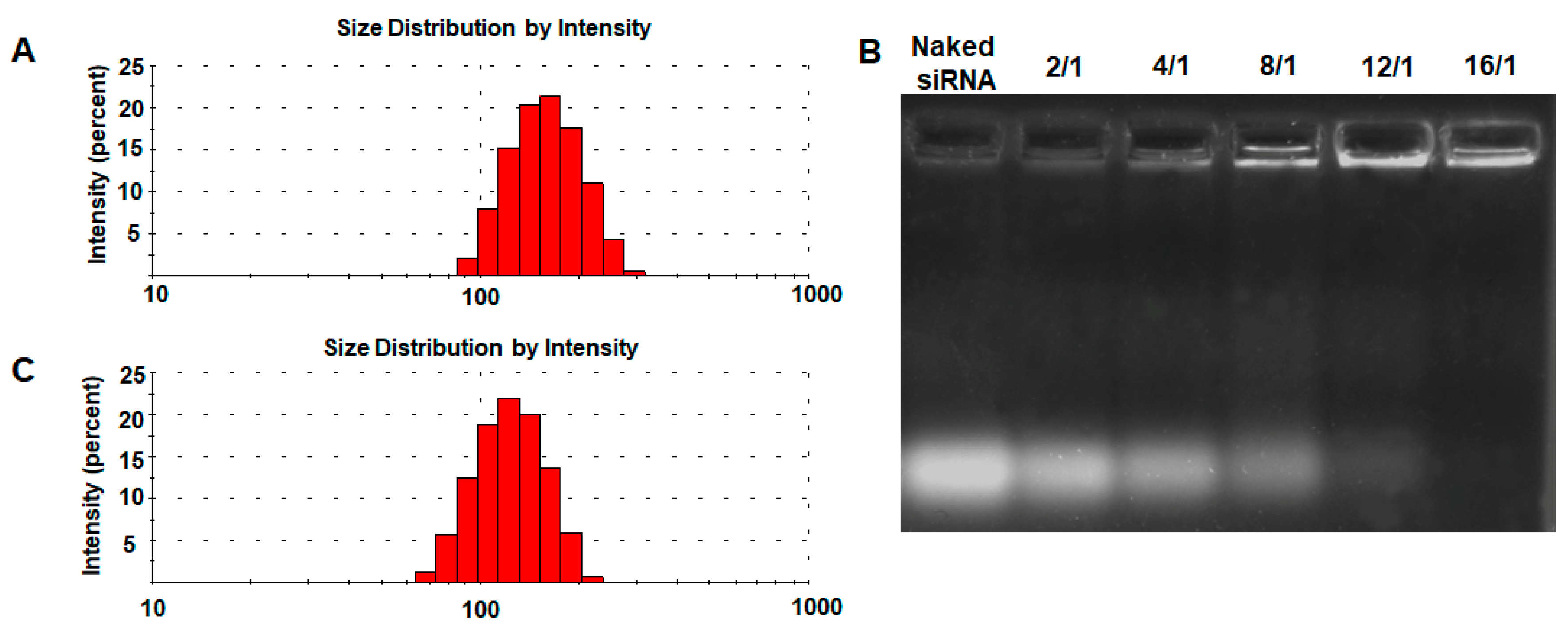
M-MTX was prepared by the self-assembly of MTX-bPEI-LA and mPEG-LA. A specified volume of MTX-bPEI-LA and mPEG-LA solution (with a molar ratio of 1:200) dissolved in chloroform (Sinopharm Chemical Reagent Co., Ltd., Shanghai, China) was mixed together and sonicated for 3 min. Then, the mixture solution was evaporated by a rotary evaporator to remove the chloroform at 37 °C and further under vacuum for 2 h to remove the residual organic solvent and to obtain a film on the flask. To prepare M-MTX, diethyl pyrocarbonate (DEPC)-treated water (Coolaber, Beijing, China) was added to the flask and sonicated for 2 min. The particle size, zeta potential, and polydispersity index (PDI) of M-MTX were measured on a Zeta-sizer Nano ZS90 from Malvern Instruments (Malvern, UK) at 25 °C.
2.4. Preparation and Characterization of M-MTX/siRNA Complexes
Gel retardation assays were performed to investigate the ability of M-MTX to complex siRNA using agarose gel electrophoresis. M-MTX with different concentrations and survivin siRNA solutions were first diluted to prepare the M-MTX/siRNA complexes with different N/P ratios. The desired amount of siRNA solution was then mixed with an equal volume of the M-MTX solution by gentle pipetting. The complexes were incubated for 10 min at room temperature before use. Then, 10 μL of the M-MTX/siRNA complexes with different N/P ratios were mixed with 2 μL of 6× loading dye and loaded into a 2% agarose gel. The voltage of electrophoresis (BIO-RAD Laboratories, Hercules, CA, USA) was set up at 100 V and run for 10 min in a Tris-acetate-EDTA (TAE) buffer (Beijing Dingguo Changsheng Biotechnology Co., Ltd., Beijing, China). After that, the gel was placed in a staining solution containing Molecular Probes SYBR® Gold nucleic acid (Invitrogen, Ltd., Willow Creek Road, Eugene, OR, USA) for 30 min. Free siRNA in the complexes could be detected as a band on the gel with a GelDoc-It Ts Imaging System (Analytik Jena US LLC., Upland, CA, USA). The particle size and the zeta potential of the M-MTX/siRNA complexes were measured by Nano ZS90 (Malvern, UK).
2.5. In Vitro siRNA Release
The in vitro siRNA release curve of FAM-siRNA loaded M-MTX (M-MTX/FAM-siRNA) in phosphate buffer saline (PBS) was studied. 1 mL M-MTX/FAM-siRNA complexes were transferred into a dialysis bag (MWCO 100 KDa, Shanghai Yuanye Biological Technology, Shanghai, China). The dialysis bag was immersed in 40 mL of PBS and stirred at 37 °C at a speed of 100 rpm. At fixed time intervals, 100 μL of the external solution was withdrawn and replaced with the same volume of fresh PBS. The fluorescence intensity of FAM-siRNA was measured by Bio Tek SYNERGY4 (Winooski, VT, USA) at λex = 485 nm and λem = 535 nm, and the concentrations of FAM-siRNA were measured based on a calibration curve of FAM-labeled siRNA with known concentrations.
2.6. Cell Culture
HeLa cells with high folate receptor (FR) expression were used to evaluate the cellular uptake of the M-MTX/siRNA complexes. HeLa cells were cultured in DMEM (Carlsbad, CA, USA) which contained 10% fetal bovine serum (FBS) (Gemini, Woodland, CA, USA) and 1% penicillin-streptomycin (Carlsbad, CA, USA) at 37 °C in a humidified atmosphere of 5% CO2.
2.7. Hemolytic Analysis of M-MTX and MTX-bPEI-LA on Murine Erythrocytes
Fresh blood samples from healthy mice were collected from the orbital sinus in heparin-coated tubes. Red blood cells (RBCs) were collected by centrifuging at 3000 rpm for 5 min and washed three times with physiological saline solution. Then, the RBCs were dispersed in the physiological saline solution to obtain a 2% erythrocyte standard dispersion (v/v). Then, 200 μL various concentrations of MTX and MTX-bPEI-LA (equivalent to MTX-bPEI-LA at concentrations of 0, 40, 80, 100, 150, and 200 μg/mL) were incubated with 1 mL erythrocyte standard dispersion for 3 h. The suspensions were centrifuged, and the absorbance of supernatant (100 µL) was measured at 450 nm.
2.8. The Viability of Cell Cultures Exposed to MTX-bPEI-LA and M-MTX
The carrier cytotoxicity of the MTX-bPEI-LA and M-MTX was studied. The HeLa cells were plated in 96-well microtiter plates (5000 cells per well) and cultured overnight. M-MTX and MTX-bPEI-LA at 3 concentrations (1, 5, and 20 µg/mL) were added. After another 24 h, 20 µL of the MTT solution (5 mg/mL) was added and incubated for 4 h to form formazan crystals. The medium was removed, and the formazan crystals were dissolved by adding 150 µL DMSO and were incubated for 15 min at 37 °C. Absorbance values at 490 nm were measured on Bio Tek SYNERGY4 (Winooski, VT, USA). Relative cell viability was determined and was presented as a viability percentage of the untreated cells.
2.9. Cellular Uptake of the M-MTX/Cy3-Labeled siRNA Complexes
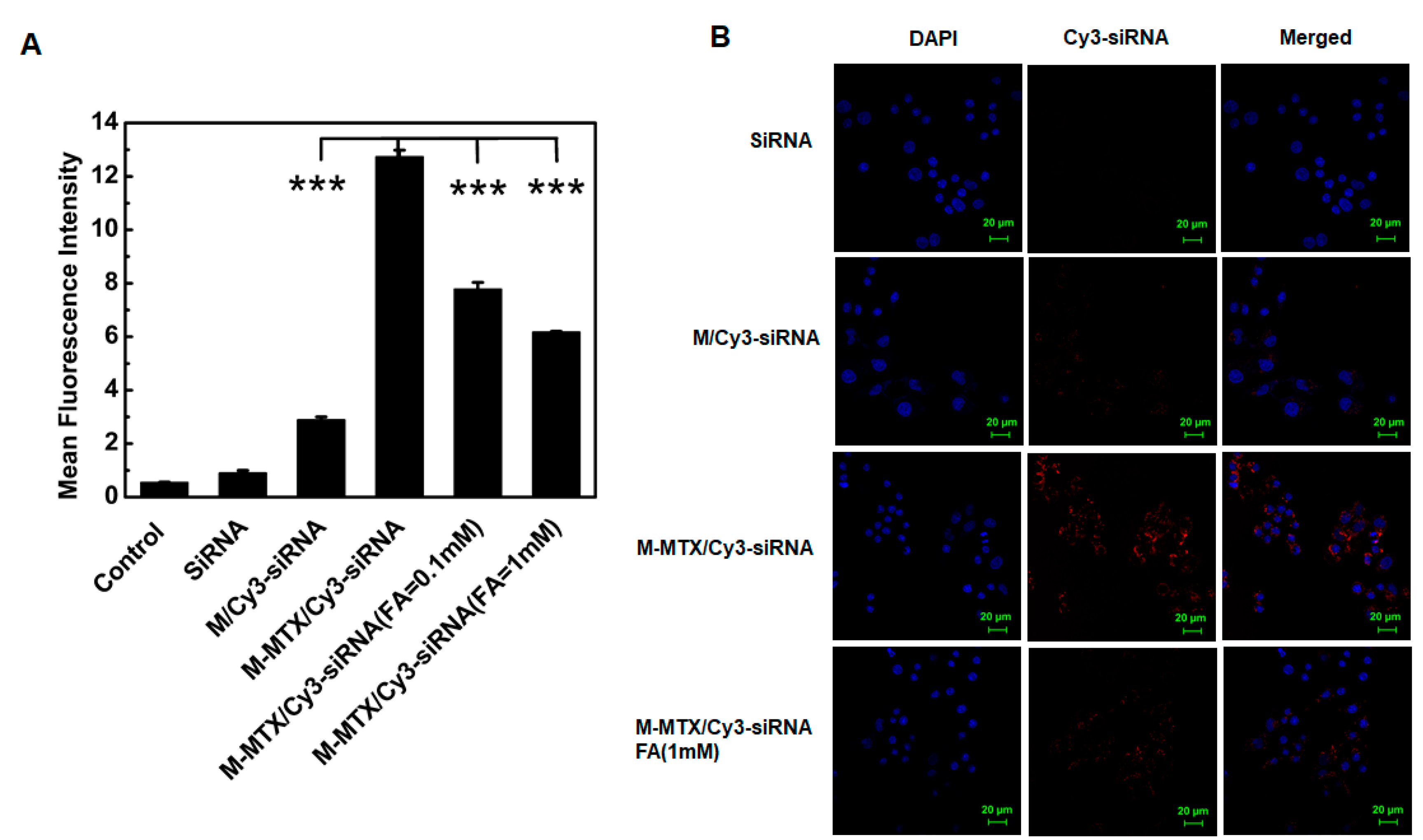
Flow cytometry was first used to investigate the cellular uptake of M-MTX/siRNA complexes. The HeLa cells were seeded in 12-well cell culture plates at a concentration of 1 × 10⁵ per well and cultured for 24 h to be attached to the plates. In order to study the FR targeting ability of the M-MTX/Cy3-labeled siRNA (Cy3-siRNA) complexes, the HeLa cells were first preincubated with free FA (0.1 mM and 1 mM) for 1 h to competitively bind to FR. Mixed micelles without an MTX conjugation but loaded with Cy3-siRNA (M/Cy3-siRNA) were set as a non-FR targeting control. Naked Cy3-siRNA, M/Cy3-siRNA, and M-MTX/Cy3-siRNA with an equivalent siRNA concentration of 100 nM were then added to the plates. After 4 h of incubation, the HeLa cells treated with different formulations were trypsinized, harvested by centrifugation, washed with cold PBS, and resuspended with 4% formaldehyde solution (w/v) (Beijing Dingguo Changsheng Biotechnology Co., Ltd., Beijing, China). The fluorescence intensity of the cells was measured on a Beckman Coulter EPICS XL flow cytometer (Brea, CA, USA). The cellular uptake of M-MTX/Cy3-siRNA was further visualized on a confocal laser scanning microscopy (CLSM). The HeLa cells were collected, counted, and then seeded at the bottom of glass flasks for 12 h. The medium was replaced with fresh opti-MEM (Thermo scientific, Rockford, IL, USA) and preincubated with free folic acid (1 mM) for 1 h. The cells were then treated with naked siRNA, M/Cy3-siRNA, and M-MTX/Cy3-siRNA complexes with an equivalent siRNA concentration of 100 nM at 37 °C. After 4 h of incubation, the medium was removed and the cells were washed gently three times with PBS (0.01 M, pH 7.4). Then, the cells were fixed with 4% (w/v) formaldehyde for 15 min at room temperature and washed repeatedly with PBS three times to remove the formaldehyde. Subsequently, the nuclei were stained with DAPI for 10 min, and the cells were collected after washing with PBS to remove the residual dye. The uptake of the M-MTX/Cy3-siRNA complexes in the HeLa cells was observed using an LSM710 microscope from Carl Zeiss (Oberkochen, Germany).
2.10. Internalization and Endosome Escape of M-MTX/FAM-siRNA Complexes in HeLa Cells
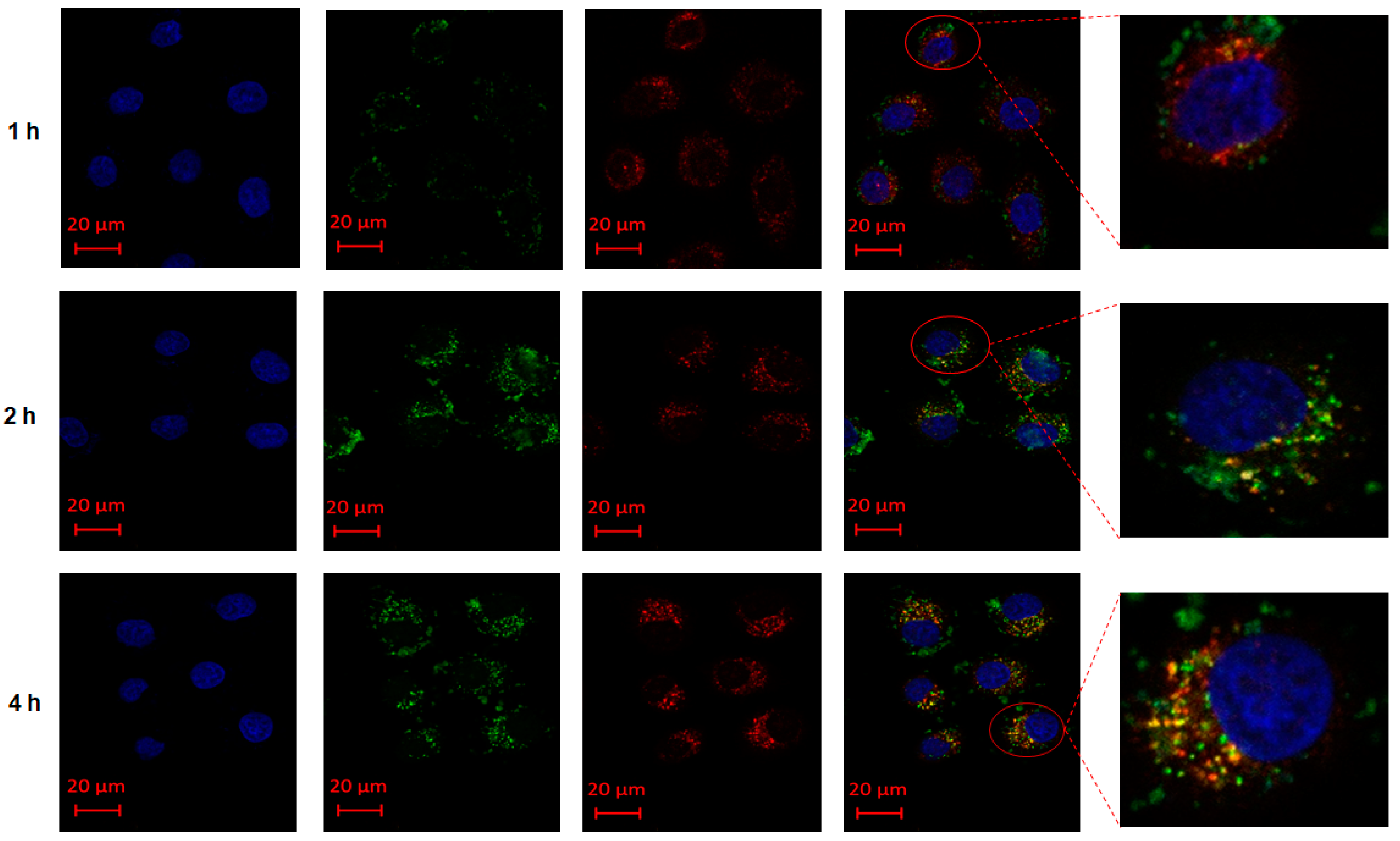
The HeLa cells were seeded at the bottom of glass flasks at a cell concentration of 1 × 10⁵ and cultured for 24 h. The medium was replaced with fresh opti-MEM and incubated with M-MTX/ FAM-siRNA complexes (100 nM) for 1 h, 2 h, and 4 h, respectively. After washing with PBS, the cells were then incubated with Lyso Tracker™ Red DND-99 for 30 min. Then, the supernatant was removed, and the cells were gently washed. The cells were then sequentially fixed with 4% (w/v) formaldehyde and stained with DAPI. After washing away the residual dye, the internalization and endosome escape of M-MTX/ FAM-siRNA complexes was observed on CLSM.
2.11. Cell Cytotoxicity of the M-MTX/Survivin-siRNA
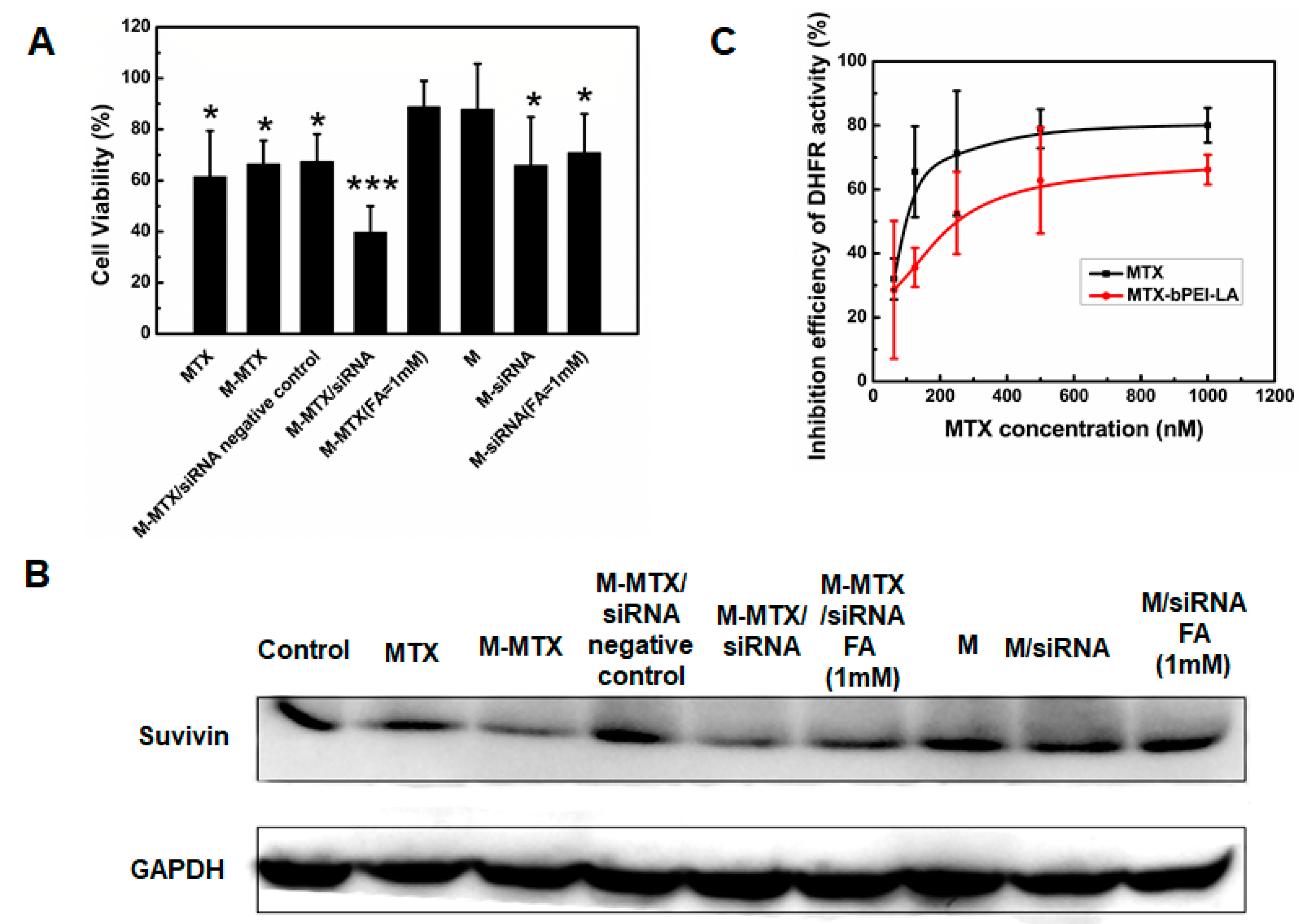
The cytotoxicity of the M-MTX/survivin-siRNA complexes was investigated. The HeLa cells were plated in 96-well microtiter plates (1 × 10⁴ cells per well) and cultured overnight. The cells of the designed wells were pretreated with free FA (1 mM) for 1 h. MTX, M-MTX, M-MTX/survivin siRNA negative control, M, M/survivin-siRNA, and M-MTX/survivin-siRNA were added to the wells (survivin siRNA 50 nM, MTX 0.242 μg/mL). M-MTX loaded with a survivin siRNA negative control was defined as an M-MTX/siRNA negative control. After being cultured for 48 h, 20 μL of the MTT solution (5 mg/mL) was added and incubated for 4 h to form formazan crystals. The medium was removed, and the formazan crystals were dissolved by adding 150 μL dimethyl sulfoxide (DMSO, Sinopharm Chemical Reagent Co., Ltd., Shanghai, China). The absorbance values at 490 nm were measured on Bio Tek SYNERGY4 (Winooski, VT, USA). The relative cell viabilities were presented as a viability percentage of the cells treated with different formulations compared to the untreated cell samples which the viabilities were referred to be 100%.
2.12. Western Blot Test
The survivin expression was analyzed by western blot. The HeLa cells were seeded in 6-well cell culture plates at a concentration of 1.5 × 10⁵ cells per well for 24 h at 37 °C in a 5% CO2 humidified atmosphere. Free FA (1 mM) was preincubated with the cells in the desired wells. MTX, M-MTX, M-MTX/siRNA, M-MTX/siRNA negative control, and M, M/siRNA with an equivalent siRNA concentration to 50 nM were then added to the wells. After 4 h of incubation, the medium was replaced with a fresh medium. After 48 h, the cells of different groups were collected, lysed in radio immunoprecipitation assay (RIPA) lysis buffer containing 1% protease inhibitor cocktail (Sigma-Aldrich, St. Louis, MO, USA) and 2% phenylmethanesulfonyl fluoride (Sigma-Aldrich, St. Louis, MO, USA) and plated on ice for 15 min. Protein fractions were collected by centrifugation at 10,000 rpm at 4 °C for 10 min, quantified by a bicinchoninic acid (BCA) Protein Assay Kit (Thermo scientific, Rockford, IL, USA), subjected to polyacrylamide gel electrophoresis containing 10% sodium dodecyl sulfate (SDS-PAGE), and then transferred to polyvinylidene fluoride (PVDF) membranes (0.45 m, Merck Millipore, Billerica, MA). The membranes were blocked with a 5% bovine serum albumin (BSA) (Sigma-Aldrich, St. Louis, MO, USA) solution (w/v) for 4 h at room temperature and incubated with survivin rabbit mAb (71G4B7E, Cell Signaling Technology Inc, Danvers, MA, USA) and GAPDH (ab181602, Abcam, cambridgeshire, UK) antibodies at 4 °C overnight, respectively. Horseradish peroxidase (HPR)-conjugated secondary antibody (Beijing Dingguo Changsheng Biotechnology Co., Ltd., Beijing, China) was added and incubated with the membranes at 4 °C for 4 h. The corresponding protein expression was measured by an electrochemiluminescence (ECL) detection kit (Merck Millipore, Billerica, MA, USA) and visualized by an imaging system (BioSpectrum 600, Analytik Jena US LLC., Upland, CA, USA).
2.13. Dose-Dependent Inhibition Efficiency of MTX and M-MTX on Dihydrofolate Reductase (DHFR) Activity
The inhibition efficiency of MTX and M-MTX on DHFR activity was carried out in accordance to the protocol of the Dihydrofolate Reductase Activity Kit (Colorimetric) from BioVision (S. Milpitas Blvd., Milpitas, CA, USA). Briefly, 40 μL of NADPH (500 μM), 60 μL of the DHFR substrate (15-fold dilution), and 50 μL of a series of concentrations of MTX or MTX-bPEI-LA (an equivalent MTX concentration 62.5 nM to 1000 nM) were added to each well of a 96-well clear plate. Finally, 50 μL of DHFR (250-fold dilution) was added to the wells to initiate the reaction with a total volume of 200 μL per well. The absorbance values at 340 nm were measured for 10 min at room temperature. The wells without MTX and MTX-bPEI-LA were set as the positive controls, and the wells without the DHFR enzyme were set as the negative controls. The Inhibition efficiency was defined as follows:
2.14. Establishment of Tumor Model
The animal experimental protocol was in compliance with the institutional guidelines and was approved by the Experimental Animal Ethics Committee of the School of Life Sciences, Jilin University. The number for the permit for the animal experiment was 201805003 from the Experimental Animal Ethics Committee of the School of Life Sciences, Jilin University. BALB/c nude mice (female, 6–8 weeks) were obtained from Beijing Vital River Laboratory Animal Technology Co., Ltd. (Beijing, China). Tumor-bearing mice were established through the subcutaneous injection of 5 × 10⁶ HeLa cells into the right rear leg of nude mice after the mice were adapted to the new environment for a week.
2.15. Accumulation of M-MTX/Cy5-Labeled siRNA (Cy5-siRNA) Complexes in Tumor Tissue
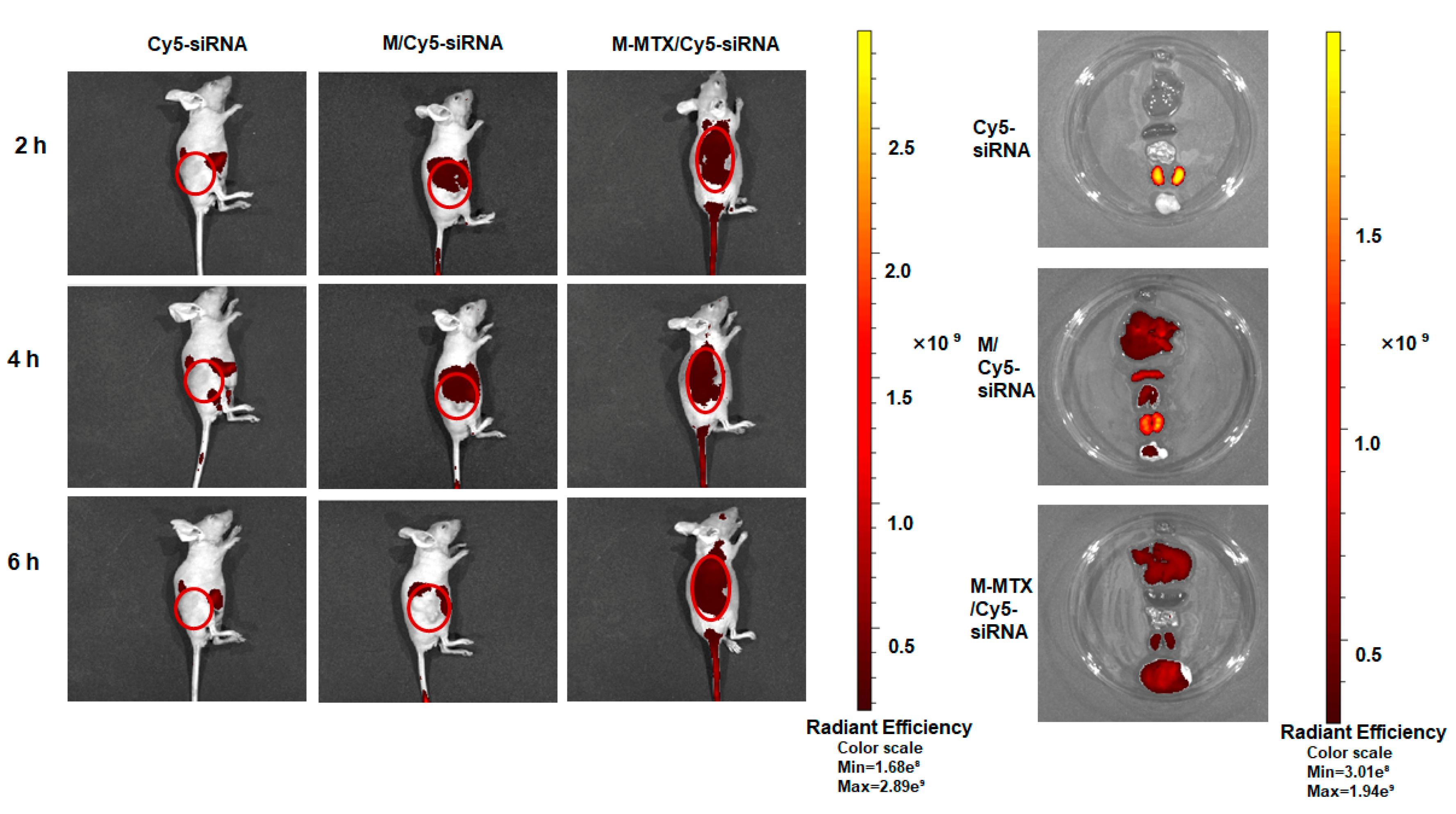
Tumor-bearing mice were injected with Cy5-siRNA, M/Cy5-siRNA, and M-MTX/Cy5-siRNA via the tail vein with an equivalent amount of siRNA (1 nmol). The biodistribution of the complexes was visualized by an IVIS® spectrum system from Caliper Life Sciences (Hopkinton, MA, USA) at the second, fourth, and sixth hour after administration. The mice were anesthetized by administrating a 1% (w/v) pentobarbital sodium solution to the abdomen, and the optimized parameter (excitation, 640 nm; emission, 680 nm) was set up for image acquisition at various time points. At the sixth hour after administration, the internal organs (Heart, Liver, Spleen, Lung, and Kidney) and tumors were dissected and then visualized by the In Vivo Imaging System.
2.16. In Vivo Antitumor Efficacy of M-MTX/Survivin siRNA Complexes
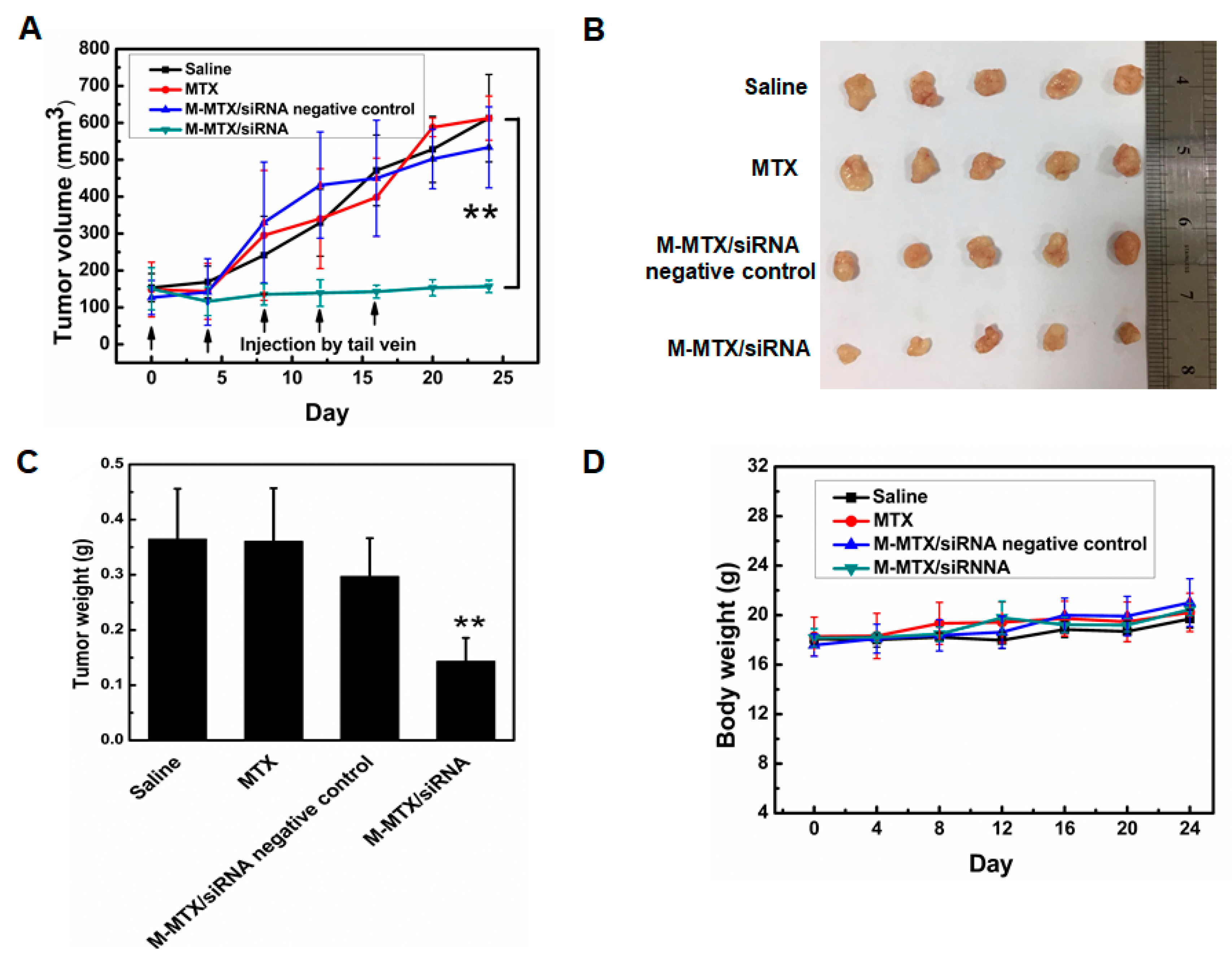
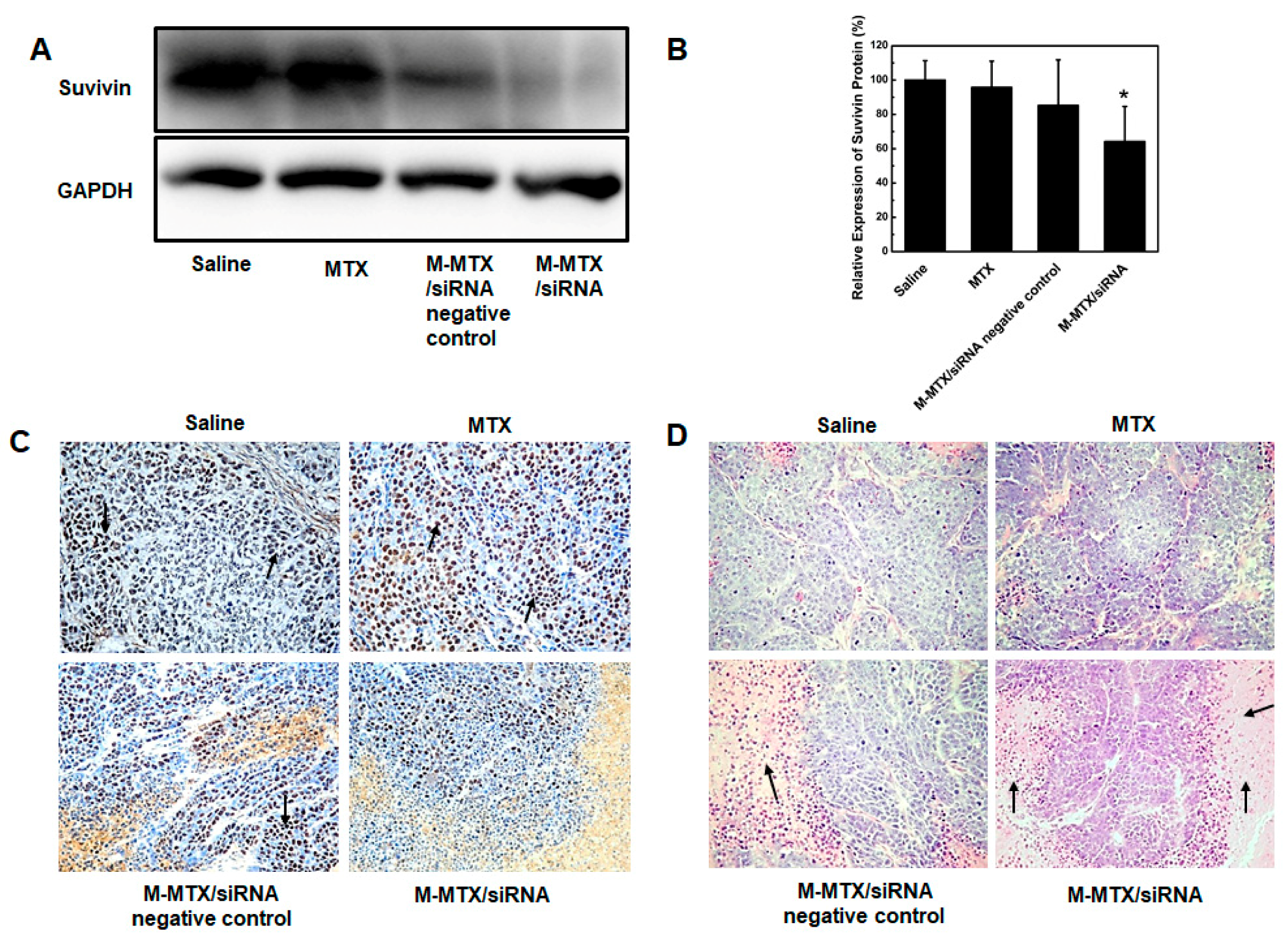
When the average tumor volume of nude mice was grown to approximately 100–150 mm3 (Day 0), the animals were randomized into four groups, each group containing 5 nude mice. Tumor-bearing mice were then injected with saline, free MTX, the M-MTX/siRNA negative control, and M-MTX/siRNA via the tail vein, respectively. The formulations (MTX 500 μg/kg, siRNA 2 nmol) were administered every 3 days. Simultaneously, the volume of the tumor and body weight were measured every 4 days using a vernier caliper and scale. On day 24, the mice were anesthetized with sodium pentobarbital and sacrificed. The tumors were dissected, weighed, and fixed for hematoxylin and eosin (H&E) and immunohistochemistry staining. The expression of survivin in tumor tissue was also analyzed by a western blot assay. The pixel density of the survivin bands compared to the GAPDH bands was quantified for each sample using Image J software (National Institutes of Health, Bethesda, MD, USA).
2.17. Histopathologic Analysis
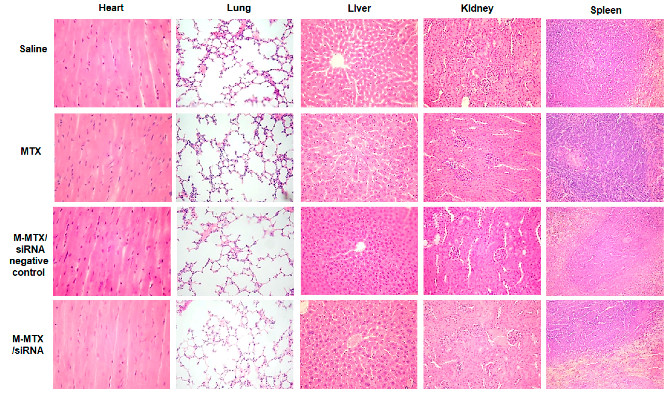
All mice were euthanized with a 1% pentobarbital sodium solution on day 24. Vital organs (Heart, liver, Spleen, Lung, and Kidney) and tumors were dissected in each group and fixed with 4% paraformaldehyde for histopathologic analysis. Tissue sections were cut into 5 microns thick and stained with H&E. For the immunohistochemistry analysis, the tumor tissue sections were first incubated with survivin rabbit mAb.
2.18. Statistical Analysis
Data were expressed as mean ± SEM and graphed by Origin 8.0 (OriginLab Corp., Northampton, MA, USA). The statistical analysis of two group differences and correlations was determined using Student’s t-test. * p < 0.05 was considered statistically significant. ** p < 0.01 and *** p < 0.001 were considered highly significant.
4. Discussion
MTX, a FA analog, has long been used for cancer therapy but is often associated with severe systemic toxicity, bone marrow suppression, and drug resistance [
6,
7,
43]. In addition, MTX monotherapy has limited effectiveness. Therefore, the new combination strategy of MTX and siRNA as well as a novel drug delivery system that reduces MTX toxicity and improves cancer efficacy is desirable [
8,
9]. As one of the strongest tumor apoptosis inhibitors, survivin not only promotes tumor cell proliferation but also is closely related to the development of tumor resistance [
14,
44,
45]. Thus, the combination therapy of MTX and survivin-siRNA demonstrated a new possibility for enhanced tumor efficacy. In order to efficiently deliver MTX and survivin-siRNA into the cells, modified cationic polymer-based mixed micelles having an MTX targeting ability were designed due to their simplicity in synthesis and preparation.
LA is a polyunsaturated fatty acid that is essential for the body and is nontoxic. Moreover, recent studies reported that LA also inhibited tumor cell growth and metastasis [
46,
47,
48]. Therefore, LA was selected to be separately conjugated to the b-PEI and mPEG to form two amphiphilic polymers of bPEI-LA and mPEG-LA in this work. The hydrophobic molecule of LA conjugated to bPEI was designed to reduce the high toxicity of bPEI [
38]. MTX was then conjugated to the bPEI-LA by an esterase-stable amide linkage (
Figure S1). MTX was conjugated to bPEI-LA by an amide linker to prevent and avoid MTX release in the blood and to enhance the targeting ability of MTX to the FA receptor. Then, M-MTX self-assembled by MTX-bPEI-LA and mPEG-LA was prepared and applied to efficiently co-deliver MTX and siRNA. M-MTX was aimed to target the tumor cells overexpressing FR and successfully release loadings into the cytoplasm as shown in
Scheme 1. Actually, some researchers had revealed that MTX conjugated to the dendrimers showed a potential FR targeting ability through the tight-binding of MTX to the folate binding protein [
49]. From the reversed cellular uptake (
Figure 2) and biological activities (
Figure 4) of M-MTX/Cy5-siRNA-treated groups preincubated with free FA and the high cellular uptake (
Figure 2), biological activities (
Figure 4) in vitro, and accumulation of M-MTX/Cy5-siRNA in the solid tumor (
Figure 5), we point out that M-MTX nanocarrier exhibits an FR targeting ability and a much higher internalization efficiency via FA receptor-mediated endocytosis than M/siRNA complexes [
50]. In
Figure 4C, when M-MTX and MTX were incubated with cells for 4 h, the M-MTX-treated group exhibited a lower protein expression compared to free MTX. This also indicated that M-MTX entered more into the cells and had a higher internalization efficiency compared to free methotrexate. The siRNA and MTX-bPEI-LA conjugates with stable chemical linkage achieved endosome escape (
Figure 3) and were released probably by the proton sponge effect of cationic polymer of bPEI [
33,
34]. As expected, M-MTX/survivin siRNA achieved tumor growth inhibition and disease remission with a low MTX and siRNA dose in tumor-bearing mice. There was no significant difference in the antitumor effect among saline, MTX, and the M-MTX/siRNA negative control with a low dose of MTX, which was not sufficient to achieve disease remission. However, the M-MTX/siRNA complexes exhibited an unexpectedly potent effect compared to MTX and the M-MTX/siRNA negative control. This may largely depend on the specific targeting and efficient delivery of siRNA by the bifunctional vector of M-MTX. Moreover, previous studies have shown that survivin siRNA may have a positive effect on chemotherapeutic drug sensitivity and may reduce drug resistance [
14]. Thus, we propose that the effect of mutual promotion and the sensitivity of the two drugs at a low dose also contribute to the desired efficacy. The M-MTX carrier exhibited good biocompatibility and low toxicity as a non-viral carrier for siRNA co-delivery in vivo. However, the specific and detailed mechanisms and synergy of MTX and siRNA warrant further investigation.

 ,
, 


