1. Introduction
Over the past several decades, remarkable progress has been made in the fabrication of polymer nanoparticles (NPs) with a diameter in the range of 1 to 1000 nm for applications in the controlled release and delivery of drugs [
1,
2]. In particular, polymer hollow NPs, which are characterized by interior voids and outer shells, have sparked a growing interest because their hollow interiors can highly encapsulate bioactive drugs, and their shells can efficiently protect the bioactivity of encapsulated drugs from deactivation [
3,
4]. Furthermore, the shells function as diffusion barriers against the undesired noticeable release of drugs at the initial stage and allow their linear release over time, which is called zero-order release [
5,
6,
7].
Although this release pattern is beneficial for chemotherapy, many clinical situations require the drugs to be released on-demand rather than continuously [
8,
9,
10]. This has led to the development of stimuli-responsive polymer hollow NPs, which allow a controlled release of drugs in response to environmental stimuli [
11,
12,
13,
14,
15]. The stimuli responsiveness is achieved by including functional monomers or inorganic components in the hollow NPs [
4]. As an example, Yang and coworkers demonstrated the fabrication of double-walled hollow nanoparticles with uniform size through the synthesis of silica/polymer hybrids by alternating sol–gel and distillation–precipitation polymerization processes, followed by the selective removal of the silica component. The resulting NPs were composed of a poly(methacrylic acid) (PMAA) inner shell and a poly(
N-isopropylacrylamide) (PNIPAM) outer shell with independent sensitivity to external pH and temperature [
16]. In a similar way, poly(
N,
N′-methylene bisacrylamide-
co-methacrylic acid) (P(MBAAm-
co-MAA)) hollow NPs were prepared from PMAA/P(MBAAm-
co-MAA) core-shell particles through the selective removal of the non-crosslinked PMAA core in ethanol. The release of the anticancer drug molecules encapsulated into the core was strongly dependent on pH [
17]. Chiu and coworkers used hydrogel hollow NPs incorporated with superparamagnetic iron oxide NPs to accelerate the anticancer drug release in response to both pH and temperature [
15]. Recently, polymer hollow NPs responsive to typical physiological stimuli such as pH and redox potential were also fabricated via the self-assembling of a novel amphiphilic diblock copolymer, allowing the release of tetraphenylporphyrin tetrasulfonic acid hydrate for photodynamic therapy [
18]. However, most of these systems consist of non-biocompatible, non-biodegradable components, which have hindered their practical use in biomedical applications. Moreover, since the closed shells of these particles make it difficult to load drug molecules in the interior voids through diffusion, their drug-loading capacity is very low, and the encapsulation of macromolecular drugs is limited.
In this study, we introduce biocompatible and biodegradable polymer hollow NPs for the easy loading and near infrared (NIR) light-triggered release of drugs. The fabrication of the hollow NPs involves the use of soft lithography to pattern a composite film made of poly(ε-caprolactone) (PCL), a naturally occurring biodegradable fatty acid (FA) with phase-change ability, and the Food and Drug Administration (FDA)-approved, biocompatible indocyanine green (ICG) as a photothermal agent to an array of discrete rings, followed by their transformation into spherical hollow NPs with surface openings. The openings, which can enhance the connectivity between the interior and the exterior, enable the easy loading of drug molecules into the interior voids, and their successive sealing ensures a stable encapsulation of the drug. Upon exposure to an external NIR source, the photothermal agent entrapped into the shells of these hollow NPs can generate heat that raises the local temperature of the NPs above the melting point of the constituent FA, leading to the formation of nanopores on their shells, and consequently, the instant release of the encapsulated drug molecules through the pores. Thus, a noticeable anticancer activity can be achieved through the combination of the hyperthermia effect from the photothermal agent and the NIR light-triggered release of the drug molecules.
3. Results and Discussion
In order to fabricate biocompatible and biodegradable hollow NPs for the easy loading and NIR light-triggered release of drugs, we chose to work with PCL and FAs due to their attractive features. Along with excellent biocompatibility and biodegradability, PCL has a slow degradation rate that is advantageous in the stable encapsulation of payloads without their undesired release [
23]. FAs are naturally occurring biocompatible and biodegradable materials that are capable of reversible solid–liquid transitions in response to small variations in temperature [
10,
24]. In particular, the eutectic mixture made of LA and SA has increasingly found biomedical applications because its melting point (i.e., 38–40 °C) is close to the physiological temperature of human bodies [
25,
26,
27,
28].
Figure 1A schematizes the fabrication procedure for the hollow NPs consisting of PCL and the eutectic FA mixture. Four major steps are involved: the generation of discrete PCL/FA composite rings on top of a PVP layer; the release of the rings into an aqueous solution of PVP; the conversion of the rings into solid nanospheres through solvent treatment; and their transformation into hollow NPs with openings on their shells via freeze-drying.
Figure 1B shows an SEM image of an array of the composite rings consisting of PCL and the eutectic mixture of LA and SA, supported on the PVP-coated substrate. The initial PCL/FA composite layer was 4.1 nm in thickness, which was prepared by spin-coating its solution with 0.02 wt% of the FA mixture and 0.2 wt% of PCL. The FA mixture and PCL were melted and dewetted by heating up to 80 °C, being transformed to the droplets that were randomly distributed on the surface of PVP (
Figure S1, Supplementary Materials). The recessed regions on the PDMS mold guided this dewetting process, since the capillary force pushed the melted composite into the cylindrical wells on the mold [
5], leading to the generation of an array of PCL/FA composite rings. The ring shape resulted from the production of composite meniscus along the PDMS walls, which is a signature mark of the capillary rise [
29].
The generated rings were released into the aqueous solution of PVP, followed by addition of a small amount of toluene.
Figure 1C shows an SEM image of the resultant sample after the release, solvent treatment for 1 h, and its evaporation in air. Upon the addition of toluene, which is highly miscible with PCL and the FA mixture but immiscible with water, the solvent diffused into the rings to have the composite molecules migrated even at room temperature. Thus, the rings were transformed into nanospheres, reducing the contact area with water. The TEM image in the inset demonstrates that these spheres had a solid structure with uniform size. They had a
ξ-potential of −29.8 mV, and their mean diameter measured from these images was 284 ± 12 nm, which was comparable to the result obtained by DLS (301 ± 18 nm) and the theoretically calculated value (
Figure S2, Supplementary Materials). The diameter can be easily controlled by varying the thickness of the composite layer used to generate the discrete rings, because there is a one-to-one correspondence between the solid NP and ring [
5].
Figure 1D shows an SEM image of the NPs with well-defined openings on their shells, which were fabricated by releasing the composite rings and treating with toluene, followed by freeze-drying in vacuum. Each particle exhibited an opening of ca. 165 nm in diameter on its shell and an increased outer diameter, compared with the solid nanospheres of
Figure 1C, of 340 ± 20 nm. A TEM image is shown in the inset, confirming that these NPs were hollow. The formation of the interior voids and openings was because of the strong evaporation flux of toluene [
10]. The solvent treatment led to the conversion of the rings into the solid NPs, as shown in
Figure 1C, but the solvent was still present in the NPs due to its greater affinity to the composite rather than to water. After freezing into an LN
2 bath, the NPs was slowly warmed up in vacuum to reach a temperature above the melting point (−95 °C) of the solvent. As driven by the strong evaporation flux of the solvent, the polymer chains and FA molecules moved toward the surface of each particle, leading to the formation of a cavity in the interior and an opening on the shell.
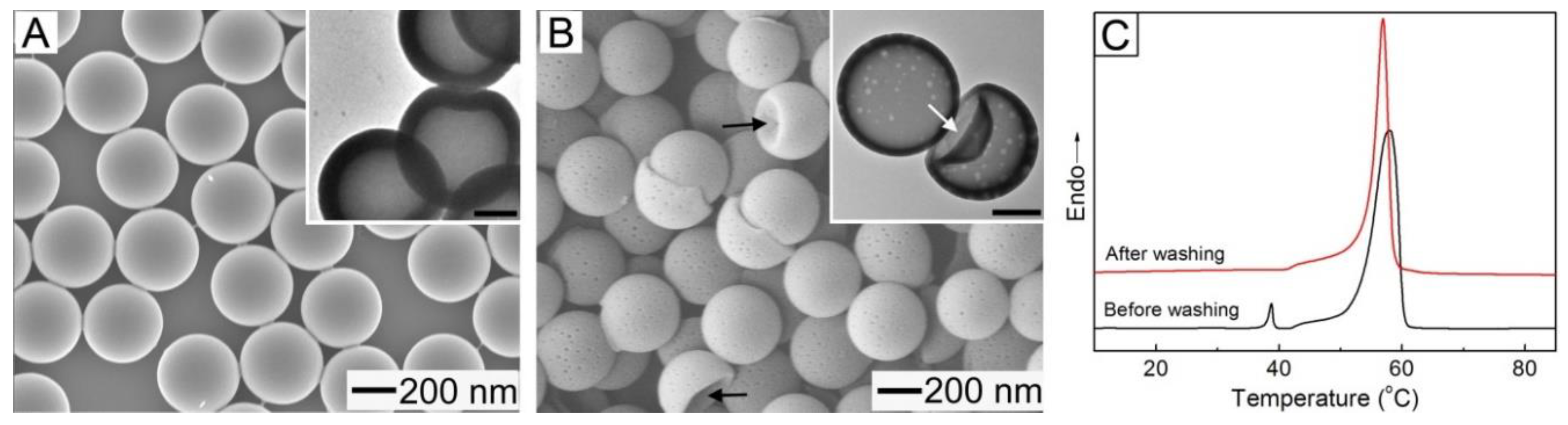
The openings could be completely sealed at room temperature through solvent annealing using a good solvent for the FA mixture and PCL.
Figure 2A shows an SEM image of the sample obtained after suspending the NPs of
Figure 1D in water, followed by the addition of a small amount of toluene as a plasticizer. The solvent treatment decreased the mean diameter of the resultant particles to 310 ± 15 nm and completely closed the openings on their shells. Furthermore, as shown in the TEM image of the inset, the hollow interiors of the particles were retained after the openings were closed. In order to investigate the driving force of the sealing, we theoretically calculated the interfacial free energy (
E) for a hollow particle with an open hole on its surface using a simple model (
Figure S3, Supplementary Materials).
E can be expressed by the interfacial tension between the inner surface and water (
γinner/water), the interfacial tension between the outer surface and water (
γouter/water), the outer (
douter) and inner (
dinner) diameters, and the two angles (
θinner and
θouter) [
30,
31]:
The relation among
dinner,
douter,
θinner, and
θouter is established by considering the diameter of opening (
DH), which has a form of
DH =
dinner sin
θinner =
douter sin
θouter [
31,
32]. By substituting the values of
dinner,
douter, and
DH measured by SEM and TEM characterizations into the relation, we obtained the theoretical values of
θinner and
θouter. The interfacial tension could be estimated using a harmonic mean equation given as:
γsurface/water =
γsurface +
γwater − 4
γsurfacedγwaterd/(
γsurfaced +
γwaterd) − 4
γsurfacepγwaterp/(
γsurfacep +
γwaterp). By assuming a PVP-coated outer surface and a PCL inner one for the hollow particle, we could obtain
γouter/water from the surface tensions for water and PVP available in literature [
31], while
γinner/water was calculated using the surface tension for PCL instead of that for PVP [
33]. The calculated
E for a hollow NP with a closed shell (
Figure 2A) was calculated to be 5.39 × 10
−15 J, which was smaller than the value (6.95 × 10
−15 J) for a NP with a surface opening (
Figure 1D). This result suggests that the sealing of the opening was due to the reduction in
E.
Figure 2B shows an SEM image of the NPs obtained after washing the sample of
Figure 2A with water at 40 °C, which is slightly above the melting point of the FA mixture, confirming the generation of nanopores with a diameter of ca. 15 nm on the shell of each particle. The NPs marked by arrows exhibited a deformed structure (with a dimple), which implies that they were still hollow. The TEM image in the inset clearly demonstrates the generation of the nanopores on the shell and the presence of the hollow interior. In order to understand the formation of these pores, we conducted a DSC analysis for the hollow NPs before and after the washing (
Figure 2C). The non-washed sample exhibited two sharp peaks at 39 °C and 58 °C that corresponded to the FA mixture and PCL, respectively, but the peak from the FA mixture was not observed for the washed sample. These results indicate that the formation of the nanopores was due to the removal of the FA mixture phase-separated with PCL from the shell. The loading content of the FA mixture in the non-washed NPs (defined as a weight percentage of the mixture relative to the NPs) was 7.3%.
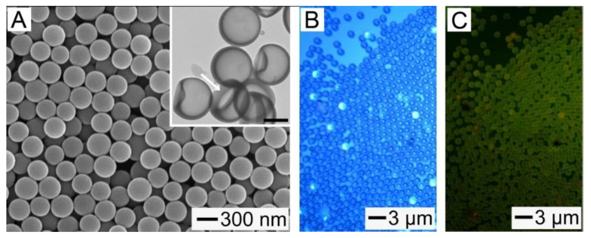
The formation and sealing of the openings on the shells of the hollow NPs can be advantageous for the quick and stable encapsulation of various types of drugs. In order to demonstrate the efficient encapsulation of macromolecular drugs, the NPs shown in
Figure 1D were mixed with an aqueous solution of BSA, followed by solvent annealing for 30 min. We used 1,4-dioxane, instead of toluene, for the annealing because the aqueous dioxane did not cause the denaturation of the protein [
7]. The use of 1,4-dioxane resulted in the complete closing of the surface openings, and consequently allowed the stable loading of BSA, as shown in
Figure 3A.
Figure 3B,C show the optical and fluorescence micrographs of the hollow particles encapsulating FITC-BSA in their interior voids. For an easy optical microscopy observation, the hollow particles with
douter of 1.2 μm were used (
Figure S4, Supplementary Materials). Each particle exhibited a fluorescence due to FITC-BSA (
Figure 3C), which confirms the effective encapsulation of the protein in the hollow interiors of all the particles. The amount of BSA encapsulated in a hollow particle is given by:
MBSA = π(4/3)(
dinner/2)
3·
CBSA, where
CBSA is the concentration of FITC-BSA solution. The mass of a hollow particle encapsulated with the protein is expressed as:
MHollow = π(4/3)[(
douter/2)
3 − (
dinner/2)
3]
ρHollow +
MBSA, where
ρHollow is the density of the particle. Based on the values of
douter (1.2 μm) and
dinner (0.9 μm) obtained from the TEM image in
Figure S4 along with the assumption of
ρHollow ≈
ρPCL = 1.1 g/mL [
34], the calculated encapsulation content (
MBSA/
MHollow × 100) was 1.7%, which was in reasonable agreement with the experimental value of 2.2%. This result suggests that the amount of drug encapsulated in the hollow particles could be readily controlled by varying the concentration of the drug solution for the loading process. In a similar way, we loaded DOX, which is an anticancer drug, into the interior voids of the hollow particles to demonstrate the convenient encapsulation of small molecular drugs (
Figure 3D,E). The encapsulation of the drug was also confirmed using DSC characterization. As shown in
Figure 3F, the hollow NPs encapsulated with DOX exhibited a sharp peak at 213 °C that corresponded to the drug, indicating the successful encapsulation of DOX in the NPs.
In order to provide the hollow NPs with a responsiveness to NIR light, we added them with ICG, which is a NIR light absorbing photothermal agent [
21]. Its inclusion was accomplished through a series of processes involving the formation of a composite layer consisting of the FA mixture, PCL, and hydrophobic ICG-tetrabutylamine, the generation of corresponding composite rings, their conversion into hollow particles with openings on the shells, and the sealing of the openings. The SEM and TEM (inset) images in
Figure 4A show the NPs fabricated from a mixture solution containing PCL (0.2 wt%), eutectic FA mixture (0.02 wt%), and hydrophobic ICG (0.0004 wt%), demonstrating that they had hollow voids and closed shells. Their mean outer diameter, obtained from these images, was 313 ± 18 nm, which is similar to the result measured by DLS (326 ± 23 nm), and they had a
ξ-potential of −30.3 mV. The loading content of the FA mixture in the hollow NPs was 7.1%, which was consistent with the result for a similar sample without the photothermal agent (
Figure 2A). As for the result in
Figure 2B, this hollow structure allowed the NPs to be mechanically deformed, as marked with a white arrow in the inset. These results collectively suggest that the inclusion of ICG did not disturb the formation of the hollow NPs.
Figure 4B,C shows the optical micrographs of the ICG-loaded hollow particles with closed shells. The fluorescence from ICG was observed in all the particles, which suggests its successful inclusion in each hollow particle.
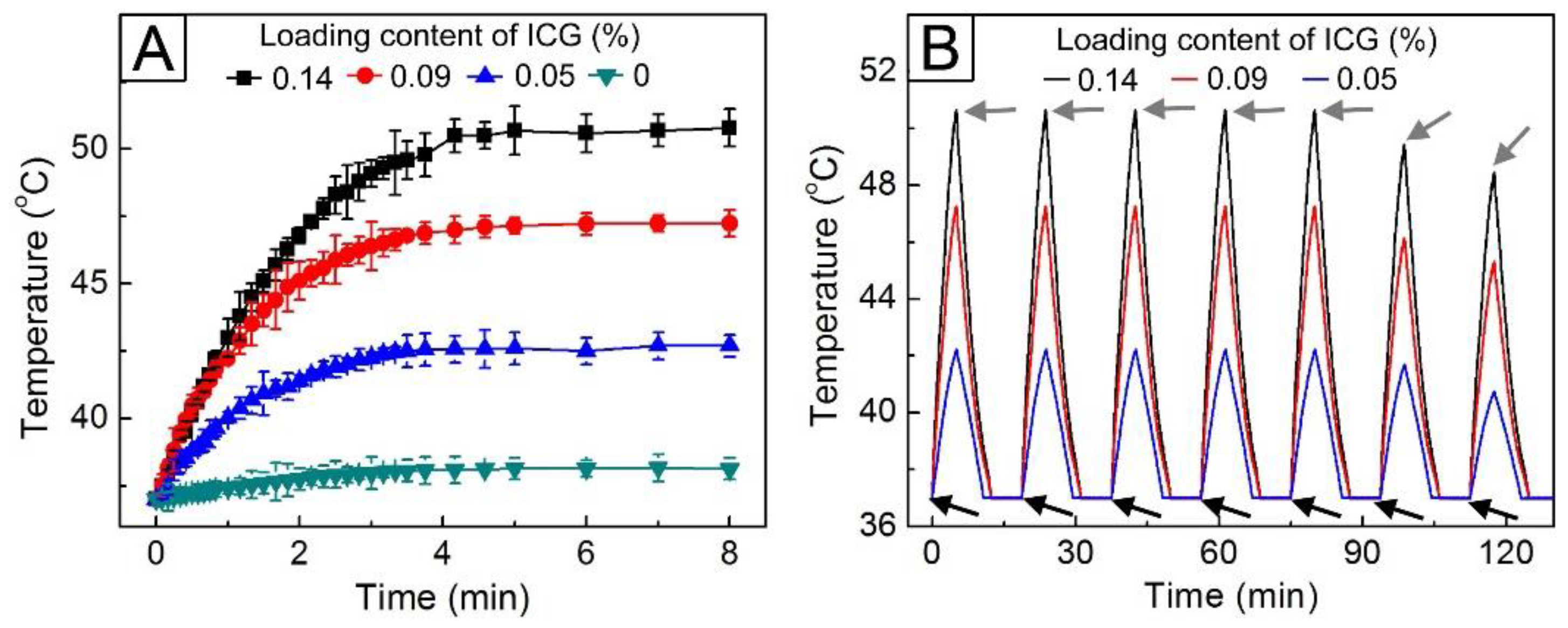
The inclusion of ICG in the hollow NPs was further verified by investigating their photothermal ability.
Figure 5A shows the time-dependent temperature curves for 5 mL of an aqueous suspension containing 5 mg of the hollow NPs under 0.7 W/cm
2 NIR light. The sample without the inclusion of ICG exhibited no change in temperature, whereas the temperature of the samples containing ICG readily increased in response to NIR light irradiation because of the photothermal effect by ICG itself. This increase in temperature was strongly dependent on the loading content of ICG. The temperature of the NPs with a loading content of 0.05% (
Figure 4A) was raised up to 42.5 °C. As the loading content increased to 0.14%, the temperature rise was even larger. The change in temperature could occur in an on–off manner when the NIR light was applied in an on–off way (
Figure 5B). This reversible manner in temperature change was maintained when the light was switched on and off five times under the same condition. However, the temperature increase after the fifth cycle was not as large as that in the previous runs, which was due to the photobleaching of ICG [
35,
36]. Based on these results, the NPs with an ICG loading content of 0.05 wt% were used to further investigate the NIR light-triggered drug release.
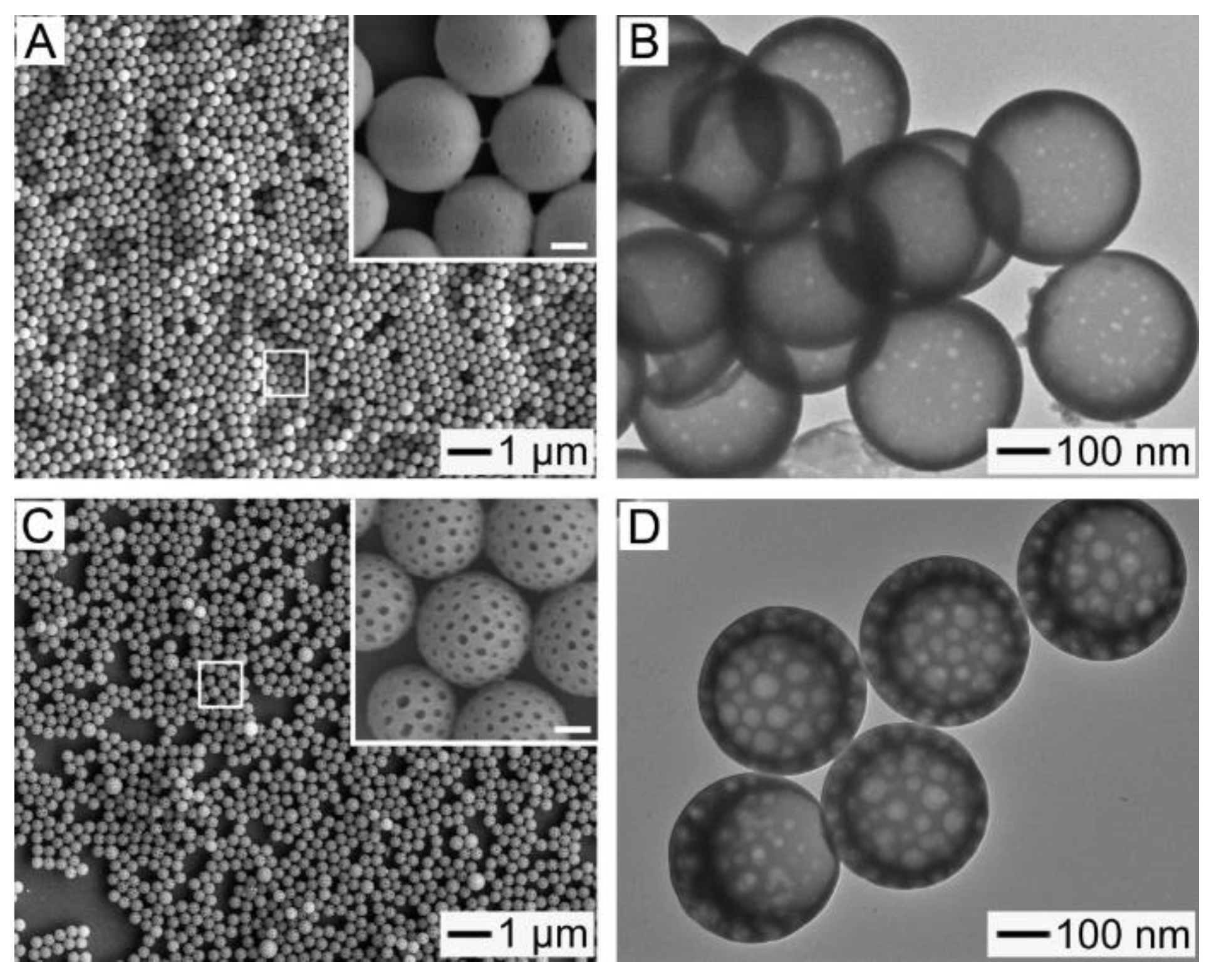
Figure 6A shows an SEM image of the resultant NPs after treating the sample in
Figure 4A with 0.7 W/cm
2 NIR light. It is clear that they were still spherical without any change in size. The inset is a magnified view of the area marked by the white box and demonstrates the formation of nanopores with a diameter of ca. 15 nm on the shell of each NP, as further confirmed by the TEM image shown in
Figure 6B. Under NIR light irradiation, the photothermal conversion by ICG included in the hollow NPs increased their local temperature above the melting point of the embedded FA mixture, leading to the melting away of the FA mixture and thereby the formation of nanopores on the shells of the NPs. The size of these pores could be increased by raising the amount of FA mixture in the hollow NPs. By using a mixture solution of PCL (0.2 wt%), FA mixture (0.04 wt%), and hydrophobic ICG (0.0004 wt%), we could obtain hollow NPs with a higher loading content (14.1%) of the FA mixture.
Figure 6C shows an SEM image of the sample obtained after exposing the NPs to 0.7 W/cm
2 NIR light. The image indicates that the resultant particles still had a spherical structure and a uniform size after the NIR treatment. The magnified view in the inset clearly demonstrates the formation of nanopores with a diameter of ca. 40 nm. However, despite the formation of these larger pores, the hollow interiors of the NPs were maintained without any destruction (
Figure 6D).
In order to investigate the NIR light-sensitive release behavior of DOX from the NPs encapsulating the drug, we prepared and tested the four types of hollow NPs: poly(ε-caprolactone) (PCL)/DOX NPs, PCL/ICG/DOX NPs, PCL/FA/DOX NPs, and PCL/FA/ICG/DOX NPs. The diameter,
ξ-potential, and composition for each sample are summarized in
Table S1 (Supplementary Materials), indicating that these aspects were similar for all the samples.
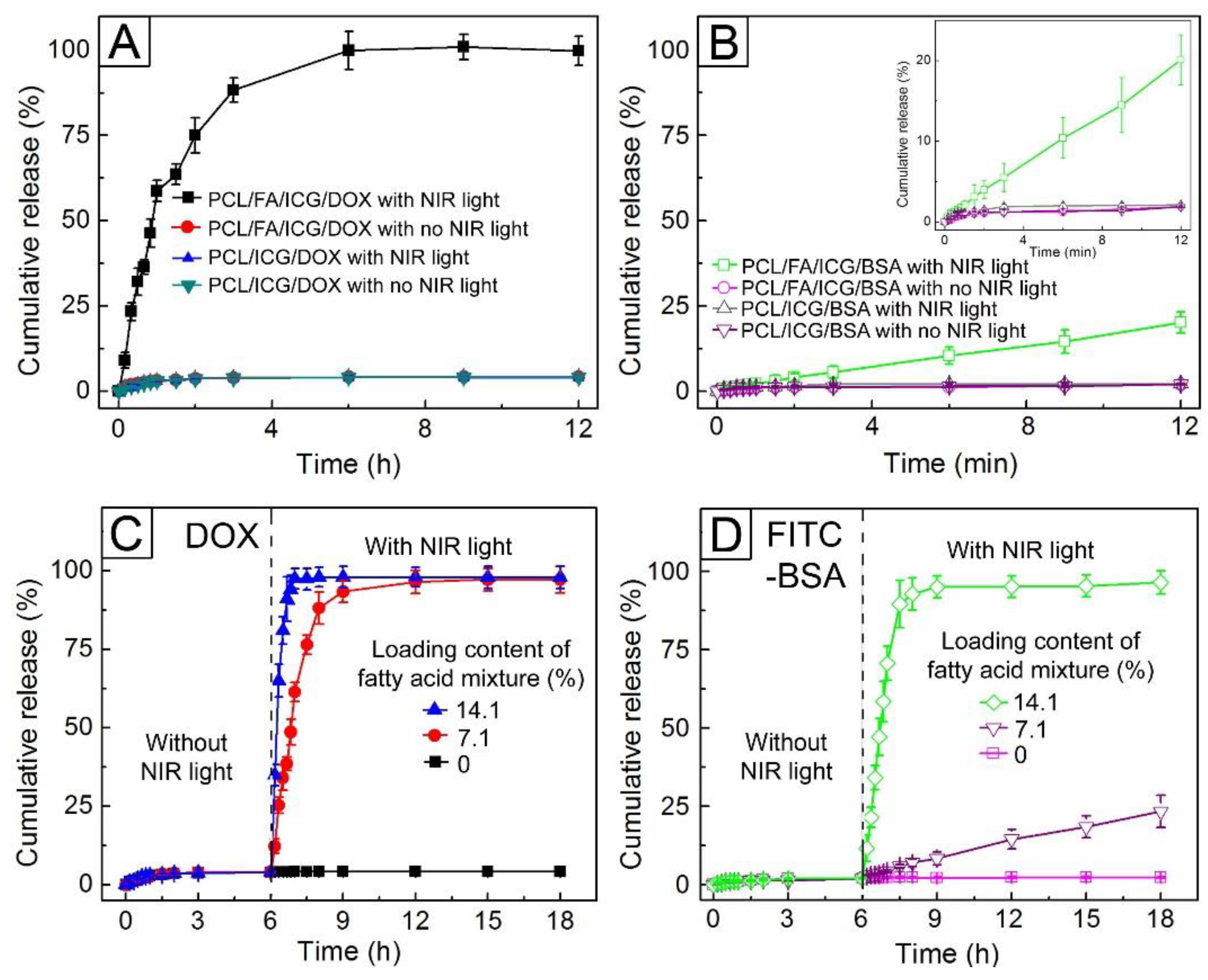
Figure 7A shows the release profiles of DOX from PCL/FA/ICG/DOX NPs and PCL/ICG/DOX NPs. Without NIR light irradiation, these two samples released only 4.2% of the drug molecules over 12 h. On the other hand, when 0.7 W/cm
2 NIR light was applied, their DOX release behaviors differed from each other, although the heat generation by ICG included in the NPs raised both their temperature up to 42.5 °C. The former allowed 100% of the drug to be released over 6 h, whereas the latter achieved a DOX release of 4.1% over the same period, similarly to the case without NIR light irradiation. Any change in particle size, which can affect the release behavior, was not observed during the release test. Thus, we attributed the NIR light-triggered release exhibited by the PCL/FA/ICG/DOX NPs to the nanopores formed by the melting away of the FA mixture, as shown in
Figure 6. To support this conclusion, we investigated the release behaviors of DOX from PCL/DOX NPs and PCL/FA/DOX NPs, which could not form nanopores on their shells. Regardless of the NIR light irradiation, the two samples released approximately 4% DOX (
Figure S5A, Supplementary Materials). These results suggest that the pores generated by NIR light functioned as effective channels for the quick release of the encapsulated drug molecules.
We also investigated the release behaviors of FITC-BSA from the four types of hollow NPs, whose diameters,
ξ-potential values, and compositions are summarized in
Table S2 (Supplementary Materials). As for the case of DOX, only the PCL/FA/ICG/BSA NPs exhibited NIR light-triggered release of the protein (
Figure 7B and
Figure S5B). However, the release of the protein was much slower than that of DOX, which was attributed to the large size of the protein molecules. It has been reported that the protein molecule has a dimension of 14 nm × 4 nm × 4 nm or 9 nm × 5.5 nm × 5.5 nm [
37,
38]. Assuming that the molecule was spherical and had the same volume as a rectangular block, its diameter was calculated to be ca. 7.6 nm. The value is half the diameter of the formed nanopores (
Figure 6A), suggesting a long time required for the macromolecules to be released through them.
Figure 7C shows the release profiles of DOX from PCL/FA/ICG/DOX NPs with loading contents of 7.1% and 14.1% for the FA mixture. All the samples released approximately 4% of the drug molecules during the first 6 h without NIR light treatment. On the other hand, 0.7 W/cm
2 NIR light irradiation led to the instant release of DOX from the hollow NPs, and the release profiles were strongly dependent on the loading content of the FA mixture. The NPs with a loading content of 7.1% achieved 100% release of the drug in 6 h after the NIR irradiation, whereas the ones with a loading content of 14.1% achieved the same result at only 1.5 h since the NIR treatment, which was due to the formation of the larger pores on the shells, as shown in
Figure 6C. The effect of the pore size on the release behavior was more distinctly observed in the systems encapsulating FITC-BSA (
Figure 7D). Without NIR light irradiation, the protein was not released from any of the types of NPs tested. On the contrary, after NIR light irradiation, the NPs with a loading content of 7.1% released 20% of the protein for 12 h since the NIR irradiation, and those with a loading content of 14.1% attained 100% release within 3 h of the NIR treatment, because their pores were large enough to let the protein molecules through.
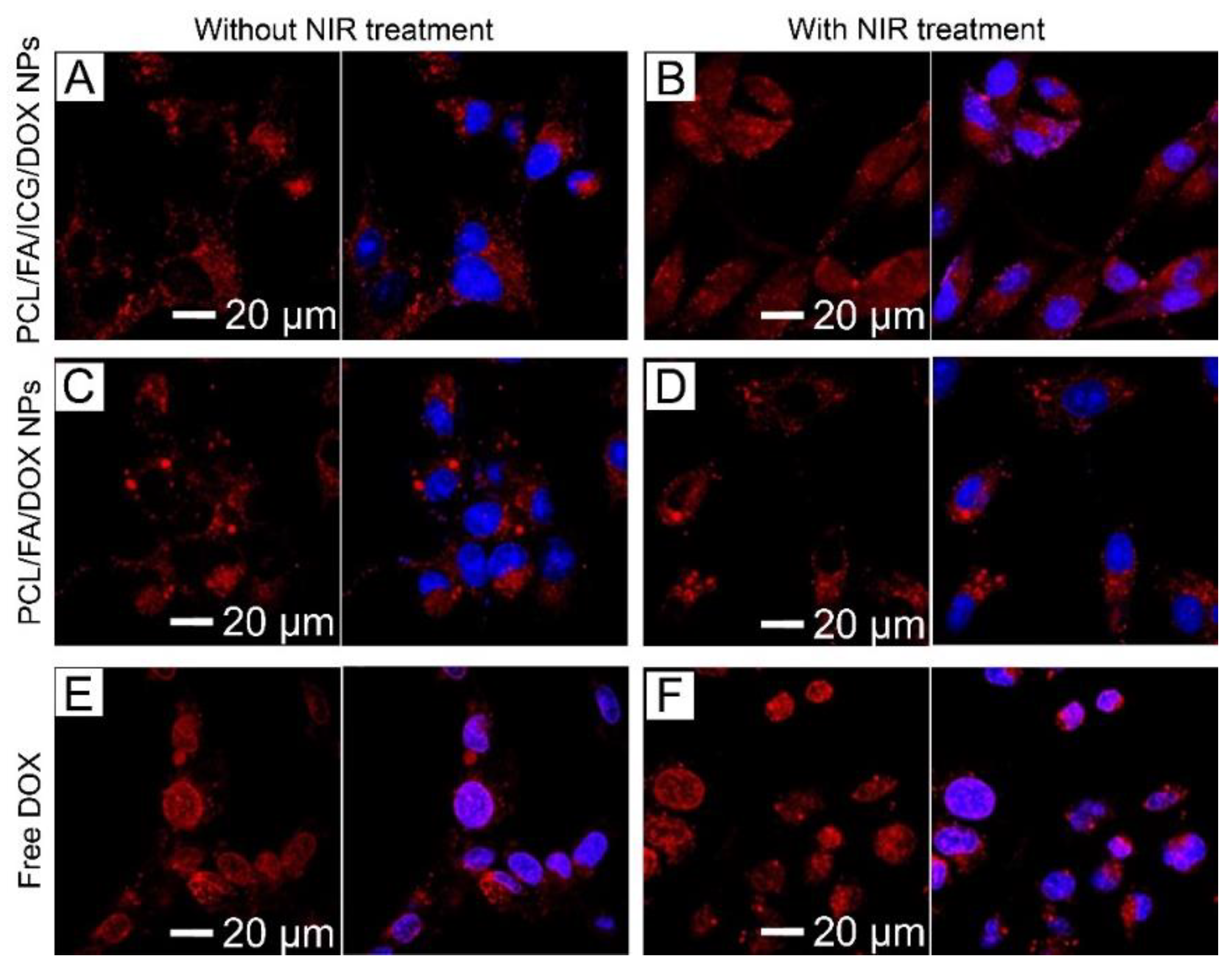
The CLS micrographs shown in
Figure 8 demonstrate the intracellular localization of DOX from PCL/FA/ICG/DOX NPs, PCL/FA/DOX NPs, and free DOX internalized by human breast cancer SK-BR3 cells. The PCL/FA/ICG/DOX NPs were exclusively localized in the cytoplasm, as represented by the strong red fluorescence (
Figure 8A). A similar result was observed in the cells treated with PCL/FA/DOX NPs (
Figure 8C). However, the cells treated with free DOX mainly exhibited the strong red fluorescence in the nuclei (blue fluorescence) of the cells, as shown in
Figure 8E, due to its diffusion through the cell membrane [
39]. These results suggest that the NPs were internalized by endocytosis, and the drug molecules were encapsulated in the hollow voids of the NPs without their undesired release. When 0.7 W/cm
2 NIR light was irradiated, a noticeable increase in red fluorescence was observed in the nuclei for the cells treated with PCL/FA/ICG/DOX NPs (
Figure 8B), whereas the cells treated with PCL/FA/DOX NPs or free DOX did not exhibit such an increase (
Figure 8D,F). This NIR light-triggered intranuclear release of the tested anticancer drug suggests that the presented NIR light-sensitive hollow NPs could act as drug-carrier candidates for on-demand cancer therapy.
We evaluated the anticancer activity of the fabricated NIR light-sensitive hollow NPs via the WST-1 assay. The cytotoxicities of the hydrophobic ICG and PCL/FA NPs were preliminarily investigated. We did not observe significant changes in the viabilities of the SK-BR3 and NHDF cells in all the cases, relative to the control, where the NPs were not added (
Figure S6, Supplementary Materials). In the case of the hydrophobic ICG, both types of the cells exhibited the viabilities above 90% up to a concentration of 1 mg/mL, compared with the control with no addition of the photothermal agent (
Figure S7, Supplementary Materials). These results suggest the good biocompatibility of the NIR light-sensitive hollow NPs. Moreover, the cytotoxicity of NIR light was also tested, and the result confirmed that it was negligible (
Figure S8, Supplementary Materials).
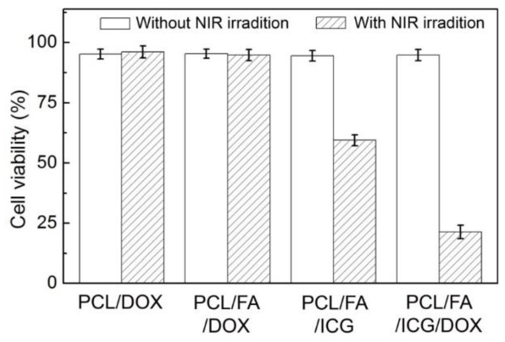
Figure 9 shows the viabilities of SK-BR-3 cells treated with the four types of hollow NPs. Without NIR light irradiation, all the samples exhibited the viabilities above 90%. Such high values for the samples encapsulated with DOX (PCL/DOX NPs, PCL/FA/DOX NPs, and PCL/FA/ICG/DOX NPs) confirm the stable encapsulation of the drug without its undesired release from the NPs, which was consistent with the results of the in vitro release test shown in
Figure 7. On the other hand, after 0.7 W/cm
2 NIR light irradiation, the cell viability was significantly reduced only for the samples treated with the ICG-entrapped samples (PCL/FA/ICG NPs and PCL/FA/ICG/DOX NPs). In the case of the PCL/FA/ICG NPs, the cell viability decreased to 59% because of the hyperthermia effect by the entrapped ICG in response to the NIR light irradiation, while the PCL/FA/ICG/DOX NPs exhibited even more improved anticancer activity under NIR irradiation (viability of 21%). This improvement could be explained by the accumulation of DOX in the nuclei of cells (
Figure 8B), where the drug molecules exerted their anticancer effect by intercalating with DNA [
40]. These results imply that the NIR light-sensitive hollow NPs can have the feasibility of noninvasive, spatiotemporal control of drug release for cancer therapy, in conjunction with the NIR light-triggered photothermal effect.

 and
and 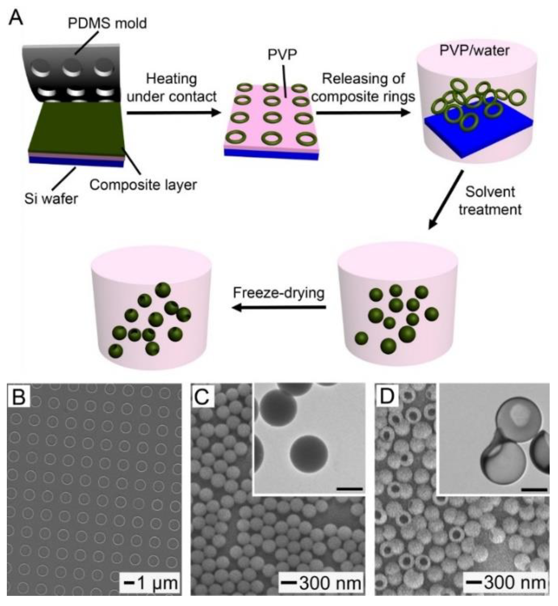


