Next Issue
Volume 9, May
Previous Issue
Volume 9, March


Viruses, Volume 9, Issue 4 (April 2017) – 31 articles

Cover Story (view full-size image): 
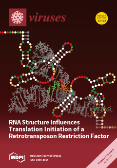
The Ty1 restriction factor p22 is encoded by internally initiated Ty1i RNA. It contains two functional initiation codons embedded within a distinct structural domain. Mutational analyses suggest that the p22 level decreases when this RNA domain is destabilized at the secondary or tertiary structural levels. Also, our data support the idea that Gag itself and possibly other cellular proteins interact with the AUG-containing structural motif of Ty1i RNA to modulate its stability and translation. View this paper
  • Issues are regarded as officially published after their release is announced to the table of contents alert mailing list.
  • You may sign up for e-mail alerts to receive table of contents of newly released issues.
  • PDF is the official format for papers published in both, html and pdf forms. To view the papers in pdf format, click on the "PDF Full-text" link, and use the free Adobe Reader to open them.
Order results
Result details
Section
Select all
Export citation of selected articles as:
4540 KiB  
Article
Deep Sequencing Analysis of RNAs from Citrus Plants Grown in a Citrus Sudden Death-Affected Area Reveals Diverse Known and Putative Novel Viruses
by Emilyn E. Matsumura, Helvecio D. Coletta-Filho, Shahideh Nouri, Bryce W. Falk, Luca Nerva, Tiago S. Oliveira, Silvia O. Dorta and Marcos A. Machado
Viruses 2017, 9(4), 92; https://doi.org/10.3390/v9040092 - 24 Apr 2017
Cited by 48 | Viewed by 7472
Abstract
Citrus sudden death (CSD) has caused the death of approximately four million orange trees in a very important citrus region in Brazil. Although its etiology is still not completely clear, symptoms and distribution of affected plants indicate a viral disease. In a search [...] Read more.
Citrus sudden death (CSD) has caused the death of approximately four million orange trees in a very important citrus region in Brazil. Although its etiology is still not completely clear, symptoms and distribution of affected plants indicate a viral disease. In a search for viruses associated with CSD, we have performed a comparative high-throughput sequencing analysis of the transcriptome and small RNAs from CSD-symptomatic and -asymptomatic plants using the Illumina platform. The data revealed mixed infections that included Citrus tristeza virus (CTV) as the most predominant virus, followed by the Citrus sudden death-associated virus (CSDaV), Citrus endogenous pararetrovirus (CitPRV) and two putative novel viruses tentatively named Citrus jingmen-like virus (CJLV), and Citrus virga-like virus (CVLV). The deep sequencing analyses were sensitive enough to differentiate two genotypes of both viruses previously associated with CSD-affected plants: CTV and CSDaV. Our data also showed a putative association of the CSD-symptomatic plants with a specific CSDaV genotype and a likely association with CitPRV as well, whereas the two putative novel viruses showed to be more associated with CSD-asymptomatic plants. This is the first high-throughput sequencing-based study of the viral sequences present in CSD-affected citrus plants, and generated valuable information for further CSD studies. Full article
(This article belongs to the Section Viruses of Plants, Fungi and Protozoa)
Show Figures

Figure 1

5184 KiB  
Review
Dengue Virus Non-Structural Protein 5
by Abbas El Sahili and Julien Lescar
Viruses 2017, 9(4), 91; https://doi.org/10.3390/v9040091 - 24 Apr 2017
Cited by 94 | Viewed by 13232
Abstract
The World Health Organization estimates that the yearly number of dengue cases averages 390 million. This mosquito-borne virus disease is endemic in over 100 countries and will probably continue spreading, given the observed trend in global warming. So far, there is no antiviral [...] Read more.
The World Health Organization estimates that the yearly number of dengue cases averages 390 million. This mosquito-borne virus disease is endemic in over 100 countries and will probably continue spreading, given the observed trend in global warming. So far, there is no antiviral drug available against dengue, but a vaccine has been recently marketed. Dengue virus also serves as a prototype for the study of other pathogenic flaviviruses that are emerging, like West Nile virus and Zika virus. Upon viral entry into the host cell and fusion of the viral lipid membrane with the endosomal membrane, the viral RNA is released and expressed as a polyprotein, that is then matured into three structural and seven non-structural (NS) proteins. The envelope, membrane and capsid proteins form the viral particle while NS1-NS2A-NS2B-NS3-NS4A-NS4B and NS5 assemble inside a cellular replication complex, which is embedded in endoplasmic reticulum (ER)-derived vesicles. In addition to their roles in RNA replication within the infected cell, NS proteins help the virus escape the host innate immunity and reshape the host-cell inner structure. This review focuses on recent progress in characterizing the structure and functions of NS5, a protein responsible for the replication and capping of viral RNA that represents a promising drug target. Full article
(This article belongs to the Section Animal Viruses)
Show Figures

Figure 1

2222 KiB  
Article
Replication and Oncolytic Activity of an Avian Orthoreovirus in Human Hepatocellular Carcinoma Cells
by Robert A. Kozak, Larissa Hattin, Mia J. Biondi, Juan C. Corredor, Scott Walsh, Max Xue-Zhong, Justin Manuel, Ian D. McGilvray, Jason Morgenstern, Evan Lusty, Vera Cherepanov, Betty-Anne McBey, David Leishman, Jordan J. Feld, Byram Bridle and Éva Nagy
Viruses 2017, 9(4), 90; https://doi.org/10.3390/v9040090 - 24 Apr 2017
Cited by 18 | Viewed by 5776
Abstract
Oncolytic viruses are cancer therapeutics with promising outcomes in pre-clinical and clinical settings. Animal viruses have the possibility to avoid pre-existing immunity in humans, while being safe and immunostimulatory. We isolated an avian orthoreovirus (ARV-PB1), and tested it against a panel of hepatocellular [...] Read more.
Oncolytic viruses are cancer therapeutics with promising outcomes in pre-clinical and clinical settings. Animal viruses have the possibility to avoid pre-existing immunity in humans, while being safe and immunostimulatory. We isolated an avian orthoreovirus (ARV-PB1), and tested it against a panel of hepatocellular carcinoma cells. We found that ARV-PB1 replicated well and induced strong cytopathic effects. It was determined that one mechanism of cell death was through syncytia formation, resulting in apoptosis and induction of interferon stimulated genes (ISGs). As hepatitis C virus (HCV) is a major cause of hepatocellular carcinoma worldwide, we investigated the effect of ARV-PB1 against cells already infected with this virus. Both HCV replicon-containing and infected cells supported ARV-PB1 replication and underwent cytolysis. Finally, we generated in silico models to compare the structures of human reovirus- and ARV-PB1-derived S1 proteins, which are the primary targets of neutralizing antibodies. Tertiary alignments confirmed that ARV-PB1 differs from its human homolog, suggesting that immunity to human reoviruses would not be a barrier to its use. Therefore, ARV-PB1 can potentially expand the repertoire of oncolytic viruses for treatment of human hepatocellular carcinoma and other malignancies. Full article
(This article belongs to the Section Animal Viruses)
Show Figures

Figure 1

598 KiB  
Correction
Correction: Ruiz, E. et al. Emerging Interaction Patterns in the Emiliania Huxleyi-EhV System. Viruses 2016, 9, 61
by Eliana Ruiz, Monique Oosterhof, Ruth-Anne Sandaa, Aud Larsen and António Pagarete
Viruses 2017, 9(4), 89; https://doi.org/10.3390/v9040089 - 24 Apr 2017
Viewed by 3374
Abstract
The authors wish to make the following change to their paper [...] Full article
(This article belongs to the Special Issue Marine Viruses 2016)
Show Figures

Figure 6

1743 KiB  
Review
Chloroviruses Have a Sweet Tooth
by James L. Van Etten, Irina Agarkova, David D. Dunigan, Michela Tonetti, Christina De Castro and Garry A. Duncan
Viruses 2017, 9(4), 88; https://doi.org/10.3390/v9040088 - 22 Apr 2017
Cited by 31 | Viewed by 7005
Abstract
Chloroviruses are large double-stranded DNA (dsDNA) viruses that infect certain isolates of chlorella-like green algae. They contain up to approximately 400 protein-encoding genes and 16 transfer RNA (tRNA) genes. This review summarizes the unexpected finding that many of the chlorovirus genes encode proteins [...] Read more.
Chloroviruses are large double-stranded DNA (dsDNA) viruses that infect certain isolates of chlorella-like green algae. They contain up to approximately 400 protein-encoding genes and 16 transfer RNA (tRNA) genes. This review summarizes the unexpected finding that many of the chlorovirus genes encode proteins involved in manipulating carbohydrates. These include enzymes involved in making extracellular polysaccharides, such as hyaluronan and chitin, enzymes that make nucleotide sugars, such as GDP-L-fucose and GDP-D-rhamnose and enzymes involved in the synthesis of glycans attached to the virus major capsid proteins. This latter process differs from that of all other glycoprotein containing viruses that traditionally use the host endoplasmic reticulum and Golgi machinery to synthesize and transfer the glycans. Full article
(This article belongs to the Special Issue Viruses of Microbes)
Show Figures

Figure 1

907 KiB  
Conference Report
“French Phage Network”—Second Meeting Report
by Clara Torres-Barceló, Oliver Kaltz, Rémy Froissart, Sylvain Gandon, Nicolas Ginet and Mireille Ansaldi
Viruses 2017, 9(4), 87; https://doi.org/10.3390/v9040087 - 21 Apr 2017
Cited by 4 | Viewed by 5590
Abstract
The study of bacteriophages (viruses of bacteria) includes a variety of approaches, such as structural biology, genetics, ecology, and evolution, with increasingly important implications for therapeutic and industrial uses. Researchers working with phages in France have recently established a network to facilitate the [...] Read more.
The study of bacteriophages (viruses of bacteria) includes a variety of approaches, such as structural biology, genetics, ecology, and evolution, with increasingly important implications for therapeutic and industrial uses. Researchers working with phages in France have recently established a network to facilitate the exchange on complementary approaches, but also to engage new collaborations. Here, we provide a summary of the topics presented during the second meeting of the French Phage Network that took place in Marseille in November 2016 Full article
(This article belongs to the Special Issue Viruses of Microbes)
Show Figures

Figure 1

2815 KiB  
Review
Classical Swine Fever—An Updated Review
by Sandra Blome, Christoph Staubach, Julia Henke, Jolene Carlson and Martin Beer
Viruses 2017, 9(4), 86; https://doi.org/10.3390/v9040086 - 21 Apr 2017
Cited by 174 | Viewed by 23450
Abstract
Classical swine fever (CSF) remains one of the most important transboundary viral diseases of swine worldwide. The causative agent is CSF virus, a small, enveloped RNA virus of the genus Pestivirus. Based on partial sequences, three genotypes can be distinguished that do not, [...] Read more.
Classical swine fever (CSF) remains one of the most important transboundary viral diseases of swine worldwide. The causative agent is CSF virus, a small, enveloped RNA virus of the genus Pestivirus. Based on partial sequences, three genotypes can be distinguished that do not, however, directly correlate with virulence. Depending on both virus and host factors, a wide range of clinical syndromes can be observed and thus, laboratory confirmation is mandatory. To this means, both direct and indirect methods are utilized with an increasing degree of commercialization. Both infections in domestic pigs and wild boar are of great relevance; and wild boars are a reservoir host transmitting the virus sporadically also to pig farms. Control strategies for epidemic outbreaks in free countries are mainly based on classical intervention measures; i.e., quarantine and strict culling of affected herds. In these countries, vaccination is only an emergency option. However, live vaccines are used for controlling the disease in endemically infected regions in Asia, Eastern Europe, the Americas, and some African countries. Here, we will provide a concise, updated review on virus properties, clinical signs and pathology, epidemiology, pathogenesis and immune responses, diagnosis and vaccination possibilities. Full article
(This article belongs to the Special Issue Porcine Viruses)
Show Figures

Figure 1

14372 KiB  
Review
Deciphering the Multifactorial Susceptibility of Mucosal Junction Cells to HPV Infection and Related Carcinogenesis
by Michael Herfs, Thing R. Soong, Philippe Delvenne and Christopher P. Crum
Viruses 2017, 9(4), 85; https://doi.org/10.3390/v9040085 - 20 Apr 2017
Cited by 28 | Viewed by 7760
Abstract
Human papillomavirus (HPV)-induced neoplasms have long been considered to originate from viral infection of the basal cell layer of the squamous mucosa. However, this paradigm has been recently undermined by accumulating data supporting the critical role of a discrete population of squamo-columnar (SC) [...] Read more.
Human papillomavirus (HPV)-induced neoplasms have long been considered to originate from viral infection of the basal cell layer of the squamous mucosa. However, this paradigm has been recently undermined by accumulating data supporting the critical role of a discrete population of squamo-columnar (SC) junction cells in the pathogenesis of cervical (pre)cancers. The present review summarizes the current knowledge on junctional cells, discusses their high vulnerability to HPV infection, and stresses the potential clinical/translational value of the novel dualistic model of HPV-related carcinogenesis. Full article
(This article belongs to the Special Issue Expert Views on HPV Infection)
Show Figures

Figure 1

1942 KiB  
Article
Seasonal Dynamics of Haptophytes and dsDNA Algal Viruses Suggest Complex Virus-Host Relationship
by Torill Vik Johannessen, Aud Larsen, Gunnar Bratbak, António Pagarete, Bente Edvardsen, Elianne D. Egge and Ruth-Anne Sandaa
Viruses 2017, 9(4), 84; https://doi.org/10.3390/v9040084 - 20 Apr 2017
Cited by 25 | Viewed by 6134
Abstract
Viruses influence the ecology and diversity of phytoplankton in the ocean. Most studies of phytoplankton host–virus interactions have focused on bloom-forming species like Emiliania huxleyi or Phaeocystis spp. The role of viruses infecting phytoplankton that do not form conspicuous blooms have received less [...] Read more.
Viruses influence the ecology and diversity of phytoplankton in the ocean. Most studies of phytoplankton host–virus interactions have focused on bloom-forming species like Emiliania huxleyi or Phaeocystis spp. The role of viruses infecting phytoplankton that do not form conspicuous blooms have received less attention. Here we explore the dynamics of phytoplankton and algal viruses over several sequential seasons, with a focus on the ubiquitous and diverse phytoplankton division Haptophyta, and their double-stranded DNA viruses, potentially with the capacity to infect the haptophytes. Viral and phytoplankton abundance and diversity showed recurrent seasonal changes, mainly explained by hydrographic conditions. By 454 tag-sequencing we revealed 93 unique haptophyte operational taxonomic units (OTUs), with seasonal changes in abundance. Sixty-one unique viral OTUs, representing Megaviridae and Phycodnaviridae, showed only distant relationship with currently isolated algal viruses. Haptophyte and virus community composition and diversity varied substantially throughout the year, but in an uncoordinated manner. A minority of the viral OTUs were highly abundant at specific time-points, indicating a boom-bust relationship with their host. Most of the viral OTUs were very persistent, which may represent viruses that coexist with their hosts, or able to exploit several host species. Full article
(This article belongs to the Special Issue Marine Viruses 2016)
Show Figures

Figure 1

422 KiB  
Review
Porcine Circoviruses and Xenotransplantation
by Joachim Denner and Annette Mankertz
Viruses 2017, 9(4), 83; https://doi.org/10.3390/v9040083 - 20 Apr 2017
Cited by 43 | Viewed by 7604
Abstract
Allotransplantation and xenotransplantation may be associated with the transmission of pathogens from the donor to the recipient. Whereas in the case of allotransplantation the transmitted microorganisms and their pathogenic effect are well characterized, the possible influence of porcine microorganisms on humans is mostly [...] Read more.
Allotransplantation and xenotransplantation may be associated with the transmission of pathogens from the donor to the recipient. Whereas in the case of allotransplantation the transmitted microorganisms and their pathogenic effect are well characterized, the possible influence of porcine microorganisms on humans is mostly unknown. Porcine circoviruses (PCVs) are common in pig breeds and they belong to porcine microorganisms that still have not been fully addressed in terms of evaluating the potential risk of xenotransplantation using pig cells, tissues, and organs. Two types of PCVs are known: porcine circovirus (PCV) 1 and PCV2. Whereas PCV1 is apathogenic in pigs, PCV2 may induce severe pig diseases. Although most pigs are subclinically infected, we do not know whether this infection impairs pig transplant functionality, particularly because PCV2 is immunosuppressive. In addition, vaccination against PCV2 is able to prevent diseases, but in most cases not transmission of the virus. Therefore, PCV2 has to be eliminated to obtain xenotransplants from uninfected healthy animals. Although there is evidence that PCV2 does not infect—at least immunocompetent—humans, animals should be screened using sensitive methods to ensure virus elimination by selection, Cesarean delivery, vaccination, or embryo transfer. Full article
(This article belongs to the Special Issue Porcine Viruses)
Show Figures

Figure 1

1021 KiB  
Article
Global Transmission Dynamics of Measles in the Measles Elimination Era
by Yuki Furuse and Hitoshi Oshitani
Viruses 2017, 9(4), 82; https://doi.org/10.3390/v9040082 - 16 Apr 2017
Cited by 16 | Viewed by 6350
Abstract
Although there have been many epidemiological reports of the inter-country transmission of measles, systematic analysis of the global transmission dynamics of the measles virus (MV) is limited. In this study, we applied phylogeographic analysis to characterize the global transmission dynamics of the MV [...] Read more.
Although there have been many epidemiological reports of the inter-country transmission of measles, systematic analysis of the global transmission dynamics of the measles virus (MV) is limited. In this study, we applied phylogeographic analysis to characterize the global transmission dynamics of the MV using large-scale genetic sequence data (obtained for 7456 sequences) from 115 countries between 1954 and 2015. These analyses reveal the spatial and temporal characteristics of global transmission of the virus, especially in Australia, China, India, Japan, the UK, and the USA in the period since 1990. The transmission is frequently observed, not only within the same region but also among distant and frequently visited areas. Frequencies of export from measles-endemic countries, such as China, India, and Japan are high but decreasing, while the frequencies from countries where measles is no longer endemic, such as Australia, the UK, and the USA, are low but slightly increasing. The world is heading toward measles eradication, but the disease is still transmitted regionally and globally. Our analysis reveals that countries wherein measles is endemic and those having eliminated the disease (apart from occasional outbreaks) both remain a source of global transmission in this measles elimination era. It is therefore crucial to maintain vigilance in efforts to monitor and eradicate measles globally. Full article
(This article belongs to the Section Animal Viruses)
Show Figures

Figure 1

1532 KiB  
Review
Mechanisms of LTR‐Retroelement Transposition: Lessons from Drosophila melanogaster
by Lidia Nefedova and Alexander Kim
Viruses 2017, 9(4), 81; https://doi.org/10.3390/v9040081 - 16 Apr 2017
Cited by 14 | Viewed by 7218
Abstract
Long terminal repeat (LTR) retrotransposons occupy a special place among all mobile genetic element families. The structure of LTR retrotransposons that have three open reading frames is identical to DNA forms of retroviruses that are integrated into the host genome. Several lines of [...] Read more.
Long terminal repeat (LTR) retrotransposons occupy a special place among all mobile genetic element families. The structure of LTR retrotransposons that have three open reading frames is identical to DNA forms of retroviruses that are integrated into the host genome. Several lines of evidence suggest that LTR retrotransposons share a common ancestry with retroviruses and thus are highly relevant to understanding mechanisms of transposition. Drosophila melanogaster is an exceptionally convenient model for studying the mechanisms of retrotransposon movement because many such elements in its genome are transpositionally active. Moreover, two LTRretrotransposons of D. melanogaster, gypsy and ZAM, have been found to have infectious properties and have been classified as errantiviruses. Despite numerous studies focusing on retroviral integration process, there is still no clear understanding of integration specificity in a target site. Most LTR retrotransposons non‐specifically integrate into a target site. Site‐specificity of integration at vertebrate retroviruses is rather relative. At the same time, sequence‐specific integration is the exclusive property of errantiviruses and their derivatives with two open reading frames. The possible basis for the errantivirus integration specificity is discussed in the present review. Full article
Show Figures

Figure 1

1302 KiB  
Review
Protein-Coding Genes’ Retrocopies and Their Functions
by Magdalena Regina Kubiak and Izabela Makałowska
Viruses 2017, 9(4), 80; https://doi.org/10.3390/v9040080 - 13 Apr 2017
Cited by 48 | Viewed by 9227
Abstract
Transposable elements, often considered to be not important for survival, significantly contribute to the evolution of transcriptomes, promoters, and proteomes. Reverse transcriptase, encoded by some transposable elements, can be used in trans to produce a DNA copy of any RNA molecule in the [...] Read more.
Transposable elements, often considered to be not important for survival, significantly contribute to the evolution of transcriptomes, promoters, and proteomes. Reverse transcriptase, encoded by some transposable elements, can be used in trans to produce a DNA copy of any RNA molecule in the cell. The retrotransposition of protein-coding genes requires the presence of reverse transcriptase, which could be delivered by either non-long terminal repeat (non-LTR) or LTR transposons. The majority of these copies are in a state of “relaxed” selection and remain “dormant” because they are lacking regulatory regions; however, many become functional. In the course of evolution, they may undergo subfunctionalization, neofunctionalization, or replace their progenitors. Functional retrocopies (retrogenes) can encode proteins, novel or similar to those encoded by their progenitors, can be used as alternative exons or create chimeric transcripts, and can also be involved in transcriptional interference and participate in the epigenetic regulation of parental gene expression. They can also act in trans as natural antisense transcripts, microRNA (miRNA) sponges, or a source of various small RNAs. Moreover, many retrocopies of protein-coding genes are linked to human diseases, especially various types of cancer. Full article
Show Figures

Figure 1

7805 KiB  
Article
Comparative Study on the Antivirus Activity of Shuang–Huang–Lian Injectable Powder and Its Bioactive Compound Mixture against Human Adenovirus III In Vitro
by Qinhai Ma, Dedong Liang, Shuai Song, Qintian Yu, Chunyu Shi, Xuefeng Xing and Jia-Bo Luo
Viruses 2017, 9(4), 79; https://doi.org/10.3390/v9040079 - 12 Apr 2017
Cited by 22 | Viewed by 5255
Abstract
Shuang–Huang–Lian injectable powder (SHL)—a classical purified herbal preparation extracted from Scutellaria baicalensis, Lonicera japonica, and Forsythia suspense—has been used against human adenovirus III (HAdV3) for many years. The combination herb and its major bioactive compounds, including chlorogenic acid, [...] Read more.
Shuang–Huang–Lian injectable powder (SHL)—a classical purified herbal preparation extracted from Scutellaria baicalensis, Lonicera japonica, and Forsythia suspense—has been used against human adenovirus III (HAdV3) for many years. The combination herb and its major bioactive compounds, including chlorogenic acid, baicalin, and forsythia glycosides A, are effective inhibitors of the virus. However, no comprehensive studies are available on the antiviral effects of SHL against HAdV3. Moreover, it remains unclear whether the mixture of chlorogenic acid, baicalin, and forsythia glycosides A (CBF) has enhanced antiviral activity compared with SHL. Therefore, a comparative study was performed to investigate the combination which is promising for further antiviral drug development. To evaluate their antivirus activity in parallel, the combination ratio and dose of CBF were controlled and consistent with SHL. First, the fingerprint and the ratio of CBF in SHL were determined by high performance liquid chromatography. Then, a plaque reduction assay, reverse transcription polymerase chain reaction (PCR), real-time polymerase chain reaction (qPCR), and enzyme-linked immunosorbent assay (ELISA) were used to explore its therapeutic effects on viral infection and replication, respectively. The results showed that SHL and CBF inhibited dose- and time-dependently HAdV3-induced plaque formation in A549 and HEp-2 cells. SHL was more effective than CBF when supplemented prior to and after viral inoculation. SHL prevented viral attachment, internalization, and replication at high concentration and decreased viral levels within and out of cells at non-toxic concentrations in both cell types. Moreover, the expression of tumor necrosis factor alpha (TNF)-α, interleukin (IL)-1ß, and IL-6 was lower and the expression of interferon (IFN)-γ was higher in both cell types treated with SHL than with CBF. In conclusion, SHL is much more effective and slightly less toxic than CBF. Full article
(This article belongs to the Section Viral Immunology, Vaccines, and Antivirals)
Show Figures

Graphical abstract

3021 KiB  
Article
LTR-Retrotransposons from Bdelloid Rotifers Capture Additional ORFs Shared between Highly Diverse Retroelement Types
by Fernando Rodriguez, Aubrey W. Kenefick and Irina R. Arkhipova
Viruses 2017, 9(4), 78; https://doi.org/10.3390/v9040078 - 11 Apr 2017
Cited by 10 | Viewed by 5219
Abstract
Rotifers of the class Bdelloidea, microscopic freshwater invertebrates, possess a highlydiversified repertoire of transposon families, which, however, occupy less than 4% of genomic DNA in the sequenced representative Adineta vaga. We performed a comprehensive analysis of A. vaga retroelements, and found that [...] Read more.
Rotifers of the class Bdelloidea, microscopic freshwater invertebrates, possess a highlydiversified repertoire of transposon families, which, however, occupy less than 4% of genomic DNA in the sequenced representative Adineta vaga. We performed a comprehensive analysis of A. vaga retroelements, and found that bdelloid long terminal repeat (LTR)retrotransposons, in addition to conserved open reading frame (ORF) 1 and ORF2 corresponding to gag and pol genes, code for an unusually high variety of ORF3 sequences. Retrovirus-like LTR families in A. vaga belong to four major lineages, three of which are rotiferspecific and encode a dUTPase domain. However only one lineage contains a canonical envlike fusion glycoprotein acquired from paramyxoviruses (non-segmented negative-strand RNA viruses), although smaller ORFs with transmembrane domains may perform similar roles. A different ORF3 type encodes a GDSL esterase/lipase, which was previously identified as ORF1 in several clades of non-LTR retrotransposons, and implicated in membrane targeting. Yet another ORF3 type appears in unrelated LTR-retrotransposon lineages, and displays strong homology to DEDDy-type exonucleases involved in 3′-end processing of RNA and single-stranded DNA. Unexpectedly, each of the enzymatic ORF3s is also associated with different subsets of Penelope-like Athena retroelement families. The unusual association of the same ORF types with retroelements from different classes reflects their modular structure with a high degree of flexibility, and points to gene sharing between different groups of retroelements. Full article
Show Figures

Figure 1

251 KiB  
Review
T Cell-Mediated Immunity towards Yellow Fever Virus and Useful Animal Models
by Alan M. Watson and William B. Klimstra
Viruses 2017, 9(4), 77; https://doi.org/10.3390/v9040077 - 11 Apr 2017
Cited by 25 | Viewed by 6665
Abstract
The 17D line of yellow fever virus vaccines is among the most effective vaccines ever created. The humoral and cellular immunity elicited by 17D has been well characterized in humans. Neutralizing antibodies have long been known to provide protection against challenge with a [...] Read more.
The 17D line of yellow fever virus vaccines is among the most effective vaccines ever created. The humoral and cellular immunity elicited by 17D has been well characterized in humans. Neutralizing antibodies have long been known to provide protection against challenge with a wild-type virus. However, a well characterized T cell immune response that is robust, long-lived and polyfunctional is also elicited by 17D. It remains unclear whether this arm of immunity is protective following challenge with a wild-type virus. Here we introduce the 17D line of yellow fever virus vaccines, describe the current state of knowledge regarding the immunity directed towards the vaccines in humans and conclude with a discussion of animal models that are useful for evaluating T cell-mediated immune protection to yellow fever virus. Full article
(This article belongs to the Special Issue Advances in Flavivirus Research)
5977 KiB  
Article
Single-Round Infectious Particle Antiviral Screening Assays for the Japanese Encephalitis Virus
by Chien-Yi Lu, Mann-Jen Hour, Ching-Ying Wang, Su-Hua Huang, Wen-Xiang Mu, Yu-Chun Chang and Cheng-Wen Lin
Viruses 2017, 9(4), 76; https://doi.org/10.3390/v9040076 - 10 Apr 2017
Cited by 14 | Viewed by 6905
Abstract
Japanese Encephalitis virus (JEV) is a mosquito-borne flavivirus with a positive-sense single-stranded RNA genome that contains a big open reading frame (ORF) flanked by 5′- and 3′- untranslated regions (UTRs). Nearly 30,000 JE cases with 10,000 deaths are still annually reported in East [...] Read more.
Japanese Encephalitis virus (JEV) is a mosquito-borne flavivirus with a positive-sense single-stranded RNA genome that contains a big open reading frame (ORF) flanked by 5′- and 3′- untranslated regions (UTRs). Nearly 30,000 JE cases with 10,000 deaths are still annually reported in East Asia. Although the JEV genotype III vaccine has been licensed, it elicits a lower protection against other genotypes. Moreover, no effective treatment for a JE case is developed. This study constructed a pBR322-based and cytomegaloviruses (CMV) promoter-driven JEV replicon for the production of JEV single-round infectious particles (SRIPs) in a packaging cell line expressing viral structural proteins. Genetic instability of JEV genome cDNA in the pBR322 plasmid was associated with the prokaryotic promoter at 5′ end of the JEV genome that triggers the expression of the structural proteins in E. coli. JEV structural proteins were toxic E. coli, thus the encoding region for structural proteins was replaced by a reporter gene (enhanced green fluorescent protein, EGFP) that was in-frame fused with the first eight amino acids of the C protein at N-terminus and the foot-and-mouth disease virus (FMDV) 2A peptide at C-terminus in a pBR322-based JEV-EGFP replicon. JEV-EGFP SRIPs generated from JEV-EGFP replicon-transfected packaging cells displayed the infectivity with cytopathic effect induction, self-replication of viral genomes, and the expression of EGFP and viral proteins. Moreover, the combination of JEV-EGFP SRIP plus flow cytometry was used to determine the half maximal inhibitory concentration (IC50) values of antiviral agents according to fluorescent intensity and positivity of SRIP-infected packaging cells post treatment. MJ-47, a quinazolinone derivative, significantly inhibited JEV-induced cytopathic effect, reducing the replication and expression of JEV-EGFP replicon in vitro. The IC50 value of 6.28 µM for MJ-47 against JEV was determined by the assay of JEV-EGFP SRIP infection in packaging cells plus flow cytometry that was more sensitive, effective, and efficient compared to the traditional plaque assay. Therefore, the system of JEV-EGFP SRIPs plus flow cytometry was a rapid and reliable platform for screening antiviral agents and evaluating antiviral potency. Full article
(This article belongs to the Section Viral Immunology, Vaccines, and Antivirals)
Show Figures

Figure 1

1893 KiB  
Review
HBV DNA Integration: Molecular Mechanisms and Clinical Implications
by Thomas Tu, Magdalena A. Budzinska, Nicholas A. Shackel and Stephan Urban
Viruses 2017, 9(4), 75; https://doi.org/10.3390/v9040075 - 10 Apr 2017
Cited by 254 | Viewed by 20964
Abstract
Chronic infection with the Hepatitis B Virus (HBV) is a major cause of liver-related morbidity and mortality. One peculiar observation in cells infected with HBV (or with closely‑related animal hepadnaviruses) is the presence of viral DNA integration in the host cell genome, despite [...] Read more.
Chronic infection with the Hepatitis B Virus (HBV) is a major cause of liver-related morbidity and mortality. One peculiar observation in cells infected with HBV (or with closely‑related animal hepadnaviruses) is the presence of viral DNA integration in the host cell genome, despite this form being a replicative dead-end for the virus. The frequent finding of somatic integration of viral DNA suggests an evolutionary benefit for the virus; however, the mechanism of integration, its functions, and the clinical implications remain unknown. Here we review the current body of knowledge of HBV DNA integration, with particular focus on the molecular mechanisms and its clinical implications (including the possible consequences of replication-independent antigen expression and its possible role in hepatocellular carcinoma). HBV DNA integration is likely to influence HBV replication, persistence, and pathogenesis, and so deserves greater attention in future studies. Full article
(This article belongs to the Special Issue Recent Advances in Hepatitis B Virus Research)
Show Figures

Figure 1

11752 KiB  
Article
Structure of Ty1 Internally Initiated RNA Influences Restriction Factor Expression
by Leszek Błaszczyk, Marcin Biesiada, Agniva Saha, David J. Garfinkel and Katarzyna J. Purzycka
Viruses 2017, 9(4), 74; https://doi.org/10.3390/v9040074 - 10 Apr 2017
Cited by 8 | Viewed by 4803
Abstract
The long-terminal repeat retrotransposon Ty1 is the most abundant mobile genetic element in many Saccharomyces cerevisiae isolates. Ty1 retrotransposons contribute to the genetic diversity of host cells, but they can also act as an insertional mutagen and cause genetic instability. Interestingly, retrotransposition occurs [...] Read more.
The long-terminal repeat retrotransposon Ty1 is the most abundant mobile genetic element in many Saccharomyces cerevisiae isolates. Ty1 retrotransposons contribute to the genetic diversity of host cells, but they can also act as an insertional mutagen and cause genetic instability. Interestingly, retrotransposition occurs at a low level despite a high level of Ty1 RNA, even though S. cerevisiae lacks the intrinsic defense mechanisms that other eukaryotes use to prevent transposon movement. p22 is a recently discovered Ty1 protein that inhibits retrotransposition in a dose-dependent manner. p22 is a truncated form of Gag encoded by internally initiated Ty1i RNA that contains two closely-spaced AUG codons. Mutations of either AUG codon compromise p22 translation. We found that both AUG codons were utilized and that translation efficiency depended on the Ty1i RNA structure. Structural features that stimulated p22 translation were context dependent and present only in Ty1i RNA. Destabilization of the 5′ untranslated region (5′ UTR) of Ty1i RNA decreased the p22 level, both in vitro and in vivo. Our data suggest that protein factors such as Gag could contribute to the stability and translational activity of Ty1i RNA through specific interactions with structural motifs in the RNA. Full article
Show Figures

Figure 1

1827 KiB  
Article
The Transmembrane Morphogenesis Protein gp1 of Filamentous Phages Contains Walker A and Walker B Motifs Essential for Phage Assembly
by Belinda Loh, Maximilian Haase, Lukas Mueller, Andreas Kuhn and Sebastian Leptihn
Viruses 2017, 9(4), 73; https://doi.org/10.3390/v9040073 - 09 Apr 2017
Cited by 18 | Viewed by 6951
Abstract
In contrast to lytic phages, filamentous phages are assembled in the inner membrane and secreted across the bacterial envelope without killing the host. For assembly and extrusion of the phage across the host cell wall, filamentous phages code for membrane-embedded morphogenesis proteins. In [...] Read more.
In contrast to lytic phages, filamentous phages are assembled in the inner membrane and secreted across the bacterial envelope without killing the host. For assembly and extrusion of the phage across the host cell wall, filamentous phages code for membrane-embedded morphogenesis proteins. In the outer membrane of Escherichia coli, the protein gp4 forms a pore-like structure, while gp1 and gp11 form a complex in the inner membrane of the host. By comparing sequences with other filamentous phages, we identified putative Walker A and B motifs in gp1 with a conserved lysine in the Walker A motif (K14), and a glutamic and aspartic acid in the Walker B motif (D88, E89). In this work we demonstrate that both, Walker A and Walker B, are essential for phage production. The crucial role of these key residues suggests that gp1 might be a molecular motor driving phage assembly. We further identified essential residues for the function of the assembly complex. Mutations in three out of six cysteine residues abolish phage production. Similarly, two out of six conserved glycine residues are crucial for gp1 function. We hypothesise that the residues represent molecular hinges allowing domain movement for nucleotide binding and phage assembly. Full article
(This article belongs to the Special Issue Viruses of Microbes)
Show Figures

Figure 1

1324 KiB  
Article
MimiLook: A Phylogenetic Workflow for Detection of Gene Acquisition in Major Orthologous Groups of Megavirales
by Sourabh Jain, Arup Panda, Philippe Colson, Didier Raoult and Pierre Pontarotti
Viruses 2017, 9(4), 72; https://doi.org/10.3390/v9040072 - 07 Apr 2017
Cited by 2 | Viewed by 5578
Abstract
With the inclusion of new members, understanding about evolutionary mechanisms and processes by which members of the proposed order, Megavirales, have evolved has become a key area of interest. The central role of gene acquisition has been shown in previous studies. However, the [...] Read more.
With the inclusion of new members, understanding about evolutionary mechanisms and processes by which members of the proposed order, Megavirales, have evolved has become a key area of interest. The central role of gene acquisition has been shown in previous studies. However, the major drawback in gene acquisition studies is the focus on few MV families or putative families with large variation in their genetic structure. Thus, here we have tried to develop a methodology by which we can detect horizontal gene transfers (HGTs), taking into consideration orthologous groups of distantly related Megavirale families. Here, we report an automated workflow MimiLook, prepared as a Perl command line program, that deduces orthologous groups (OGs) from ORFomes of Megavirales and constructs phylogenetic trees by performing alignment generation, alignment editing and protein-protein BLAST (BLASTP) searching across the National Center for Biotechnology Information (NCBI) non-redundant (nr) protein sequence database. Finally, this tool detects statistically validated events of gene acquisitions with the help of the T-REX algorithm by comparing individual gene tree with NCBI species tree. In between the steps, the workflow decides about handling paralogs, filtering outputs, identifying Megavirale specific OGs, detection of HGTs, along with retrieval of information about those OGs that are monophyletic with organisms from cellular domains of life. By implementing MimiLook, we noticed that nine percent of Megavirale gene families (i.e., OGs) have been acquired by HGT, 80% OGs were Megaviralespecific and eight percent were found to be sharing common ancestry with members of cellular domains (Eukaryote, Bacteria, Archaea, Phages or other viruses) and three percent were ambivalent. The results are briefly discussed to emphasize methodology. Also, MimiLook is relevant for detecting evolutionary scenarios in other targeted phyla with user defined modifications. It can be accessed at following link 10.6084/m9.figshare.4653622. Full article
(This article belongs to the Special Issue Viruses of Protozoa)
Show Figures

Figure 1

2167 KiB  
Article
A Chinese Variant Marek’s Disease Virus Strain with Divergence between Virulence and Vaccine Resistance
by Guo-rong Sun, Yan-ping Zhang, Hong-chao Lv, Lin-yi Zhou, Hong-yu Cui, Yu-long Gao, Xiao-le Qi, Yong-qiang Wang, Kai Li, Li Gao, Qing Pan, Xiao-mei Wang and Chang-jun Liu
Viruses 2017, 9(4), 71; https://doi.org/10.3390/v9040071 - 03 Apr 2017
Cited by 24 | Viewed by 5960
Abstract
Marek’s disease (MD) virus (MDV) has been evolving continuously, leading to increasing vaccination failure. Here, the MDV field strain BS/15 was isolated from a severely diseased Chinese chicken flock previously vaccinated with CVI988. To explore the causes of vaccination failure, specific-pathogen free (SPF) [...] Read more.
Marek’s disease (MD) virus (MDV) has been evolving continuously, leading to increasing vaccination failure. Here, the MDV field strain BS/15 was isolated from a severely diseased Chinese chicken flock previously vaccinated with CVI988. To explore the causes of vaccination failure, specific-pathogen free (SPF) chickens vaccinated with CVI988 or 814 and unvaccinated controls were challenged with either BS/15 or the reference strain Md5. Both strains induced MD lesions in unvaccinated chickens with similar mortality rates of 85.7% and 80.0% during the experimental period, respectively. However, unvaccinated chickens inoculated with BS/15 exhibited a higher tumor development rate (64.3% vs. 40.0%), but prolonged survival and diminished immune defects compared to Md5-challenged counterparts. These results suggest that BS/15 and Md5 show a similar virulence but manifest with different pathogenic characteristics. Moreover, the protective indices of CVI988 and 814 were 33.3 and 66.7 for BS/15, and 92.9 and 100 for Md5, respectively, indicating that neither vaccine could provide efficient protection against BS/15. Taken together, these data suggest that MD vaccination failure is probably due to the existence of variant MDV strains with known virulence and unexpected vaccine resistance. Our findings should be helpful for understanding the pathogenicity and evolution of MDV strains prevalent in China. Full article
(This article belongs to the Section Animal Viruses)
Show Figures

Figure 1

363 KiB  
Communication
How to Name and Classify Your Phage: An Informal Guide
by Evelien Adriaenssens and J. Rodney Brister
Viruses 2017, 9(4), 70; https://doi.org/10.3390/v9040070 - 03 Apr 2017
Cited by 277 | Viewed by 14738
Abstract
With this informal guide, we try to assist both new and experienced phage researchers through two important stages that follow phage discovery; that is, naming and classification. Providing an appropriate name for a bacteriophage is not as trivial as it sounds, and the [...] Read more.
With this informal guide, we try to assist both new and experienced phage researchers through two important stages that follow phage discovery; that is, naming and classification. Providing an appropriate name for a bacteriophage is not as trivial as it sounds, and the effects might be long-lasting in databases and in official taxon names. Phage classification is the responsibility of the Bacterial and Archaeal Viruses Subcommittee (BAVS) of the International Committee on the Taxonomy of Viruses (ICTV). While the BAVS aims at providing a holistic approach to phage taxonomy, for individual researchers who have isolated and sequenced a new phage, this can be a little overwhelming. We are now providing these researchers with an informal guide to phage naming and classification, taking a “bottom-up” approach from the phage isolate level. Full article
(This article belongs to the Special Issue Viruses of Microbes)
Show Figures

Figure 1

891 KiB  
Review
Identifying and Characterizing Interplay between Hepatitis B Virus X Protein and Smc5/6
by Christine M. Livingston, Dhivya Ramakrishnan, Michel Strubin, Simon P. Fletcher and Rudolf K. Beran
Viruses 2017, 9(4), 69; https://doi.org/10.3390/v9040069 - 03 Apr 2017
Cited by 35 | Viewed by 8216
Abstract
Hepatitis B X protein (HBx) plays an essential role in the hepatitis B virus (HBV) replication cycle, but the function of HBx has been elusive until recently. It was recently shown that transcription from the HBV genome (covalently-closed circular DNA, cccDNA) is inhibited [...] Read more.
Hepatitis B X protein (HBx) plays an essential role in the hepatitis B virus (HBV) replication cycle, but the function of HBx has been elusive until recently. It was recently shown that transcription from the HBV genome (covalently-closed circular DNA, cccDNA) is inhibited by the structural maintenance of chromosome 5/6 complex (Smc5/6), and that a key function of HBx is to redirect the DNA-damage binding protein 1 (DDB1) E3 ubiquitin ligase to target this complex for degradation. By doing so, HBx alleviates transcriptional repression by Smc5/6 and stimulates HBV gene expression. In this review, we discuss in detail how the interplay between HBx and Smc5/6 was identified and characterized. We also discuss what is known regarding the repression of cccDNA transcription by Smc5/6, the timing of HBx expression, and the potential role of HBx in promoting hepatocellular carcinoma (HCC). Full article
(This article belongs to the Special Issue Recent Advances in Hepatitis B Virus Research)
Show Figures

Figure 1

2997 KiB  
Article
ICAM-1 Binding Rhinoviruses Enter HeLa Cells via Multiple Pathways and Travel to Distinct Intracellular Compartments for Uncoating
by Haleh Ganjian, Christin Zietz, Diana Mechtcheriakova, Dieter Blaas and Renate Fuchs
Viruses 2017, 9(4), 68; https://doi.org/10.3390/v9040068 - 01 Apr 2017
Cited by 13 | Viewed by 6419
Abstract
Of the more than 150 human rhinovirus (RV) serotypes, 89 utilize intercellular adhesion molecule-1 (ICAM-1) for cell entry. These belong either to species A or B. We recently demonstrated that RV-B14 and RV-A89, despite binding this same receptor, are routed into distinct endosomal [...] Read more.
Of the more than 150 human rhinovirus (RV) serotypes, 89 utilize intercellular adhesion molecule-1 (ICAM-1) for cell entry. These belong either to species A or B. We recently demonstrated that RV-B14 and RV-A89, despite binding this same receptor, are routed into distinct endosomal compartments for release of their RNA into the cytosol. To gain insight into the underlying mechanism we now comparatively investigate the port of entry, temperature-dependence of uncoating, and intracellular routing of RV-B3, RV-B14, RV-A16, and RV-A89 in HeLa cells. The effect of various drugs blocking distinct stages on the individual pathways was determined via comparing the number of infected cells in a TissueFaxs instrument. We found that RV-B14 and RV-A89 enter via clathrin-, dynamin-, and cholesterol-dependent pathways, as well as by macropinocytosis. Drugs interfering with actin function similarly blocked entry of all four viruses, indicating their dependence on a dynamic actin network. However, uniquely, RV-A89 was able to produce progeny when internalized at 20 °C followed by neutralizing the endosomal pH and further incubation at 37 °C. Blocking dynein-dependent endosomal transport prevented uncoating of RV-A16 and RV-A89, but not of RV-B3 and RV-B14, indicative for routing of RV-A16 and RV-A89 into the endocytic recycling compartment for uncoating. Our results call for caution when developing drugs aimed at targeting entry or intracellular trafficking of all rhinovirus serotypes. Full article
Show Figures

Figure 1

2500 KiB  
Brief Report
Identification of HIV-1 Tat-Associated Proteins Contributing to HIV-1 Transcription and Latency
by Maxime Junior Jean, Derek Power, Weili Kong, Huachao Huang, Netty Santoso and Jian Zhu
Viruses 2017, 9(4), 67; https://doi.org/10.3390/v9040067 - 01 Apr 2017
Cited by 20 | Viewed by 6211
Abstract
Human immunodeficiency virus type 1 (HIV-1) Tat is a virus-encoded trans-activator that plays a central role in viral transcription. We used our recently developed parallel analysis of in vitro translated open reading frames (ORFs) (PLATO) approach to identify host proteins that associate with [...] Read more.
Human immunodeficiency virus type 1 (HIV-1) Tat is a virus-encoded trans-activator that plays a central role in viral transcription. We used our recently developed parallel analysis of in vitro translated open reading frames (ORFs) (PLATO) approach to identify host proteins that associate with HIV-1 Tat. From this proteomic assay, we identify 89 Tat-associated proteins (TAPs). We combine our results with other datasets of Tat or long terminal repeat (LTR)-associated proteins. For some of these proteins (NAT10, TINP1, XRCC5, SIN3A), we confirm their strong association with Tat. These TAPs also suppress Tat-mediated HIV-1 transcription. Removing suppression of HIV-1 transcription benefits the reversal of post-integrated, latent HIV-1 proviruses. We demonstrate that these transcriptionally suppressing TAPs contribute to HIV-1 latency in Jurkat latency (J-LAT) cells. Therefore, our proteomic analysis highlights the previously unappreciated TAPs that play a role in maintaining HIV-1 latency and can be further studied as potential pharmacological targets for the “shock and kill” HIV-1 cure strategy. Full article
Show Figures

Figure 1

3135 KiB  
Article
MTase Domain of Dendrolimus punctatus cypovirus VP3 Mediates Virion Attachment and Interacts with Host ALP Protein
by Lan Su, Congrui Xu, Chuangang Cheng, Chengfeng Lei and Xiulian Sun
Viruses 2017, 9(4), 66; https://doi.org/10.3390/v9040066 - 01 Apr 2017
Cited by 5 | Viewed by 4576
Abstract
Dendrolimus punctatus cypovirus (DpCPV) is an important pathogen of D. punctatus, but little is known about the mechanisms of DpCPV infection. Here, we investigated the effects of VP3, VP4 and VP5 structural proteins on the viral invasion. Both the C-terminal of VP3 [...] Read more.
Dendrolimus punctatus cypovirus (DpCPV) is an important pathogen of D. punctatus, but little is known about the mechanisms of DpCPV infection. Here, we investigated the effects of VP3, VP4 and VP5 structural proteins on the viral invasion. Both the C-terminal of VP3 (methyltransferase (MTase) domain) and VP4 (A-spike) bound to Spodoptera exigua midgut brush border membrane vesicles (BBMVs) in a dose-dependent manner, and the binding was inhibited by purified DpCPV virions. Importantly, anti-MTase and anti-VP4 antibodies inhibited viral binding to S. exigua BBMVs. Using far-Western blots, a 65 kDa protein in Bombyx mori BBMVs, identified as alkaline phosphatase protein (BmALP) by mass spectrometry, specifically interacted with DpCPV MTase. The interaction between MTase and BmALP was verified by co-immunoprecipitation in vitro. Pretreatment of B. mori BBMVs with an anti-ALP antibody or incubation of DpCPV virions with prokaryotically expressed BmALP reduced viral attachment. Additionally, BmALP inhibited DpCPV infection in S. exigua larvae. Our data provide evidence that the MTase domain and A-spike function as viral attachment proteins during the DpCPV infection process, and ALP is the ligand that interacts with DpCPV via the MTase domain. These results augment our understanding of the mechanisms used by cypoviruses to enter their hosts. Full article
(This article belongs to the Section Insect Viruses)
Show Figures

Figure 1

1067 KiB  
Review
Free-Living Amoebae as Hosts for and Vectors of Intracellular Microorganisms with Public Health Significance
by Carsten Balczun and Patrick L. Scheid
Viruses 2017, 9(4), 65; https://doi.org/10.3390/v9040065 - 01 Apr 2017
Cited by 96 | Viewed by 9131
Abstract
Free-living amoebae (FLA) are parasites within both humans and animals causing a wide range of symptoms and act as hosts of, and vehicles for phylogenetically diverse microorganisms, called endocytobionts. The interaction of the FLA with sympatric microorganisms leads to an exceptional diversity within [...] Read more.
Free-living amoebae (FLA) are parasites within both humans and animals causing a wide range of symptoms and act as hosts of, and vehicles for phylogenetically diverse microorganisms, called endocytobionts. The interaction of the FLA with sympatric microorganisms leads to an exceptional diversity within FLA. Some of these bacteria, viruses, and even eukaryotes, can live and replicate intracellularly within the FLA. This relationship provides protection to the microorganisms from external interventions and a dispersal mechanism across various habitats. Among those intracellularly-replicating or -residing organisms there are obligate and facultative pathogenic microorganisms affecting the health of humans or animals and are therefore of interest to Public Health Authorities. Mimiviruses, Pandoraviruses, and Pithoviruses are examples for interesting viral endocytobionts within FLA. Future research is expected to reveal further endocytobionts within free-living amoebae and other protozoa through co-cultivation studies, genomic, transcriptomic, and proteomic analyses. Full article
(This article belongs to the Special Issue Viruses of Protozoa)
Show Figures

Figure 1

1180 KiB  
Review
Targeting TNF and TNF Receptor Pathway in HIV-1 Infection: from Immune Activation to Viral Reservoirs
by Sébastien Pasquereau, Amit Kumar and Georges Herbein
Viruses 2017, 9(4), 64; https://doi.org/10.3390/v9040064 - 30 Mar 2017
Cited by 88 | Viewed by 14533
Abstract
Several cellular functions such as apoptosis, cellular proliferation, inflammation, and immune regulation involve the tumor necrosis factor-α (TNF)/TNF receptor (TNFR) pathway. Human immunodeficiency virus 1 (HIV-1) interacts with the TNF/TNFR pathway. The activation of the TNF/TNFR pathway impacts HIV-1 replication, and the TNF/TNFR [...] Read more.
Several cellular functions such as apoptosis, cellular proliferation, inflammation, and immune regulation involve the tumor necrosis factor-α (TNF)/TNF receptor (TNFR) pathway. Human immunodeficiency virus 1 (HIV-1) interacts with the TNF/TNFR pathway. The activation of the TNF/TNFR pathway impacts HIV-1 replication, and the TNF/TNFR pathway is the target of HIV-1 proteins. A hallmark of HIV-1 infection is immune activation and inflammation with increased levels of TNF in the plasma and the tissues. Therefore, the control of the TNF/TNFR pathway by new therapeutic approaches could participate in the control of immune activation and impact both viral replication and viral persistence. In this review, we will describe the intricate interplay between HIV-1 proteins and TNF/TNFR signaling and how TNF/TNFR activation modulates HIV-1 replication and discuss new therapeutic approaches, especially anti-TNF therapy, that could control this pathway and ultimately favor the clearance of infected cells to cure HIV-infected patients. Full article
(This article belongs to the Section Animal Viruses)
Show Figures

Figure 1

3302 KiB  
Article
Simultaneous Detection of Both RNA and DNA Viruses Infecting Dry Bean and Occurrence of Mixed Infections by BGYMV, BCMV and BCMNV in the Central-West Region of Mexico
by Elizabeth Chiquito-Almanza, Jorge A. Acosta-Gallegos, Nadia C. García-Álvarez, Eduardo R. Garrido-Ramírez, Victor Montero-Tavera, Lorenzo Guevara-Olvera and José L. Anaya-López
Viruses 2017, 9(4), 63; https://doi.org/10.3390/v9040063 - 30 Mar 2017
Cited by 20 | Viewed by 5318
Abstract
A multiplex reverse transcription polymerase chain reaction (RT-PCR) assay was developed to simultaneously detect bean common mosaic virus (BCMV), bean common mosaic necrotic virus (BCMNV), and bean golden yellow mosaic virus (BGYMV) from common bean leaves dried with silica gel using a single [...] Read more.
A multiplex reverse transcription polymerase chain reaction (RT-PCR) assay was developed to simultaneously detect bean common mosaic virus (BCMV), bean common mosaic necrotic virus (BCMNV), and bean golden yellow mosaic virus (BGYMV) from common bean leaves dried with silica gel using a single total nucleic acid extraction cetyl trimethyl ammonium bromide (CTAB) method. A mixture of five specific primers was used to amplify three distinct fragments corresponding to 272 bp from the AC1 gene of BGYMV as well as 469 bp and 746 bp from the CP gene of BCMV and BCMNV, respectively. The three viruses were detected in a single plant or in a bulk of five plants. The multiplex RT-PCR was successfully applied to detect these three viruses from 187 field samples collected from 23 municipalities from the states of Guanajuato, Nayarit and Jalisco, Mexico. Rates of single infections were 14/187 (7.5%), 41/187 (21.9%), and 35/187 (18.7%), for BGYMV, BCMV, and BCMNV, respectively; 29/187 (15.5%) samples were co-infected with two of these viruses and 10/187 (5.3%) with the three viruses. This multiplex RT-PCR assay is a simple, rapid, sensitive, and cost-effective method for detecting these viruses in the common bean and can be used for routine molecular diagnosis and epidemiological studies. Full article
(This article belongs to the Section Viruses of Plants, Fungi and Protozoa)
Show Figures

Figure 1

Previous Issue
Next Issue
Back to TopTop