Article

Low Replication Efficiency of a Japanese Rabbit Hepatitis E Virus Strain in the Human Hepatocarcinoma Cell Line PLC/PRF/5

1
Department of Virology II, National Institute of Infectious Diseases, Gakuen 4-7-1, Musashi-murayama, Tokyo 208-0011, Japan
2
Department of Veterinary Science, National Institute of Infectious Diseases, Toyama 1-23-1, Shinjuku-ku, Tokyo 162-8640, Japan
3
Division of Experimental Animals Research, National Institute of Infectious Diseases, Tokyo 208-0011, Japan
4
Center for Emergency Preparedness and Response, National Institute of Infectious Diseases, Tokyo 208-0011, Japan
*
Author to whom correspondence should be addressed.
Viruses 2023, 15(6), 1322; https://doi.org/10.3390/v15061322
Submission received: 10 May 2023 / Revised: 31 May 2023 / Accepted: 1 June 2023 / Published: 5 June 2023
(This article belongs to the Section Animal Viruses)

Abstract

:
A Japanese rabbit hepatitis E virus (HEV) strain, JP-59, has been identified in a feral rabbit. When this virus was transmitted to a Japanese white rabbit, it caused persistent HEV infection. The JP-59 strain shares an <87.5% nucleotide sequence identity with other rabbit HEV strains. Herein, to isolate JP-59 by cell culture, we used a 10% stool suspension recovered from a JP-59-infected Japanese white rabbit and contained 1.1 × 10⁷ copies/mL of the viral RNA and using it to infect a human hepatocarcinoma cell line, PLC/PRF/5. No sign of virus replication was observed. Although long-term virus replication was observed in PLC/PRF/5 cells inoculated with the concentrated and purified JP-59 containing a high titer of viral RNA (5.1 × 10⁸ copies/mL), the viral RNA of JP-59c that was recovered from the cell culture supernatants was <7.1 × 10⁴ copies/mL during the experiment. The JP-59c strain did not infect PLC/PRF/5 cells, but its intravenous inoculation caused persistent infection in rabbits. The nucleotide sequence analyses of the virus genomes demonstrated that a total of 18 nucleotide changes accompanying three amino acid mutations occurred in the strain JP-59c compared to the original strain JP-59. These results indicate that a high viral RNA titer was required for JP-59 to infect PLC/PRF/5 cells, but its replication capability was extremely low. In addition, the ability of rabbit HEVs to multiply in PLC/PRF/5 cells varied depending on the rabbit HEV strains. The investigations of cell lines that are broadly susceptible to rabbit HEV and that allow the efficient propagation of the virus are thus needed.

1. Introduction

The Hepatitis E virus (HEV) is a positive-sense single-strand RNA virus that is classified in the family Hepeviridae, which includes the two subfamilies: Orthohepevirinae and Parahepevirinae (ictv.global/taxonomy). The subfamily Orthohepevirinae includes at least four genera: Paslahepevirus, Rocahepevirus, Chirohepevirus, and Avihepevirus [1]. The genus Paslahepevirus was classified into two species, alci and balayani, and the species balayani included eight viral genotypes, HEV-1 to HEV-8 [1]. It has been confirmed that HEV-1 to HEV-4, HEV-7, and rat HEV transmit to humans and cause hepatitis E and rat HEV belonging to the genus Rocahepevirus [2,3,4].
Rabbit HEV was originally identified in farmed rex rabbits and is genetically close to HEV-3 [5,6]. However, rabbit HEV shares an <80% nucleotide sequence identity with other HEV-3 strains and forms a distinct group; rabbit HEV is provisionally assigned as a subtype of HEV-3, i.e., HEV-3ra, by the International Committee on the Taxonomy of Viruses (ICTV) Hepeviridae Study Group [7,8]. Rabbit HEV contains approx. 7.2-kb RNA genomes encoding three discontinuous and partially overlapped open reading frames (ORFs). ORF1 encodes a large non-structure polyprotein with several putative functional proteins. ORF2 encodes the major capsid protein. ORF3 encodes a phosphoprotein with multiple functions [5,9].
To date, a large number of rabbit HEV strains have been detected worldwide in farmed rabbits, wild rabbits, and specific pathogen-free rabbits, suggesting that (i) virus infection is common in rabbits and (ii) rabbits are a natural host of HEV [10,11,12,13,14,15,16]. Cell culture for growing rabbit HEV has been established using the human hepatocarcinoma cell line PLC/PRF/5 and the human lung carcinoma cell line A549 [17], and infectious viruses have been produced by a reverse genetics system [18].
Rabbit HEV is also capable of infecting pigs and cynomolgus monkeys under experimental conditions, suggesting that this virus has a potential for cross-species transmission [19,20]. In fact, rabbit HEV is known to transmit to humans and causes zoonotic infection, and numerous cases of zoonotic infections in humans have been reported in France, Switzerland, and Ireland [21,22,23].
A rabbit HEV strain, JP-59, was identified in a feral rabbit in Japan [16]. It was confirmed that the strain JP-59 shared an <87.5% nucleotide sequence identity with other known rabbit HEVs and caused persistent infection when it was transmitted to a Japanese white rabbit [16]. In the present study, we characterized this strain JP-59 by inoculating PLC/PRF/5 cells with the virus. We observed that although JP-59 replicated in the cells, the virus titer was lower than that of rbIM223LR: a rabbit HEV strain that was originally identified in a farmed rabbit in Inner Mongolia, China, and then produced by a reverse genetic system, suggesting that the replication ability of rabbit HEV in cell cultures varies depending on the strain.

2. Materials and Methods

2.1. Rabbit HEV Strains and Preparation of the Virus Suspensions

A rabbit HEV strain JP-59 (GenBank accession no. LC535077) was identified in a feral rabbit in 2018 in Japan and isolated by transmitting it to a Japanese white rabbit [16]. Another rabbit HEV strain, rbIM223LR (accession no. LC484431), was identified in 2011 in Inner Mongolia, China, and infectious viruses were produced by a reverse genetics system [17,18]. Both JP-59 and rbIM223LR caused persistent infection when administered to Japanese white rabbits. We, thus, used rbIM223LR as a positive control in this study.
A total of 5 g of the fecal specimens collected from a JP-59-infected rabbit [16] on day 520 post-infection (p.i.) were diluted with 50 mL of a 10 mM phosphate-buffered saline (PBS) to prepare a 10% (w/v) suspension, which was shaken at 4 °C for 1 h, clarified by centrifugation at 10,000× g for 30 min, and then passed through a 0.45-µm membrane filter (Millipore, Bedford, MA, USA). This suspension contained 1.1 × 10⁷ copies/mL of viral RNAs, and we named it JP-59s.
The fecal specimens derived from a rbIM223LR-infected rabbit on day 680 p.i. were treated similarly, adjusted to the same RNA copy number, and we named the suspension “LRs”. The final suspensions were stored at −80 °C until their use for the inoculation of PLC/PRF/5 cells.

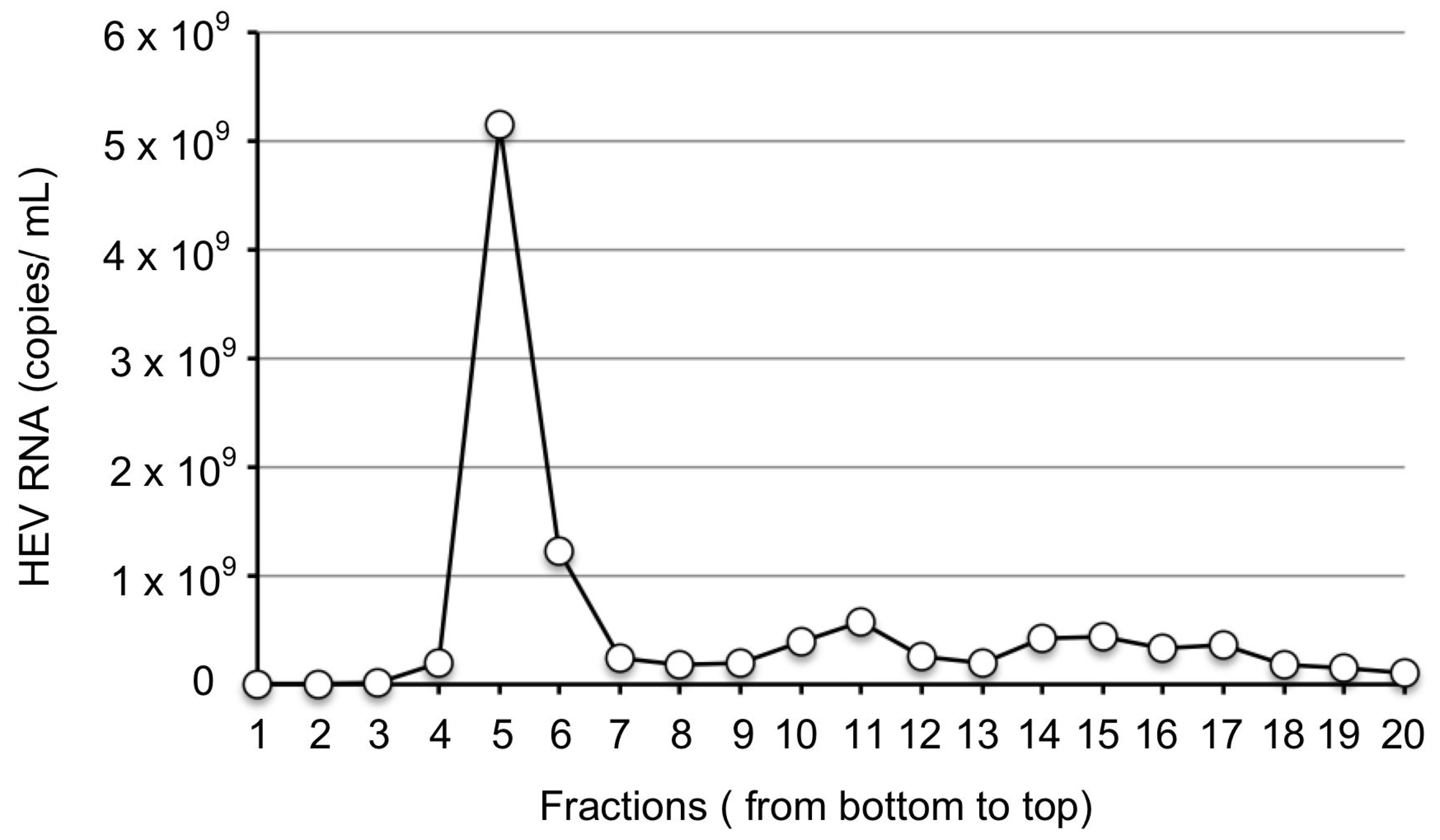
2.2. Purification of JP-59 from Stool Suspension

To raise the viral titer and remove potential virus inhibitors and toxic substances from the fecal specimens, a total of 200 g of the fecal specimens was collected from a JP-59-infected rabbit [16] on days 736–738 p.i. and a 10% stool suspension was prepared as described above. This suspension was concentrated by centrifugation at 126,000× g for 3 h at 4 °C in a Beckman SW32Ti rotor (Beckman Coulter. Inc. Brea, CA, USA), and the pellet was suspended with PBS. The virus was purified by CsCl gradient centrifugation. Briefly, 4.5 mL of the samples were mixed with 2.1 g of CsCl and centrifuged at 100,000× g for 24 h at 10 °C in the Beckman SW55Ti rotor. This gradient was fractionated into 250-µL aliquots, and the viral RNA in each fraction was examined by a quantitative real-time reverse transcription-polymerase chain reaction (RT-qPCR).
As shown in Figure 1, a peak of the viral RNA appeared in fraction 5; the copy number was 5.2 × 10⁹ copies/mL. The copy number in fraction 6 was 1.3 × 10⁹ copies/mL. For the removal of CsCl, fractions 5 and 6 were diluted with PBS and centrifuged at 100,000× g for 2 h at 4 °C in the Beckman SW55Ti rotor (Beckman). The pellets were resuspended in 1.5 mL of PBS and passed through a 0.45-µm membrane filter (Millipore). We named the viruses purified from fractions 5 and 6 JP-59F5 and JP-59F6, respectively. As shown by RT-qPCR, the final RNA copy number of JP-59F5 was 5.1 × 10⁸ copies/mL, and that of JP-59F6 was 1.2 × 10⁸ copies/mL.

2.3. Cell Culture and Virus Infection

The human hepatocarcinoma cell line PLC/PRF/5 (JCRB0406) and the human lung carcinoma cell line A549 (IF0505153) were obtained from the Health Science Research Resources Bank and grown in Dulbecco’s modified Eagle’s medium (DMEM) supplemented with 10% (v/v) heat-inactivated fetal bovine serum (FBS; Nichirei Biosciences, Tokyo, Japan), 100 U penicillin, and 100 mg streptomycin (Gibco, Grand Island, NY, USA) at 37 °C in a humidified 5% CO2 atmosphere. For virus inoculation, the confluent cells were trypsinized and cultured in a 25-cm2 culture bottle (5 × 10⁵ cells/bottle).
The next day, PLC/PRF/5 cells were inoculated with stool suspension or purified viruses and were adsorbed at 37 °C for 1 h. The cells were washed three times with PBS before being supplemented with a 10 mL maintenance medium consisting of medium 199 (Invitrogen, Carlsbad, CA, USA) containing 2% (v/v) heat-inactivated FBS and 10 mM MgCl2. The incubation was conducted at 36 °C, and the culture medium was replaced with a new medium every 4 days.

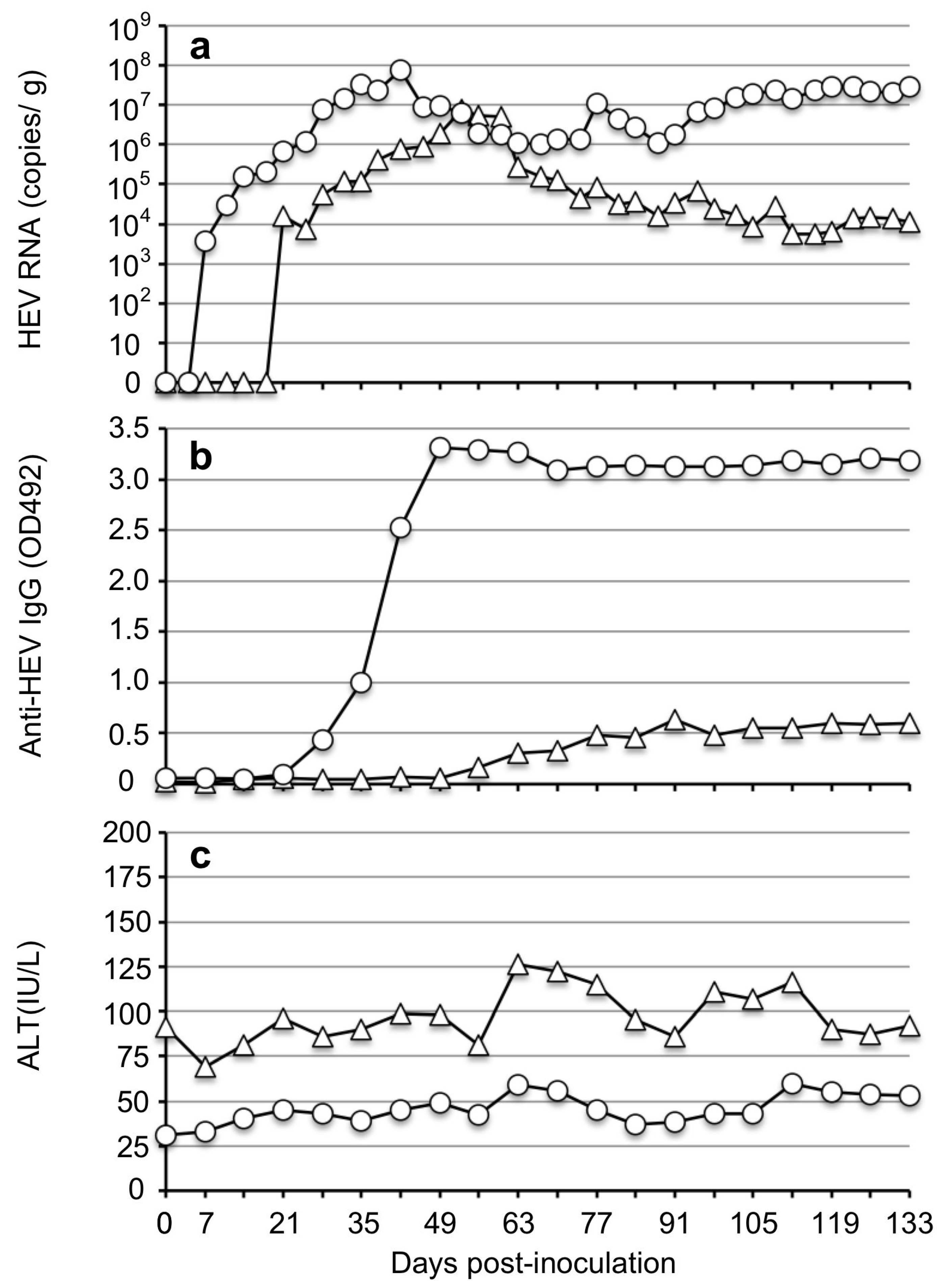
2.4. Inoculation of Rabbits and Sample Collection

The cell culture supernatant containing 7.1 × 10⁴ copies/mL of the viral RNA was collected from JP-59F5-infected PLC/PRF/5 cells on day 60 p.i., clarified by centrifugation at 10,000× g for 30 min, and then passed through a 0.45-µm membrane filter (Millipore). Two 1-year-old female Japanese white rabbits (Rab59-1 and Rab59-2) (SLC, Shizuoka, Japan) were intravenously inoculated with 1 mL of the supernatant through an ear vein.
Serum samples were collected weekly from the rabbits and used for the detection of rabbit HEV RNA, anti-rabbit HEV IgG antibodies, and alanine aminotransferase (ALT) values. Fecal specimens were collected every 3 or 4 days, and 10% (w/v) stool suspensions were used for the detection of the viral RNA.
The animal experiments were reviewed and approved by the institutional ethics committee and were performed according to the ‘Guides for Animal Experiments at the National Institute of Infectious Diseases, Tokyo, Japan, under codes 119083 (15 August 2019) and 119149 (9 January 2020). The rabbits were individually housed in Biosafety Level-2 facilities. They were negative for anti-HEV IgG antibodies and rabbit HEV RNA before inoculation.

2.5. Detection of Rabbit HEV RNA

Viral RNA was extracted from 200 µL of the cell culture supernatants or 10% stool suspensions by a MagNA Pure 96 System (Roche Applied Science, Mannheim, Germany) with a MagNA Pure 96 DNA and Viral NA Small Volume Kit (Roche Applied Science), and the copy numbers were examined by a one-step RT-qPCR using a TaqMan Fast Virus 1-step Master Mix (Applied Biosystems, Foster City, CA, USA) and a QuantStudio 3 Real-Time PCR System (Applied Biosystems) as described [24]. A 10-fold serial dilution (107 to 101 copies) of the synthetic entire genome RNA of rbIM223LR was used as the standard for the quantitation of the viral genome copy numbers.
The partial genome of the viral RNA was amplified by a nested RT-PCR with nine sets of primers (Table 1). Both the first and second PCR amplifications were carried out under the following conditions: inoculation at 96 °C for 60 s, followed by 35 cycles of 30 s at 95 °C, 30 s at 52 °C, and 60 s at 72 °C, and a final extension at 72 °C for 7 min. The PCR products were purified using a QIAquick PCR purification kit (Qiagen, Hilden, Germany), and nucleotide sequencing was carried out with the primers used for the second PCR.

2.6. Detection of Rabbit Anti-HEV IgG Antibodies

The IgG antibodies against rabbit HEV were detected by an in-house enzyme-linked immunosorbent assay (ELISA) using virus-like particles (VLPs) of rabbit HEV as the antigen. The cutoff value for the anti-HEV IgG was 0.151 [25].

2.7. Liver Enzyme Level

ALT values in the sera from rabbits were monitored weekly using a Fuji Dri-Chem Slide GPT/ALT-PIII kit (Fujifilm, Saitama, Japan) according to the manufacturer’s recommendations.

2.8. Next-Generation Sequencing (NGS) for the Entire JP-59 Genome

The fecal suspension and the infected cell culture supernatant were concentrated by ultracentrifugation at 126,000× g for 3 h in a Beckman SW32Ti rotor (Beckman), and the pellet was resuspended with PBS and used for NGS. The entire genome sequence of JP-59 was determined by NGS as described [26]. Briefly, viral RNA was extracted from 200 µL of the 10% stool suspensions, and a 200-base pair (bp) fragment library was constructed for each sample with an NEBNext Ultra RNA Library Prep Kit for Illumina ver. 1.2 (New England Biolabs, Ipswich, MA, USA) according to the manufacturer’s instructions. Samples were bar-coded for multiplexing with the use of NEBNext Multiplex Oligos for Illumina and Index Primer Sets 1 and 2 (New England Biolabs). Library purification was conducted with Agencourt AMPure XP magnetic beads (Beckman Coulter, Pasadena, CA, USA) as recommended in the NEBNext protocol. A 151-cycle paired-end read sequencing run was carried out on a MiSeq desktop sequencer (Illumina, San Diego, CA, USA) using the MiSeq Reagent Kit ver. 2 (300 cycles).

3. Results

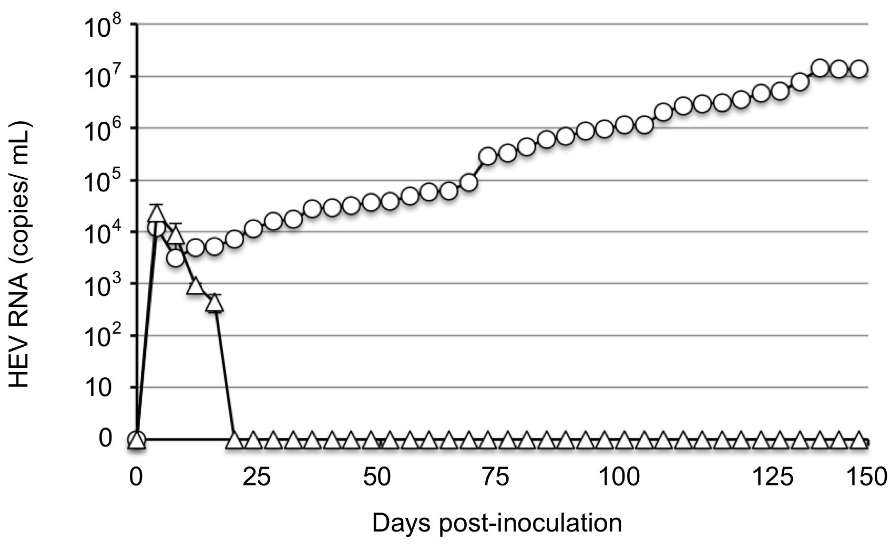
3.1. No Virus Was Recovered from PLC/PRF/5 Cells Inoculated with JP-59s

To isolate the JP-59 by using PLC/PRF/5 cells, we inoculated the cells with 1 mL of JP-59s: a stool suspension derived from a persistently JP-59-infected rabbit that contained 1.1 × 10⁷ copies/mL of viral RNA. Triplicate samples were used for the inoculation. In total, with 1 mL of LRs, the virus suspension derived from a rbIM223LR-infected rabbit contained the same RNA copy number and was used as a positive control. The viral RNA in the LRs-infected cells was first detected on day 4 p.i. at 1.21 × 10⁴ copies/mL; it decreased to 3.12 × 10³ copies/mL on day 8 p.i. and then increased gradually, reaching 1.3 × 10⁷ copies/mL on day 140 p.i. By contrast, the viral RNA in the supernatants of JP-59s-infected cells decreased rapidly and became undetectable after 20 days p.i., although viral RNA was detected on day 4 p.i. at 2.29 × 10⁴ copies/mL (Figure 2). This observation was performed until day 148 p.i., and no evidence of virus replication was observed.
A549 cells have been confirmed to be susceptible to rabbit HEV including Inner Mongolia isolates such as rbIM214L and rbIM223L [17]; however, we observed no evidence of virus replication in the case of JP-59. These results indicate that direct inoculation of a 10% stool suspension of the virus onto the existing susceptible cells is not sufficient for the further propagation of the JP-59s strain.

3.2. JP-59 Replicates in PLC/PRF/5 Cells When a Purified High-Titer Virus Was Used

In an attempt to investigate whether the purified high-titer JP-59 infected PLC/PRF/5 cells, we inoculated JP-59F5 or JP-59F6 onto the cells and monitored the viral RNAs (Figure 3). In JP-59F5-infected cells, viral RNA was detected in the culture supernatant on day 4 p.i., and the copy number was 3.7 × 10⁴ copies/mL. The RNA gradually decreased to 5.4 × 10³ copies/mL on day 28 p.i. and increased gradually from day 32 p.i., reaching 5.0 × 10⁴ copies/mL on day 40 p.i. and staying at a plateau between 2.5 × 10⁴ copies/mL and 7.1 × 10⁴ copies/mL until day 120 p.i. We named the virus recovered from the supernatants of JP-59F5-infected cells JP-59c. In the case of JP-59F6, the viral RNAs showed a similar pattern until day 60 p.i., although these copy numbers were lower than those of JP-59F5.
For the determination of whether the cell passage affected the viral replication, JP-59F6-inoculated cells were trypsinized and passaged at day 60 p.i. The cells were washed three times with PBS and incubated with 1 mL of 0.05% trypsin at 37 °C for 10 min. After the trypsin was removed, the cells were diluted with a 20 mL maintenance medium, separated into two bottles and incubated at 36 °C. The viral RNA in the supernatants rapidly decreased after the passage and became undetectable on day 8 post-passage.
These results indicated that the JP-59 strain grew in PLC/PRF/5 cells if the purified high-titer virus was used and a constant growth condition was maintained. However, the maximum copy number of the viral RNA was >100 times lower than that of the control rbIM223LR strain.

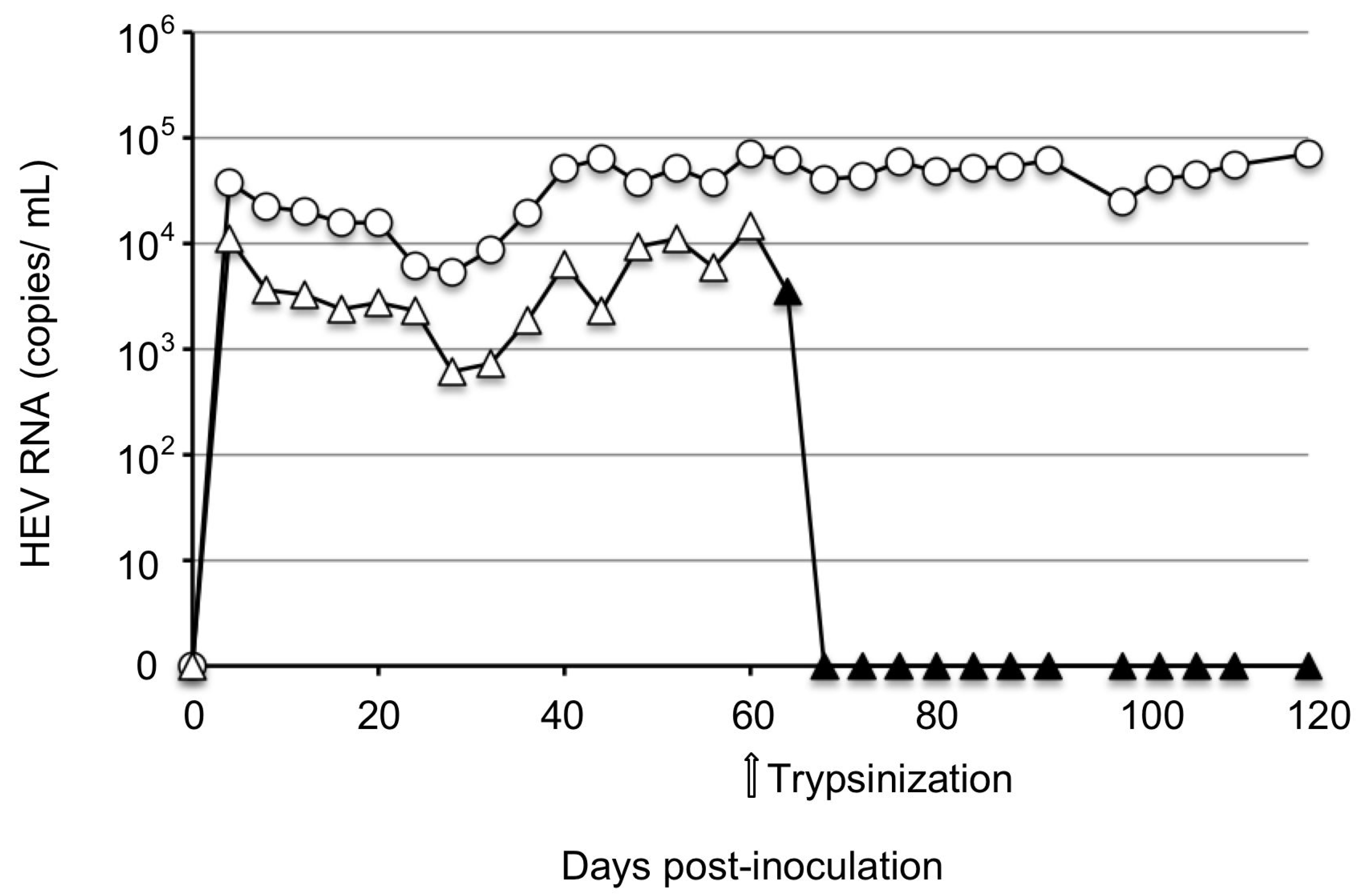
3.3. Infectivity of JP59c

To examine the infectivity of JP-59c recovered from the cell culture, we prepared two samples for inoculation: (i) an un-concentrated supernatant, i.e., the supernatant of JP-59F5-inoculated cells collected on day 60 p.i., containing 7.1 × 10⁴ copies/mL of the viral RNA, and (ii) a concentrated supernatant, in which a total of 110 mL of the supernatants collected from day 40 to 120 p.i. were mixed and concentrated at 126,000× g for 3 h in a Beckman SW32Ti rotor (Beckman), and this pellet was then suspended in 1.5 mL of PBS and then passed through a 0.45-µm membrane filter (Millipore). The concentrated supernatant contained 1.8 × 10⁶ copies/mL of viral RNA. However, both the un-concentrated and concentrated supernatants did not show any sign of replication upon their infection to PLC/PRF/5 cells.
For the examination of the in vivo infectivity of JP-59c, two Japanese white rabbits (Rab59-1 and Rab59-2) were inoculated with 1 mL of the supernatant containing viral RNA at 7.1 × 10⁴ copies/mL. After inoculation via the ear vein, the viral RNA was detected in the feces of Rab59-1 on day 7 p.i. at 3.76 × 10³ copies/g; it gradually increased to 7.85 × 10⁷ copies/g on day 42 p.i. and was then maintained at approx. 1.01 × 10⁶ to 2.96 × 10⁷ copies/g until day 133 p.i. We named the virus recovered from Rab59-1’s fecal specimens JP-59cr.
In the case of Rab59-2, the viral RNA was first detected on day 21 p.i. at 1.59 × 10⁴ copies/g; it had gradually increased to 7.82 × 10⁶ copies/g on day 53 p.i. and then gradually decreased but was detected at approx. 1.12 × 10⁴ copies/g until day 133 p.i. (Figure 4a). The viral RNA in all serum samples was under detection limits by the RT-qPCR.
The IgG antibody was first detected on day 28 in Rab59-1 with an optical density (OD) value of 0.429 and on day 56 p.i. in Rab59-2 with an OD value of 0.161. The IgG peaked on day 49 p.i. in Rab59-1 with an OD value of 3.309 and then stayed at similar levels until day 133 p.i. In contrast, the peak value of the IgG was 0.601 in Rab59-2 on day 91 p.i. (Figure 4b). Although the ALT levels in these two rabbits differed, no significant elevation was observed during the infection period (Figure 4c). These results indicated that the JP-59c isolated by the cell culture was infectious in vivo.

3.4. Nucleotide Sequence Analyses of JP-59 Strains

The entire genome sequences of JP-59F5, JP-59F6, and JP-59cr were analyzed by NGS, which revealed that all the viruses consisted of 7282 nucleotides plus a poly (A) tail with an undetermined length. The 5’ terminal untranslated region (5’UTR) and 3’UTR contained 26 nucleotides (nucleotide sequence nos. 1–26) and 70 nucleotides (7213–7282), respectively. ORF1 (27–5195) encoded 1723 amino acids (aa), ORF3 (5192–5560) encoded 122 aa, and ORF2 (5230–7212 nt) encoded 660 aa.
JP-59F5 and JP-59F6 were collected on days 736–738 (approx. 2 years) p.i., and both entire genome sequences were identical to that of the original JP-59 collected from the same rabbit at 12 weeks p.i. [16], suggesting that JP-59, which replicated in the rabbit, was genetically stable. In contrast, a total of 18 nucleotide changes (15 in ORF1, two in ORF2, and one in ORF3) were identified in JP-59cr (Table 2). These nucleotide changes were accompanied by two aa mutations (Thr566Ile and Gly749Ser) in ORF1 and one aa mutation (Ser4Phe) in ORF3 (Table 2). The mutation Thr566Ile was observed in the hepevirus unique domain and Gly749Ser is located in the hypervariable polyproline region of ORF1. The amino acid, 566Ile, was identified in rbIM223LR, but 749Ser and 4Phe were not present in rbIM223LR. No mutation was found in the 5’UTR or 3’UTR.
Since the virus titer of JP-59c was quite low, the entire genome sequence could not be determined by NGS. For the confirmation of whether the mutation occurred during the replication in cell culture, nine partial genome fragments of JP-59c that covered all the positions of these 18 mutations were amplified by RT-PCR using the primers shown in Table 1. The nucleotide sequence analyses confirmed that all 18 mutations were present in JP-59c, demonstrating that these mutations in JP-59cr were derived from JP-59c and occurred during replication in PLC/PRF/5 cells.

4. Discussion

Although a cell culture system that multiplied rabbit HEV was established using virus strains such as rbIM214L and rbIM223L, which were isolated in Inner Mongolia [17], it is unknown whether other strains were capable of replicating in the cell culture. JP-59 is the only rabbit HEV strain identified in Japan [16]. To establish a cell culture for growing this strain, we inoculated the virus onto PLC/PRF/5 cells because the PLC/PRF/5 was broadly susceptible to HEV infection, and the cell culture of rabbit HEV was established using this cell line. However, the ability of JP-59 to replicate in the cell culture was significantly lower than that of an Inner Mongolia isolate, rbIM223LR, suggesting that the ability of rabbit HEV to propagate in the cell culture varied. In other words, PLC/PRF/5 cells were not the best choice for this purpose, and a novel cell culture system for rabbit HEV was required. It is of interest to examine whether the cell lines derived from rabbits were susceptible to rabbit HEV infection.
We observed the replication of JP-59 in the cells only when the virus solution containing >108 copies/mL of the viral RNA was used, demonstrating that a high titer of JP-59 was required to initiate an infection in PLC/PRF/5 cells. Although the cell culture is a convenient method for HEV isolation, it was difficult to maintain the original viral sequence by the cell culture due to the unexpected mutations that often occurred during the long-term virus multiplication in the cell culture. Our findings demonstrate that it is difficult to isolate viruses with a low replication capacity, such as strain JP-59. In contrast, we observed that JP-59 recovered from the cell culture supernatants, which had low copy numbers of the viral RNA, could infect rabbits, suggesting that the isolation of the virus by injecting rabbits is another option for rabbit HEV isolation. Since the JP-59 that was replicated in the rabbits was genetically stable, these rabbits might provide a useful animal model to obtain rabbit HEV with the original sequence.
The nucleotide acid sequence identity between the JP-59 and rbIM223LR strains was 87.4%, and it is of interest to identify the nucleotide acid and amino acid changes that are responsible for better rabbit HEV replication.

Author Contributions

Conceptualization: W.Z. and T.L. Methodology: W.Z. and T.L. Investigation: M.V.M., Y.S., Y.A. NGS analyses: Y.H.D. Writing—original draft preparation: W.Z. Writing—review and editing: T.L. Supervision: K.M. Funding acquisition: T.L. All authors have read and agreed to the published version of the manuscript.

Funding

This research was supported by grants from the Research Program on Hepatitis (nos. JP23fk0210109, JP23fk0210132, JP22wm0225009) from the Japan Agency for Medical Research and Development (AMED) and Health Labour Sciences Research Grant (no. 21KA1003).

Institutional Review Board Statement

The animal experiments were reviewed and approved by the institutional ethics committee and performed according to the Guides for Animal Experiments at the National Institute of Infectious Diseases, Tokyo, Japan under codes 119083 (15 August 2019) and 119149 (9 January 2020).

Informed Consent Statement

Not applicable.

Data Availability Statement

The sequences of HEV used in the study were assigned (GenBank accession nos. LC535077 and LC484431).

Acknowledgments

We thank Miyuki Oizumi for their technical assistance and Naokazu Takeda for revising the manuscript.

Conflicts of Interest

The authors declare no conflict of interest.

References

  1. Purdy, M.A.; Drexler, J.F.; Meng, X.-J.; Norder, H.; Okamoto, H.; Van der Poel, W.H.M.; Reuter, G.; de Souza, W.M.; Ulrich, R.G.; Smith, D.B. ICTV virus taxonomy profile: Hepeviridae 2022. J. Gen. Virol. 2022, 103, 001778. [Google Scholar] [CrossRef]
  2. Meng, X.J. Hepatitis E virus: Animal reservoirs and zoonotic risk. Vet. Microbiol. 2010, 140, 256–265. [Google Scholar] [CrossRef] [PubMed] [Green Version]
  3. Lee, G.-H.; Tan, B.-H.; Teo, E.C.-Y.; Lim, S.-G.; Dan, Y.-Y.; Wee, A.; Aw, P.P.K.; Zhu, Y.; Hibberd, M.; Tan, C.-K.; et al. Chronic infection with camelid hepatitis E virus in a liver transplant recipient who regularly consumes camel meat and milk. Gastroenterology 2016, 150, 355–357.e3. [Google Scholar] [CrossRef] [Green Version]
  4. Sridhar, S.; Yip, C.C.; Wu, S.; Chew, N.F.; Leung, K.; Chan, J.F.; Zhao, P.S.; Chan, W.; Poon, R.W.; Tsoi, H.; et al. Transmission of Rat hepatitis E virus infection to humans in Hong Kong: A clinical and epidemiological analysis. Hepatology 2021, 73, 10–22. [Google Scholar] [CrossRef] [PubMed]
  5. Zhao, C.; Ma, Z.; Harrison, T.J.; Feng, R.; Zhang, C.; Qiao, Z.; Fan, J.; Ma, H.; Li, M.; Song, A.; et al. A novel genotype of hepatitis E virus prevalent among farmed rabbits in China. J. Med. Virol. 2009, 81, 1371–1379. [Google Scholar] [CrossRef] [PubMed]
  6. Smith, D.B.; Simmonds, P.; International Committee on Taxonomy of Viruses Hepeviridae Study Group; Jameel, S.; Emerson, S.U.; Harrison, T.J.; Meng, X.J.; Okamoto, H.; Van der Poel, W.H.; Purdy, M.A. Consensus proposals for classification of the family Hepeviridae. J. Gen. Virol. 2014, 95, 2223–2232. [Google Scholar] [CrossRef] [PubMed]
  7. Smith, D.B.; Simmonds, P.; Izopet, J.; Oliveira-Filho, E.F.; Ulrich, R.G.; Johne, R.; Koenig, M.; Jameel, S.; Harrison, T.J.; Meng, X.-J.; et al. Proposed reference sequences for hepatitis E virus subtypes. J. Gen. Virol. 2016, 97, 537–542. [Google Scholar] [CrossRef]
  8. Smith, D.B.; Izopet, J.; Nicot, F.; Simmonds, P.; Jameel, S.; Meng, X.-J.; Norder, H.; Okamoto, H.; van der Poel, W.H.; Reuter, G.; et al. Update: Proposed reference sequences for subtypes of hepatitis E virus (species Orthohepevirus A). J. Gen. Virol. 2020, 101, 692–698. [Google Scholar] [CrossRef]
  9. Geng, J.; Fu, H.; Wang, L.; Bu, Q.; Liu, P.; Wang, M.; Sui, Y.; Wang, X.; Zhu, Y.; Zhuang, H. Phylogenetic analysis of the full genome of rabbit hepatitis E virus (rbHEV) and molecular biologic study on the possibility of cross species transmission of rbHEV. Infect. Genet. Evol. 2011, 11, 2020–2025. [Google Scholar] [CrossRef]
  10. Cossaboom, C.M.; Córdoba, L.; Cao, D.; Ni, Y.-Y.; Meng, X.-J. Complete genome sequence of hepatitis E virus from rabbits in the United States. J. Virol. 2012, 86, 13124–13125. [Google Scholar] [CrossRef] [Green Version]
  11. Geng, Y.; Zhang, H.; Li, J.; Huang, W.; Harrison, T.J.; Zhao, C.; Zhou, Y.; Lian, H.; Wang, Y. Comparison of hepatitis E virus genotypes from rabbits and pigs in the same geographic area: No evidence of natural cross-species transmission between the two animals. Infect. Genet. Evol. 2013, 13, 304–309. [Google Scholar] [CrossRef] [PubMed]
  12. Burt, S.A.; Veltman, J.; Der Honing, R.H.-V.; Schmitt, H.; Van Der Poel, W.H.M. Hepatitis E virus in farmed rabbits, wild rabbits and petting farm rabbits in the Netherlands. Food Environ. Virol. 2016, 8, 227–229. [Google Scholar] [CrossRef] [Green Version]
  13. Di Bartolo, I.; De Sabato, L.; Marata, A.; Martinelli, N.; Magistrali, C.F.; Monini, M.; Ponterio, E.; Ostanello, F.; Ruggeri, F.M. Serological survey of hepatitis E virus infection in farmed and pet rabbits in Italy. Arch. Virol. 2016, 161, 1343–1346. [Google Scholar] [CrossRef] [PubMed]
  14. Hammerschmidt, F.; Schwaiger, K.; Dähnert, L.; Vina-Rodriguez, A.; Höper, D.; Gareis, M.; Groschup, M.H.; Eiden, M. Hepatitis E virus in wild rabbits and European brown hares in Germany. Zoonoses Public Health 2017, 64, 612–622. [Google Scholar] [CrossRef] [PubMed]
  15. Han, S.-H.; Park, B.-J.; Ahn, H.-S.; Kim, Y.-H.; Go, H.-J.; Kim, D.-H.; Lee, J.-B.; Park, S.-Y.; Song, C.-S.; Lee, S.-W.; et al. Evidence of hepatitis E virus infection in specific pathogen-free rabbits in Korea. Virus Genes 2018, 54, 587–590. [Google Scholar] [CrossRef] [PubMed]
  16. Mendoza, M.V.; Yonemitsu, K.; Ishijima, K.; Minami, S.; Supriyono; Tran, N.T.; Kuroda, Y.; Tatemoto, K.; Inoue, Y.; Okada, A.; et al. Characterization of rabbit hepatitis E virus isolated from a feral rabbit. Vet. Microbiol. 2021, 263, 109275. [Google Scholar] [CrossRef]
  17. Jirintai, S.; Jinshan; Tanggis; Manglai, D.; Mulyanto; Takahashi, M.; Nagashima, S.; Kobayashi, T.; Nishizawa, T.; Okamoto, H. Molecular analysis of hepatitis E virus from farm rabbits in Inner Mongolia, China and its successful propagation in A549 and PLC/PRF/5 cells. Virus Res. 2012, 170, 126–137. [Google Scholar] [CrossRef]
  18. Zhang, W.; Ami, Y.; Suzaki, Y.; Doan, Y.H.; Jirintai, S.; Takahashi, M.; Okamoto, H.; Takeda, N.; Muramatsu, M.; Li, T. Persistent infection with a rabbit hepatitis E virus created by a reverse genetics system. Transbound. Emerg. Dis. 2020, 8, 615–625. [Google Scholar] [CrossRef]
  19. Cossaboom, C.M.; Córdoba, L.; Sanford, B.J.; Piñeyro, P.; Kenney, S.P.; Dryman, B.A.; Wang, Y.; Meng, X.-J. Cross-species infection of pigs with a novel rabbit, but not rat, strain of hepatitis E virus isolated in the United States. J. Gen. Virol. 2012, 93, 1687–1695. [Google Scholar] [CrossRef]
  20. Liu, P.; Bu, Q.-N.; Wang, L.; Han, J.; Du, R.-J.; Lei, Y.-X.; Ouyang, Y.-Q.; Li, J.; Zhu, Y.-H.; Lu, F.-M.; et al. Transmission of hepatitis E virus from rabbits to cynomolgus macaques. Emerg. Infect. Dis. 2013, 19, 559–565. [Google Scholar] [CrossRef]
  21. Abravanel, F.; Lhomme, S.; El Costa, H.; Schvartz, B.; Peron, J.-M.; Kamar, N.; Izopet, J. Rabbit hepatitis E virus infections in humans, France. Emerg. Infect. Dis. 2017, 23, 1191–1193. [Google Scholar] [CrossRef] [PubMed] [Green Version]
  22. Sahli, R.; Fraga, M.; Semela, D.; Moradpour, D.; Gouttenoire, J. Rabbit HEV in immunosuppressed patients with hepatitis E acquired in Switzerland. J. Hepatol. 2019, 70, 1023–1025. [Google Scholar] [CrossRef] [Green Version]
  23. Lhomme, S.; Dubois, M.; Abravanel, F.; Top, S.; Bertagnoli, S.; Guerin, J.-L.; Izopet, J. Risk of zoonotic transmission of HEV from rabbits. J. Clin. Virol. 2013, 58, 357–362. [Google Scholar] [CrossRef] [PubMed]
  24. Jothikumar, N.; Cromeans, T.L.; Robertson, B.H.; Meng, X.J.; Hill, V.R. A broadly reactive one-step real-time RT-PCR assay for rapid and sensitive detection of hepatitis E virus. J. Virol. Methods 2006, 131, 65–71. [Google Scholar] [CrossRef]
  25. Bai, H.; Kataoka, M.; Ami, Y.; Suzaki, Y.; Takeda, N.; Muramatsu, M.; Li, T.-C. Immunogenicity and antigenicity of rabbit hepatitis E virus-like particles produced by recombinant baculoviruses. Viruses 2021, 13, 1573. [Google Scholar] [CrossRef] [PubMed]
  26. Li, T.-C.; Yang, T.; Yoshizaki, S.; Ami, Y.; Suzaki, Y.; Ishii, K.; Haga, K.; Nakamura, T.; Ochiai, S.; Takaji, W.; et al. Construction and characterization of an infectious cDNA clone of rat hepatitis E virus. J. Gen. Virol. 2015, 96, 1320–1327. [Google Scholar] [CrossRef]
Figure 1. Purification of the JP-59 viruses. Fecal specimens were collected from the JP-59-infected rabbit on days 736–738 p.i., and a 10% suspension was prepared. The virus was concentrated by an ultracentrifugation and then purified by a CsCl equilibrium density gradient ultracentrifugation. The copy numbers of the virus RNA in each fraction were determined by RT-qPCR (○).
Viruses 15 01322 g001
Figure 2. The replication of strains JP-59 and rbIM223LR in PLC/PRF/5 cells. PLC/PRF/5 cells were cultured in 25-cm2 bottles. The stool suspensions (JP-59s) derived from a JP-59-infected rabbit containing 1.1 × 10⁷ copies/mL of the viral RNA were inoculated onto three bottles of PLC/PRF/5 cells (△). The stool suspension (LRs) derived from the rbIM223LR-infected rabbit containing the same copy number of the viral RNA was inoculated onto one bottle of PLC/PRF/5 cells and used as a positive control (○). The culture supernatants were collected every 4 days and used for the detection of viral RNA by RT-qPCR.
Viruses 15 01322 g002
Figure 3. The replication of strain JP-59 in PLC/PRF/5 cells. One milliliter of the concentrated purified JP-59F5 (○) and JP-59F6 (△), respectively, was inoculated onto PLC/PRF/5 cells. The JP-59F6-infected cells were harvested, trypsinized, and passaged on day 60 p.i. and maintained similarly thereafter (▲). Arrow: the time point of the trypsinization. The culture supernatants were collected every 4 days and used for the detection of viral RNA by RT-qPCR.
Viruses 15 01322 g003
Figure 4. The infectivity of strain JP-59c in vivo. Two Japanese white rabbits, Rab59-1 (○) and Rab59-2 (△) were intravenously inoculated with the cell culture supernatants containing 7.1 × 10⁴ copies/mL of the viral RNAs (JP-59c), which were derived from the JP-59F5-inoculated cells on day 60 p.i. The fecal specimens were collected every 3 or 4 days, and the serum samples were collected weekly. The kinetics of the viral RNA in the feces were determined by RT-qPCR (a). The serum anti-rabbit HEV IgG antibodies were determined by ELISA (b). ALT values were measured by a commercial kit (c).
Viruses 15 01322 g004
Table 1. Primers used to amplify the partial genome of JP-59c.
NameFor the First PCRNameFor the Second PCRProducts,
nt
F38TCGATGCCATGGAGGCCCAF105GCCTTGGCGAATGCTGTGG105–469
R560CATTGCCTCTGCCACATCAGR469TCAAAGCAATAGGTTCGATC
F373GCGATGGTATTCTGCCCCTAF540CTGATGTGGCAGAGGCAATG373–1100
R1128CATCTTCAGAGGCATTCCAGR1110AGCTACAAGAGCACCAACAG
F1337GCTTTCTGCCGGCTTTCATTF1378TTGACGAATCTGTGCACTGC1378–1759
R1805CTGAGCAGTACGTTCTCTCAR1759TCAGTGATTGTGGTGCGGAA
F2089GGGAGTCTACTAACCCGTTTF2160AGCGACTTTTCACCACCTGA2160–2439
R2519CTTGGTATGGTCGAAAGACTR2439ATGCGTTAACCAGCCAGTCA
F2501GTCTTTCGACCATACCAAGTF2581TCGCCCCTGATTATAGAGTT2581–3040
R3074CTGCTGTATGGACCTCGATTR3040AACGCCTGCAGTGAATTGATA
F3221CCACCTGCTTTTGTTGCATAF3301TTGAGCATGCCGGTCTAGTC3301–3699
R3840GATAGGCACTAATCTGGCAGR3699ATATGCCCACCTCACGAAGT
F4048GTATGGGCGTCGAACAAAGTF4141CTACAACGTGCGAGCTTTAT4141–4359
R4440CAGAGTCTTCATAGGCGTCAR4359GCCCAAATAAGGCGCAGAAT
F5020CGAATGTTGCTCAGGTGTGTF5091CTTATTGGCATGTTACAGAC5091–5440
R5280CAGCATAGGCAAACACACGAR5440CGGAATGTGAGTCAACGTCA
F5986TATCGTAACCAGGGGTGGCGF6022GGTGTAGCCGAGGAGGAGGC6022–6650
R6702AGGGTTAGTTGACGAGCCATR6650GAGAGCCACAACACATCGTT
Table 2. Nucleotide and amino acid changes in rabbit HEV recovered from a JP-59c-infected rabbit.
Nucleotide ChangesAmino Acid Changes
a PositionJP-59JP-59crb PositionORF1ORF2ORF3
249CT
368CT
551GA
800TC
1040GA
1439CT
1613AG
1652GA
1723CT566Thr/Ile
2261CT749Gly/Ser
2271GA
2690CT
2930CT
3455TC
4304CT
5205CT4 Ser/Phe
6351CT
6594AG
a Nucleotide positions in the genomic RNA. b Amino acid positions in each ORF.
Disclaimer/Publisher’s Note: The statements, opinions and data contained in all publications are solely those of the individual author(s) and contributor(s) and not of MDPI and/or the editor(s). MDPI and/or the editor(s) disclaim responsibility for any injury to people or property resulting from any ideas, methods, instructions or products referred to in the content.

