Article

Genomic Analysis of Amphioxus Reveals a Wide Range of Fragments Homologous to Viral Sequences

1
Agricultural Bioinformatics Key Laboratory of Hubei Province and 3D Genomics Research Centre, College of Informatics, Huazhong Agricultural University, Wuhan 430070, China
2
School of Biomedical Sciences, The Chinese University of Hong Kong, Hong Kong, China
3
Hong Kong Bioinformatics Centre, The Chinese University of Hong Kong, Hong Kong, China
4
Stanford Cardiovascular Institute, Stanford University School of Medicine, Stanford, CA 94305, USA
*
Author to whom correspondence should be addressed.
These authors contributed equally to this work.
Viruses 2023, 15(4), 909; https://doi.org/10.3390/v15040909
Submission received: 18 January 2023 / Revised: 11 March 2023 / Accepted: 28 March 2023 / Published: 31 March 2023
(This article belongs to the Special Issue An Interdisciplinary Approach to Virology Research)

Abstract

:
Amphioxus species are considered living fossils and are important in the evolutionary study of chordates and vertebrates. To explore viral homologous sequences, a high-quality annotated genome of the Beihai amphioxus (Branchiostoma belcheri beihai) was examined using virus sequence queries. In this study, 347 homologous fragments (HFs) of viruses were identified in the genome of B. belcheri beihai, of which most were observed on 21 genome assembly scaffolds. HFs were preferentially located within protein-coding genes, particularly in their CDS regions and promoters. A range of amphioxus genes with a high frequency of HFs is proposed, including histone-related genes that are homologous to the Histone or Histone H2B domains of viruses. Together, this comprehensive analysis of viral HFs provides insights into the neglected role of viral integration in the evolution of amphioxus.

1. Introduction

Many studies involving viral homologous sequences in the genomes of higher animals have been reported, and sequences homologous to viral oncogenes have been identified in most vertebrates [1,2,3]. For example, it has been demonstrated that a sequence in the human genome is homologous to the v-myc oncogene of the Avian myelocytomatosis virus [4], and wild bats host sequences homologous to sequences in various eukaryotic viruses [5]. The analysis of the viral homologous sequences in animals helps in revealing the occurrence of endogenous viral integration and viral invasion, as well as genetic material exchanges between viruses and hosts [5,6,7,8,9]. The endogenous integration of the virus into higher animal genomes is believed to play an important role in evolution [10,11]. Endogenous retroviruses (ERVs) shape the evolution of transcriptional networks, and some ERVs can encode complete proteins and play physiological functions within hosts [12,13]. Endogenous bornavirus-like elements and some other nonretroviral endogenous viral elements encode functional proteins in host animals [10,14,15]. It is noteworthy that the diversity and evolution of the viruses themselves are also affected by endogenous integration [16]. Thus, viral homologous sequences provide valuable information regarding evolutionary modifications associated with hosts as well as virus–host interactions.
The common histones H2A, H2B, H3, and H4 are quintessentially eukaryotic proteins. Nevertheless, the homologs of the genes that encode these eukaryotic histones have also been identified in the DNA of virus genomes. Pandoravirus genomes have histone genes with eukaryotic homologs [5], and a Pandoravirus-related genome assembled from the marine metagenome encodes an H4-like protein that is 77% identical to human H4 [17]. Viruses in Marseilleviridae and the medusavirus in the order Pandoravirales encode distinct histone-like proteins homologous to all four core eukaryotic histones [18,19,20]. Some viral histone genes alter host gene expression by expression in the host, leading to negative changes in host development and metabolism that are beneficial to the viruses [21,22,23,24]. Viral histone H4 alters host gene expression by interacting with eukaryotic nucleosomes [23]. Bracoviruses have been shown to use their histone genes as weapons to gain an advantage by suppressing host immune responses and development [25,26,27]. Many viral histones have not been investigated to identify their functions and evolutionary roles in hosts and viruses. The group of basal chordates, amphioxus, contains some of the closest living invertebrate relatives of vertebrates [28,29,30,31,32,33]. Its members play a pivotal role in elucidating the evolution of chordates and vertebrates [34,35,36]. However, there have been few systematical studies of the viral homologous sequences and histones in amphioxus species.
A genomic analysis was performed to fully identify the viral homologous sequences in amphioxi. In this study, the adult Beihai amphioxus was suggested as a B. belcheri subspecies and was tentatively named B. belcheri beihai. A high-quality B. belcheri beihai genome was assembled and annotated by our team [37]. A total of 347 HFs in the B. belcheri beihai genome were identified, and the genomic features of the HFs were analyzed. A few amphioxus genes with high-confidence HFs and genomic regions enriched with HFs were proposed. More importantly, there were highly conserved sequences in histone-related genes between viruses and the amphioxus. The analysis of the viral HFs in the amphioxus genome could provide abundant evidence for endogenous integrations, broadening our understanding of the evolution of and interaction between amphioxus species and viruses.

2. Materials and Methods

2.1. Sample Collection

Adults of the Beihai amphioxus, B. belcheri beihai were obtained from the sea near Dianbai District, Maoming City, Guangdong Province, China, cultured at 24–28 °C with air-pumped circulating artificial seawater in Beihai Marine Station of Nanjing University in Beihai City, Guangxi Province, China, and fed with seawater and sea alga.

2.2. Genome Sequencing and De Novo Genome Assembly

Genome sequencing and de novo genome assembly of B. belcheri beihai has been reported [37]. In brief, the genomic DNA was sequenced by Illumina HiSeq 2000 in 500-bp and 3000-bp libraries to generate paired-end NGS short reads and PacBio RS system for third-generation long reads. The first draft of genome assembly was constructed with PacBio long reads using Canu v1.8 [38]. Then, the final genome assembly was generated after scaffolding and polishing of the draft assembly. The continuity of the genome assembly was assessed by QUAST v5.0.2 [39].The completeness was assessed by BUSCO v3.1.0 [40] with database metazoa_odb9.
The genome assembly size of B. belcheri beihai was 478,319,013 bp and the scaffold number was 583. The scaffold N50 length was 4,185,906 bp and the gap content was 0.156%. Regardless of the gaps, the ungapped length of B. belcheri beihai was 474,945,525 bp. As for completeness, the genome assembly of B. belcheri beihai was 97.2%.

2.3. Genome Annotation

Firstly, repeat masking was performed by de novo prediction with RepeatModeler v2.0.1 [41], and masking with RepeatMasker v4.0.8 (RepBase edition 20181026) [42]. In the de novo prediction with RepeatModeler v2.0.1 [41], the prediction of repeat families in the genome was performed using RECON v1.05 [43] and RepeatScout v1.0.6 [44]. Then, Maker pipeline v2.31 was used for genome annotation [45]. In the Maker pipeline, the alignment was supported by transcriptome assemblies and homologous proteins by Exonerate v2.4.0 [46], whereas gene prediction was accomplished by SNAP (lib v2017–03-01) [47], GeneMark v4.38 [48] and Augustus v3.3.1 [49]. The quality of genome annotation was assessed by BUSCO v3.1.0 [40] with database metazoa_odb9. The completeness of genome annotation was 95.2%. In total, 44,745 protein-coding genes were annotated in B. belcheri beihai, and most of genes were homologous to Homo sapiens and Mus musculus genes (Figure S1).

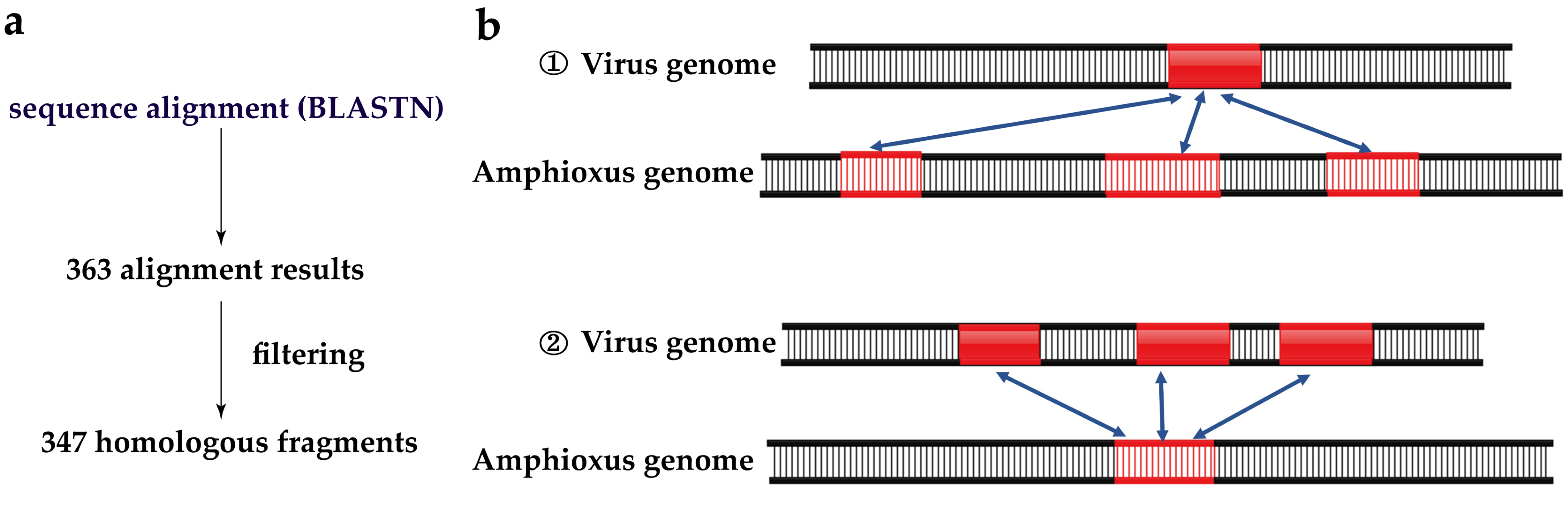
2.4. Identification of HFs in Amphioxus Genome

A total of 9569 viral genomes were searched in the genome of B. belcheri beihai by BLASTN v2.5.0 [50] at E-value cutoff of 1 × 10⁻⁵ to obtain 363 aligned fragments. The B. belcheri beihai genome was used as the reference sequence to build the library. The aligned fragments were filtered to obtain HFs.
A fragment of viral sequences might align with multiple DNA fragments in the B. belcheri beihai genome. There were also fragments of viral sequences that only aligned with a fragment of B. belcheri beihai. To make the essence of the alignment score the same, the HFs of B. belcheri beihai that were in line with the first situation were retained. There was another situation of alignments where multiple fragments of viral sequences aligned with the same DNA fragment in the B. belcheri beihai genome. The results with the minimum E-value were retained after filtering using the E-value cutoff, and finally 347 HFs were identified in the B. belcheri beihai genome.

2.5. Identification of HFs in Viral Genomes

Multiple fragments of viral sequences were less likely to align with the same DNA fragment in the B. belcheri beihai genome. BLASTN results with the minimum E-value after filtering using the E-value cutoff of 1 × 10⁻⁵ were retained, and all results with the same minimum E-value were retained. In total, 361 results were obtained from 363 alignment results through above processing. The same fragment of viral sequences aligned with multiple DNA fragments in the B. belcheri beihai genome. In this case, 69 results with the minimum E-value were retained. Because there were adjacent breakpoints in the viral genomes, a threshold of 10 bp was adopted to merge the adjacent fragments into a single fragment. The duplicate DNA fragments were removed after merging, and a total of 50 HFs were identified in 17 viral genomes.

2.6. Data Analysis

The data analysis statistics were mainly implemented by Linux shell v4.2.46 and R v4.0.5 [51] programming, and the ggplot2 [52] v3.3.6 R package undertook most of visualization tasks in this study. Statistical significance was assessed by computing either the p value or the adjusted p value (using the Bonferroni method) based on the chi-squared ( X 2 ) test. This test was selected because it enables comparison of the observed frequency distribution of a categorical variable with an expected frequency distribution that follows a specific theoretical distribution, as previously reported in the literature [53,54,55,56].
The simplified pipeline, the schematic diagram for the HFs and the schematic indicating that multiple fragments of viral DNA were homologous to one fragment in the B. belcheri beihai genome were manually drawn in PowerPoint 2019. The schematic diagram for HFs containing the longest length with the highest alignment quality were also manually drawn through Illustrator for Biological Sequences v1.0 [57,58] online. The Circos image for the gene analysis and functional annotation of the viral HFs was completed by using the OmicStudio tools at https://www.omicstudio.cn/tool (accessed on 22 November 2022). The images for general profiles of HFs and distribution of HFs in 100 kb sliding windows were generated in SVG format based on Perl v5.10 (http://www.perl.org/, accessed on 25 September 2022).

3. Results

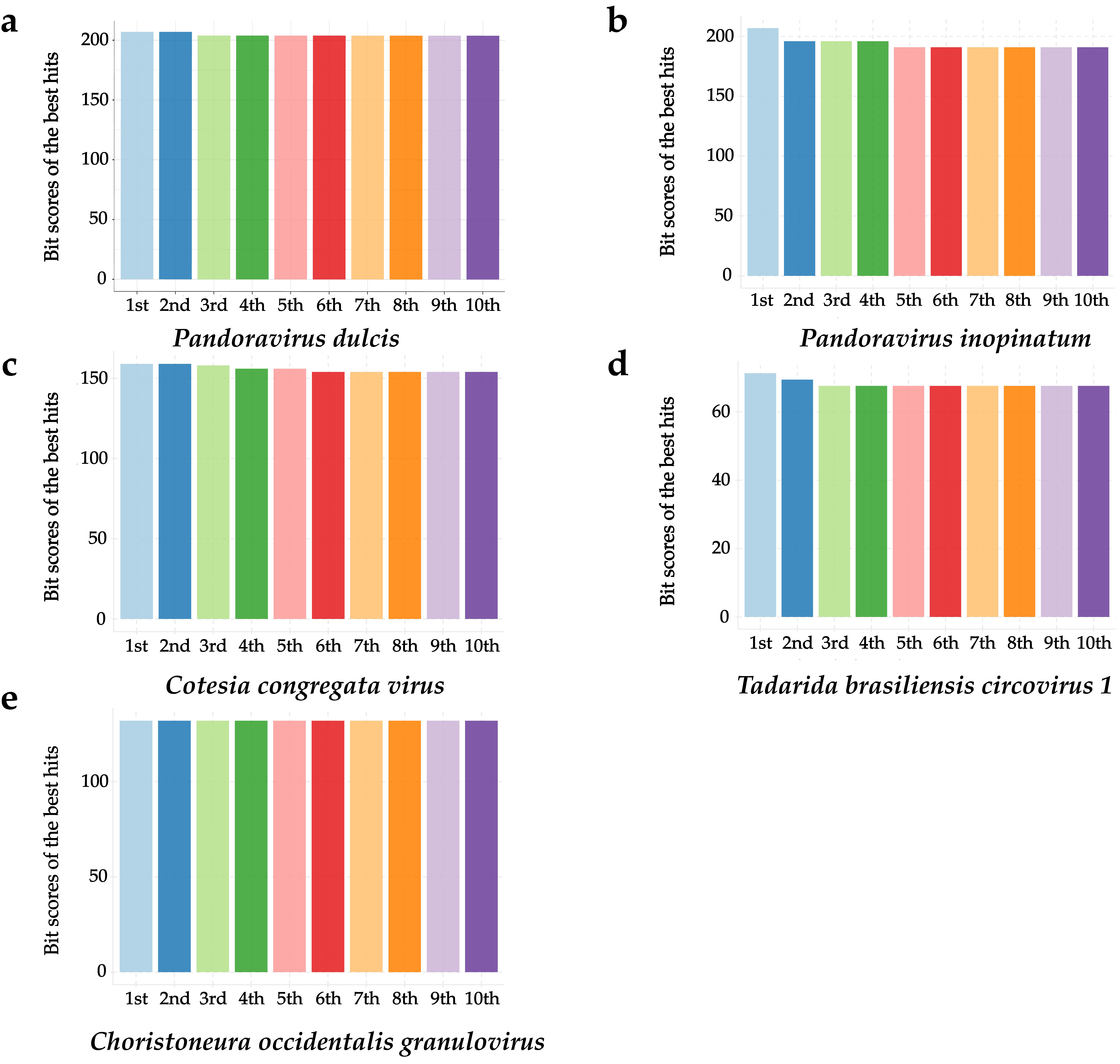
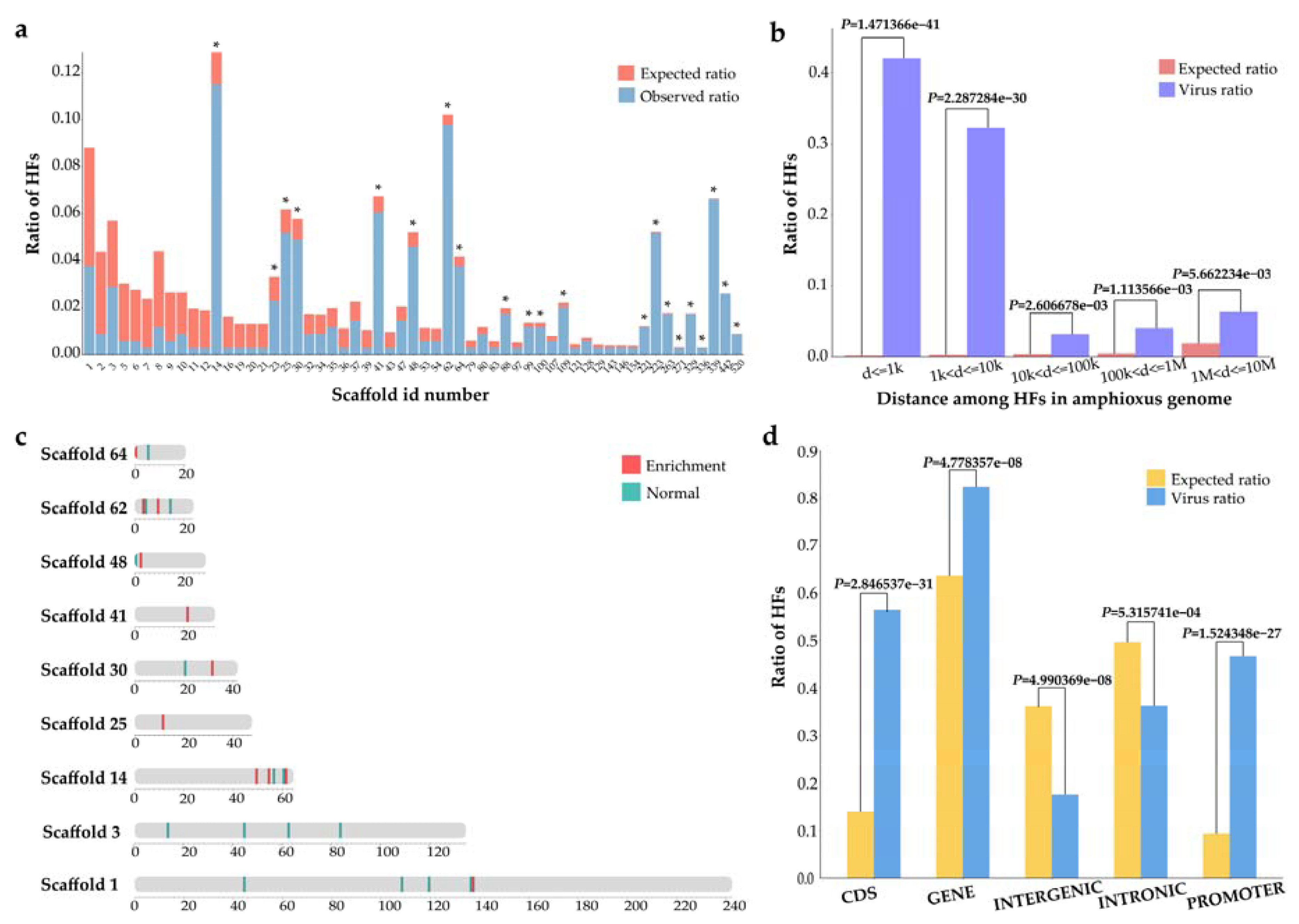
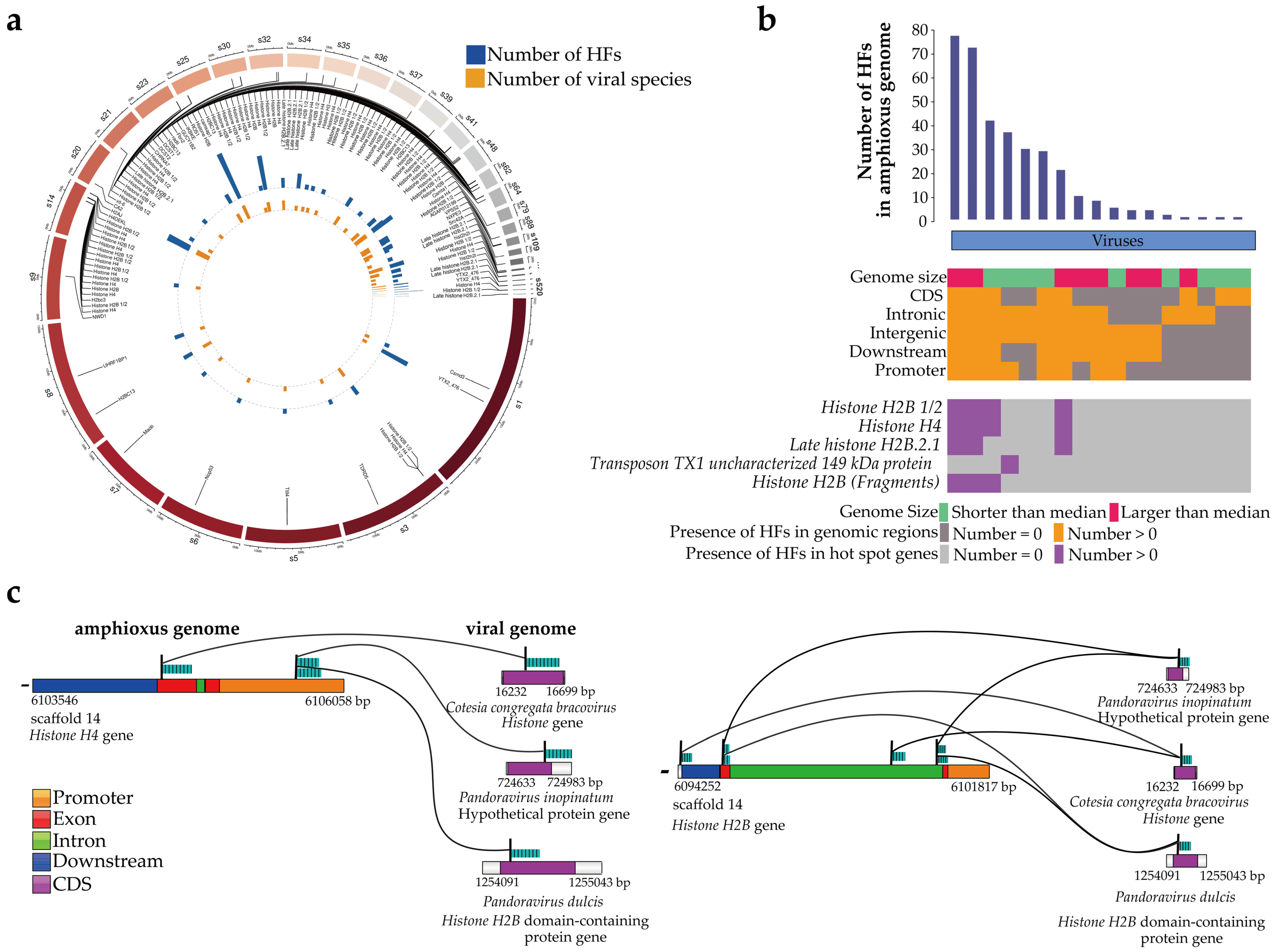
3.1. Identification of Viral HFs in the Amphioxus Genome

To search for viral HFs, viral sequences were mapped to the B. belcheri beihai genome and 347 HFs between B. belcheri beihai and 17 viral genomes were obtained after filtering (Table 1, Figure 1 and Figure 2). The average length of these HFs was 174 bp (range 33–277 bp). The viruses were from Pandoravirus, Bracovirus, Bat associated circovirus 4, Choristoneura fumiferana granulovirus, Betaretrovirus, Alpharetrovirus, herpesvirus, Pygoscelis adeliae polyomavirus 1, Myoviridae and Phycodnaviridae. Y73 sarcoma virus and Mason-Pfizer monkey virus (MPMV) are retroviruses. The viruses with the most HFs with the amphioxus belonged to the genus Pandoravirus with large genomes and morphologies. A total of 172 HFs from three pandoraviruses were observed. Notably, aquatic viruses and herpesvirus, in this study, were homologous with B. belcheri beihai, which is consistent with previously published findings.
To decode the HF features in the B. belcheri beihai genome, we surveyed the distribution of HFs. The HFs were not uniformly distributed on 57 of the 583 B. belcheri beihai genome scaffolds. The HFs were observed to be enriched on 21 scaffolds (adjusted p value < 0.05, X 2 test) (Figure 3a), suggesting that the location preference of HFs exists at the scaffold level. The average length of these scaffolds was 1,696,183 bp (range 29,688–6,451,831 bp). On each scaffold enriched with HFs, an average of five genes was observed, each of which contained nine HFs on average. The most genes (22) with HFs and the most HFs (43) were identified on scaffold 62. There were no genes with HFs on scaffolds 100, 221, 271, and 336. Notably, the majority of the genes on the scaffolds enriched with HFs were related to histone.
The analysis of the distance of neighboring HFs across the B. belcheri beihai genome further demonstrates the prevalence of HFs. The HFs were significantly enriched near each other (p < 0.01, X 2 test) (Figure 3b). Strikingly, 43.0% of HFs were located within 1 kilobase (kb) of one another (p = 1.471366 × 10−41). Subsequently, the enrichment of HFs in 100 kb sliding windows in the B. belcheri beihai genome was verified, and it was discovered that the genomic windows enriched with HFs were primarily distributed in scaffolds 1, 14, 25, 30, 41, 48, 62, and 64 (Figure 3c). A total of 59 amphioxus genes with HFs were observed in the genomic regions enriched with HFs. Of these, 91.5% were primarily associated with histone. We propose that a potential preference for HFs exists at selective target genes.
Among the HFs in the B. belcheri beihai genome, 56.5% were in the CDS regions, 36.3% were in introns, and 46.7% were in the promoter region. Statistically significant enrichment of HFs in the genes, CDS, and promoter regions of B. belcheri beihai was noted (Figure 3d; p < 0.05, X 2 test). Notably, the enrichment of HFs in promoters underscores the potential influence of HFs on the transcription of specific genes.
Next, the HFs in viral genomes were investigated. A total of 50 HFs in 17 viral genomes were identified (Table S1), suggesting the influence of the repeat content in the B. belcheri beihai genome on the homologous count. Another 21 HFs were observed in the CDS regions of the viral genomes (Table S2). However, the observed numbers of HFs in the CDS regions were significantly less than the expected numbers (p < 0.01, X 2 test) (Figure S2), suggesting that HFs are not prone to be located in CDS regions in viral genomes. The five viruses with the greatest number of HFs in the viral genomes were Cyprinid herpesvirus 1, Pandoravirus spp. (Pandoravirus dulcis and Pandoravirus inopinatum), Choristoneura occidentalis granulovirus, Cotesia congregata bracovirus and Equid gammaherpesvirus 5. The five viruses with the greatest number of HFs in the amphioxus genomes were Pandoravirus spp. (P. dulcis and P. inopinatum), C. congregata virus, Tadarida brasiliensis circovirus 1, C. occidentalis granulovirus and MPMV, among which pandoraviruses had the largest genome in the various environments [59]. Tadarida brasiliensis circovirus 1, which was detected in a bat species taxonomically, is a new species in the genus Circovirus [60]. Circoviridae viruses have been reported to infect many vertebrates [61]. MPMV is a primate retrovirus and can encode a protease. Viral structure proteins and viral enzymes are formed by processing virus-encoded polyprotein precursors through this protease [62,63].

3.2. Gene and Functional Annotation of the Viral HFs

Within the annotated genomes of B. belcheri beihai, 36 genes with 286 HFs were identified and analyzed. A number of hot-spot genes with HFs were discovered (Figure 4a). The 10 genes with the most HFs were Histone H2B 1/2, Histone H4, Late histone H2B.2.1, Transposon TX1 uncharacterized 149 kDa protein, Histone H2B (Fragments), DCST1, H2BC13, DCST2, hist2h2l and Histone H2B (Table S3), of which seven genes were histone related. There were 14 HFs in the Transposon TX1-related gene, encoding the uncharacterized 149 kDa protein, Transposon TX1. Interspersed repeats were transposable elements divided into DNA transposons and retrotransposons. Notably, the repeat contents of B. belcheri beihai were 37.21%, of which most repeats were interspersed repeats.
In the analysis of the general profile of HFs at the gene level, P. dulcis, P. inopinatum, C. congregata bracovirus and Pandoravirus salinus were homologous to four histone-related genes in five hot-spot genes, with most HFs being found in the amphioxus genome (Figure 4b). T. brasiliensis circovirus 1 was homologous to the Transposon TX1-related gene. The DNA fragments homologous to P. dulcis, P. inopinatum, C. congregata bracovirus and P. salinus were in the promoters, CDS, and introns of the histone-related genes of the amphioxus, further denoting the role of these HFs in specific gene expression and transcription. Although the majority of the HFs in the B. belcheri beihai genome were homologous to only one DNA fragment in the viral genome (Figure S3), some HFs or some genes could be homologous to multiple viral DNA fragments (Figure 4a,b). Of the HFs in the B. belcheri beihai genome, 6.90% and 2.60% were homologous to either two or three viral genome DNA fragments, respectively. The number of HFs within the genes and corresponding viral species were mostly distinct (Figure 4a), indicating the diversity and heterogeneity of HFs. Therefore, we propose that there is an association between viruses and the amphioxus at the individual gene level.
Subsequently, 18 amphioxus genes with HFs in CDS regions were identified, of which 13 genes were related to histones (Table S4). A total of 193 HFs in 13 histone-associated genes were noted, and the average length of these HFs was 245 bp (range 153–276 bp). These HFs accounted for an average proportion of 50.1% of the length of these histone-related genes (range 0.500–88.4%). A total of 193 HFs were from C. congregata bracovirus, P. dulcis, P. salinus and P. inopinatum (Table 2). Among these four, except for P. inopinatum, the other viruses contained DNA fragments homologous to amphioxus HFs within their Histone or Histone H2B domain (Table 2). A total of 59.0% HFs (23/39) between the histone-related genes of the amphioxus and C. congregata bracovirus accounted for >50.0% of both of their lengths. In the amphioxus, 64.3% HFs (45/70) accounted for >50.0% of histone-related gene length but accounted for an average of only 25.7% of the Histone H2B domain-containing protein gene length in P. dulcis.
Sequence alignment analysis allowed us to focus on the HFs with high confidence of high alignment quality and long length. Notably, the five HFs with the highest confidence (271–276 bp) were all located in Histone H4 and Histone H2B on scaffold 14 (Table 3). The high-confidence HFs within Histone H4 were located in the exon region (Figure 4c). When inspecting the virus side, these five amphioxus HFs were all homologous to the CDS regions of the Histone of C. congregata bracovirus. Additionally, P. inopinatum and P. dulcis contained DNA fragments homologous to the same Histone H4 and Histone H2B on scaffold 14 of the amphioxus, although these HFs were not among the five HFs (Figure 4c and Figure S4). Specifically, two amphioxus HFs within the promoter of Histone H4 were homologous to the intergenic and CDS regions of a hypothetical P. inopinatum gene and the CDS region of the Histone H2B domain-containing protein of P. dulcis, respectively (Figure 4c and Figure S4a). Notably, the viral DNA fragments within the CDS region of the Histone H2B domain-containing protein in P. dulcis were homologous with the DNA fragments within the exon region of Histone H2B in the amphioxus (Figure 4c and Figure S4c). In summary, highly conserved sequences of histone-related genes between the amphioxus and viruses were observed.

4. Discussion

As a crucial group of invertebrate chordates, amphioxus is considered an appropriate subject for studying the evolution of vertebrates and chordates. HFs are viewed as partial evidence of viral endogenous integration, which plays a key role in host genome evolution. However, the HFs of viruses in the amphioxus genome are not yet fully understood. Our study identified 347 confident HFs of viral sequences in the genome of Beihai amphioxus, B. belcheri beihai. We comprehensively investigated viral HFs in the B. belcheri beihai genome using the annotated genomes of amphioxus and viruses. The investigation revealed the preference distribution of HFs and the list of related genes.
We identified 17 viruses that are homologous to amphioxus, including aquatic viruses and herpesvirus. These findings are consistent with previous publications that suggest that aquatic viruses and herpesvirus have integrated into the amphioxus genome [6,64,65,66]. The habitat of amphioxus is typically in temperate or tropical ocean [67], which could facilitate the fusion of aquatic viruses with the amphioxus genome. Among the 10 genes in amphioxus with the most HFs, the Transposon TX1-related genes, DCST1 and DCST2 were identified, in addition to seven histone-related genes. Transposable elements (TEs) have been discovered in the amphioxus genome [68], and some TEs in animals have been reported to have homology with viruses [69,70]. DCST1 has been identified as a regulator of Type I interferon signaling through its interaction with STAT2 [71]. Type I interferon mediates the innate immune response to control virus infections in invertebrates [71,72]. New members of the signal transducer and activator of transcription (STAT) family have been reported in the chordate amphioxus, which can exert similar biological functions to vertebrate STATs [73]. DCST2 is an important paralog of DCST1. A significant proportion of HFs showed a clear preference for genes, specifically CDS and promoter regions. Furthermore, the histone-related genes with a high frequency of HFs were homologous to the Histone or Histone H2B domain of viruses.
We suggest that most of these HFs possibly resulted from ancient integrations of viral DNA into the amphioxus genome. The functional and evolutionary influence of endogenous viral integration into the host genome has been reported. Consistent with previous conclusions, and our results, we speculate that viruses could interfere with the expression of genes in B. belcheri beihai, especially Histone H4 and Histone H2B, through changes in transcript structure or cis-regulatory patterns [74,75,76,77]. Along with other published findings, and our results [78,79], viruses appear to hijack genes or manipulate the mechanism of B. belcheri beihai to help themselves achieve gene expression, favoring their survival and establishing dominance in the host. This may be because viruses required various proteins to favor their own survival in ancient times when the fusions or integrations occurred. In this study, the sequences of histone-related genes of P. dulcis, C. congregata bracovirus and P. salinus are highly homologous to those of amphioxus, which could play an important role in the origin of the nucleus of modern eukaryotic cells [80]. Viral HFs were also searched for in genomes of Erpetoichthys calabaricus (Vertebrata), Aplidium turbinatum (Urochordata), Lytechinus variegatus (Echinodermata), Saccoglossus kowalevskii (Hemichordate), Drosophila melanogaster (Protostomia), Dendronephthya gigantea (Cnidaria) and Amphimedon queenslandica (Porifera) using discontiguous megablast of NCBI’s BLASTN programs. Many of the viral HFs were also present in the genomes of these animal species, but the matching similarities were mostly lower than those observed in amphioxus regarding the bit-scores and identities. Therefore, we speculate that part of the viral HFs may not have entered the amphioxus genome directly through endogenous integration events, but had passed through several species during evolution, finally being inherited into the amphioxus genome. A large number of viral histones and their similarity to eukaryotic histones have not been thoroughly investigated, and further research is needed to better understand the origins of viral histones and their relationship with eukaryotes. Although the transcriptome data and amino acid sequences of amphioxus were not analyzed in this study, and we did not perform experimental validation of candidate integrations or confirm the timing of the integration events, we expect that this research will make a significant contribution to the future study of eukaryotic and viral evolution.
We believe that further investigation of viral homologous sequences in amphioxus and other animal genomes, particularly histone genes, will be valuable for advancing our understanding of the evolution of amphioxus and viruses. Together, our study offers an overview of the homologous locations and annotations shared between the genomes of amphioxus and viruses. We propose that the viral genomic elements identified in the amphioxus genome in this study may have played a crucial role in the evolution of both amphioxus and viruses, particularly in relation to histone genes. This study sheds light on the ancient history and evolution of these organisms and provides a foundation for further research in this area.

Supplementary Materials

The following supporting information can be downloaded at: https://www.mdpi.com/article/10.3390/v15040909/s1, Figure S1: The species of top BLAST hits in functional annotation of the amphioxus genome; Figure S2: The distribution of HFs in CDS regions of viral genomes; Figure S3: The number of viral DNA fragments which were homologous to each HF in the B. belcheri beihai genome; Figure S4: The HFs of amphioxus genes containing the HFs with the highest confidence of long length and high alignment quality; Table S1: The 17 viruses with viral HFs; Table S2: The HFs in CDS regions of viral genomes; Table S3: The 10 amphioxus genes with the most HFs; Table S4: The amphioxus genes with HFs in CDS regions.

Author Contributions

Conceptualization, X.Z. and Q.X.; methodology, X.Z., Q.D. and F.P.; analysis, Q.D. and F.P.; data curation, K.Y.Y., M.W., X.R. and Y.W.; writing—original draft preparation, Q.D.; writing—review and editing, Q.D., F.P. and Q.X.; visualization, Q.D., K.X., S.L., X.C. and Z.W.; project administration, S.K.-W.T. and Q.X. All authors have read and agreed to the published version of the manuscript.

Funding

This work was supported by the National Natural Science Foundation of China (31900479, 82072759) and Shanghai rising star program (20QA1412000).

Institutional Review Board Statement

Not applicable.

Informed Consent Statement

Not applicable.

Data Availability Statement

The genome sequencing data, assembly, and annotation of B. belcheri beihai have been uploaded to NCBI database under the BioProject accession: PRJNA804338. The transcriptome data of B. belcheri beihai were from the BioProject accession: PRJNA310680. The viral genome sequence data used in this study were deposited in the NCBI (https://ftp.ncbi.nih.gov/genomes/Viruses/all.fna.tar.gz, accessed on 20 February 2022).

Conflicts of Interest

The authors declare no conflict of interest.

References

  1. Corrin, B.; Nicholson, A.G. Chapter 12—Tumours. In Pathology of the Lungs, 3rd ed.; Corrin, B., Nicholson, A.G., Eds.; Churchill Livingstone: Edinburgh, UK, 2011; pp. 531–705. [Google Scholar] [CrossRef]
  2. Shilo, B.Z.; A Weinberg, R. DNA sequences homologous to vertebrate oncogenes are conserved in Drosophila melanogaster. Proc. Natl. Acad. Sci. USA 1981, 78, 6789–6792. [Google Scholar] [CrossRef] [PubMed]
  3. Vennström, B.; Bishop, J.M. Isolation and characterization of chicken DNA homologous to the two putative oncogenes of avian erythroblastosis virus. Cell 1982, 28, 135–143. [Google Scholar] [CrossRef]
  4. Colby, W.W.; Chen, E.Y.; Smith, D.H.; Levinson, A.D. Identification and nucleotide sequence of a human locus homologous to the v-myc oncogene of avian myelocytomatosis virus MC29. Nature 1983, 301, 722–725. [Google Scholar] [CrossRef] [PubMed]
  5. Iida, A.; Takemae, H.; Tarigan, R.; Kobayashi, R.; Kato, H.; Shimoda, H.; Omatsu, T.; Supratikno; Basri, C.; Mayasari, N.L.P.I.; et al. Viral-derived DNA invasion and individual variation in an Indonesian population of large flying fox Pteropus vampyrus. J. Veter.-Med. Sci. 2021, 83, 1068–1074. [Google Scholar] [CrossRef]
  6. Savin, K.W.; Cocks, B.G.; Wong, F.; Sawbridge, T.; Cogan, N.; Savage, D.; Warner, S. A neurotropic herpesvirus infecting the gastropod, abalone, shares ancestry with oyster herpesvirus and a herpesvirus associated with the amphioxus genome. Virol. J. 2010, 7, 308. [Google Scholar] [CrossRef] [PubMed]
  7. Belyi, V.A.; Levine, A.J.; Skalka, A.M. Unexpected Inheritance: Multiple Integrations of Ancient Bornavirus and Ebolavirus/Marburgvirus Sequences in Vertebrate Genomes. PLoS Pathog. 2010, 6, e1001030. [Google Scholar] [CrossRef]
  8. Rappoport, N.; Linial, M. Viral Proteins Acquired from a Host Converge to Simplified Domain Architectures. PLoS Comput. Biol. 2012, 8, e1002364. [Google Scholar] [CrossRef]
  9. Kapoor, A.; Simmonds, P.; Lipkin, W.I. Discovery and Characterization of Mammalian Endogenous Parvoviruses. J. Virol. 2010, 84, 12628–12635. [Google Scholar] [CrossRef]
  10. Horie, M.; Honda, T.; Suzuki, Y.; Kobayashi, Y.; Daito, T.; Oshida, T.; Ikuta, K.; Jern, P.; Gojobori, T.; Coffin, J.M.; et al. Endogenous non-retroviral RNA virus elements in mammalian genomes. Nature 2010, 463, 84–87. [Google Scholar] [CrossRef]
  11. Zilber-Rosenberg, I.; Rosenberg, E. Role of microorganisms in the evolution of animals and plants: The hologenome theory of evolution. FEMS Microbiol. Rev. 2008, 32, 723–735. [Google Scholar] [CrossRef]
  12. Wolf, G.; Greenberg, D.; Macfarlan, T.S. Spotting the enemy within: Targeted silencing of foreign DNA in mammalian genomes by the Krüppel-associated box zinc finger protein family. Mob. DNA 2015, 6, 1–20. [Google Scholar] [CrossRef]
  13. Chuong, E.B.; Elde, N.C.; Feschotte, C. Regulatory evolution of innate immunity through co-option of endogenous retroviruses. Science 2016, 351, 1083–1087. [Google Scholar] [CrossRef]
  14. Geisler, C.; Jarvis, D.L. Rhabdovirus-like endogenous viral elements in the genome of Spodoptera frugiperda insect cells are actively transcribed: Implications for adventitious virus detection. Biologicals 2016, 44, 219–225. [Google Scholar] [CrossRef]
  15. Lequime, S.; Lambrechts, L. Discovery of flavivirus-derived endogenous viral elements in Anopheles mosquito genomes supports the existence of Anopheles-associated insect-specific flaviviruses. Virus Evolut. 2017, 3, vew035. [Google Scholar] [CrossRef]
  16. Harvey, E.; Holmes, E.C. Diversity and evolution of the animal virome. Nat. Rev. Genet. 2022, 20, 321–334. [Google Scholar] [CrossRef]
  17. Moniruzzaman, M.; Martinez-Gutierrez, C.A.; Weinheimer, A.R.; Aylward, F.O. Dynamic genome evolution and complex virocell metabolism of globally-distributed giant viruses. Nat. Commun. 2020, 11, 1–11. [Google Scholar] [CrossRef]
  18. Aylward, F.O.; Moniruzzaman, M.; Ha, A.D.; Koonin, E.V. A phylogenomic framework for charting the diversity and evolution of giant viruses. PLoS Biol. 2021, 19, e3001430. [Google Scholar] [CrossRef] [PubMed]
  19. Thomas, V.; Bertelli, C.; Collyn, F.; Casson, N.; Telenti, A.; Goesmann, A.; Croxatto, A.; Greub, G. Lausannevirus, a giant amoebal virus encoding histone doublets. Environ. Microbiol. 2011, 13, 1454–1466. [Google Scholar] [CrossRef]
  20. Yoshikawa, G.; Blanc-Mathieu, R.; Song, C.; Kayama, Y.; Mochizuki, T.; Murata, K.; Ogata, H.; Takemura, M. Medusavirus, a Novel Large DNA Virus Discovered from Hot Spring Water. J. Virol. 2019, 93, e02130-18. [Google Scholar] [CrossRef] [PubMed]
  21. Joseph, S.R.; Palfy, M.; Hilbert, L.; Kumar, M.; Karschau, J.; Zaburdaev, V.; Shevchenko, A.; Vastenhouw, N.L. Competition between histone and transcription factor binding regulates the onset of transcription in zebrafish embryos. eLife 2017, 6, e23326. [Google Scholar] [CrossRef] [PubMed]
  22. Gad, W.; Kim, Y. N-terminal tail of a viral histone H4 encoded in Cotesia plutellae bracovirus is essential to suppress gene expression of host histone H4. Insect Mol. Biol. 2009, 18, 111–118. [Google Scholar] [CrossRef]
  23. Hepat, R.; Song, J.-J.; Lee, D.; Kim, Y. A Viral Histone H4 Joins to Eukaryotic Nucleosomes and Alters Host Gene Expression. J. Virol. 2013, 87, 11223–11230. [Google Scholar] [CrossRef]
  24. Hepat, R.; Kim, Y. Transient expression of a viral histone H4 inhibits expression of cellular and humoral immune-associated genes in Tribolium castaneum. Biochem. Biophys. Res. Commun. 2011, 415, 279–283. [Google Scholar] [CrossRef]
  25. Gad, W.; Kim, Y. A viral histone H4 encoded by Cotesia plutellae bracovirus inhibits haemocyte-spreading behaviour of the diamondback moth, Plutella xylostella. J. Gen. Virol. 2008, 89, 931–938. [Google Scholar] [CrossRef]
  26. Kumar, S.; Gu, X.; Kim, Y. A viral histone H4 suppresses insect insulin signal and delays host development. Dev. Comp. Immunol. 2016, 63, 66–77. [Google Scholar] [CrossRef]
  27. Kim, J.; Kim, Y. A viral histone H4 suppresses expression of a transferrin that plays a role in the immune response of the diamondback moth, Plutella xylostella. Insect Mol. Biol. 2010, 19, 567–574. [Google Scholar] [CrossRef]
  28. Chen, J.-Y.; Dzik, J.; Edgecombe, G.D.; Ramsköld, L.; Zhou, G.-Q. A possible Early Cambrian chordate. Nature 1995, 377, 720–722. [Google Scholar] [CrossRef]
  29. Chen, J.-Y.; Huang, D.-Y.; Li, C.-W. An early Cambrian craniate-like chordate. Nature 1999, 402, 518–522. [Google Scholar] [CrossRef]
  30. Mallatt, J.; Chen, J.-Y. Fossil sister group of craniates: Predicted and found. J. Morphol. 2003, 258, 1–31. [Google Scholar] [CrossRef]
  31. Blair, J.E.; Hedges, S.B. Molecular Phylogeny and Divergence Times of Deuterostome Animals. Mol. Biol. Evol. 2005, 22, 2275–2284. [Google Scholar] [CrossRef]
  32. Delsuc, F.; Brinkmann, H.; Chourrout, D.; Philippe, H. Tunicates and not cephalochordates are the closest living relatives of vertebrates. Nature 2006, 439, 965–968. [Google Scholar] [CrossRef] [PubMed]
  33. Vienne, A.; Pontarotti, P. Metaphylogeny of 82 gene families sheds a new light on chordate evolution. Int. J. Biol. Sci. 2006, 2, 32–37. [Google Scholar] [CrossRef] [PubMed]
  34. Putnam, N.H.; Butts, T.; Ferrier, D.E.K.; Furlong, R.F.; Hellsten, U.; Kawashima, T.; Robinson-Rechavi, M.; Shoguchi, E.; Terry, A.; Yu, J.-K.; et al. The amphioxus genome and the evolution of the chordate karyotype. Nature 2008, 453, 1064–1071. [Google Scholar] [CrossRef]
  35. Schubert, M.; Escriva, H.; Xavier-Neto, J.; Laudet, V. Amphioxus and tunicates as evolutionary model systems. Trends Ecol. Evol. 2006, 21, 269–277. [Google Scholar] [CrossRef] [PubMed]
  36. Holland, L.Z.; Laudet, V.; Schubert, M. The chordate amphioxus: An emerging model organism for developmental biology. Cell. Mol. Life Sci. 2004, 61, 2290–2308. [Google Scholar] [CrossRef]
  37. Xiong, Q.; Yang, K.Y.; Zeng, X.; Wang, M.; Ng, P.K.-S.; Zhou, J.-W.; Ng, J.K.-W.; Law, C.T.-Y.; Du, Q.; Xu, K.; et al. Massive Horizontal Gene Transfer in Amphioxus Illuminates the Early Evolution of Deuterostomes. bioRxiv 2022. [Google Scholar] [CrossRef]
  38. Koren, S.; Walenz, B.P.; Berlin, K.; Miller, J.R.; Bergman, N.H.; Phillippy, A.M. Canu: Scalable and accurate long-read assembly via adaptive k-mer weighting and repeat separation. Genome Res. 2017, 27, 722–736. [Google Scholar] [CrossRef]
  39. Walker, B.J.; Abeel, T.; Shea, T.; Priest, M.; Abouelliel, A.; Sakthikumar, S.; Cuomo, C.A.; Zeng, Q.; Wortman, J.; Young, S.K.; et al. Pilon: An Integrated Tool for Comprehensive Microbial Variant Detection and Genome Assembly Improvement. PLoS ONE 2014, 9, e112963. [Google Scholar] [CrossRef]
  40. Simão, F.A.; Waterhouse, R.M.; Ioannidis, P.; Kriventseva, E.V.; Zdobnov, E.M. BUSCO: Assessing genome assembly and annotation completeness with single-copy orthologs. Bioinformatics 2015, 31, 3210–3212. [Google Scholar] [CrossRef]
  41. Flynn, J.M.; Hubley, R.; Goubert, C.; Rosen, J.; Clark, A.G.; Feschotte, C.; Smit, A.F. RepeatModeler2 for automated genomic discovery of transposable element families. Proc. Natl. Acad. Sci. USA 2020, 117, 9451–9457. [Google Scholar] [CrossRef]
  42. Tarailo-Graovac, M.; Chen, N. Using RepeatMasker to Identify Repetitive Elements in Genomic Sequences. Curr. Protoc. Bioinform. 2009, 25, 4.10.1–4.10.14. [Google Scholar] [CrossRef]
  43. Bao, Z.; Eddy, S.R. Automated De Novo Identification of Repeat Sequence Families in Sequenced Genomes. Genome Res. 2002, 12, 1269–1276. [Google Scholar] [CrossRef]
  44. Price, A.L.; Jones, N.C.; Pevzner, P.A. De novo identification of repeat families in large genomes. Bioinformatics 2005, 21, i351–i358. [Google Scholar] [CrossRef]
  45. Cantarel, B.L.; Korf, I.; Robb, S.M.C.; Parra, G.; Ross, E.; Moore, B.; Holt, C.; Alvarado, A.S.; Yandell, M. MAKER: An easy-to-use annotation pipeline designed for emerging model organism genomes. Genome Res. 2008, 18, 188–196. [Google Scholar] [CrossRef]
  46. Slater, G.S.C.; Birney, E. Automated generation of heuristics for biological sequence comparison. BMC Bioinform. 2005, 6, 31. [Google Scholar] [CrossRef]
  47. Korf, I. Gene finding in novel genomes. BMC Bioinform. 2004, 5, 59. [Google Scholar] [CrossRef]
  48. Borodovsky, M.; Lomsadze, A. Eukaryotic Gene Prediction Using GeneMark.hmm-E and GeneMark-ES. Curr. Protoc. Bioinform. 2011, 35, Unit 4.6.1–Unit 4.6.10. [Google Scholar] [CrossRef]
  49. Hoff, K.J.; Stanke, M. Predicting Genes in Single Genomes with AUGUSTUS. Curr. Protoc. Bioinform. 2018, 65, e57. [Google Scholar] [CrossRef]
  50. Scott, M.G.; Madden, T.L. BLAST: At the core of a powerful and diverse set of sequence analysis tools. Nucleic Acids Res. 2004, 32 (Suppl. S2), W20–W25. [Google Scholar]
  51. R Team. R: A language and environment for statistical computing. Computing 2011, 1, 12–21. [Google Scholar]
  52. Wickham, H. Ggplot2: Elegant Graphics for Data Analysis; Springer: New York, NY, USA, 2009. [Google Scholar]
  53. Zhao, L.-H.; Liu, X.; Yan, H.-X.; Li, W.-Y.; Zeng, X.; Yang, Y.; Zhao, J.; Liu, S.P.; Zhuang, X.-H.; Lin, C.; et al. Genomic and oncogenic preference of HBV integration in hepatocellular carcinoma. Nat. Commun. 2016, 7, 12992, Erratum in Nat. Commun. 2016, 7, 13591. [Google Scholar] [CrossRef] [PubMed]
  54. Zhao, L.; Wang, Y.; Tian, T.; Rao, X.; Dong, W.; Zhang, J.; Yang, Y.; Tao, Q.; Peng, F.; Shen, C.; et al. Analysis of viral integration reveals new insights of oncogenic mechanism in HBV-infected intrahepatic cholangiocarcinoma and combined hepatocellular-cholangiocarcinoma. Hepatol. Int. 2022, 16, 1339–1352. [Google Scholar] [CrossRef] [PubMed]
  55. Zeng, X.; Tsui, J.C.-C.; Shi, M.; Peng, J.; Cao, C.Y.; Kan, L.L.-Y.; Lau, C.P.-Y.; Liang, Y.; Wang, L.; Liu, L.; et al. Genome-Wide Characterization of Host Transcriptional and Epigenetic Alterations During HIV Infection of T Lymphocytes. Front. Immunol. 2020, 11, 2131. [Google Scholar] [CrossRef] [PubMed]
  56. Hu, Z.; Zhu, D.; Wang, W.; Li, W.; Jia, W.; Zeng, X.; Ding, W.; Yu, L.; Wang, X.; Wang, L.; et al. Genome-wide profiling of HPV integration in cervical cancer identifies clustered genomic hot spots and a potential microhomology-mediated integration mechanism. Nat. Genet. 2015, 47, 158–163. [Google Scholar] [CrossRef]
  57. Liu, W.; Xie, Y.; Ma, J.; Luo, X.; Nie, P.; Zuo, Z.; Lahrmann, U.; Zhao, Q.; Zheng, Y.; Zhao, Y.; et al. IBS: An illustrator for the presentation and visualization of biological sequences. Bioinformatics 2015, 31, 3359–3361. [Google Scholar] [CrossRef]
  58. Ren, J.; Wen, L.; Gao, X.; Jin, C.; Xue, Y.; Yao, X. DOG 1.0: Illustrator of protein domain structures. Cell Res. 2009, 19, 271–273. [Google Scholar] [CrossRef]
  59. Akashi, M.; Takemura, M. Co-Isolation and Characterization of Two Pandoraviruses and a Mimivirus from a Riverbank in Japan. Viruses 2019, 11, 1123. [Google Scholar] [CrossRef]
  60. Lima, F.E.S.; Cibulski, S.P.; Bello, A.G.D.; Mayer, F.Q.; Witt, A.A.; Roehe, P.M.; D’Azevedo, P.A. A Novel Chiropteran Circovirus Genome Recovered from a Brazilian Insectivorous Bat Species. Genome Announc. 2015, 3, e01393-15. [Google Scholar] [CrossRef]
  61. Wiederkehr, M.A.; Qi, W.; Schoenbaechler, K.; Fraefel, C.; Kubacki, J. Virus Diversity, Abundance, and Evolution in Three Different Bat Colonies in Switzerland. Viruses 2022, 14, 1911. [Google Scholar] [CrossRef]
  62. Rhee, S.S.; Hunter, E. Myristylation is required for intracellular transport but not for assembly of D-type retrovirus capsids. J. Virol. 1987, 61, 1045–1053. [Google Scholar] [CrossRef]
  63. Sonigo, P.; Barker, C.; Hunter, E.; Wain-Hobson, S. Nucleotide sequence of Mason-Pfizer monkey virus: An immunosuppressive D-type retrovirus. Cell 1986, 45, 375–385. [Google Scholar] [CrossRef]
  64. Hanson, L.; Dishon, A.; Kotler, M. Herpesviruses that Infect Fish. Viruses 2011, 3, 2160–2191. [Google Scholar] [CrossRef]
  65. Zhang, Q.-Y.; Gui, J.-F. Diversity, evolutionary contribution and ecological roles of aquatic viruses. Sci. China Life Sci. 2018, 61, 1486–1502. [Google Scholar] [CrossRef]
  66. Crane, M.; Hyatt, A. Viruses of Fish: An Overview of Significant Pathogens. Viruses 2011, 3, 2025–2046. [Google Scholar] [CrossRef]
  67. Carvalho, J.E.; Lahaye, F.; Schubert, M. Keeping amphioxus in the laboratory: An update on available husbandry methods. Int. J. Dev. Biol. 2017, 61, 773–783. [Google Scholar] [CrossRef]
  68. Etchegaray, E.; Naville, M.; Volff, J.-N.; Haftek-Terreau, Z. Transposable element-derived sequences in vertebrate development. Mob. DNA 2021, 12, 1–24. [Google Scholar] [CrossRef]
  69. Nikiforov, M.A.; Gudkov, A.V. ART-CH: A VL30 in chickens? J. Virol. 1994, 68, 846–853. [Google Scholar] [CrossRef]
  70. Mount, S.M.; Rubin, G.M. Complete nucleotide sequence of the Drosophila transposable element copia: Homology between copia and retroviral proteins. Mol. Cell. Biol. 1985, 5, 1630–1638. [Google Scholar] [CrossRef]
  71. Nair, S.; Bist, P.; Dikshit, N.; Krishnan, M.N. Global functional profiling of human ubiquitome identifies E3 ubiquitin ligase DCST1 as a novel negative regulator of Type-I interferon signaling. Sci. Rep. 2016, 6, 36179. [Google Scholar] [CrossRef]
  72. Bartl, S.; Baish, M.; Weissman, I.L.; Diaz, M. Did the molecules of adaptive immunity evolve from the innate immune system? Integr. Comp. Biol. 2003, 43, 338–346. [Google Scholar] [CrossRef]
  73. Cao, Y.; Fang, T.; Fan, M.; Wang, L.; Lv, C.; Song, X.; Jin, P.; Ma, F. Functional characterization of STATa/b genes encoding transcription factors from Branchiostoma belcheri. Dev. Comp. Immunol. 2020, 114, 103838. [Google Scholar] [CrossRef] [PubMed]
  74. Horie, M. The biological significance of bornavirus-derived genes in mammals. Curr. Opin. Virol. 2017, 25, 1–6. [Google Scholar] [CrossRef]
  75. Feschotte, C.; Gilbert, C. Endogenous viruses: Insights into viral evolution and impact on host biology. Nat. Rev. Genet. 2012, 13, 283–296. [Google Scholar] [CrossRef] [PubMed]
  76. Cohen, C.J.; Lock, W.M.; Mager, D.L. Endogenous retroviral LTRs as promoters for human genes: A critical assessment. Gene 2009, 448, 105–114. [Google Scholar] [CrossRef] [PubMed]
  77. Jern, P.; Coffin, J.M. Effects of Retroviruses on Host Genome Function. Annu. Rev. Genet. 2008, 42, 709–732. [Google Scholar] [CrossRef]
  78. Bézier, A.; Annaheim, M.; Herbinière, J.; Wetterwald, C.; Gyapay, G.; Bernard-Samain, S.; Wincker, P.; Roditi, I.; Heller, M.; Belghazi, M. Polydnaviruses of braconid wasps derive from an ancestral nudivirus. Science 2009, 323, 926–930. [Google Scholar] [CrossRef]
  79. Debyser, Z.; Christ, F.; De Rijck, J.; Gijsbers, R. Host factors for retroviral integration site selection. Trends Biochem. Sci. 2015, 40, 108–116. [Google Scholar] [CrossRef]
  80. Talbert, P.B.; Armache, K.-J.; Henikoff, S. Viral histones: Pickpocket’s prize or primordial progenitor? Epigenet. Chromatin 2022, 15, 21. [Google Scholar] [CrossRef]
Figure 1. The detection method for HFs and the schematic diagram for HFs. (a) The simplified pipeline for detecting HFs. (b) The concrete schematic diagram for HFs. There were 2 situations of alignment. A fragment of viral sequences aligned with multiple DNA fragments in the B. belcheri beihai genome. Multiple fragments of viral sequences aligned with the same DNA fragment in the B. belcheri beihai genome.
Viruses 15 00909 g001
Figure 2. The alignment quality of 10 HFs in 5 virus species with the most HFs in the B. belcheri beihai genome. The averages of the Bit scores of the 10 HFs with the smallest E-values were shown.
Viruses 15 00909 g002
Figure 3. The distribution of HFs in the B. belcheri beihai genome. (a) The distribution of HFs on scaffold level. Black star represents statistically significant difference (adjusted p value < 0.05) between the observed number of HFs and expected (random) number of HFs in each scaffold. Adjusted p value were calculated by X 2   test (using the Bonferroni method). (b) The distance between neighbor HFs in the B. belcheri beihai genome. The number of HFs within multiple distances was calculated. A uniformly random distribution of HFs across the entire B. belcheri beihai genome was used to calculate the expected ratio. Pink bar shows the expected ratio of HFs. Purple bar shows the observed ration of HFs. These p values were calculated by X 2 test. (c) Distribution of HFs in each 100 kb sliding windows on scaffolds. Scaffolds with no less than 10 HFs and length greater than 1000 kb were shown in figure. Sliding windows enriched with HFs were colored as red (p < 0.01, X 2 test); sliding windows without enrichment of HFs were colored as green. (d) The distribution of HFs in functional genomic regions of the B. belcheri beihai genome. The expected (random distribution, yellow) and the observed (actual numbers, blue) ratios of fragments in the CDS, gene, intergenic, intron and promoter are shown. These p values were calculated by X 2 test.
Viruses 15 00909 g003
Figure 4. Gene and functional annotation of HFs in the B. belcheri beihai genome. (a) The number of HFs and species of virus corresponding to each gene in the B. belcheri beihai genome. The number in the outer circle represents the scaffold id number of the amphioxus genome, and the inner circle shows the amphioxus gene name. The height of the yellow bar represents the number of viruses species. The height of the blue bar represents the number of HFs in amphioxus. (b) General profile of HF in the B. belcheri beihai genome when mapped to viruses. Each blue vertical track represents a virus species. The height of blue vertical track represents the number of HFs in amphioxus. All panels are aligned with vertical tracks. The data are sorted by virus type, genome size, and the presence of HFs in the amphioxus CDS, intron, intergenic, downstream, and promoter. The bottom heat map shows the presence of HF in the hot spot homologous genes of the B. belcheri beihai genome. The hot spot genes were the 5 genes with the most HFs in the B. belcheri beihai genome. (c) The HFs of amphioxus genes containing the HFs with the highest confidence of long length and high alignment quality. The 5 HFs with the highest confidence were located in Histone H4 and Histone H2B of amphioxus and were homologous to C. congregata bracovirus. In addition to the HFs with the top longest length and the highest alignment quality, other HFs within the gene were also shown. There were multiple viral DNA fragments were homologous to one DNA fragment of amphioxus and one viral DNA fragment were possibly homologous to multiple DNA fragments of amphioxus. The bars on the left represent the amphioxus genome; different colors represent different genomic regions of amphioxus; the dark green bars with vertical line represent the HFs of amphioxus. The bars on the right represent viral genomes; different colors represent different genomic regions of virus; the dark green bars with vertical line represent viral HFs. Yellow represents promoter regions in the amphioxus genome; red represents exon regions in the amphioxus genome; green represents intron regions in the amphioxus genome; blue represents downstream regions in the amphioxus genome; purple represents CDS regions in viral genomes.
Viruses 15 00909 g004
Table 1. Viruses homologous to B. belcheri beihai genome.
Virus AccessionNumber of HFs in AmphioxusVirus Name
NC_021858.178Pandoravirus dulcis
NC_026440.173Pandoravirus inopinatum
NC_006639.142Cotesia congregata bracovirus
NC_028045.137Tadarida brasiliensis circovirus 1
NC_008168.130Choristoneura occidentalis granulovirus
NC_001550.129Mason-Pfizer monkey virus
NC_022098.121Pandoravirus salinus
NC_026421.110Equid gammaherpesvirus 5
NC_028094.18Chrysochromulina ericina virus
NC_026141.25Adelie penguin polyomavirus
NC_008603.14Paramecium bursaria Chlorella virus FR483
NC_008724.14Acanthocystis turfacea Chlorella virus 1
NC_019491.12Cyprinid herpesvirus 1
NC_000852.51Paramecium bursaria Chlorella virus 1
NC_001716.21Human betaherpesvirus 7
NC_008094.11Y73 sarcoma virus
NC_023006.11Pseudomonas phage PPpW-3
Table 2. HFs in histone genes of amphioxus.
Virus NameNumber of HFs in VirusesNumber of HFs in AmphioxusVirus GeneRatio of HFs in VirusesRatio of HFs in Amphioxus
Cotesia congregate bracovirus939Histone55.4%
(32.5%–59.1%)
50.4%
(0.500%–88.4%)
Pandoravirus dulcis570Histone H2B domain-containing protein25.7%
(17.0%–26.4%)
49.5%
(1.60%–77.7%)
Pandoravirus salinus116Histone H2B domain23.4%12.4%
(7.80%–76.4%)
Pandoravirus inopinatum368hypothetical protein----
Ratio of HFs in viruses was the ratio of the length of HFs in the viral histone genes. Ratio of HFs in the amphioxus was the ratio of the length of HFs in histone genes of the amphioxus. Outside brackets are the average ratios, and inside brackets are the highest and lowest ratios.
Table 3. The 5 HFs with highest confidence of longest length and the highest alignment quality.
Scaffold IdThe Length of HFs (bp)Amphioxus GenesVirus NameVirus Genes
Scaffold 14276Histone H4Cotesia congregata bracovirusHistone
scaffold 14275Histone H4Cotesia congregata bracovirusHistone
scaffold 14275Histone H2BCotesia congregata bracovirusHistone
scaffold 14272Histone H4Cotesia congregata bracovirusHistone
scaffold 14271Histone H2BCotesia congregata bracovirusHistone
Disclaimer/Publisher’s Note: The statements, opinions and data contained in all publications are solely those of the individual author(s) and contributor(s) and not of MDPI and/or the editor(s). MDPI and/or the editor(s) disclaim responsibility for any injury to people or property resulting from any ideas, methods, instructions or products referred to in the content.

