Next Article in Journal
Genomic Analysis of Amphioxus Reveals a Wide Range of Fragments Homologous to Viral Sequences
Previous Article in Journal
Laboratory Evaluation and Field Testing of Dengue NS1 and IgM/IgG Rapid Diagnostic Tests in an Epidemic Context in Senegal
 
 
viruses-logo
Article Menu

Article Menu

Article

Multifactorial White Matter Damage in the Acute Phase and Pre-Existing Conditions May Drive Cognitive Dysfunction after SARS-CoV-2 Infection: Neuropathology-Based Evidence

by
Ellen Gelpi
1,2,*,
Sigrid Klotz
1,2,
Miriam Beyerle
3,4,
Sven Wischnewski
5,6,
Verena Harter
7,
Harald Kirschner
7,
Katharina Stolz
8,
Christoph Reisinger
8,
Elisabeth Lindeck-Pozza
9,
Alexander Zoufaly
10,11,
Marlene Leoni
12,
Gregor Gorkiewicz
12,
Martin Zacharias
12,
Christine Haberler
1,2,
Johannes Hainfellner
1,2,
Adelheid Woehrer
1,2,
Simon Hametner
1,2,
Thomas Roetzer
1,2,
Till Voigtländer
1,2,
Gerda Ricken
1,2,
Verena Endmayr
1,2,
Carmen Haider
1,2,
Judith Ludwig
1,2,
Andrea Polt
1,2,
Gloria Wilk
1,2,
Susanne Schmid
1,2,
Irene Erben
1,2,
Anita Nguyen
1,2,
Susanna Lang
13,
Ingrid Simonitsch-Klupp
13,
Christoph Kornauth
13,14,
Maja Nackenhorst
13,
Johannes Kläger
13,
Renate Kain
13,
Andreas Chott
15,
Richard Wasicky
15,
Robert Krause
16,
Günter Weiss
17,
Judith Löffler-Rag
17,
Thomas Berger
1,2,18,
Patrizia Moser
19,
Afshin Soleiman
19,
Martin Asslaber
12,
Roland Sedivy
7,
Nikolaus Klupp
8,
Martin Klimpfinger
7,13,
Daniele Risser
8,
Herbert Budka
1,2,
Lucas Schirmer
4,5,20,
Anne-Katrin Pröbstel
3,4 and
Romana Höftberger
1,2,*
add Show full author list remove Hide full author list
1
Division of Neuropathology and Neurochemistry, Department of Neurology, Medical University of Vienna, 1090 Vienna, Austria
2
Comprehensive Center for Clinical Neurosciences & Mental Health, Medical University of Vienna, 1090 Vienna, Austria
3
Departments of Neurology, Biomedicine and Clinical Research, University Hospital and University of Basel, 4031 Basel, Switzerland
4
Research Center for Clinical Neuroimmunology and Neuroscience Basel (RC2NB), Department of Clinical Research, University Hospital and University of Basel, 4031 Basel, Switzerland
5
Department of Neurology, Medical Faculty Mannheim, Heidelberg University, 68167 Mannheim, Germany
6
Mannheim Center for Translational Neuroscience and Institute for Innate Immunoscience, Medical Faculty Mannheim, Heidelberg University, 68167 Mannheim, Germany
7
Department of Pathology, Klinik Favoriten, 1100 Vienna, Austria
8
Department of Forensic Medicine, Medical University of Vienna, 1090 Vienna, Austria
9
Department of Neurology, Klinik Favoriten, 1100 Vienna, Austria
10
Intensive Care Unit, Klinik Favoriten, 1100 Vienna, Austria
11
Faculty of Medicine, Sigmund Freud University, 1020 Vienna, Austria
12
D&F Institute of Pathology, Neuropathology, Medical University Graz, 8036 Graz, Austria
13
Department of Pathology, Medical University of Vienna, 1090 Vienna, Austria
14
Münchner Leukämielabor, 81377 Munich, Germany
15
Institute of Pathology, Klinik Ottakring, 1160 Vienna, Austria
16
Division of Infectious Diseases, Department of Internal Medicine, Medical University of Graz, 8036 Graz, Austria
17
Department of Internal Medicine and Pulmonology, Medical University of Innsbruck, 6020 Innsbruck, Austria
18
Department of Neurology, Medical University of Vienna, 1090 Vienna, Austria
19
Department of Neuropathology, Tirol Kliniken GmbH, 6020 Innsbruck, Austria
20
Interdisciplinary Center for Neurosciences, Heidelberg University, 69120 Heidelberg, Germany
*
Authors to whom correspondence should be addressed.
Viruses 2023, 15(4), 908; https://doi.org/10.3390/v15040908
Submission received: 20 February 2023 / Revised: 24 March 2023 / Accepted: 27 March 2023 / Published: 31 March 2023
(This article belongs to the Section SARS-CoV-2 and COVID-19)

Abstract

:
Background: There is an urgent need to better understand the mechanisms underlying acute and long-term neurological symptoms after COVID-19. Neuropathological studies can contribute to a better understanding of some of these mechanisms. Methods: We conducted a detailed postmortem neuropathological analysis of 32 patients who died due to COVID-19 during 2020 and 2021 in Austria. Results: All cases showed diffuse white matter damage with a diffuse microglial activation of a variable severity, including one case of hemorrhagic leukoencephalopathy. Some cases revealed mild inflammatory changes, including olfactory neuritis (25%), nodular brainstem encephalitis (31%), and cranial nerve neuritis (6%), which were similar to those observed in non-COVID-19 severely ill patients. One previously immunosuppressed patient developed acute herpes simplex encephalitis. Acute vascular pathologies (acute infarcts 22%, vascular thrombosis 12%, diffuse hypoxic–ischemic brain damage 40%) and pre-existing small vessel diseases (34%) were frequent findings. Moreover, silent neurodegenerative pathologies in elderly persons were common (AD neuropathologic changes 32%, age-related neuronal and glial tau pathologies 22%, Lewy bodies 9%, argyrophilic grain disease 12.5%, TDP43 pathology 6%). Conclusions: Our results support some previous neuropathological findings of apparently multifactorial and most likely indirect brain damage in the context of SARS-CoV-2 infection rather than virus-specific damage, and they are in line with the recent experimental data on SARS-CoV-2-related diffuse white matter damage, microglial activation, and cytokine release.

1. Introduction

The COVID-19 pandemic is placing a severe burden on socioeconomic, physical, and mental health. The mid- and long-term consequences are expected to be challenging for the coming years. Neurological symptoms and complications have been a focus of attention for both acute-phase symptoms (e.g., headache, hyposmia, cerebrovascular disease with ischemic and hemorrhagic lesions, symptoms associated with encephalitis mimics, seizures, and cranial and peripheral nerve damage, including Guillain–Barré syndrome) and the sequel of a SARS-CoV-2 infection, termed “Long- or Post-COVID-19“ syndrome that includes various symptoms such as fatigue, memory, and attention problems (“brain fog”), potentially progressive cognitive decline [1,2,3] and autonomic dysfunction.
Neuropathological investigations have contributed to a better understanding of the underlying mechanisms and, thus, the symptoms and complications which occur during the acute phase of the disease [1,2,3,4,5,6,7,8,9,10,11,12,13,14,15,16,17].
We therefore performed a postmortem neuropathological assessment of a series of Austrian patients, who died of/with COVID-19 infection, with the aim of identifying underlying brain, spinal, and neuromuscular pathologies, and to discuss potential substrate(s) of mid- and long-term cognitive alterations as have been reported for post-COVID-19 patients [18,19,20,21].

2. Materials and Methods

The study cohort included 32 patients (19 male, 13 female), aged 21 to 97 years at death (mean 65.8, median 66.5), who died during the pandemic in 2020 and 2021. Inclusion criteria were having had a positive PCR for SARS-CoV-2 before death and an autopsy including brain extraction. No exclusion criteria were applied. Details on the premorbid conditions of the patients and cause of death are summarized in Table 1.
Brains were obtained at autopsy at several Austrian Pathology Departments (Klinik Favoriten and Klinik Ottakring in Vienna, University Hospital Graz, University Hospital Innsbruck), at the Department for Forensic Medicine in Vienna, and by Dr. Denk (unaffiliated forensic medicine expert). Ethical approval for the use of human postmortem brain tissue for research purposes was obtained from the Medical University of Vienna (EK1454/2018). The study was performed in accordance with the World Medical Association Declaration of Helsinki.
For the neuropathological examination, we assessed formalin-fixed and paraffin-embedded brain tissues from multiple brain areas, including olfactory bulb and olfactory tract, frontal, temporal, parietal, and occipital cortex, anterior and posterior basal ganglia, amygdala, anterior, and posterior hippocampus, posterior hypothalamus, including the corpora mamillaria, thalamus, and supra- and infratentorial white matter, midbrain, pons, medulla oblongata, cervical spinal cord, cerebellar vermis, and hemispheres with white matter and dentate nucleus. In a subgroup of patients, skeletal muscle and the peripheral nerve were also obtained. As muscles were obtained at autopsy from different departments, only formalin-fixed muscle tissue was available for the study. Muscle type differentiation was performed by immunohistochemistry, with antibodies directed against slow (type I) and fast (type II) myosin, acknowledging that some fibers co-express both.
Five-micrometer-thick sections were stained with hematoxylin-eosin, luxol fast blue (LFB), and, in selected cases and brain areas, Elastica van Gieson combined stain. Immunohistochemistry was performed in selected brain areas using a panel of primary antibodies (see Supplementary Table S1) on a DAKO autostainer (Dako, Glostrup, Denmark). For the visualization of an antigen–antibody reaction, we applied the Dako Envision Kit and diaminobenzidine as chromogen.
A total of 32 cases had a PCR-confirmed SARS-CoV-2 infection either in vivo (30 cases) or postmortem (2 cases), and two thirds had a clinical diagnosis of COVID-19 pneumonia that required intensive care unit treatment (Table 1); no cases with “Post-COVID-19” were studied. We also assessed three additional brains of patients who died in 2009/2010 due to influenza virus (H1N1) infection and compared the distribution of T- and B-cells, microglial reactivity, and additional neuropathological changes.
The assessment of the inflammatory reaction (anti-CD8, anti-CD20, and anti-HLA-DR) was graded in each brain area separately for perivascular spaces and within the parenchyma in a semiquantitative manner, as follows: 0 = absent (no cellular infiltrates); 1 = mild (<5 cells in 1 mm2); 2 = moderate (5–10 cells/mm2); 3 = abundant (>10 cells/mm2). This scale refers to the density of the labeled cells for CD8 and CD20, and for HLA-DR, in addition to the density, we assessed the change in the morphology: in the perivascular space: 1 = mild (<50 cells in 1 mm2, rod to delicate ramified morphology); 2 = moderate (50–100 cells/mm2, moderate enlargement with coarse branching); and 3 = abundant (>50–100 cells/mm2, broad amoeboid morphology). In the parenchyma: 1 = mild (<200 cells/mm2, delicate ramified morphology); 2 = moderate (200–500 cells/mm2, moderate enlargement with broad branching); and 3 = abundant (>200–500 cells/mm2, broad amoeboid morphology). A comparison between patients who died from COVID-19 and patients who died from other conditions who additionally tested positive for SARS-CoV-2 was performed.
The evaluation of white matter changes was based on HE, Luxol Fast Blue, and HLA-DR stains and it was semi-quantitatively assessed in the regions of interest (see below) as: 0 = absent (no obvious vacuoles; no white matter pallor and homogeneous intense blue myelin staining in LFB), + = mild (<5 vacuoles/mm2, patchy and smooth discoloration in LFB stain), ++ = moderate (5–10 vacuoles/mm2, partly confluent areas of white matter discoloration in LFB stain), and +++ = severe (>10 vacuoles/mm2, diffuse confluent reduction in blue intensity in LFB). On HE and LFB, we assessed the degree of pallor and vacuolation, on HLA-DR the density and morphology of microglia in the white matter of the main lobes, frontal, temporal, parietal, and occipital, corpus callosum, and cerebellum, and integrated them in an overall score of “white matter damage”. CD8, CD20, and HLA-DR were assessed for their perivascular and parenchymal compartment separately. As these patients were in the acute phase of disease and most of them were in the intensive care unit, a detailed correlation with neurological symptoms was not possible.
In one patient with concomitant herpes simplex virus type 1 infection, we performed an ultrastructural analysis of the viral particles with a Zeiss electron microscope. For this, a small fragment of formalin-fixed temporal cortex was embedded in Epon resin, cut ultrathin, and contrasted with uranyl acetate.

3. Results

A summary of the results is presented in Table 2, Table 3, Table 4 and Table 5 and Supplementary Tables S1 and S2, as well as in Figure 1, Figure 2 and Figure 3.

3.1. Global Findings

All cases (case #1 to case #32) showed signs of diffuse edema with pericellular and perivascular tissue rarefaction. This was observed at different degrees of severity ranging from mild to severe. There was a spectrum of pathologies that ranged from a complete absence of focal lesions to acute ischemic infarcts, acute hemorrhages, single vascular thromboses, mild olfactory and/or cranial nerve neuritis, or nodular brainstem encephalitis. No signs of obvious vascular or meningeal inflammation were identified (see Table 2, Table 3, Table 4 and Table 5). All cases showed a variable degree of microglial activation, which was predominantly and diffusely affecting the white matter.

3.2. Specific Alterations

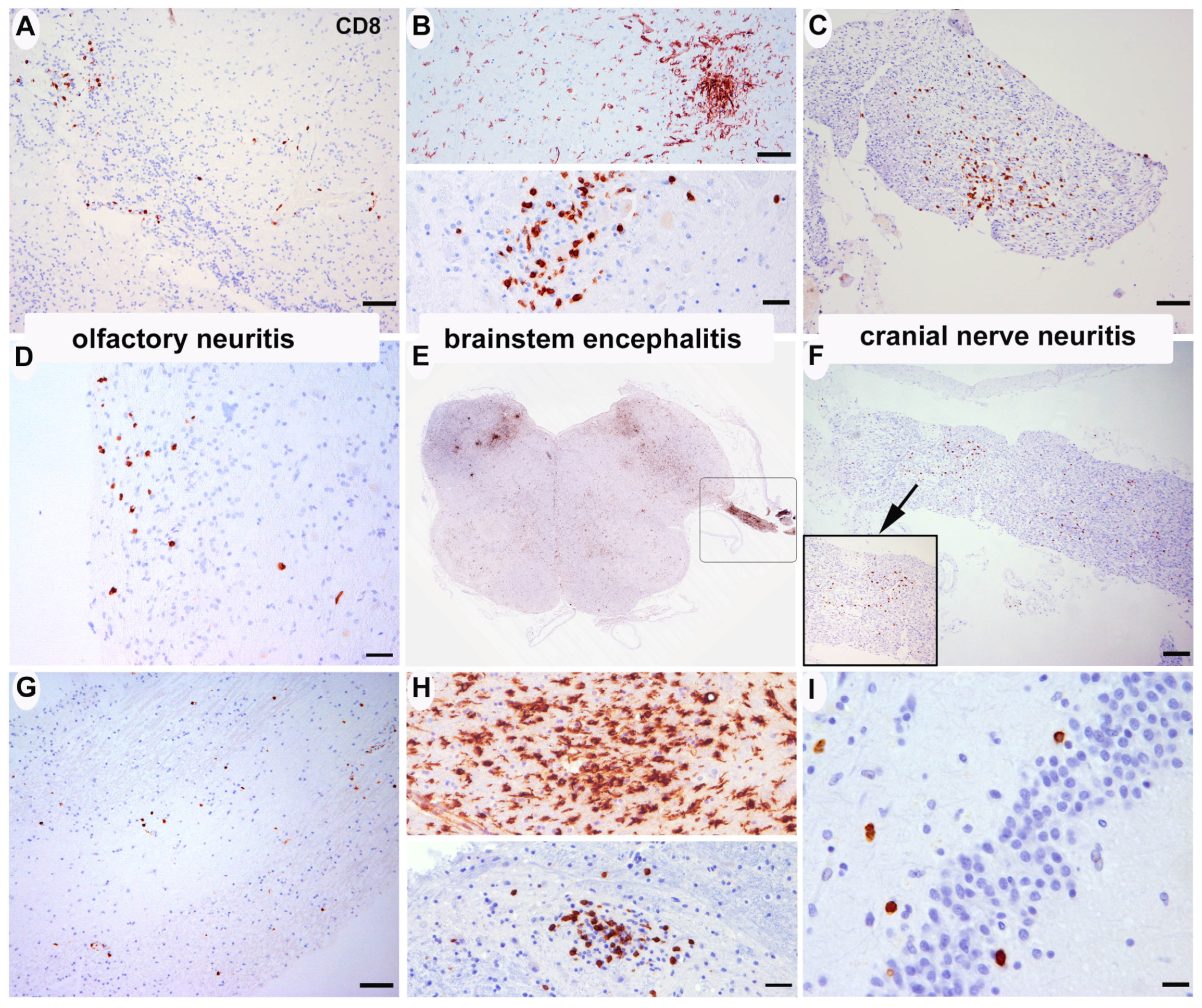
3.2.1. Inflammatory Changes (Figure 1)

A: Olfactory system: eight patients showed mild CD8-positive T lymphocyte-dominated inflammatory infiltrates along the olfactory bulb and tract (cases #2, #3, #5, #6, #7, #12, #21, #22). In one patient, mild infiltrates were also noted within the entorhinal area adjacent to the hippocampus and amygdala (case #1). These infiltrates were accompanied by relatively marked microglial activation in both grey and white matter. All other cases showed diffuse, unspecific microglial activation but no obvious inflammatory nodules (Figure 1A,D,G). In H1N1-infected cases, olfactory bulb was unfortunately not available for analysis, but entorhinal areas also showed relatively prominent microglial activation in 2 of 3 cases (cases #1 and #2), one of them with small microglial nodules (Table 2, case #2).
B: (Micronodular) brain stem encephalitis: seven cases showed mild (cases #2, #6, #12, #16, #19, #20, #21, #32) and three cases moderate (cases #3, #4, #5) inflammatory nodules in the midbrain, pons, and/or medulla oblongata. The motor nucleus of the vagal nerve and its emergence from the brainstem was involved in most cases. The cellular infiltrates were mainly composed by CD8-positive T lymphocytes and microglia/macrophages (Figure 1B–D). No CD20- or CD79A-positive B-cells were identified in any brain region in the parenchyma, only isolated intravascular or isolated perivascular cells were seen in single cases. The distribution of pathology was comparable to that observed in three brains of H1N1-infected patients (Table 2). The spinal cord was analyzed in 10 cases, and no signs of myelitis were identified. No signs of meningeal inflammation were detected in any case.
C: Cranial nerve neuritis: two patients with brainstem inflammation (patient #2 and #5, Table 2) also showed moderate inflammatory infiltrates along the emerging 10th cranial nerve. This infiltrate was also composed by CD8-positive T-cells and macrophages without B-cells (Figure 1C,F).
We additionally analyzed the peripheral nerves (n. medianus) of 10 patients. We found no signs of neuritis, perineuritis, endotheliitis, vasculitis, or of demyelination suggestive of GBS/CIDP. Only one elderly patient showed a moderate axonal neuropathy. Spinal ganglia were not available.
Moreover, we investigated the formalin-fixed skeletal muscle of the forearm of 10 cases and found no signs of myositis, endotheliitis, vasculitis, necrotizing myopathy, or metabolic damage. Only two patients showed a tendency to type 2 fiber atrophy by immunohistochemistry (cases #17, #29), which could be attributed to the prolonged immobilization at the intensive care unit.
D: Co-occurring infections: one patient (case #32) with prior immunosuppression due to treated breast cancer presented an acute herpes simplex encephalitis (HSV1) in the context of severe respiratory distress due to bilateral SARS-CoV-2 pneumonia (Figure 3C1–C4).
Although there were no major differences in respect to inflammatory changes between patients who died of SARS-CoV2-related pneumonia and those with COVID-19 who died of other conditions, patients with longer intensive care and septic state had, in general, more perivascular CD8-positive cells and diffuse microglial activation than patients with shorter disease duration and acute death, caused by myocardial complications or the pulmonary embolism. We could not identify a specific immunoreactivity for SARS-CoV-2 antigens.
Figure 1. Inflammatory changes. (A,D,G) Focal cellular infiltrates of CD8+ T-lymphocytes in the olfactory bulb/tract (A), case 2; (D), case 5; (G), case 22) (anti-CD8 immunohistochemistry). (B,E,H) Focal nodular accumulation of CD8+ T-lymphocytes and HLA-DR+ microglia in the medulla oblongata (B), upper panel anti-HLA-DR, lower panel anti-CD8, case 3; (E), anti-HLA-DR, case 5; (H), upper panel anti-HLA-DR, lower panel anti-CD8, case 4). (C,F) Focal cellular infiltrates of CD8+ T-lymphocytes along emerging cranial nerves (vagus) (C), case 2; (F), case 5). (I) Isolated CD8+ T-cells adjacent to the dentate gyrus of the hippocampus (case 4). Scale bars: (A,B) (upper panel), (C,G) 50 μm; (B) lower panel, (D,H) 20 μm; (E) ×0.7; (F) 100 μm; (I) 10 μm.
Viruses 15 00908 g001
Table 2. Distribution of inflammatory infiltrates in selected brain areas in SARS-CoV-2 and H1N1 infection (0 = absent, i = isolated, + = mild, ++ = moderate, +++ abundant; for scoring details please see Section 2).
Olfactory BulbHippocampusCortical Areas GlobalBasal Ganglia GlobalMedulla OblongataCerebellum Global
CD8HLA-DRCD20CD8HLA-DRCD20CD8HLA-DRCD20CD8HLA-DRCD20CD8HLA-DRCD20CD8HLA-DRCD20
SARS-CoV-2Perivascular/ParenchymaPerivascular/ParenchymaPerivascular/ParenchymaPerivascular/ParenchymaPerivascular/ParenchymaPerivascular/ParenchymaPerivascular/ParenchymaPerivascular/ParenchymaPerivascular/ParenchymaPerivascular/ParenchymaPerivascular/ParenchymaPerivascular/ParenchymaPerivascular/ParenchymaPerivascular/ParenchymaPerivascular/ParenchymaPerivascular/ParenchymaPerivascular/ParenchymaPerivascular/Parenchyma
Case 1nanana+/0 +++/+++ 0/0 +/0 ++/++ 0/0 +/0 ++/++ 0/0 nananananana
Case 2+/+++/++ 0/0 ++/i +++/++ 0/0 ++/0+++/+++ i/0+/0 ++/++ 0/0 ++/+ +++/+++ i/0 ++/i +++/++ 0/0
Case 3+/++ ++/++ 0/0 +/0 ++/++ 0/0 +/0 ++/++ 0/0 +/0 ++/+ 0/0 ++/++ ++/++ 0/0 ++/+ ++/++ 0/0
Case 4i/0+++/+++ i/0+/++++/++ i/0+/0 +++/+++ i/0+/0 +++/+++ i/0+/++ +++/+++ 0/0 +/0 +++/+++ 0/0
Case 5+/+++++/+++ 0/0 +/0 +++/++ 0/0 +/0 +++/++ 0/0 +/0 +++/++ 0/0 +/++ +++/+++ +/0 +/0 +++/++ 0/0
Case 6+/+++/++ 0/0 +/0 +++/++ 0/0 +/0 +++/++ 0/0 +/0 ++/++ 0/0 ++/++ +++/++ 0/0 +/0 +++/++ 0/0
Case 7+/+++/++ 0/0 i/0 ++/++ 0/0 +/0 ++/++ 0/0 +/0 +/+ 0/0 ++/+++++/++ i/0 +/0 ++/++0/0
Case 8i/i+/+0/0 +/0 +/+0/0 +/0 +/+0/0 +/0 +/+0/0 +/0 +/+0/0 +/0 +/+0/0
Case 9i/0++/++ 0/0 i/0+++/++ 0/0 i/0++/++ 0/0 i/0++/++ 0/0 i/0+++/+++ 0/0 i/0++/++ 0/0
Case 10nana0/0 +/0 +/+0/0 +/0 +/+0/0 +/0 +/+0/0 +/0 +/+0/0 +/0 +/+0/0
Case 11+/0 ++/+0/0 +/0 +/+0/0 +/0 ++/+i/0+/0 +/+0/0+/0 ++/+0/0 +/0 ++/+0/0
Case 12+/+++/++ 0/0 +/0 +++/++ 0/0 +/0 +++/+++i/0+/0 ++/++ 0/0+/+ ++/++0/0 +/0 +++/+++ 0/0
Case 13+/0 +/+0/0 +/0 +/+0/0 +/0 +/+0/0 +/0 +/+0/0 +/0 +/+0/0 +/0 +/+0/0
Case 14i/0++/++ 0/0 +/0 ++/++ *0/0 +/0 ++/++ 0/0 +/0 ++/++ 0/0+/0 +++/+++ 0/0 +/0 ++/++ 0/0
Case 15i/0++/++ 0/0 +/0 ++/++ 0/0 +/0 ++/++ 0/0 +/0 ++/++ 0/0 +/0 ++/++ 0/0 +/0 ++/++ 0/0
Case 16i/0++/++ 0/0 +/0 ++/++ 0/0 +/0 ++/+ 0/0 +/0 ++/++ 0/0 +/0 ++/+++ 0/0 +/0 ++/++ 0/0
Case 17i/0++/++ 0/0 i/0++/++ 0/0 i/0++/++ 0/0 i/0++/++ 0/0 i/0++/++ 0/0 i/0++/++ 0/0
Case 18i/0++/++ 0/0 +/0 +/+0/0 +/0 +/+0/0 +/0 +/+i/0+/0 ++/++ 0/0 +/0 ++/++ 0/0
Case 19+/0 ++/++ i/0+/0 +++/++ i/0+/0 +/+0/0 +/0 ++/++ 0/0 ++/+ +++/+++ 0/0 +/0 +++/++ 0/0
Case 20+/0 +/+ i/0+/0 ++/+ 0/0 +/0 +/+ i/0+/0 +/+ i/0+/0 ++/++ 0/0 +/0++/+ 0/0
Case 21+/+++/++ 0/0 +/0 +/+0/0 +/0 +/+ 0/0 +/0 +/+ 0/0 ++/++++/++ 0/0 +/0 +/+ 0/0
Case 22+/++ ++/++ 0/0 +/0 ++/++/0 +/0 +/+ 0/0 +/0 ++/++ +/+ +/i++/+ i/0 ++/0++/+ i/0
Case 23i/0++/++ 0/0 +/0 ++/++/0 +/0 ++/++ 0/0 +/0 ++/++ 0/0 +/0 ++/++0/0 +/0 ++/++0/0
Case 24i/0++/++ 0/0 +/0 ++/+ 0/0 +/0 ++/++ 0/0 +/0 ++/++ 0/0 +/0 ++/++ 0/0 +/0 ++/++ 0/0
Case 25+/0++/++ 0/0 +/0 +++/++ 0/0 +/0 +++/+++ 0/0 +/0 +++/+++0/0 +/0 +++/+++0/0 +/0 +++/+++0/0
Case 26i/0+/+ 0/0 i/0++/+0/0 i/0+/+ 0/0 +/0 ++/++ 0/0 +/0 ++/++ i/0 +/0 ++/++ 0/0
Case 27i/0++/++ 0/0 i/0++/++ 0/0 i/0+/+ 0/0 i/0++/++ 0/0 +/0 ++/++ 0/0 +/0 ++/++ 0/0
Case 28+/0++/++ 0/0 +/0++/++ 0/0 +/0 ++/+0/0 +/0++/++ 0/0 i/0 ++/++ 0/0 i/0 ++/++ 0/0
Case 29i/0++/++ 0/0 +/0++/+0/0 i/0 ++/++ 0/0 i/0 ++/++ 0/0 i/0 ++/++ 0/0 i/0 ++/++ 0/0
Case 30+/i++/++ 0/0 i/0+++/+++ 0/0 i/0+++/+++ 0/0 i/0 +++/+++0/0 +/++++/+++ 0/0 +/++++/+++ 0/0
Case 31i/0+/++ 0/0 i/0++/+ 0/0 i/0+/+ 0/0 i/0+/+ 0/0 i/0++/++ 0/0 i/0++/+ 0/0
Case 32+/+++/++ 0/0 ++/+++++/+++0/0 ++/+ ++/++ 0/0 ++/+ +++/+++0/0 +/++++/+++ 0/0 +/0 ++/++ 0/0
H1N1
Case 1nanana +/0 ++/+ 0/0 i/0 ++/+ 0/0 +/0 +/+ 0/0 +/0 +/++ 0/0 i/0 +/+ 0/0
Case 2nanana +/0 +/++ 0/0 i/0 +/++ 0/0 +/0 ++/++ 0/0 +/0 ++/++ 0/0 +/0 ++/++ 0/0
Case 3nanana +/0 +/+ 0/0 i/0 +/+ 0/0 +/0 +/+ 0/0 +/i +/++ 0/0 i/0 +/+ 0/0
* around ßA4-amyloid plaques.

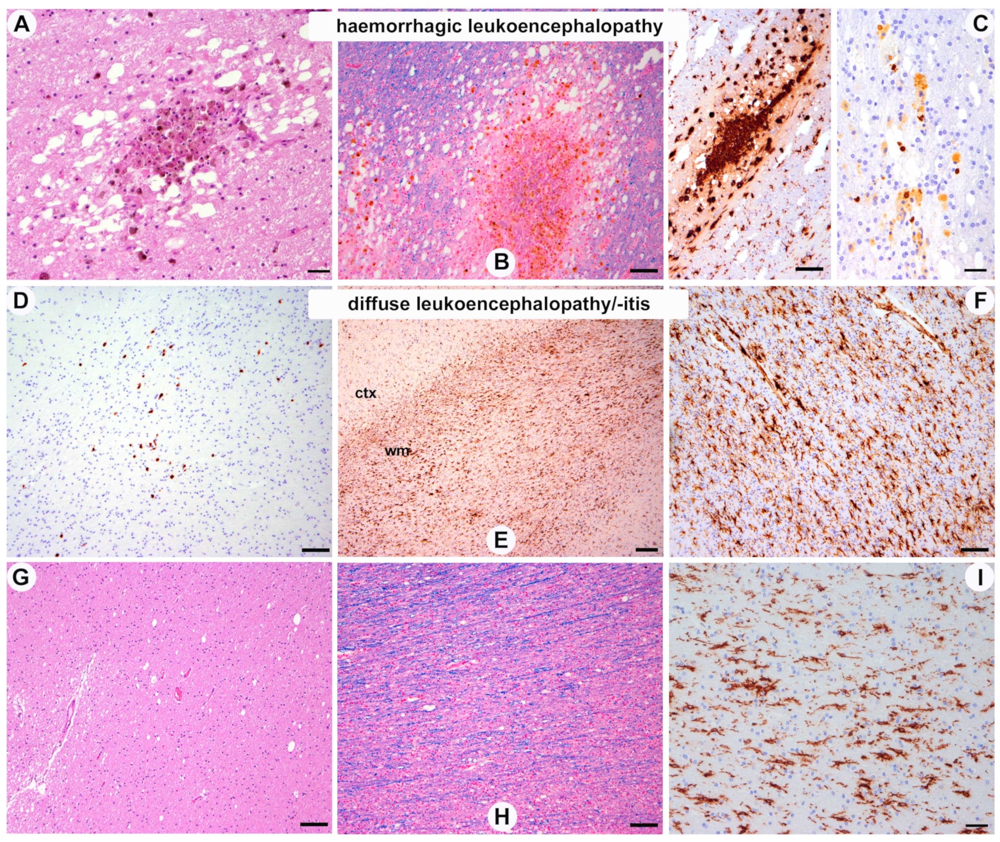
3.2.2. White Matter Pathology (Figure 2)

A: Hemorrhagic leukoencephalopathy: one patient (case #4) showed multiple subacute white matter hemorrhages that were distributed throughout the supratentorial and infratentorial white matter and affected all cerebral lobes, the basal ganglia, brainstem, and cerebellum. The lesions showed a peripheral rim of hemosiderin pigment and central red blood cells in different stages of degradation (Figure 2A–C). These lesions were mostly associated with white matter vessels, but without signs of vasculitis, fibrinoid necrosis of the vascular wall, or thrombosis.
B: Leukoencephalitis: in one patient with olfactory neuritis and brain stem encephalitis, we further identified the mild foci of CD8-positive lymphocytes diffusely distributed within the white matter (Figure 2D–F, case #2 Table 2); however, these were identified without signs of selective demyelination.
C: Diffuse white matter damage: this was a nearly constant feature in all cases and ranged from subtle changes in the LFB stain to the formation of relatively prominent white matter vacuoles (Figure 2D–I), likely representing intramyelinic oedema. These changes were more prominent in posterior, parieto-occipital areas. Moreover, there was a diffuse, white matter predominant microglial activation with clear perivascular accentuation (Figure 2E,F,I, Table 3), which also involved the corpus callosum and the anterior commissure, and was associated with a perivascular accumulation of CD8-positive lymphocytes. No obvious phagocytosis of perivascular myelin such as in acute demyelinating encephalomyelitis (ADEM) was observed. Moreover, we could not identify demyelinating plaques in the white matter or cortical demyelination. No prominent white matter gliosis was detected. Axons were comparatively well preserved.
Figure 2. White matter pathology. (AC) Multiple subacute hemorrhagic lesions throughout the white matter with hemosiderin-laden macrophages, surrounding Waller degeneration with axonal and myelin vacuolation ((B), luxol fast blue), proliferation of macrophages and activated microglia ((C), left panel anti-HLA-DR immunohistochemistry) and isolated CD8+ T-lymphocytes ((C), right panel; case 4). (D) Focal cellular infiltrates of CD8+ T-lymphocytes in the white matter (case 2) and prominent diffuse microglial activation, particularly in the white matter, independently of T-cell infiltrates ((E,F), anti-HLA-DR immunohistochemistry (ctx: cortex; wm: white matter). (GI) mild to moderate diffuse vacuolation of white matter, better identified on luxol-fast-blue-stained sections, without signs of demyelination (H) and diffuse microglial activation, accentuated at the perivascular spaces ((I), anti-HLA-DR immunohistochemistry) ((GI), case 5). Scale bars: (A,C) right panel, (I) 20 μm; (B,C) left panel, (D,FH) 50 μm; (E) 100 μm.
Viruses 15 00908 g002
Table 3. Global distribution of inflammatory cells in supra- and infratentorial white matter (0 = absent, i = isolated, + = mild, ++ = moderate, +++ abundant; for scoring details please see Section 2).
White Matter SupratentorialWhite Matter Cerebellum
CD8HLA-DRCD20CD8HLA-DRCD20
Perivascular/ParenchymaPerivascular/ParenchymaPerivascular/ParenchymaPerivascular/ParenchymaPerivascular/ParenchymaPerivascular/Parenchyma
Case 1+/0++/+++0/0nanana
Case 2+/++++/+++0/0++/i+++/++0/0
Case 3+/0++/+++0/0++/+++/++0/0
Case 4+/0++/+++i/0+/0+++/+++0/0
Case 5+/0+++/++0/0+/0+++/++0/0
Case 6i/0++/+++0/0+/0+++/++0/0
Case 7+/0++/++0/0+/0++/++0/0
Case 8+/0+/+0/0+/0+/+0/0
Case 9i/0++/++0/0i/0++/++0/0
Case 10+/0+/+0/0+/0+/+0/0
Case 11+/0+/+0/0+/0++/+0/0
Case 12+/0++/+++0/0+/0+++/+++0/0
Case 13+/0+/+0/0+/0+/+0/0
Case 14+/0++/++0/0+/0++/++0/0
Case 15+/0++/++0/0+/0++/++0/0
Case 16+/0++/++0/0+/0++/++0/0
Case 17i/0++/++0/0i/0++/++0/0
Case 18+/0++/++0/0+/0++/++0/0
Case 19+/0++/++0/0+/0+++/++0/0
Case 20+/0++/+i/0+/0++/+0/0
Case 21+/0++/++0/0+/0+/+0/0
Case 22+/0++/+0/0++/0++/+i/0
Case 23+/0++/++0/0+/0++/++0/0
Case 24+/0++/++0/0+/0++/++0/0
Case 25+/0+++/++0/0+/0+++/+++0/0
Case 26+/0++/+0/0+/0++/++0/0
Case 27+/0++/+0/0+/0++/++0/0
Case 28+/0++/+0/0i/0++/++0/0
Case 29i/0++/++0/0i/0++/++0/0
Case 30i/0+++/+++0/0+/++++/+++0/0
Case 31i/0+/+0/0i/0++/+0/0
Case 32+/0+++/+++0/0+/0++/++0/0

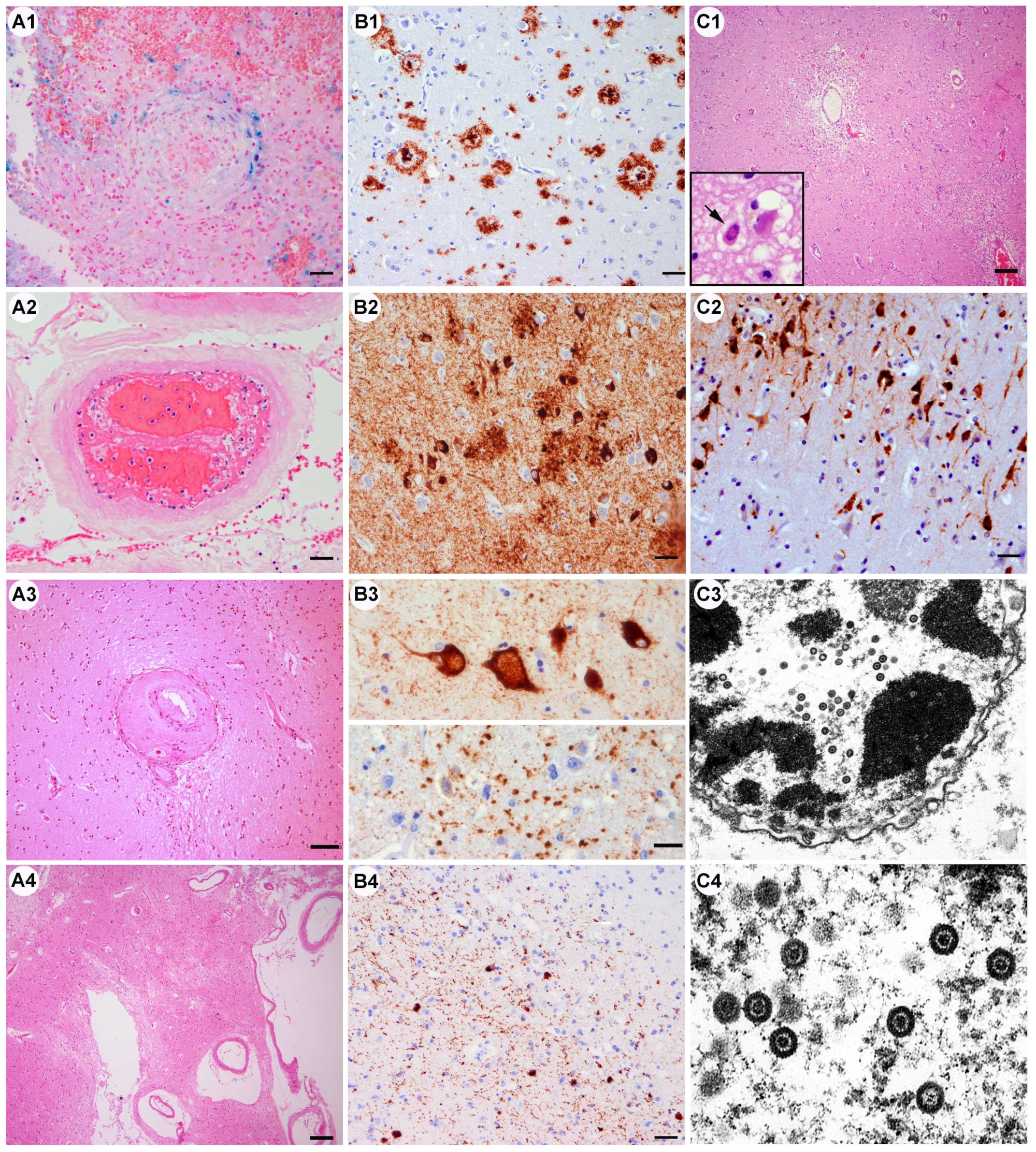
3.2.3. Vascular Pathology (Figure 3A, Table 4 and Table 5)

A: Vascular thrombosis: four cases: venous and arterial thrombosis was identified in a patient who also had a sinus vein thrombosis in the context of a generalized thrombotic event (case #9). One patient showed small arterial thrombosis in a meningeal vessel (case #19, Figure 3A2) and another a fibrin clot in the dural sinus (case #25). In case #26, we observed hemorrhagic lesions around a centrally located fresh venous thrombus in the frontal cortex. In the temporo-basal cortex, in an area of an apparently old traumatic injury, we found an occluded vessel with bleeding and hemosiderin deposits (Figure 3A1). The thrombotic events were associated with acute ischemic or hemorrhagic lesions.
We frequently found stagnant vessels with abundant entrapped leukocytes throughout most of the cases of our series, without the direct attachment of CD8-positive T-cells to endothelial cells, thrombosis, the disruption of the endothelial cell layer, and no signs of endotheliitis or vasculitis. This phenomenon is frequently observed in postmortem brains in the context of diffuse stagnation and sepsis, independent of the cause. The adherence of some neutrophils on the vessel wall should therefore not be considered a specific inflammation of the endothelium.
B: Diffuse hypoxic–ischemic damage: this type of damage was observed in several patients, either in relation to the venous or arterial thrombosis or in the context of a cardiac dysfunction (Table 5). Moreover, most patients suffered from severe pneumonia, a frequent cause of brain hypoxia. Hippocampus CA4 and CA1 sectors, thalamic neurons, cerebellar Purkinje cells, and dentate nucleus neurons appeared to be most vulnerable. Areas of acute or subacute cortical laminar necrosis were also identified (cases #6, #12, #13, #20, #25).
C: Acute hemorrhages: one patient suffered acute cerebellar bleeding in the context of arterial hypertension and dialysis (patient #23, Table 2).
D: Small vessel disease: most of the patients over 60 had pre-existing vascular risk factors (see Table 1). The brain vessels in the white matter and basal ganglia showed diffuse fibrosis of the wall, perivascular tissue rarefaction, and frequent yellowish perivasal lipopigment. In the basal ganglia cribriform, the change was moderate or prominent in nine cases (30%, cases #1, #7, #8, #10, #11, #14, #15, #28, #29)) (Figure 3A3,A4), while small lacunar and cerebellar infarcts were detected in five (16%, cases #1, #10, #11, #13, #29). Diffuse white matter rarefaction, particularly around fibrotic vessels and around the lateral ventricles, was also observed in these cases.
E: Large territorial infarcts: these were only observed in single patients who had focal neurological symptoms or seizures and were associated with vascular thrombosis (cases #25, #26). Neurologically asymptomatic patients did not show “silent” infarcts.
F: Incidental findings: in one patient, we identified a small vascular malformation—a cavernoma—in the frontal white matter (case #5).

3.2.4. Pre-Existing Neurodegenerative Pathology (Figure 3B, Table 4)

A: Alzheimer’s disease neuropathological change: the combination of tau-positive neurofibrillary pathology and ßA4-amyloid/neuritic plaques +/− amyloid angiopathy was identified in 10 patients (32%) (Figure 3B1,B2), most of them aged over 70 years (mean 79, median 80, range 62–97) (cases #1,#3,#5,#10,#14, #17, #18, #26, #27, #28), but not in younger patients. The alterations consisted of neurofibrillary tangles and neuropil threads in the transentorhinal region and the limbic system (Braak stages I–IV), and they affected in some patients also neocortical areas (stages V and VI). ß-Amyloid pathology was in the form of diffuse and compact/cored amyloid plaques in mild, moderate, or abundant densities (CERAD plaque scores A, B, C, respectively) throughout the cortical areas, the limbic system, basal ganglia, brainstem, and cerebellum (Thal amyloid phases 1–5; severity scores ranging from A1, B1, and C1 to A3, B3, and C3 (Supplementary Table S2). Amyloid angiopathy was prominent in two of four cases. There was no increased inflammatory reaction around amyloid plaques and there were no cases of ß-amyloid-related angiitis (ABRA). Primary age-related tauopathy (PART) without significant ßA4-plaque pathology was identified in five patients below 70 years of age (16%, cases #2, #4, #11, #15, #16).
B: Argyrophilic grain disease: this limbic-predominant four-repeat tau pathology was observed in six patients (aged 64 to 86; cases #6, #8, #18, #25, #26, #28). Interestingly, the highest burden of tau pathology was identified in the youngest patient (Figure 3B3, case #25).
C: Lewy body pathology: alpha-synuclein immunoreactive Lewy bodies and Lewy neurites were identified in three patients in early brainstem or olfactory stages (cases #4, #10, #11), mainly affecting the dorsal motor nucleus of the vagal nerve, the olfactory bulb and tract, and the olfactory area around the amygdala. As these patients had not been clinically diagnosed as having Parkinson’s disease, they most likely represent an “incidental” Lewy body pathology (Figure 3B4).
D: TDP43 proteinopathy: two elderly patients (88 and 86 years, case #1 and #26) showed pTDP-43 protein aggregates in the limbic system representing LATE (limbic age-related TDP43 encephalopathy), with neither hippocampal sclerosis nor signs of motor neuron disease or extensive fronto-temporal involvement.
E: Prion disease: not a single case of (co-)incidental Creutzfeldt–Jakob disease or other type of prion disease was observed.
Figure 3. Other conditions. (A1A4) Alteration of the vessel wall and vascular lumen. (A1) Vascular occlusion surrounded by fresh and old blood remnants (Pearl’s blue) and reactive tissue changes with gliosis, microglia, and macrophages (case 26). (A2) Early and partial fibrin clot in a meningeal vessel with entrapped leukocytes and erythrocytes. (A3) Eccentric thickening and fibrosis of small sized arterioles and perivascular tissue rarefaction (case 25) characteristic of small vessel disease that occurs in patients with chronic vascular risk factors. (A4) Prominent perivascular tissue rarefaction status cribosus of the basal ganglia (case 1) (all HE-stained sections). Scale bars: (A1,A2) 20 μm; (A3) 50 μm; (A4) 100 μm. (B1B4) Pre-existing neurodegenerative pathology. (B1) Alzheimer’s disease neuropathological change. ßA4-amyloid deposits in brain parenchyma in form of compact and diffuse plaques (case 1) and within the wall of leptomeningeal and cortical vessels (inset, amyloid angiopathy; case 3). (B2) Tau-positive neurofibrillary pathology with abundant fine neuropil threads, intraneuronal neurofibrillary tangles, and dystrophic neurites surrounding amyloid plaques (case 1). (B3) Tau positive argyrophilic grain pathology with several enlarged “ballooned” neurons in the amygdala (upper panel) and grain-like structures in the neuropil (lower panel; case 25). (B4) Alpha-synuclein immunoreactive Lewy bodies and Lewy neurites in the olfactory bulb (case 4). Scale bars: (B1,B2,B4) 20 μm; (B3) 10 μm. (C1C4) Concomitant HSV-1 infection. (C1) Diffuse hypoxic cortical damage with tissue pallor, endothelial activation, and diffuse inflammatory infiltrates. Inset: intranuclear neuronal inclusion. (C2) Immunohistochemistry shows abundant HSV1 viral antigens in cortical neurons. (C3,C4) Ultrastructural identification of neuronal intranuclear viral particles with typical morphology of Herpes simplex. (case #32). Scale bars: (C1) 100 μm (C2) 20 μm; (C3) original magnification ×30,000, (C4) ×80,000.
Viruses 15 00908 g003

3.2.5. Other

One patient suffered from a traumatic subdural hemorrhage and severe brain contusion that led to death (case #29); their death was not causally linked to COVID-19.
Table 4. Summary of neuropathological findings.
Pathology (n = 32)n° of Cases (%)
Vascular
Small vessel disease, pre-exisiting11 (34%)
Large territorial infarct7 (22%)
Vascular thrombosis4 (12.5%)
Vasculitis/endotheliitis0
Diffuse hypoxic–ischemic damage13 (40%)
Acute hemorrhage1 (3%)
Inflammation
Olfactory neuritis8 (25%)
Brainstem encephalitis micronodular10 (31%)
Cranial nerve neuritis2 (6%)
Encephalitis0
Myelitis0
Meningitis/ependymitis/plexitis0
Peripheral neuritis0
Myositis0
Vasculitis/endotheliitis0
White matter pathology
Hemorrhagic leukoencephalopathy1 (3%)
Diffuse leukoencephalopathy/microglial activation32 (100%)
ADEM0
Neurodegenerative changes preexisting
ADNC10 (32%)
PART5 (16%)
ARTAG2 (6%)
Lewy bodies3 (9%)
TDP-43 proteinopathy2 (6%)
Argyrophilic grain disease6 (18%)
Other
Fulminant Herpes simplex encephalitis1 (3%)
Acute traumatic brain injury1 (3%)
Table 5. Individual neuropathological diagnoses.
CaseAgeSexInflammationVascular Pathology/OtherNeurodeg Pathology
Case 188F-SVD, lacunar infarctADNC A2B3C2, LATE
Case 279MOlfactory neuritis, micronodular brainstem encephalitis, cranial nerve neuritisPatchy acute hypoxic–ischemic neuronal damagePART II, ARTAG
Case 380FOlfactory neuritis, micronodular brainstem encephalitis-ADNC A3B3C2 + CAA
Case 462Fmicronodular brainstem encephalitisHemorrhagic leukoencephalopathyiLBD 3, PART II
Case 569MOlfactory neuritis, micronodular brainstem encephalitis, cranial nerve neuritis Incidental cavernoma frontalADNC A3B2C2 + CAA
Case 679MOlfactory neuritis, micronodular brainstem encephalitisLaminar occipital cortical necrosis older bilat (Morel)AgD II
Case 758MOlfactory neuritisSVD-
Case 871F-Patchy acute hypoxic–ischemic neuronal damage, SVD AgD I
Case 958M-Severe diffuse posthypoxic–postischemic pan encephalopathy with dural sinus thrombosis and meningeal vessel thrombosis; brainstem hemorrhage (Duret)ADNC A1B0C0
Case 1080M-Old infarct cerebellum, SVDLBD 4, ADNC A2B1C2
Case 1180F-Old lacunar infarct pons, SVD, patchy acute hypoxic–ischemic neuronal damageiLBD 1, PART III
Case 1266MOlfactory neuritis, micronodular brainstem encephalitisAcute cortical laminar necrosis occipital and parietal-
Case 1354M-Subacute infarct cerebellum-
Case 1497F-SVDADNC A3B3C3 + CAA
Case 1571F-SVD, patchy acute hypoxic–ischemic neuronal damagePART II
Case 1657FMicronodular brainstem encephalitisPatchy acute hypoxic–ischemic neuronal damagePART II
Case 1781M-Patchy acute hypoxic–ischemic neuronal damageADNC A3B1C1
Case 1873M--ADNC A1B1C1, AgD I
Case 1964MMicronodular brainstem encephalitis Small thrombus meningeal vessels frontalARTAG
Case 2066MMicronodular brainstem encephalitis Acute partly hemorrhagic cortical infarcts cingulum and parietal ctx-
Case 2138MOlfactory neuritis, micronodular brainstem encephalitis Diffuse acute hypoxic–ischemic neuronal damage, CO intoxication-
Case 2234MOlfactory neuritisPatchy acute hypoxic–ischemic neuronal damage-
Case 2372M-Old SDH and SAB, patchy acute hypoxic–ischemic neuronal damage-
Case 2452F-Perivascular fibrosis (systemic sclerosis known)-
Case 2564MMild hypothalamusMultiple bilateral old cortical laminar necrosis and cerebellar necrosis; sinus vein thrombosisAgD III
Case 2686M-Hemorrhagic infarcts frontal and frontobasal, vascular thrombosisAgD I, LATE, ADNC A2B1C1
Case 2762F-Severe venous congestion and SAB occipitalADNC A2B0C1 + CAA
Case 2882F-SVD, acute traumatic SDH + SAB + acute hypoxic damage + diffuse axonal injuryAgD II, ADNC A3B2C2
Case 2967F-Old thalamic lacunar infarct, SVD, metabolic gliosisADNC A1B0C0
Case 3049M-Multiple subacute infarcts cingulum, amygdala, occipitomental, pons, medulla dorsolateral, cerebellum; Wernicke-like changes (alcohol abuse)-
Case 3121M-Patchy acute hypoxic–ischemic neuronal damage-
Case 3257FHSV-related encephalitisRed neurons in encephalitis regions

3.2.6. H1N1-Infected Cases (Table 2)

The three patients who died of acute respiratory distress syndrome after H1N1 infection had similar neuropathological changes to those described here for SARS-CoV-2 (see also Table 2). Case #1 (f, 57) and Case #2 (m, 57) showed a disseminated microangiopathy with fibrinoid necrosis of several vessels with disseminated microhemorrhages in white matter hemorrhagic leukoencephalopathy, associated with moderate hypoxic–ischemic damage and diffuse edema, the latter also present in case #3. Moreover, disseminated fungal microabscesses (fungal pneumonia and sepsis, case #2) and a fungal septic necrotic focus in the occipital white matter (septic shock, case #1) were identified. Both cases also had brainstem microglial nodules and case #3 diffuse and brainstem predominant microglial activation. Patient #2 also showed a mild metabolic encephalopathy with increased Alzheimer type II astrocytes. No viral antigens could be demonstrated by PCR in any of the three cases in brain tissue.

4. Discussion

We present the neuropathological findings in a series of SARS-CoV-2-infected patients who died during the pandemic in 2020–2021 in Austria; two thirds of these patients had severe COVID-19 and received ICU treatment. We observed that one common neuropathological feature is the development of a diffuse white matter damage with intramyelinic edema and prominent diffuse and perivascular-accentuated microglial activation associated with the mild accumulation of perivascular CD8-positive T-cells and comparatively less axonal damage. The intensity of these alterations varied between cases, but it was globally moderate to severe and also included single cases with prominent vacuolar leukoencephalopathy, partly reminiscent of posterior reversible encephalopathy syndrome (PRES-like) [22], and one case of a micro-hemorrhagic HURST-like leukoencephalopathy [23]. The latter was also observed in one of three patients with H1N1 influenza infection from 2009 to 2010 and could be also related to disseminated intravascular coagulation, among other factors.
These changes seemed to be unrelated to the presence of acute/focal lesions and were accentuated in the parieto-occipital regions. Diffuse white matter damage is a non-specific finding and may be caused by several afflictions. Aside from inherited conditions causing, for example, hereditary leukodystrophies or CADASIL, acquired toxic or metabolic derangements may also alter the myelin sheath. We propose that a (severe) SARS-CoV-2 infection as in most of our cases may induce myelin damage not by the virus itself, but by a combination of an excessive cytokine release by the immune system and a metabolic/hydroelectrolytic derangement caused by the functional damage of vital organs and/or toxic damage by poly-medication (e.g., anticoagulation during extracorporeal membrane oxygenation).
During the acute phase of the disease, around 34% of patients experienced impaired memory, concentration, or attention [24]. Some recent studies have examined the immune cell profile in the CSF of patients with “Neuro-COVID-19” by single-cell sequencing [25,26] and found signs of local immune overactivation with a broad clonal T-cell expansion and reduced interferon response [25]. Another recent study performed a multidimensional characterization of immune mediators in the CSF and plasma of patients belonging to different Neuro-COVID-19 severity classes and identified a distinctive set of CSF and plasma mediators associated with blood–brain barrier (BBB) impairment, elevated microglia activation markers, and a polyclonal B-cell response targeting self-antigens and non-self-antigens underlying post-acute COVID-19 syndrome [27]. Altered BBB biomarkers have been also described in infected patients with neurological complications by other authors [28]. These results from other studies support the existence of immune-mediated mechanisms in severe COVID-19 that may contribute to neurological alterations, and that there might be a compromised antiviral response [25,26,29]. While one group did not observe an expansion of B-cells in CSF, findings that are also in line with our postmortem study where a B-cell component was nearly absent, others did. These differences between previous studies may have resulted from the analysis at different time points of the disease. Yet, another study analyzed several cyto/chemokines in the CSF and/or sera of patients with COVID-19 and neurological symptoms and found predominantly an alteration of the neurovascular unit rather than a prominent COVID-19-related neuroinflammatory profile [30]. Furthermore, a recent study performed in mice and humans observed a white matter-selective microglia reactivity after SARS-CoV-2 infection. The authors also observed a persistently impaired hippocampal neurogenesis, decreased oligodendrocyte counts, and signs of myelin loss in the context of increased CSF cytokines/chemokine levels, including CCL11 [31]. These findings were also detected after H1N1 influenza infection. These previous experimental findings do also support our postmortem findings of diffuse white matter microglial activation and likely intramyelinic edema.
In the post-illness stage, 15–80% of patients complained of cognitive dysfunction as part of a long-term clinical manifestation called post-COVID-19 syndrome [32]. This syndrome encompasses symptoms that persist after about 12 weeks of infection and include fatigue and concentration, attention, and memory deficits, among others. As white matter plays an important role in cognition [33], it might well be that acute and diffuse, probably multifactorial, white matter damage contributes to the development of post-COVID-19 cognitive dysfunction. In addition to the possible consequences of altered cytokine/chemokine levels, acute vascular pathologies, including thrombotic events, hypoxic/ischemic/hemorrhagic lesions in strategic brain areas, and/or the presence of pre-existing small vessel disease with/without leukoencephalopathy in patients with cardiovascular risk factors (altogether roughly observed in about one third of our cohort) may contribute to short-, mid-, or even long-term memory/attention problems in post-COVID-19 syndrome [34,35,36,37]. Additionally, important are underlying “preclinical” neurodegenerative morbidities in elderly people (e.g., Alzheimer type changes, limbic TDP-43 proteinopathy, premotor Lewy body pathology, argyrophilic grain pathology) might act as additional factors. These frequently develop in strategic limbic and brainstem nuclei (in our study, in about two thirds of patients, including patients in their late 50s/early 60s) and may also contribute to cognitive deficits after SARS-CoV-2 infection.
The reversibility of these changes will probably depend on the severity of the myelin damage and axonal integrity as well as on the host-dependent immune system, cognitive reserve, and several yet unknown factors. Of note, cognitive dysfunction has been also repeatedly reported after critical illness [38].
As we learnt from previous pandemics, such as the one caused by H1N1 virus in 1915–1930, long-term neurological consequences might appear even decades after the infection. A good example is linked to postencephalitic parkinsonism, a neurodegenerative tauopathy that developed years after influenza infection and an episode of encephalitis lethargica. In those cases, however, no viral antigens have yet been demonstrated in the brains [39]. It has been therefore postulated that an immunological process would be more likely the trigger of neurodegeneration rather than the virus per se. Therefore, it will be of utmost importance to perform future neuropathological studies in patients who survive COVID-19 and die years later of another condition, to investigate whether there are any sequelae, including unusual neurodegenerative features. It will be also particularly important to perform well-designed and controlled morphometric studies.
We observed a mild inflammatory CD8-positive T-cell infiltration of the bulbus/tractus olfactorius in eight cases. In addition, we identified a mild to moderate micronodular brainstem encephalitis in ten patients. In two of those, a cranial nerve inflammation was also identified. It is a matter of debate whether these changes are directly related to the viral infection and contribute to some of the symptoms, including the prominent brainstem dysfunction and respiratory problems the patients present, or whether this is a consequence of severe, end-stage diseases [40,41,42,43]. As in some other studies [44,45], we could not identify a specific signal for SARS-CoV-2 antigens by conventional immunohistochemistry, but this does not exclude the presence of viral particles. Some authors have detected small viral sequences by RT-PCR and RNAseq in a small fraction of patients [46], but it is still unclear whether they represent true CNS infection. Further studies with other, more sensitive modalities are currently in progress. The micronodular brain stem encephalitis that we observed in about one third of the SARS-CoV-2-infected patients was comparable to that seen in the three H1N1-infected patients of our study, and might therefore not necessarily be a direct consequence of a SARS-CoV-2 infection itself. Microglial activation in the postmortem human brain is a frequent, non-specific finding and can be observed in a vast range of conditions, particularly in the context of critical illness, where an interplay of hypoxic–systemic–metabolic–infectious conditions generally contribute to death [17,47,48]. An alternative cause of these findings could be a reactivation of a latent viral infection of another type. This is frequently observed in autopsy series in the context of sepsis or severe terminal disease [49] and is not unique to SARS-CoV-2 infection. Interestingly, a single patient with chemotherapy-related immunosuppression developed a fulminant necrotizing herpes simplex virus encephalitis in the context of SARS and bilateral pneumonia. Moreover, two patients had a non-CNS reactivation of herpes simplex virus infection, two patients of Epstein–Barr virus, one patient suffered from aspergillosis, and one from mucormycosis. These co-morbid conditions might have contributed to patients’ immune response in the brain and underscores the importance of ruling out potential co-occurring infections. Per contra, patients were critically ill and received several medications, including antiviral, antibiotic, antimycotic, and anti-inflammatory drugs, including corticosteroids, which likely altered the inflammatory milieu in the brain, influencing the postmortem neuropathological findings, which is another note of caution for the interpretation of our results.
The limitations of the study include its retrospective nature, the analysis of postmortem brain tissue of a clinically heterogeneous patients group with different pre-existing conditions and risk factors, different clinical evolutions and treatment strategies, and the lack of patients with “post-COVID-19” syndrome.
In summary, our results support some previous neuropathological findings of an apparently multifactorial brain damage in the context of SARS-CoV-2 infection and are in line with recent experimental data on diffuse white matter damage, microglial activation, and cytokine release. At least, the alpha and/or delta SARS-CoV-2 variant that affected most of the patients of our study seem not to be what is classically considered a “neuro(no)tropic virus” in a strict sense [26,46,50,51]. Such neurotropism combines neuroinvasiveness, neurovirulence, and neuroreplication, the direct infection of neural cells/neurons by the virus and its replication there, causing an acute polioencephalitis-associated variable T-cell-mediated inflammation and neuronal loss, as known for well-established neurotropic viruses, e.g., flaviviruses [52]. In contrast, we believe that the prominent immunological reaction induced by the SARS-CoV-2 virus and subsequent cytokine boost, potential immunological mimicry of SARS-CoV-2 antibodies with neuronal/glial antigens [53], the septic condition, and the dysfunction of vital organs with subsequent hypoxic brain damage, mitochondrial/metabolic/hydroelectrolytic derangements, are all likely contributors to a diffuse myelin damage, as already observed in other viral diseases where a pathogenetic network co-orchestrates virus-specific as well as -unspecific factors, e.g., in HIV infection [54]. Such alterations may decompensate or accelerate pre-existing “silent” brain conditions, in particular, underlying subtle neurodegenerative pathologies, chronic vascular pathologies, and latent viral infections, that might alter synaptic and neurotransmitter functions in the short-, mid-, and long-term.
While those who died are not necessarily representative of the majority of people who survive and develop long COVID-19/post COVID-19 and cognitive sequelae, the observed white matter damage in postmortem studies might be an important contributing factor to the long-term effects of COVID-19 infection.
Whether new virus variants will change or broaden the spectrum of neurological complications remains unclear, but it is likely. More recent COVID-19 variants that are likely to have a lower mortality may perhaps have less neuropathological impact. Further interdisciplinary studies are essential, and neuropathological expertise and brain banks can provide a valuable resource of tissue-based information to further elucidate the neurological, and particularly the cognitive consequences/sequelae of a severe infection, with multiorgan involvement, as exemplified by SARS-CoV-2.

Supplementary Materials

The following supporting information can be downloaded at: https://www.mdpi.com/article/10.3390/v15040908/s1, Table S1: List of primary antibodies; Table S2: Semiquantitative assessment and mapping of inflammatory infiltrates across the different brain regions.

Author Contributions

General statement: Autopsies during COVID-19 pandemics have been possible only in selected academic and non-academic centers, and postmortem neuropathological investigations have been still rarer. These centers have been distributed all over Austria and these investigations have been only possible with the participation of the listed authors, who represent the different involved centers and departments in Austria. Conceptualization: E.G., K.S., C.H. (Christine Haberler), J.H., A.W., S.H., T.R., G.R., N.K., D.R., H.B., S.L., A.-K.P. and R.H.; formal analysis: E.G., K.S., M.B., S.W., S.K., C.R., G.R., P.M., M.A., S.L., A.-K.P. and R.H.; pathological investigation and clinical data: E.G., K.S., M.B., S.W., V.H., H.K., S.K., C.R., E.L.-P., A.Z., M.K., M.L., G.G., M.Z., C.H. (Christine Haberler), J.H., A.W., S.H., T.R., T.V., G.R., V.E., C.H. (Carmen Haider), J.L., A.P., G.W. (Gloria Wilk), S.S., I.E., A.N., L.S., I.S.-K., C.K., M.N., J.K., R.K. (Renate Kain), A.C., R.W., R.K. (Robert Krause), G.W. (Günther Weiss), J.L.-R., T.B., P.M., A.S., M.A., R.S., N.K., D.R., H.B., S.L., A.-K.P. and R.H.; project administration: E.G. and R.H.; resources and funding acquisition: E.G., S.L., A.-K.P. and R.H.; visualization: E.G., K.S. and R.H.; writing—original draft preparation: E.G., H.B., S.L., A.-K.P. and R.H.; writing—review and editing by all authors. All authors have read and agreed to the published version of the manuscript.

Funding

This research was supported by the Austrian Fonds of the Major of Vienna (Bürgermeisterfonds der Stadt Wien) to RH (code COVID038), Austrian Science Fund FWF, SYNABS (code I4685-B) to R.H. and V.E., the German Academic Exchange Service (PROMOS scholarship to M.B.), German Academic Scholarship Foundation (Scholarship to M.B.), the Goldschmidt-Jacobson Foundation (to A.-K.P.), the National Multiple Sclerosis Society (Kathleen C. Moore Fellowship: FG-1708-28871, to A.-K.P.), the Swiss National Science Foundation (PCEPF3_194609 to A.-K.P.), intramural funding from the University of Basel (to A.-K.P.), intramural funding provided by the Medical Faculty Mannheim of Heidelberg University (research grants to S.L. and scholarship to S.W.), research grants from the Hertie Foundation (medMS MyLab, P1180016 to S.L.), the European Research Council (“DecOmPress” ERC StG, to S.L.), and the German Research Foundation (SCHI 1330/2-1/4-1, to S.L.).

Institutional Review Board Statement

Ethical approval for the use of human postmortem brain tissue for research purposes was obtained from the Medical University of Vienna (EK1454/2018). The study was performed in accordance with the World Medical Association Declaration of Helsinki.

Informed Consent Statement

Individual patient consent was waived due to the presentation of anonymized data of the postmortem study in the frame of the routine neuropathological diagnostic setting.

Data Availability Statement

Data are available upon reasonable request to the corresponding authors.

Acknowledgments

We are grateful to all clinical and laboratory personell for their unconditional engagement during the COVID-19 pandemic. We particularly thank Alexander Grasmugg, Tamara Lang and Andreas Jurkowitsch from the Division of Neuropathology and Neurochemistry, Department of Neurology, Medical University of Vienna, for technical assistance and Wolfgang Denk, Forensic Medicine, Irmgard Brandl and Mario Karolyi, from Klinik Favoriten in Vienna (Patholgy and Intensive Care Unit), for their valuable contributions to this study. We thank Daniel Bradley for English editing.

Conflicts of Interest

The authors declare no conflict of interest. The funders had no role in the design of the study; in the collection, analyses, or interpretation of data; in the writing of the manuscript; or in the decision to publish the results.

References

  1. Ritchie, K.; Chan, D.; Watermeyer, T. The cognitive consequences of the COVID-19 epidemic: Collateral damage? Brain Commun. 2020, 2, fcaa069. [Google Scholar] [CrossRef] [PubMed]
  2. Yachou, Y.; El Idrissi, A.; Belapasov, V.; Ait Benali, S. Neuroinvasion, neurotropic, and neuroinflammatory events of SARS-CoV-2: Understanding the neurological manifestations in COVID-19 patients. Neurol. Sci. 2020, 41, 2657–2669. [Google Scholar] [CrossRef]
  3. Travi, G.; Rossotti, R.; Merli, M.; D’Amico, F.; Chiappetta, S.; Giussani, G.; Panariello, A.; Corradin, M.; Vecchi, M.; Raimondi, A.; et al. Neurological manifestations in patients hospitalized with COVID-19: A retrospective analysis from a large cohort in Northern Italy. Eur. J. Neurosci. 2021, 53, 2912–2922. [Google Scholar] [CrossRef]
  4. Al-Sarraj, S.; Troakes, C.; Hanley, B.; Osborn, M.; Richardson, M.P.; Hotopf, M.; Bullmore, E.; Everall, I.P. Invited Review: The spectrum of neuropathology in COVID-19. Neuropathol. Appl. Neurobiol. 2021, 47, 3–16. [Google Scholar] [CrossRef]
  5. Maiese, A.; Manetti, A.C.; Bosetti, C.; Del Duca, F.; La Russa, R.; Frati, P.; Di Paolo, M.; Turillazzi, E.; Fineschi, V. SARS-CoV-2 and the brain: A review of the current knowledge on neuropathology in COVID-19. Brain Pathol. 2021, 31, e13013. [Google Scholar] [CrossRef]
  6. Kantonen, J.; Mahzabin, S.; Mäyränpää, M.I.; Tynninen, O.; Paetau, A.; Andersson, N.; Sajantila, A.; Vapalahti, O.; Carpén, O.; Kekäläinen, E.; et al. Neuropathologic features of four autopsied COVID-19 patients. Brain Pathol. 2020, 30, 1012–1016. [Google Scholar] [CrossRef] [PubMed]
  7. Solomon, I.H.; Normandin, E.; Bhattacharyya, S.; Mukerji, S.S.; Keller, K.; Ali, A.S.; Adams, G.; Hornick, J.L.; Padera, R.F., Jr.; Sabeti, P. Neuropathological Features of COVID-19. N. Engl. J. Med. 2020, 383, 989–992. [Google Scholar] [CrossRef]
  8. Mehta, S.; Bhandari, S.; Mehta, S. Brain autopsies in fatal COVID-19 and postulated pathophysiology: More puzzling than a Rubik’s cube. J. Clin. Pathol. 2021, 74, 612–613. [Google Scholar] [CrossRef]
  9. Matschke, J.; Lütgehetmann, M.; Hagel, C.; Sperhake, J.P.; Schröder, A.S.; Edler, C.; Mushumba, H.; Fitzek, A.; Allweiss, L.; Dandri, M.; et al. Neuropathology of patients with COVID-19 in Germany: A post-mortem case series. Lancet Neurol. 2020, 19, 919–929. [Google Scholar] [CrossRef] [PubMed]
  10. Glatzel, M.; Hagel, C.; Matschke, J.; Sperhake, J.; Deigendesch, N.; Tzankov, A.; Frank, S. Neuropathology associated with SARS-CoV-2 infection. Lancet 2021, 397, 276. [Google Scholar] [CrossRef]
  11. Nasrallah, M.P.; Mourelatos, Z.; Lee, E.B. Neuropathology associated with SARS-CoV-2 infection. Lancet 2021, 397, 277. [Google Scholar] [CrossRef] [PubMed]
  12. Egervari, K.; Thomas, C.; Lobrinus, J.A.; Kuhlmann, T.; Brück, W.; Love, S.; Crary, J.F.; Stadelmann, C.; Paulus, W.; Merkler, D. Neuropathology associated with SARS-CoV-2 infection. Lancet 2021, 397, 276–277. [Google Scholar] [CrossRef] [PubMed]
  13. Reichard, R.R.; Kashani, K.B.; Boire, N.A.; Constantopoulos, E.; Guo, Y.; Lucchinetti, C.F. Neuropathology of COVID-19: A spectrum of vascular and acute disseminated encephalomyelitis (ADEM)-like pathology. Acta Neuropathol. 2020, 140, 1–6. [Google Scholar] [CrossRef]
  14. Kirschenbaum, D.; Imbach, L.L.; Rushing, E.J.; Frauenknecht, K.B.M.; Gascho, D.; Ineichen, B.V.; Keller, E.; Kohler, S.; Lichtblau, M.; Reimann, R.R.; et al. Intracerebral endotheliitis and microbleeds are neuropathological features of COVID-19. Neuropathol. Appl. Neurobiol. 2021, 47, 454–459. [Google Scholar] [CrossRef]
  15. Lou, J.J.; Movassaghi, M.; Gordy, D.; Olson, M.G.; Zhang, T.; Khurana, M.S.; Chen, Z.; Perez-Rosendahl, M.; Thammachantha, S.; Singer, E.J.; et al. Neuropathology of COVID-19 (neuro-COVID): Clinicopathological update. Free Neuropathol. 2021, 2, 2. [Google Scholar] [PubMed]
  16. Thakur, K.T.; Miller, E.H.; Glendinning, M.D.; Al-Dalahmah, O.; Banu, M.A.; Boehme, A.K.; Boubour, A.L.; Bruce, S.S.; Chong, A.M.; Claassen, J.; et al. COVID-19 neuropathology at Columbia University Irving Medical Center/New York Presbyterian Hospital. Brain 2021, 144, awab148. [Google Scholar] [CrossRef]
  17. Deigendesch, N.; Sironi, L.; Kutza, M.; Wischnewski, S.; Fuchs, V.; Hench, J.; Frank, A.; Nienhold, R.; Mertz, K.D.; Cathomas, G.; et al. Correlates of critical illness-related encephalopathy predominate postmortem COVID-19 neuropathology. Acta Neuropathol. 2020, 140, 583–586. [Google Scholar] [CrossRef]
  18. Fisicaro, F.; Di Napoli, M.; Liberto, A.; Fanella, M.; Di Stasio, F.; Pennisi, M.; Bella, R.; Lanza, G.; Mansueto, G. Neurological Sequelae in Patients with COVID-19: A Histopathological Perspective. Int. J. Environ. Res. Public Health 2021, 18, 1415. [Google Scholar] [CrossRef]
  19. Manca, R.; De Marco, M.; Ince, P.G.; Venneri, A. Heterogeneity in Regional Damage Detected by Neuroimaging and Neuropathological Studies in Older Adults With COVID-19: A Cognitive-Neuroscience Systematic Review to Inform the Long-Term Impact of the Virus on Neurocognitive Trajectories. Front. Aging Neurosci. 2021, 13, 646908. [Google Scholar] [CrossRef]
  20. Hosp, J.A.; Dressing, A.; Blazhenets, G.; Bormann, T.; Rau, A.; Schwabenland, M.; Thurow, J.; Wagner, D.; Waller, C.; Niesen, W.D.; et al. Cognitive impairment and altered cerebral glucose metabolism in the subacute stage of COVID-19. Brain 2021, 144, 1263–1276. [Google Scholar] [CrossRef]
  21. Altuna, M.; Sanchez-Saudinos, M.B.; Lleo, A. Cognitive symptoms after COVID-19. Neurol. Perspect. 2021, 1 (Suppl. S1), s16–s24. [Google Scholar] [CrossRef]
  22. Hixon, A.M.; Thaker, A.A.; Pelak, V.S. Persistent visual dysfunction following posterior reversible encephalopathy syndrome due to COVID-19: Case series and literature review. Eur. J. Neurol. 2021, 28, 3289–3302. [Google Scholar] [CrossRef] [PubMed]
  23. Walker, J.M.; Gilbert, A.R.; Bieniek, K.F.; Richardson, T.E. COVID-19 Patients with CNS Complications and Neuropathologic Features of Acute Disseminated Encephalomyelitis and Acute Hemorrhagic Leukoencephalopathy. J. Neuropathol. Exp. Neurol. 2021, 80, 628–631. [Google Scholar] [CrossRef]
  24. Rogers, J.P.; Chesney, E.; Oliver, D.; Pollak, T.A.; McGuire, P.; Fusar-Poli, P.; Zandi, M.S.; Lewis, G.; David, A.S. Psychiatric and neuropsychiatric presentations associated with severe coronavirus infections: A systematic review and meta-analysis with comparison to the COVID-19 pandemic. Lancet Psychiatry 2020, 7, 611–627. [Google Scholar] [CrossRef]
  25. Heming, M.; Li, X.; Räuber, S.; Mausberg, A.K.; Börsch, A.L.; Hartlehnert, M.; Singhal, A.; Lu, I.N.; Fleischer, M.; Szepanowski, F.; et al. Neurological manifestations of COVID-19 feature T cell exhaustion and dedifferentiated monocytes in cerebrospinal fluid. Immunity 2021, 54, 164–175. [Google Scholar] [CrossRef] [PubMed]
  26. Song, E.; Bartley, C.M.; Chow, R.D.; Ngo, T.T.; Jiang, R.; Zamecnik, C.R.; Dandekar, R.; Loudermilk, R.P.; Dai, Y.; Liu, F.; et al. Divergent and self-reactive immune responses in the CNS of COVID-19 patients with neuro-logical symptoms. Cell Rep. Med. 2021, 2, 100288. [Google Scholar] [CrossRef]
  27. Etter, M.M.; Martins, T.A.; Kulsvehagen, L.; Pössnecker, E.; Duchemin, W.; Hogan, S.; Sanabria-Diaz, G.; Müller, J.; Chiappini, A.; Rychen, J.; et al. Severe Neuro-COVID is associated with peripheral immune signatures, autoimmunity and neurodegeneration: A prospective cross-sectional study. Nat. Commun. 2022, 13, 6777. [Google Scholar] [CrossRef]
  28. Bonetto, V.; Pasetto, L.; Lisi, I.; Carbonara, M.; Zangari, R.; Ferrari, E.; Punzi, V.; Luotti, S.; Bottino, N.; Biagianti, B.; et al. Markers of blood-brain barrier disruption increase early and persistently in COVID-19 patients with neurological manifestations. Front. Immunol. 2022, 13, 1070379. [Google Scholar] [CrossRef] [PubMed]
  29. Ferreira-Gomes, M.; Kruglov, A.; Durek, P.; Heinrich, F.; Tizian, C.; Heinz, G.A.; Pascual-Reguant, A.; Du, W.; Mothes, R.; Fan, C.; et al. SARS-CoV-2 in severe COVID-19 induces a TGF-β-dominated chronic immune response that does not target itself. Nat. Commun. 2021, 12, 1961. [Google Scholar] [CrossRef]
  30. Bernard-Valnet, R.; Perriot, S.; Canales, M.; Pizzarotti, B.; Caranzano, L.; Castro-Jiménez, M.; Epiney, J.-B.; Vijiala, S.; Salvioni-Chiabotti, P.; Anichini, A.; et al. Encephalopathies associ-ated with severe COVID-19 present neurovascular unit alterations without evidence for strong neuroinflammation. Neurol. Neuroimmunol. Neuroinflamm. 2021, 8, e1029. [Google Scholar] [CrossRef]
  31. Fernández-Castañeda, A.; Lu, P.; Geraghty, A.C.; Song, E.; Lee, M.H.; Wood, J.; Yalçın, B.; Taylor, K.R.; Dutton, S.; Acosta-Alvarez, L.; et al. Mild respiratory SARS-CoV-2 infection can cause multi-lineage cellular dysregulation and myelin loss in the brain. Cell 2022, 185, 2452–2468. [Google Scholar] [CrossRef] [PubMed]
  32. Daroische, R.; Hemminghyth, M.S.; Eilertsen, T.H.; Breitve, M.H.; Chwiszczuk, L.J. Cognitive Impairment after COVID-19-A Review on Objective Test Data. Front. Neurol. 2021, 12, 699582. [Google Scholar] [CrossRef] [PubMed]
  33. Buyanova, I.S.; Arsalidou, M. Cerebral White Matter Myelination and Relations to Age, Gender, and Cognition: A Selective Review. Front. Hum. Neurosci. 2021, 15, 662031. [Google Scholar] [CrossRef]
  34. MacLean, M.A.; Kamintsky, L.; Leck, E.D.; Friedman, A. The potential role of microvascular pathology in the neurological manifestations of coronavirus infection. Fluids Barriers CNS 2020, 17, 55. [Google Scholar] [CrossRef]
  35. Gutierrez Amezcua, J.M.; Jain, R.; Kleinman, G.; Muh, C.R.; Guzzetta, M.; Folkerth, R.; Snuderl, M.; Placantonakis, D.G.; Galetta, S.L.; Hochman, S.; et al. COVID-19-Induced Neurovascular Injury: A Case Series with Emphasis on Pathophysiological Mechanisms. SN Compr. Clin. Med. 2020, 2, 2109–2125. [Google Scholar] [CrossRef]
  36. Jaunmuktane, Z.; Mahadeva, U.; Green, A.; Sekhawat, V.; Barrett, N.A.; Childs, L.; Shankar-Hari, M.; Thom, M.; Jäger, H.R.; Brandner, S. Microvascular injury and hypoxic damage: Emerging neuropathological signatures in COVID-19. Acta Neuropathol. 2020, 140, 397–400. [Google Scholar] [CrossRef]
  37. Keller, E.; Brandi, G.; Winklhofer, S.; Imbach, L.L.; Kirschenbaum, D.; Frontzek, K.; Steiger, P.; Dietler, S.; Haeberlin, M.; Willms, J.; et al. Large and Small Cerebral Vessel Involvement in Severe COVID-19: Detailed Clinical Workup of a Case Series. Stroke 2020, 51, 3719–3722. [Google Scholar] [CrossRef] [PubMed]
  38. Kaushik, R.; Ferrante, L.E. Long-term recovery after critical illness in older adults. Curr. Opin. Crit. Care 2022, 28, 572–580. [Google Scholar] [CrossRef] [PubMed]
  39. Cadar, D.; Jellinger, K.A.; Riederer, P.; Strobel, S.; Monoranu, C.M.; Tappe, D. No Metagenomic Evidence of Causative Viral Pathogens in Postencephalitic Parkinsonism Following Encephalitis Lethargica. Microorganisms 2021, 9, 1716. [Google Scholar] [CrossRef] [PubMed]
  40. Bulfamante, G.; Bocci, T.; Falleni, M.; Campiglio, L.; Coppola, S.; Tosi, D.; Chiumello, D.; Priori, A. Brainstem neuropathology in two cases of COVID-19: SARS-CoV-2 trafficking between brain and lung. J. Neurol. 2021, 268, 4486–4491. [Google Scholar] [CrossRef] [PubMed]
  41. Bulfamante, G.; Chiumello, D.; Canevini, M.P.; Priori, A.; Mazzanti, M.; Centanni, S.; Felisati, G. First ultrastructural autoptic findings of SARS -CoV-2 in olfactory pathways and brainstem. Minerva Anestesiol. 2020, 86, 678–679. [Google Scholar] [CrossRef] [PubMed]
  42. Montini, F.; Martinelli, V.; Sangalli, F.; Callea, M.; Anzalone, N.; Filippi, M. Chronic lymphocytic inflammation with pontine perivascular enhancement responsive to steroids (CLIPPERS) after SARS-CoV-2 pneumonia. Neurol. Sci. 2021, 42, 4373–4375. [Google Scholar] [CrossRef] [PubMed]
  43. Meinhardt, J.; Radke, J.; Dittmayer, C.; Franz, J.; Thomas, C.; Mothes, R.; Laue, M.; Schneider, J.; Brünink, S.; Greuel, S.; et al. Olfactory transmucosal SARS-CoV-2 invasion as a port of central nervous system entry in individuals with COVID-19. Nat. Neurosci. 2021, 24, 168–175. [Google Scholar] [CrossRef]
  44. Aschman, T.; Mothes, R.; Heppner, F.L.; Radbruch, H. What SARS-CoV-2 does to our brains. Immunity 2022, 55, 1159–1172. [Google Scholar] [CrossRef]
  45. Li, Y.C.; Zhang, Y.; Tan, B.H. What can cerebrospinal fluid testing and brain autopsies tell us about viral neuroinvasion of SARS-CoV-2. J. Med. Virol. 2021, 93, 4247–4257. [Google Scholar] [CrossRef] [PubMed]
  46. Serrano, G.E.; Walker, J.E.; Tremblay, C.; Piras, I.S.; Huentelman, M.J.; Belden, C.M.; Goldfarb, D.; Shprecher, D.; Atri, A.; Adler, C.H.; et al. SARS-CoV-2 Brain Regional Detection, Histopathology, Gene Expression, and Immunomodulatory Changes in Decedents with COVID-19. J. Neuropathol. Exp. Neurol. 2022, 81, 666–695. [Google Scholar] [CrossRef]
  47. Poloni, T.E.; Medici, V.; Moretti, M.; Visonà, S.D.; Cirrincione, A.; Carlos, A.F.; Davin, A.; Gagliardi, S.; Pansarasa, O.; Cereda, C.; et al. COVID-19-related neuropathology and microglial activation in elderly with and without dementia. Brain Pathol. 2021, 31, e12997. [Google Scholar] [CrossRef]
  48. Pröbstel, A.K.; Schirmer, L. SARS-CoV-2-specific neuropathology: Fact or fiction? Trends Neurosci. 2021, 44, 933–935. [Google Scholar] [CrossRef]
  49. Schmidbauer, M.; Budka, H.; Ambros, P. Herpes simplex virus (HSV) DNA in microglial nodular brainstem encephalitis. J. Neuropathol. Exp. Neurol. 1989, 48, 645–652. [Google Scholar] [CrossRef]
  50. Cosentino, G.; Todisco, M.; Hota, N.; Della Porta, G.; Morbini, P.; Tassorelli, C.; Pisani, A. Neuropathological findings from COVID-19 patients with neurological symptoms argue against a direct brain invasion of SARS-CoV-2: A critical systematic review. Eur. J. Neurol. 2021, 28, 3856–3865. [Google Scholar] [CrossRef]
  51. Remmelink, M.; De Mendonça, R.; D’Haene, N.; De Clercq, S.; Verocq, C.; Lebrun, L.; Lavis, P.; Racu, M.L.; Trépant, A.L.; Maris, C.; et al. Unspecific post-mortem findings despite multiorgan viral spread in COVID-19 patients. Crit. Care 2020, 24, 495. [Google Scholar] [CrossRef] [PubMed]
  52. Budka, H. Flaviviruses 1. General Introduction and Tick Borne Encephalitis. In Infections of the Central Nervous System: Neuropathology and Genetics; Chrétien, F., Wong, K.T., Sharer, L.R., Keohane, C., Gray, F., Eds.; International Society of Neuropathology (ISN) Book Series Pathology and Genetics; Wiley: Hoboken, NJ, USA, 2020; Chapter 14; pp. 131–146. [Google Scholar]
  53. Kreye, J.; Reincke, S.M.; Prüss, H. Do cross-reactive antibodies cause neuropathology in COVID-19? Nat. Rev. Immunol. 2020, 20, 645–646. [Google Scholar] [CrossRef] [PubMed]
  54. Budka, H. HIV-Related Dementia: Pathology and Possible Pathogenesis. In The Neuropathology of HIV Infection; Scaravilli, F., Ed.; Springer: London, UK, 1993; pp. 171–185. [Google Scholar]
Table 1. Basic clinical data.
AgeSexClinical DiagnosisCause of DeathDisease DurationDays in ICUNeurological SymptomsVascular Risk FactorsAdditional co-MorbiditiesTreatments SARS-CoV-2 PCR Positivity
Case 188Femalenanananananananana *
Case 278MaleCOVID-19 pneumoniaCardiovascular failure5648Did not regain consciousness after stop of narcoticsDiabetes, hypertension, obesity, atrial fibrillationDilatative cardiomyopathy, arteriosclerosisRemdesivirblood, perianal, trachea, larynx
Case 379FemaleCOVID-19 pneumoniaPulmonary thrombosis, pulmonary bleeding, Sepsis 1811NoneHypertension, obesity-Kaletra, cortisone, 3 doses reconvalescent plasma, remdesivirLarynx
Case 462FemaleCOVID-19 pneumoniaRespiratory failurena63Critical illness polyneuropathy/myopathy, reduced vigilance, EEG diffuse slowingAtrial fibrillation, arterial hypertensionSuspicion of latent tuberculosisCortisone; positive EBV PCR- Ganciclovirna *
Case 568MaleCOVID-19 pneumoniaMulti-organ failure3127Agitation, confusion, aggression, psychosisDiabetes, hypertension, obesityPulmonary emphysema, steatosis hepatis, arteriosclerosisKaletra, remdesivir, cortisone, Roactemra, 3 doses reconvalescent plasmalarynx, trachea
Case 678MaleCOVID-19 pneumoniaMulti-organ failure2825Did not regain consciousness after stop of narcotics, anisocoria, meningismus, tetraparesisArterial hypertension, atrial fibrillationPulmonary emphysema, arteriosclerosis, chronic kidney injury, prostate hyperplasia, hyperuricemiaReconvalescent plasma, Roactemra, dialysistrachea, larynx
Case 757MaleCOVID-19 pneumoniaRespiratory failure50NoneArterial hypertension, obesitySteatosis hepatisNonenaso-pharynx
Case 871FemaleCOVID-19 pneumoniaMulti-organ failure secondary to respiratory failure due to pneumonia104naArteriosclerosisMyotonic dystrophy type 2, steatosis hepatis, asthma Cortisone, remdesivirnaso-pharynx
Case 958MaleCOVID-19 pneumoniaSinus thrombosis267Seizures, comaChronic kidney failure, arteriosclerosisPulmonary emphysemaNonetrachea, naso-pharynx
Case 1080MaleCOVID-19 pneumoniaRespiratory failure70Anxiety disorder, somnolence due to psychopharmaceuticalsArterial hypertension, arteriosclerosis, acute on chronic kidney diseaseHumerus fracture left armCeftriaxone, hydroxychloroquine, zincna *
Case 1180FemaleCOVID-19 pneumoniaCardiorespiratory failure33None Cardiac insufficiency (NYHA IV), diabetes, arterial hypertension, arteriosclerosis-None due to rapid cardiac deteriorationna *
Case 1266MaleCOVID-19 pneumoniaMulti-organ failure, hypoxic encephalopathy4235Impaired consciousness of unknown origin, potentially due to hypoxic encephalopathyArterial hypertension, hypercholesterinemiaStatus post transient ischemic attack 2014Cortisone, remdesivir; EBV, HSV and Aspergillus infection na *
Case 1354MaleCOVID-19 pneumonia, mucormycosis with pulmonary infarctionRespiratory failure2418NoneObesity, arterial hypertension, hypertrophic left ventricleMyelodysplastic syndrome, secondary acute myeloid leukemia with chemotherapy and complete hematologic remission, bone marrow hypoplasia, signs of dysplastic hematopoiesis in the peripheral bloodTocilizumabna *
Case 1496FemaleRib fractures, hemothorax, and COVID-19 pneumoniaRespiratory failurenananaArterial hypertension, hypertensive cardiomyopathy, pulmonary hypertension-Nonenaso-pharynx
Case 1571MaleCOVID-19 pneumoniaMulti-organ failure secondary to respiratory failure due to pneumonia with interstitial fibrosis 2415Ageusia, agitationArterial hypertensionArteriosclerosis, steatosis hepatis, cholecystolithiasis Cortisone, remdesivirtrachea, naso-pharynx, bronchial
Case 1656FemaleCOVID-19 pneumoniaHemorrhagic shock after ECMO implantation1512NoneDiabetes, obesityStatus post thyroid carcinomaCortisone, remdesivirnaso-pharynx, trachea
Case 1781MaleUnclear, postoperative multi-organ failureMulti-organ failurena1NoneIntermittent atrial fibrillationIPMN of the pancreasNonena *
Case 1873MaleCOVID-19 pneumoniaCardiogenic shock after ECMO implantation1611NoneArterial hypertension, hypercholesterinemia-Cortisone, remdesivirnaso-pharynx, trachea
Case 1963FemaleCOVID-19 pneumoniaRespiratory failure10NoneArterial hypertension, obesity, arteriosclerosisChronic venous insufficiency, steatosis hepatisNonenaso-pharynx
Case 2065MaleCOVID-19 pneumoniaRespiratory failure3626Hemiparesis right side (onset 1 November 2021)Obesity, sleep apnea syndrome, arterial hypertension, diabetesSteatosis hepatisCortisonenaso-pharynx
Case 2138MaleNot applicable (death at home)Carbon monoxide poisoningna0 naNone-Nonenaso-pharynx post mortem
Case 2233MaleCardiac arrestCardiovascular failure70NoneNone-Nonenaso-pharynx
Case 2371MaleCOVID-19 pneumoniaMulti-organ failure7157None/prolonged weaningDiabetes, arterial hypertension, PAOD, hyperlipidemiaStatus post TIACortisonenaso-pharynx
Case 2452FemaleCOVID-19 pneumoniaCardiogenic shock due to myocarditis, death during new cannulation of ECMO125NoneObesitySclerodermaCortisonenaso-pharynx
Case 2564MaleSepsisMulti-organ failurena34Coma, therapy-resistant status epilepticusArteriosclerosis, arterial hypertension, obesity, chronic kidney disease, hypercholesterinemia, nicotine abuseAlcohol abuse, steatosis hepatis, chronic pancreatitis, pulmonary edema and congestion, hypothyroidism, hyperuricemia, pericarditis Nonenaso-pharynx
Case 2686MaleCardiac arrestDiffuse myocardial ischemiana0DeliriumArteriosclerosis, arterial hypertensionProstate hyperplasia, status post meningioma resection left frontal lobe 9 days before deathCortisonenaso-pharynx
Case 2762FemaleCOVID-19 pneumoniaRespiratory failure144naNone-Cortisonenaso-pharynx
Case 2882FemaleSubdural hematomaSubdural hematoma80Coma, anisocoriaAtrial fibrillationAnxiety disorderNonenaso-pharynx
Case 2967FemaleNot applicable (death at home)Cardiovascular failurena0 naArterial hypertension, atrial fibrillationChronic kidney failureNonenaso-pharynx post mortem
Case 3049MaleProgressive strokeCardiovascular failure6112Stroke with visual disturbance, gait disturbance, dysphagia, dysarthria, aphasia, agitation, reduced vigilanceDiabetes, arterial hypertensionChronic kidney disease, schizoaffective disorderCortisonenaso-pharynx
Case 3121MaleMyocardial infarctionAcute myocardial infarctionna0Epileptic seizureObesityPurpura Schönlein–HenochNonenaso-pharynx
Case 3257FemaleCOVID-19 pneumoniaCardiorespiratory failurenananaArteriosclerosisInvasive mammary carcinomanana *
na *: all patients had PCR confirmed SARS-CoV-2 infection; in cases with na * exact sampling site for PCR was not available.
Disclaimer/Publisher’s Note: The statements, opinions and data contained in all publications are solely those of the individual author(s) and contributor(s) and not of MDPI and/or the editor(s). MDPI and/or the editor(s) disclaim responsibility for any injury to people or property resulting from any ideas, methods, instructions or products referred to in the content.

