Article

The Synthetic Opioid Fentanyl Increases HIV Replication and Chemokine Co-Receptor Expression in Lymphocyte Cell Lines

by
Janani Madhuravasal Krishnan
1,
Ling Kong
1,
Rebekah Karns
2,
Mario Medvedovic
3,
Kenneth E. Sherman
1,4 and
Jason T. Blackard
1,4,*
1
Division of Digestive Diseases, Department of Internal Medicine, University of Cincinnati College of Medicine, Cincinnati, OH 45267, USA
2
Digestive Health Center, Cincinnati Children’s Hospital, Cincinnati, OH 45229, USA
3
Department of Environmental & Public Health Sciences, University of Cincinnati College of Medicine, Cincinnati, OH 45267, USA
4
Center for Addiction Research, University of Cincinnati College of Medicine, Cincinnati, OH 45267, USA
*
Author to whom correspondence should be addressed.
Viruses 2023, 15(4), 1027; https://doi.org/10.3390/v15041027
Submission received: 28 March 2023 / Revised: 11 April 2023 / Accepted: 15 April 2023 / Published: 21 April 2023
(This article belongs to the Special Issue HIV and Drugs of Abuse 2.0)

Abstract

:
Background: In the United States, the illicit use of synthetic opioids such as fentanyl has led to a serious public health crisis. Synthetic opioids are known to enhance viral replication and to suppress immunologic responses, but their effects on HIV pathogenesis remain unclear. Thus, we examined the impact of fentanyl on HIV-susceptible and HIV-infected cell types. Methods: TZM-bl and HIV-infected lymphocyte cells were incubated with fentanyl at varying concentrations. Expression levels of the CXCR4 and CCR5 chemokine receptors and HIV p24 antigen were quantified with ELISA. HIV proviral DNA was quantified using SYBR RT-PCR. Cell viability was detected with the MTT assay. RNAseq was performed to characterize cellular gene regulation in the presence of fentanyl. Results: Fentanyl enhanced expression of both chemokine receptor levels in a dose-dependent manner in HIV-susceptible and infected cell lines. Similarly, fentanyl induced viral expression in HIV-exposed TZM-bl cells and in HIV-infected lymphocyte cell lines. Multiple genes associated with apoptosis, antiviral/interferon response, chemokine signaling, and NFκB signaling were differentially regulated. Conclusions: Synthetic opioid fentanyl impacts HIV replication and chemokine co-receptor expression. Increased virus levels suggest that opioid use may increase the likelihood of transmission and accelerate disease progression.

1. Introduction

The number of opioid overdose deaths in the United States has increased dramatically in recent years [1,2,3]. Fentanyl or fentanyl analogs were involved in ~90% of unintended overdoses [3,4,5,6]. Opioids are endogenous, exogenous, or synthetic substances that work by activating multiple opioid receptors, and these opioid receptors are present on lymphocytes, monocytes, macrophages, and neutrophils, as well as other immune cells [7,8,9]. As opioids alter both innate and adaptive immune functions, it is critical to understand how opioid use affects distinct immune cell types [10,11,12]. Fentanyl is ~50 times more potent than heroin and ~100 times more potent than morphine [13]. Compared with heroin, fentanyl has a rapid onset but shorter duration of action, which increases the frequency with which people inject drugs and increases drug sharing. Injection drug use increases the risk of transmission of blood-borne viruses such as HIV, hepatitis C virus, and hepatitis B virus [13,14,15,16].
HIV enters target cells by forming a complex consisting of the viral envelope glycoprotein (gp120), CD4 receptor, and members of the chemokine co-receptor family (e.g., CCR5 and CXCR4). Multicellular immune responses (e.g., natural killer cells and T regulatory cells) are regulated by CCR5 [17]. Multiple pathological conditions are associated with CXCR4, including immune system disorders, viral infections, and cancer [18]. Amphetamines, cocaine, marijuana, and opiates are all cofactors that increase the risk of HIV infection and disease progression [19,20]. For instance, morphine enhances HIV replication in vitro [21,22,23,24,25] by upregulation of the chemokine co-receptor CCR5 [26]. Morphine downregulates the level of beta chemokines, regulates the expression of pro-inflammatory markers such as TNF-α and IL-6 to block CD8-mediated anti-HIV activity, inhibits the expression of anti-HIV miRNA, and activates the HIV long terminal repeat (LTR) [27,28,29,30]. Non-opioid drugs of abuse also impact HIV replication and disease progression. For example, cocaine promotes HIV infection and replication by suppressing protective chemokines and/or upregulating HIV co-receptors [31]. Methamphetamine also enhances HIV replication by suppression of immune cells in vivo [32].
Although fentanyl, fentanyl analogs, and fentanyl metabolites are commonly detected in persons experiencing overdoses, their effects on viral replication are poorly characterized. The proliferation of viruses can be enhanced by several drugs of abuse that suppress immunological responses; the effects of fentanyl on replication of HIV and cellular gene expression have not been investigated thoroughly. Thus, we evaluated the effects of fentanyl on HIV-susceptible/infected cell types and chemokine receptor expression. As CD4+ T cells represent a major reservoir of HIV replication, we also assessed whether fentanyl affected HIV replication in CD4+ T cells.

2. Methods

2.1. Cell Lines and Reagents

TZM-bl is an indicator cell line that expresses high levels of CD4, CCR5, and CXCR4 and is susceptible to infection with a variety of HIV isolates. TZM-bl cells incorporate two HIV Tat-regulated reporter genes—firefly luciferase and b-galactosidase—that facilitate quantification of infectious HIV [33,34]. J-Lat GFP is a Jurkat-derived T lymphocyte cell line with a single HIV retroviral vector insertion site that enables LTR-driven green fluorescent protein (GFP) expression [35,36]. H9 (cat.no. ARP-400) is derived from the human T cell line HUT 78 and is infected HIV-1 IIIB. ACH-2 is a T lymphocyte cell line containing integrated LAV provirus [37,38]. All cell lines were provided by the NIH AIDS Reagent Program. TZM-bl cells were cultured in Dulbecco’s modified Eagle medium (DMEM) with 10% fetal bovine serum (FBS), 100 U/mL penicillin, 100 μg/mL streptomycin, and 1% 200 mM L-glutamine at 37 °C and 5% CO2. T cell lines were maintained in RPMI 1640 medium (Life Technologies, Grand Island, NY, USA) supplemented with 10% heat-inactivated FBS, 100 U/mL penicillin, and 100 μg/mL streptomycin (Life Technologies) at 37 °C and 5% CO2.
As a certified reference material from Cerilliant (Round Rock, TX, USA), fentanyl can be used for the testing, calibration, and quantification in analytical and research applications [39]. The Institutional Review Board at the University of Cincinnati approved the use of human blood as part of protocol 0584_2019 (approved 29 May 2019).

2.2. Quantification of Mu Opioid Receptor

Mu opioid receptor (MOR) expression was quantified in SH-5YSY, TZM-bl, J-Lat GFP, ACH-2, and H9 cells. Cell lysates were prepared from ~1 × 10⁵ cells, and 100 uL of the lysate was processed for human opioid receptor mu 1 (OPRM1) ELISA (My BioSource; San Diego, CA, USA).

2.3. Propagation of HIV

Infectious HIVYK-JRCSF and HIVNL4-3 were prepared by transfection of 1 × 10⁶ 293T cells (ATCC #CRL-3216) per well with 2 μg of the full-length HIVYK-JRCSF and HIVNL4-3 plasmids, obtained from the NIH AIDS Reagent Program, with the FuGene6 transfection reagent (Roche; Basel, Switzerland). Transfected cells were incubated at 37 °C for 48 h. The supernatant was harvested and passed through a 0.20 μm filter to remove cellular debris and then precipitated in polyethylene glycol at 4 °C. Precipitated virus was centrifuged at 3000 g for 20 min, and the virus precipitate was resuspended in phosphate-buffered saline (PBS) and preserved at −80 °C. The virus was titered using TZM-bl cells and β -galactosidase staining. HIV p24 protein in cell culture supernatants was quantified as outlined below.

2.4. HIV Infection, Drug Exposure, and p24 Protein Quantification

TZM-bl cells were seeded at ~2 × 10⁵ cells per well. After 24 h, cells were treated with HIVYK-JRCSF and HIVNL4-3 at TCID50 of 0.5 for 1 h. Prior to the exposure to fentanyl, the cells were rinsed with PBS three times to remove unbound virus and -replaced with fresh media. Working concentrations of fentanyl were prepared by dilution of the concentration stock with dH2O to obtain 1 ng/mL, 100 ng/mL, and 10 ug/mL concentrations, and added to the respective wells. After incubation with the drug for 24 h, the expression of HIV p24 protein levels was quantified in cell culture supernatants using the HIV p24 ELISA Kit (Abcam; Cambridge, MA) with a lower limit of sensitivity of 1.1 pg/mL.

2.5. Quantification of Integrated HIV DNA

TZM-bl cells were seeded at 1 × 10⁶ cells per well. After 24 h, the cells were treated with HIVNL4-3 at TCID50 of 0.5 for 1 h, rinsed with PBS three times to remove unbound virus, and - replaced with fresh media. Fentanyl at varying concentrations was added to the respective wells and incubated. After incubation with the drug for 24 h, cellular DNA was extracted from cells using the Qiagen mini kit per the manufacturer’s instructions. DNA was similarly extracted from ACH-2 cells that contain a single copy of HIV-1 proviral DNA (LAV strain) [37,38] and used as a positive control to quantify virus levels. The number of HIV-1 proviral copies was quantified with real-time PCR amplification using Brilliant III ultra-fast SYBR green QPCR master mix (Agilent), as described elsewhere [40]. Real-time PCR was performed using SYBR green 2× Master Mix with 200 nM of each oligonucleotide primer targeting the HIV-1 pol gene and DNA extracted from cells treated and untreated with HIVNL4-3 +/− fentanyl at concentrations of 1 ng/mL, 100 ng/mL, and 10 ug/mL. To quantify HIV-1 provirus, a standard curve was defined, using serial dilutions of ACH-2-derived DNA ranging from 1 to 106 copies per cell. All standard dilutions, controls, and samples were run in duplicate, and the average value ct was utilized to quantify HIV-1 DNA copies.

2.6. Cell Viability

TZM-bl, J-Lat GFP, ACH-2, and H9 cells were seeded at a concentration of 5 × 10⁴ cells per well in 100 μL of DMEM medium + 10% FBS and allowed to adhere for 24 h at 37 °C. After 24 h of incubation, fentanyl was added. At 24 h post fentanyl exposure, cell viability was evaluated with a 3-(4,5-dimethylthiazol-2-yl)-2,5-diphenyl tetrazolium bromide (MTT) assay using the MTT Cell Proliferation Assay Kit (Abcam; Cambridge, MA, USA).

2.7. Chemokine Receptor Expression

A quantity of ~2 × 10⁵ TZM-bl, J-Lat GFP, ACH-2, or H9 cells were seeded, and after 24 h fentanyl was added. Post fentanyl exposure, cells were harvested and freeze-thawed three times to prepare cell lysates. CXCR4 or CCR5 receptor protein (pg/mL) was quantified in cell lysates with ELISA (My BioSource; San Diego, CA, USA).

2.8. Primary Cell Isolation and Culture

Peripheral blood samples were collected from healthy adult donors with no self-reported HIV or HCV at the University of Cincinnati Medical Center. Patients provided written informed consent prior to any study procedures. Four mLs of blood was collected in BD Vacutainer Cell Preparation Tubes (catalog #362760). After inverting the tubes 8–10 times, the tubes were centrifuged for 25 min at 1900 RCF. Mononuclear cells were collected in 15 mL centrifuge tubes and rinsed with PBS two times. The final cell pellet was resuspended in 5 mLs of PBS, and the total number of cells in the suspension was determined by trypan blue staining.
Naïve CD4+ T cells were subsequently isolated by negative selection using the naïve CD4+ T cell isolation kit (cat. no. 130-096-533, Miltenyi Biotec GmbH, Bergisch Gladbach, Germany) according to the manufacturer’s instructions. Briefly, the PBMC cell suspension was centrifuged at 800× g for 5 min. The cell pellet was resuspended in 40 μL of MACS running buffer (PBS + 5%FBS) per 1 × 10⁷ cells. An amount of 10 μL of CD4+ T cell biotin-antibody cocktail was added per 1 × 10⁷ cells and incubated at 4 °C for 10 min. An amount of 30 uL of MACS running buffer and 20 uL of anti-biotin microbeads per 1 × 10⁷ cells were added and incubated at 4 °C for 10 min. Cells were resuspended in MACS running buffer and applied to an LS column placed in a magnet. Unlabeled CD+ T cells were collected and centrifuged at 800× g for 5 min. The cells were resuspended with 1 mL of MACS running buffer, and the total cell count was determined by trypan blue staining. Cells were centrifuged again and resuspended in 1 mL RPMI + 10% FBS + 1% antibiotics (Pen/Strep) + 1% glutamine. Purified cells were plated at 0.3 × 10⁶ cells per well in a 96-well plate with 200 ul of RPMI + 10% FBS + 1% antibiotics (Pen/Strep) + 1% glutamine. The cells were stimulated with anti-CD3/CD28 dynabeads (cat. no. 111.31D, Invitrogen, 1 cell per 3 beads) + 100 IU/mL IL−2 at 37 °C in 5% CO2 for 3 days.
After 3 days of incubation, the activated cells were collected and transferred to a 5 mL polystyrene tube and placed in a dynabead magnet to remove beads from cells. The cells were then transferred to a new tube and centrifuged at 800× g for 5 min. The cell pellet was resuspended in 1 mL of RPMI + 10% FBS + 1% antibiotics (Pen/Strep) + 1% glutamine, and the cell count was determined. An amount of ~1 × 10⁵ cells was seeded per well. Cells were infected with HIVNL4-3 at MOI of 1 for 2 h and rinsed with RPMI + 10% FBS + 1% antibiotics (Pen/Strep) + 1% glutamine three times to remove any unbound virus and - replaced with fresh media. Fentanyl at concentration of 10 ug/mL was added to the respective wells and incubated at 37 °C in 5% CO2. After incubation with the drug and virus for 72 h, HIV proviral DNA was quantified in cells with real-time PCR based on SYBR Green I detection, and HIV p24 antigen expression was estimated from the cell culture supernatant.

2.9. Cellular RNA Isolation and Purification

Total RNA from fentanyl-treated (10 μg/mL) and untreated ACH-2 cells was isolated using a commercially available miRNA Isolation Kit (miVana; Applied Biosystems; Carlsbad, CA, USA) according to the manufacturer’s protocol. A NanoDrop 2000 spectrophotometer (Thermo Fisher Scientific; Waltham, MA, USA) was used to determine total RNA concentration and purity. A 2100 Bioanalyzer (Agilent; Santa Clara, CA, USA) and agarose electrophoresis were used to assess RNA integrity.

2.10. miRNA-Seq and Data Analysis

MicroRNA-seq was performed by the Genomics, Epigenomics and Sequencing Core at the University of Cincinnati [41]. A NEBNext small RNA sample library preparation kit (NEB; Ipswich, MA, USA) was used to prepare the library with a modified approach for precise miRNA library size selection; as a result, the kit could process low-quality RNA with limited input for higher library recovery and miRNA read alignment. After 15 cycles of PCR with 100 ng total RNA as input, the libraries with unique indices were then pooled, column-cleaned, and combined with a customized DNA ladder containing 135 and 146 base pairs (bp) purified PCR amplicon. This size range corresponds to a 16–27 nt insert miRNA library that covers all miRNAs. Agarose gel electrophoresis and gel excision were performed. The library pool ranging, from 135 to 146 bp, including the DNA marker, was gel-purified and quantified using the NEBNext Library Quant kit (NEB) in the Quant Studio 5 Real-Time PCR System (Thermo Fisher Scientific; Waltham, MA, USA). The first round of sequencing was performed using an Illumina Nextseq 550 sequencer to generate a few million reads to measure the relative concentration of each library. For the final data analysis, the capacity of each library was adjusted to generate the predicted number of equal reads from each sample. Sequence reads were pre-processed to remove adapters and filter low-quality reads using the ShortRead R package [42]. Reads were aligned to the reference human genome (hg19) using the Bowtie2 aligner [43]. The reads aligning to each known mature miRNA were counted using Bioconductor packages for next-generation sequencing data analysis [44] based on miRNA definitions in the miRBase database [45]. Statistical analysis to detect differentially expressed miRNAs were performed, and p-values were calculated based on the negative binomial model of read counts as implemented in the edgeR [46] R package.

2.11. RNAseq Analysis

The Genomics, Epigenomics and Sequencing Core at the University of Cincinnati performed directed polyA RNA-seq, following established protocols [47,48]. Bioanalyzer was used to test the quality of total RNA (Agilent; Santa Clara, CA, USA). The Poly(A) mRNA Magnetic Isolation Module (NEBNext; Ipswich, MA, USA) was used to isolate polyA RNA for library preparation with 1 µg of high-quality total RNA as input. SMARTer Apollo automated NGS library prep system (Takara Bio USA; Mountain View, CA, USA) was used for enrichment of polyA RNA. For library preparation, the New England BioLabs NEBNext Ultra II Directional RNA Library Prep kit (PCR cycle number 8) was used. The individual indexed libraries were proportionally pooled and sequenced on the NextSeq 550 sequencer (Illumina; San Diego, CA) with the setting of single read 1 × 85 bp after QC and quantification via real-time qPCR (NEBNext Library Quant Kit; New England BioLabs; Ipswich, MA, USA).
Raw reads were processed using Kallisto, which employs pseudoalignment to quickly and accurately determine if reads are compatible with genomic targets. Genomic annotations were provided by the University of California, Santa Cruz (UCSC) Genome Browser, with output as transcripts per million (TPM). Raw data were log2-tranformed, the baseline was set as the median of all samples. Further filtering was performed to include only transcripts with TPM > 3 in 50% of samples (N = 10,703 transcripts). The level of differential expression was determined using moderated t-tests with a significance cutoff of p < 0.05. Significant transcripts were assessed for ontological significance using ToppGene and ToppCluster, with figures generated in Cytoscape, focusing on biological processes and pathways. To construct comprehensive gene sets for candidate biological functions, Gataca (https://gataca.cchmc.org/gataca/; accessed on 24 April 2022) and ToppGene were used (https://toppgene.cchmc.org; accessed on 24 April 2022), where the input for Gataca is a biological process or pathway and the output is related genes. Candidate gene sets were then used to generate principal components through principal component analysis (PCA), which were plotted to determine each candidate gene set’s ability to segregate samples according to drug exposure.

2.12. Statistical Analysis

The standard deviation of each experimental condition was represented by error bars for technical duplicates. An ANOVA with replication was used to evaluate the statistical significance (p < 0.05) of the different fentanyl doses compared to no drug. Statistix v10 (Analytical Software, Tallahassee, FL, USA) was used to perform all the statistical analysis.

3. Results

3.1. HIV-Susceptible and HIV-Infected Cell Lines Express Mu Opioid Receptor

The expression of opioid receptors on a cell indicates that it is capable of reactivity to opioid exposure. Therefore, we examined the expression of MOR in cell types that were susceptible to or infected with HIV before infection with the virus. Mu opioid receptor (MOR) expression was quantified in the positive control cell line SH-5YSY, HIV-susceptible cell line TZM-bl, as well as three HIV-infected lymphocyte cells, including J-Lat GFP, ACH-2, and H9. MOR was expressed in all cell lines, as shown in Figure 1.

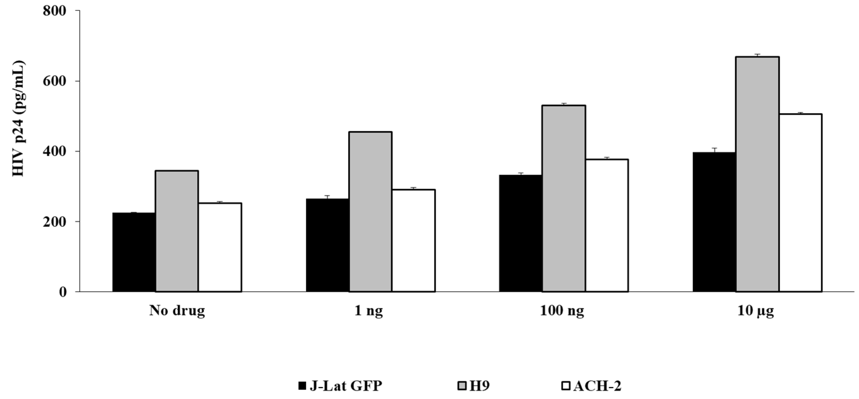
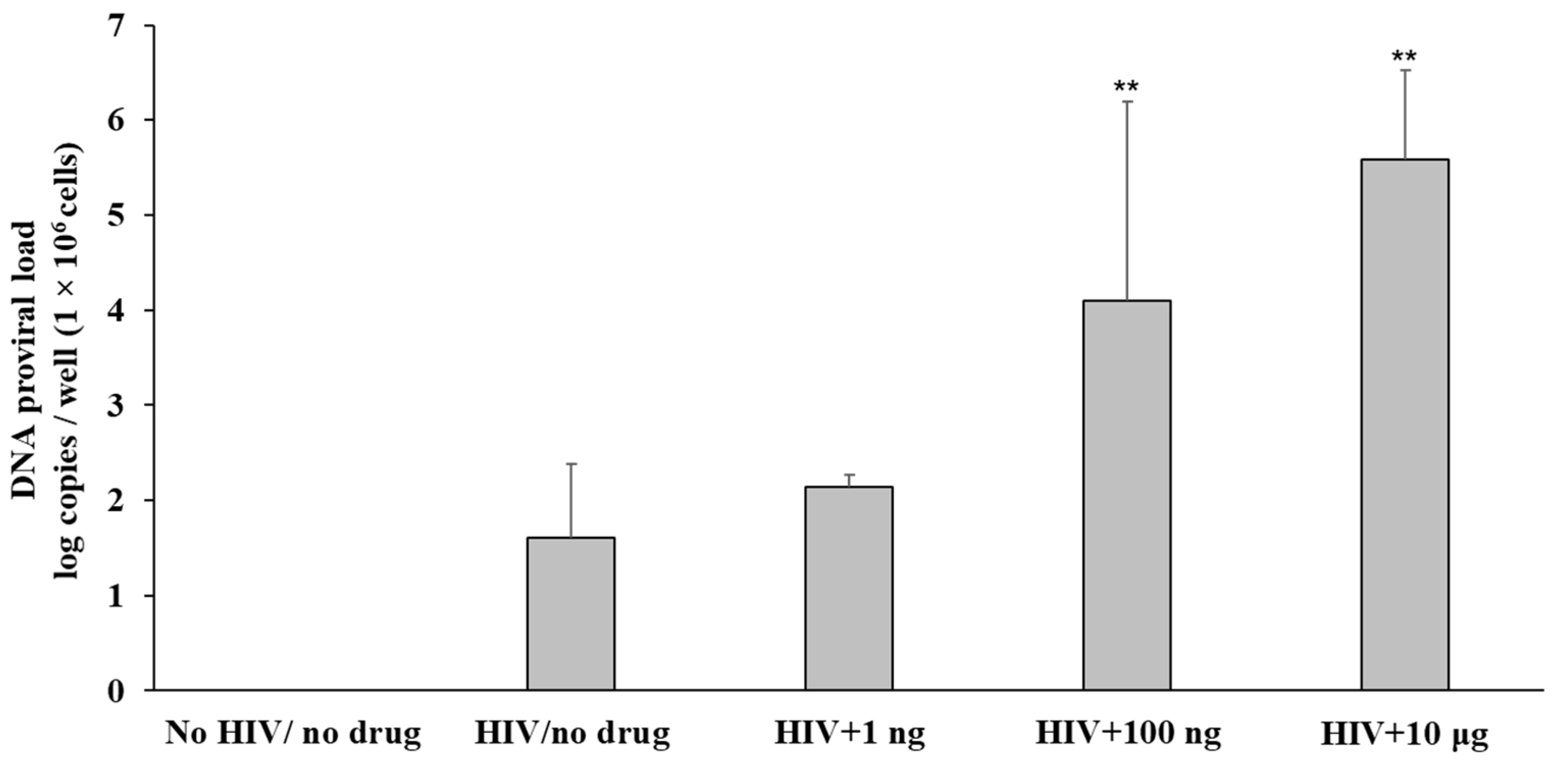
3.2. Fentanyl Enhances HIV Replication

We found expression of MOR in the HIV-susceptible and infected cells. Thus, we sought to determine whether fentanyl could modulate HIV-1 infection in vitro. The impact of fentanyl on multiple cell types was evaluated using three different concentrations: 1 ng/mL, 100 ng/mL, and 10 μg/mL. For TZM-bl cells, fentanyl exposure resulted in increased expression of the HIV p24 protein in a dose-dependent manner compared to the no-drug condition for both HIVYK-JRCSF and HIVNL4-3 (Figure 2).
To evaluate the effect of fentanyl in other cell types, HIV-infected lymphocyte cell lines were also incubated with fentanyl. As shown in Figure 3, fentanyl led to a significant dose-dependent increase in expression of HIV p24 protein in the J-Lat GFP, ACH-2, and H9 cell lines as well.
In TZM-bl cells treated with HIVNL4-3 and exposed to fentanyl at varying concentrations, log copies of proviral DNA increased with dose-dependent concentration of drug compared to the virally infected but drug-naïve cells (Figure 4).

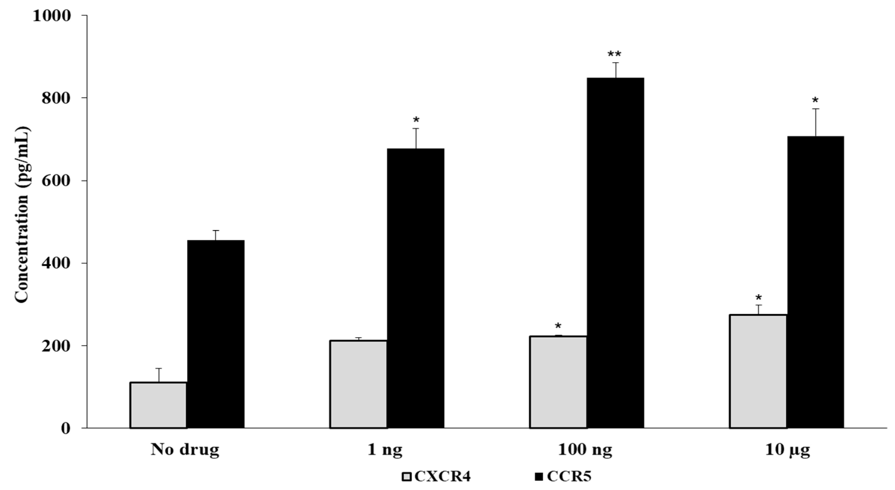
3.3. Fentanyl Increases HIV Co-Receptor Expression

Since the interaction of HIV with susceptible cells is mediated through its primary receptor (CD4) as well as chemokine co-receptors, we examined the effect of fentanyl on expression levels of CCR5 and CXCR4 proteins in uninfected cells treated with the drug. Fentanyl significantly induced the expression of CCR5 and non-significant expression of CXCR4 co-receptors in uninfected TZM-bl cells treated with fentanyl (Figure 5).
To further evaluate the impact of fentanyl on chemokine receptor expression, receptor levels were also quantified in HIV-infected cells exposed to fentanyl. All three HIV-infected cell lines showed significant increase in expression of CCR5. Expression of CXCR4 was significantly increased only in ACH-2 and H-9 cells. (Figure 6).

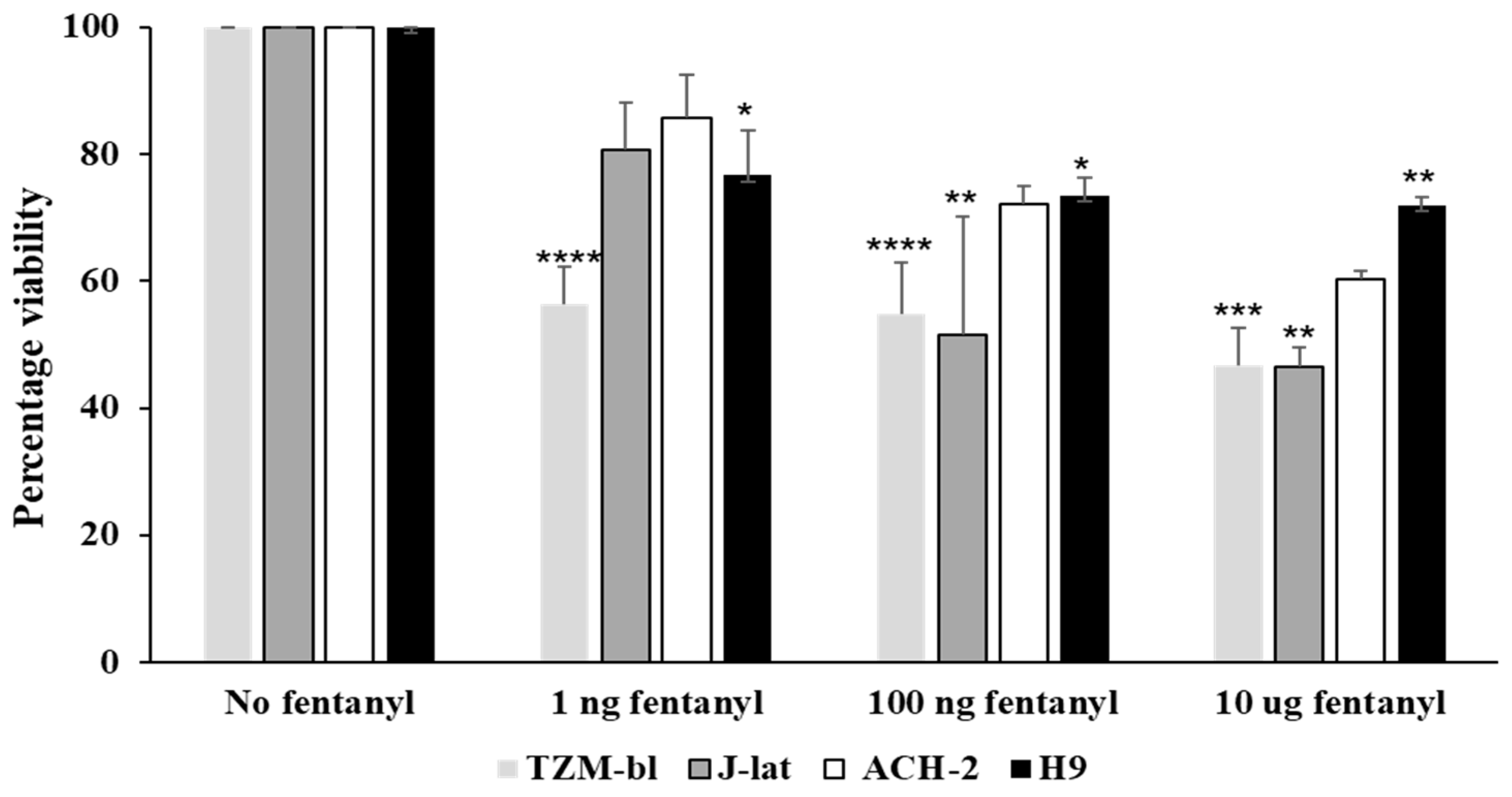
3.4. Fentanyl Alters Cell Viability

To determine the impact of fentanyl on cell viability, HIV-susceptible and HIV-infected cell lines were treated with three different concentrations (1 ng/mL, 100 ng/mL, and 10 ug/mL) of fentanyl for 24 h. A dose-dependent decrease in cell viability with increasing concentration of fentanyl was noted in all cell types tested, but there was a minimal effect on viability for ACH-2 and H9 cell lines (Figure 7). The cells were ≥60% viable even with highest dose of fentanyl (10 ug/mL) in ACH-2 and H9 cell lines.

3.5. Fentanyl Enhances HIV Replication in Primary PBMC Derived T Cells

We further tested the effect of fentanyl on HIV-1 replication in CD4+ T cells that are the primary targets of HIV. PBMCs were isolated from peripheral blood of normal human donors, and CD4+ T cells were purified. Activated CD4+ T cells were infected with HIVNL4-3 and treated with fentanyl after infection. Cells treated with fentanyl showed enhanced HIVp24 expression compared to untreated ones (Figure 8A). The finding was also consistent with increase in HIV-1 replication detected by proviral DNA in the presence of fentanyl (Figure 8B).

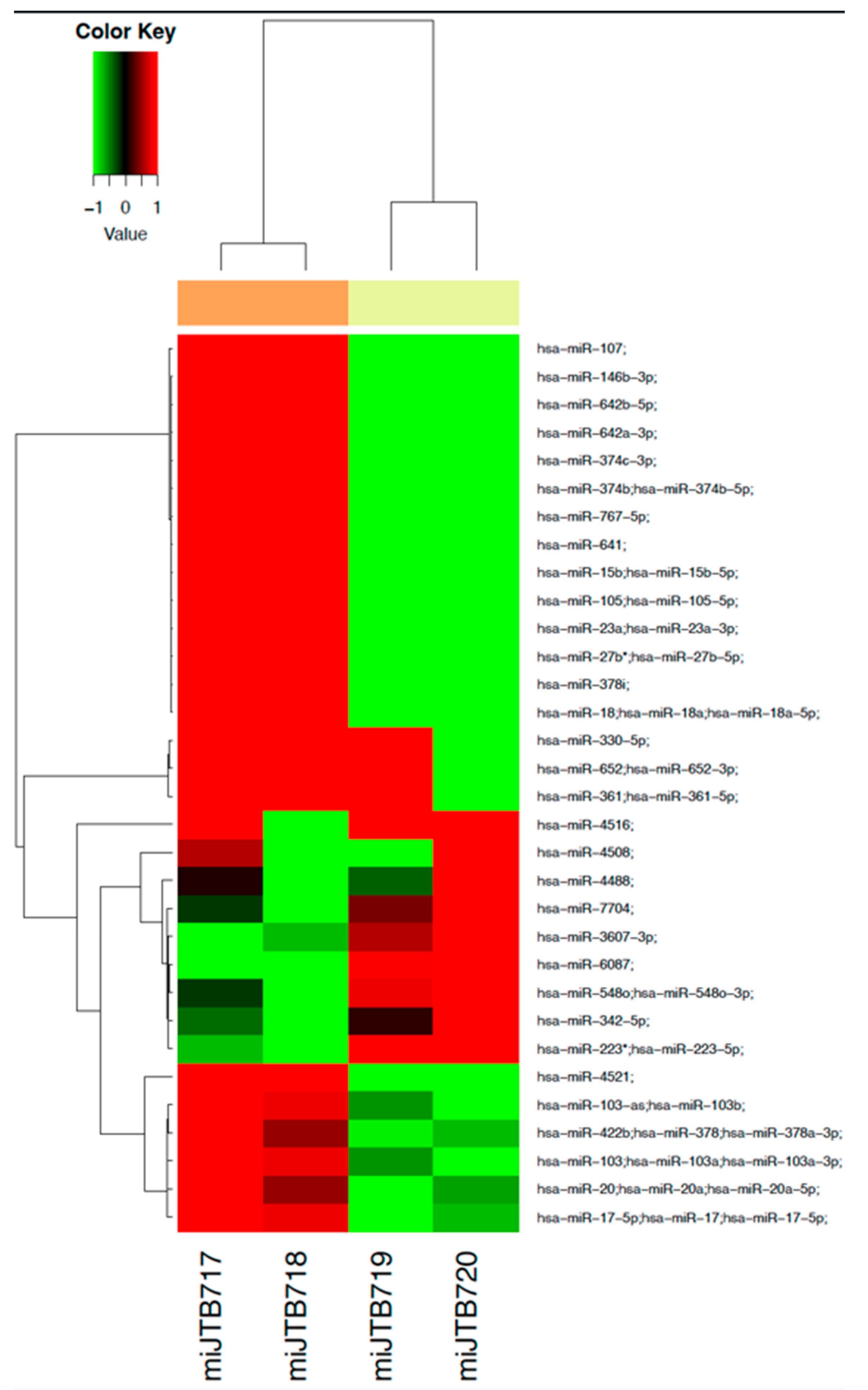
3.6. Dysregulation of miRNA Profile by Fentanyl

Total miRNA isolated from the ACH-2 cell line treated with or without fentanyl for 24 h was used to generate a comprehensive miRNA expression profile. Comparing the fentanyl-treated versus with untreated conditions, there were 32 miRNAs differentially expressed, with p-value < 0.2 as shown in Figure 9.
A list of miRNAs with their p-values and fold changes is provided in Supplementary Table S1. Of those miRNAs previously shown to impact HIV, eight mRNAs—miR-15, miR-146b, miR-378, miR-223-3p, miR-27b, miR-17, miR-3607-3p, and miR-4516—were differentially expressed in ACH-2 cells treated with fentanyl. A list of microRNAs that may play a role in HIV pathogenesis is provided in Supplementary Table S2. MicroRNAs that are differentially expressed with p-value < 0.2 are denoted by ⚫️.

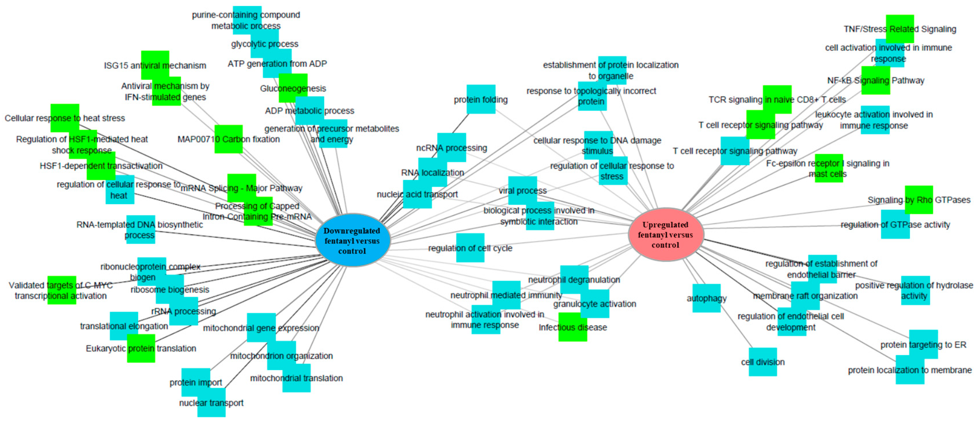
3.7. Fentanyl Alters the Cellular Transcriptome

To further understand the role of fentanyl in HIV pathogenesis, the ACH-2 cell line was utilized to characterize the effect of fentanyl on cellular gene expression. Differential analysis identified 1134 transcripts, with 619 up- and 515 downregulated transcripts in fentanyl-treated cells compared to untreated ones. Biological processes and pathways related to these genes are presented in Figure 10.
Candidate gene analysis identified 702 candidate genes involved in antiviral response, cell death or apoptosis, chemokine signaling, interferon response, and NFkB signaling (gene lists built and consolidated in gataca.cchmc.org). Of these 702 candidate genes, 49 were differentially regulated in treated ACH-2 cells (p-value < 0.05 with moderated t-test; Supplementary Table S3—labeled in green), with 21 more indicating a trend toward significance (p < 0.1 is labelled in red). As shown in Figure 11, 16 antiviral genes were significantly differently expressed in fentanyl-treated ACH-2 cells, including downregulation of APOBEC3B, APOBEC3D, TARDBP, TRIM13, TRIM28, and TRIM6 and upregulation of GSN, IFI16, INPP5K, JUN, PARP10, REST, TFAP4, TNF, TRIM26, and TRIM8 (Figure 11A). For cell-death pathways, ten genes were significantly downregulated (CASP9, ARHGAP10, E2F1, HIST1H1B, PSMB2, SATB1, CYCS, PSMB7, KPNB1, and TFDP2), and nine genes were upregulated (PRKCQ, ADD1, GSN, HMGB2, PSMB9, PSME2, TP53, UBB, and VIM) in fentanyl-treated ACH-2 cells (Figure 11B). Fentanyl treatment induced expression of 14 chemokine signaling genes: HRAS, RAC1, RAF1, and RASGRP2 were downregulated, while DOCK2, GNG10, IKBKB, ITK, MAP2K1, NFKB1, PLCB2, PREX1, VAV3, and PXN were significantly upregulated, as shown in Figure 11C, with fold changes ranging from 1.6 to 147. For interferon signaling genes, HLA-A, HLA-B, HLA-c, and IP6K2 were upregulated, with no genes significantly downregulated (Figure 11D). Finally, 17 genes involved in NFkB signaling were differentially expressed. ASH1L, EIF5A, IL23A, MAP4K2, MOB3C, NFKB2, NFAT5, and NFKBID were downregulated, while ARHGAP5, ETV6, HIVEP1, LIG1, MSN, PAN2, RND1, TNF, and TP53 were upregulated (Figure 11E). As a result of fentanyl exposure, several genes show an upregulation and differential expression, suggesting a significant effect on transcriptional regulation and immune response. Thus, fentanyl augments cell death and activates antiviral, chemokine, interferon, and NFkB signaling processes leading to suppression of the immune system.

4. Discussion

Since 2000, the number of opioid-related overdose deaths has increased steadily. With the increased use of synthetic opioids (mainly fentanyl and fentanyl analogs), the death toll reached an all-time high in 2021 [49]. The US is facing a major public health concern due to the opioid epidemic, which can be linked to HIV infections [50,51,52,53,54]. Sharing needles, syringes, or other drug injection materials directly increases the risk of HIV infection in people with opioid use disorder (OUD) [55,56,57,58]. For instance, Degenhardt et al. reported that among people who inject the drug (PWID), 17.8% live with HIV, 52.3% are HCV-seropositive, and the HBV surface antigen is positive in 9.1% [59]. People with drug addiction are at risk of exacerbating HIV-related comorbidities [60,61]. Furthermore, compared to uninfected people, long-term opiate use increases the risk of death in HIV-positive people [62,63]. As a result, it is critical to understand the link between opioid use and increased HIV infectivity to prevent the spread of HIV and its deleterious effects.
Several drugs of abuse are known to promote viral replication by suppressing innate immune responses [8,14]. HIV expression is increased by endogenous opioid peptides. For example, in microglia, endorphin increased viral protein synthesis and LTR activation [64]. Endomorphin-1 also increased HIV expression in mixed glial/neuronal and microglial cell cultures [65], whereas dynorphin promoted HIV expression in fetal brain co-cultures [66].
Expression of HIV was increased in the presence of morphine in co-cultures of promonocytes and fetal brain cells, as well as primary Kupffer cell cultures [24,26]. Prottengeier et al. found that morphine stimulated viral reactivation in latently infected cells [67]. HIV replication increased in peripheral blood lymphocytes and T cell lines in vitro upon withdrawal of morphine [68] and antagonized viral inhibitory factors in macrophages [69]. Morphine has been shown to increase HIV expression in blood monocyte-derived macrophages and neonatal macrophages, while decreasing anti-HIV microRNA expression, upregulating chemokine receptor expression, and inhibiting interferons (IFN), as well as IFN-inducible genes and regulators of the Janus kinase signal transducer and activator of transcription (JAK–STAT)-signaling pathway [20,21,49,70].ye
The semi-synthetic opioid heroin inhibits anti-HIV microRNAs in macrophages, promoting HIV expression [71]. Prottengeier et al. reported HIV reactivation in latently infected lymphocytes by heroin in vitro [67]. MicroRNA expression was also altered in peripheral blood mononuclear cells (PBMCs) and/or macrophages isolated from heroin-dependent individuals [72]. Buprenorphine was found to increase in vitro infection of PBMC with an HIV reporter virus [73]. The synthetic opioid methadone was found to promote HIV replication in fetal microglia and blood monocyte-derived macrophages by increasing the expression of CCR5 and reducing the expression of interferon, interferon stimulated genes, and anti-HIV microRNAs [74,75].
The US Drug Enforcement Administration has found counterfeit pills ranging from 0.02 to 5.1 milligrams (more than twice the lethal dose) of fentanyl per tablet. The estimated lethal dose of fentanyl in humans is 2 mg [76]. Our unpublished data collected from persons reporting to the University of Cincinnati Medical Center with HIV and opioid overdose demonstrate that blood levels of fentanyl associated with opioid overdose ranges from 1 to 25 ng/mL. Others have reported that post-mortem blood fentanyl concentration ranges from 7 to 97 ng/mL [77]. Another study by Wu et al. reported that median concentrations of fentanyl in urine and serum or plasma samples were 34 ng/mL and 10.5 ng/mL, respectively [78].
We previously reported that fentanyl increased expression of hepatitis C virus and hepatitis B virus in hepatocyte cell lines. Fentanyl also differentially regulated genes involved in the antiviral response, chemokine signaling, NFkB signaling, apoptosis, and cell death in hepatocyte cell lines [79]. These findings are supported by other studies demonstrating that opioid receptors are expressed in the liver and are important mediators of liver disease progression [80,81]. Similarly, hepatic stellate cells express several opioid receptors and contribute significantly to hepatic fibrosis [82,83]. Other in vitro studies have shown that morphine, heroin, and methamphetamine increase HCV replication [84,85,86,87]. Several research studies have proven that drugs of abuse consisting of cocaine, methamphetamine, tetrahydrocannabinol, and alcohol elevate the expression of the CCR5 and/or CXCR4 co-receptors [88,89,90,91,92,93]. The levels of chemokine co-receptors have also been found to increase with opioids such as morphine and methadone [21,22,74,94]. Increased expression of chemokine co-receptor in the presence of fentanyl is particularly intriguing in light of data showing that MOR activation leads to the heterologous desensitization of chemokine receptors [95]. We recently also proved that fentanyl induced HIV replication and chemokine co-receptor expression in human neuroblastoma cell line SH-SY5Y, resulting in decrease in TLR9 expression [96]. Yet another study by Yan J et al. showed that HIV replication and reactivation was enhanced by fentanyl in macrophages and Jurkat C11 cells [97]. The interaction of opioid receptors with chemokine receptors may have important implications for HIV pathogenesis.
Similar to the findings reported here in lymphocyte cell lines, fentanyl induces apoptosis in peripheral blood lymphocytes and human umbilical cord mononuclear cells [98]. In addition to changing the phenotype of T lymphocytes, fentanyl reduces cell proliferation, triggers apoptosis, and decreases proinflammatory cytokines [99]. However, ours is the first study to evaluate the effect of fentanyl on HIV expression and dysregulation of host cellular pathways. In addition to the impact of fentanyl on HIV replication, our study found that fentanyl promotes chemokine receptor CCR5 expression in a dose-dependent manner in all cell types and significantly enhances the expression of CXCR4 in ACH-2 and H9 cell lines. Altered or enhanced expression of chemokine receptors by various cells by fentanyl may significantly enhance the ability of HIV pathogenesis. Thus, fentanyl may facilitate enhanced viral infection by increasing the availability of HIV-1 co-receptor. Our study included the effects of fentanyl on lymphocytes as well as TZM-bl cells, which are adherent, non-immunological cells engineered to express the HIV co-receptors CD4, CCR5, and CXCR4. This cell type is crucial for the investigation of fundamental HIV biology, drug screens, and, more recently, clinical studies of HIV persistence [34]. RNA sequencing analysis of fentanyl-treated ACH-2 cells showed multiple differentially regulated genes indicating fentanyl-driven augmentation of cell death, antiviral response, chemokine, interferon, and NFkB signaling in the HIV-infected ACH-2 cell line. Fentanyl also led to dysregulation of multiple transcriptional factors that are involved in viral replication and pathogenesis. T-cell receptor signaling was found to be upregulated in ACH-2 cells treated with fentanyl. The stimulation of TCR by fentanyl could lead to the activation of T cells and the expression of viral genes via transcription factors such as NFkB. Fentanyl also downregulated various transcriptional factors, including interferon stimulating genes, which inhibit antiviral signaling and modulate immune responses in ACH-2 cells.
Limitations of the current study design should be considered. First, the experiments describe an acute, one-time exposure to fentanyl, and the long-term impact of fentanyl on viral replication is unknown. Second, all studies were performed in vitro with lab-adapted HIV isolates; thus, complementary studies on HIV replication and cellular gene expression are warranted in individuals with HIV infection and fentanyl exposure. Third, while there were several miRNAs that were differentially expressed [100,101,102,103,104,105,106,107,108,109,110,111,112,113,114,115,116,117,118,119,120,121,122,123,124,125,126,127,128], none of these differences were statistically significant, and this analysis was limited to a single cell type. Thus, further studies are needed to explore how viral infections and synthetic opioids synergize to regulate miRNA expression levels.

5. Conclusions

The opioid epidemic continues to pose a significant global health problem. More studies are needed to examine how fentanyl contributes to or limits HIV pathogenesis. Our in vitro data indicates the possibility that fentanyl induces chemokine co-receptor expression and increases HIV replication. Further, precise mechanisms by which fentanyl impairs HIV infection must also be determined through in vitro and ex vivo studies. Increased knowledge on virus-drug interactions may ultimately lead to the improvement of existing therapeutic interventions or the development of novel therapeutic approaches for this difficult-to-treat population.

Supplementary Materials

The following supporting information can be downloaded at: https://www.mdpi.com/article/10.3390/v15041027/s1, Table S1: List of miRNAs with their p-value and fold change, Table S2: List of microRNAs that may play a role in HIV pathogenesis, Table S3: List of differentially regulated genes in fentanyl treated ACH-2 cells.

Author Contributions

J.M.K.: Investigation, Writing—original draft, Data analysis; L.K.: Investigation, Formal analysis; R.K.: Data analysis and review; M.M.: Data analysis and review; K.E.S.: statistical analysis and review; J.T.B.: Conceptualization, Funding acquisition, Supervision, Writing—review and editing. All authors have read and agreed to the published version of the manuscript.

Funding

This work was supported by the National Institute on Drug Abuse (grant DA048439 to JTB).

Institutional Review Board Statement

The study protocols were approved by the Institutional Review Board at University of Cincinnati. (Approval Code: 2019_0584 and Approval Date: 05/29/2019).

Informed Consent Statement

Written informed consent was obtained from all study participants.

Data Availability Statement

All data generated during and/or analyzed during the current study are included in this article. The RNAseq and miRNAseq data are available within GEO and have been assigned the numbers GSE216124 and GSE216044, respectively. All other relevant data are within the paper and its supporting information files.

Acknowledgments

These data were presented in part at the HIV and Liver Disease meeting in Jackson, Wyoming from 9–11 September 2021. We thank Jacek Biesiada at the University of Cincinnati for help with miRNA-seq data analysis. The authors would like to thank Elizabeth Odegard and Heidi Meeds for their valuable suggestions and constructive comments.

Conflicts of Interest

The authors declare no conflict of interest.

References

  1. O’Donnell, J.K.; Gladden, R.M.; Seth, P. Trends in deaths involving heroin and synthetic opioids excluding methadone, and law enforcement drug product reports, by census region—United States, 2006–2015. MMWR Morb. Mortal. Wkly. Rep. 2017, 66, 897–903. [Google Scholar] [CrossRef] [PubMed]
  2. Mattson, C.L.; Tanz, L.J.; Quinn, K.; Kariisa, M.; Patel, P.; Davis, N.L. Trends and geographic patterns in drug and synthetic opioid overdose deaths—United States, 2013–2019. MMWR Morb. Mortal. Wkly. Rep. 2021, 70, 202. [Google Scholar] [CrossRef] [PubMed]
  3. Daniulaityte, R.; Juhascik, M.P.; Strayer, K.E.; Sizemore, I.E.; Harshbarger, K.E.; Antonides, H.M.; Carlson, R.R. Overdose deaths related to fentanyl and its analogs—Ohio, January–February 2017. MMWR Morb. Mortal. Wkly. Rep. 2017, 66, 904–908. [Google Scholar] [CrossRef] [PubMed]
  4. Peterson, A.B.; Gladden, R.M.; Delcher, C.; Spies, E.; Garcia-Williams, A.; Wang, Y.; Halpin, J.; Zibbell, J.; McCarty, C.L.; DeFiore-Hyrmer, J.; et al. Increases in fentanyl-related overdose deaths—Florida and Ohio, 2013–2015. MMWR Morb. Mortal. Wkly. Rep. 2016, 65, 844–849. [Google Scholar] [CrossRef] [PubMed]
  5. Strayer, K.E.; Antonides, H.M.; Juhascik, M.P.; Daniulaityte, R.; Sizemore, I.E. LC-MS/MS-based method for the multiplex detection of 24 fentanyl analogues and metabolites in whole blood at sub ng mL-1 concentrations. ACS Omega 2018, 3, 514–523. [Google Scholar] [CrossRef]
  6. Samji, H.; Yu, A.; Wong, S.; Wilton, J.; Binka, M.; Alvarez, M.; Bartlett, S.; Pearce, M.; Adu, P.; Jeong, D.; et al. Drug-related deaths in a population-level cohort of people living with and without hepatitis C virus in British Columbia, Canada. Int. J. Drug Policy 2020, 86, 102989. [Google Scholar] [CrossRef]
  7. Banerjee, A.; Strazza, R.; Wigdahl, B.; Pirrone, V.; Meucci, O.; Nonnemacher, M. Role of mu-opioid receptors as cofactors in human immunodeificiency virus type 1 disease progression and neuorpathogenesis. J. Neuroviral. 2011, 17, 291–302. [Google Scholar] [CrossRef]
  8. Eisenstein, T.K. The role of opioid receptors in immune system function. Front Immunol. 2019, 10, 2904. [Google Scholar] [CrossRef]
  9. Kraus, J.; Börner, C.; Giannini, E.; Höllt, V. The role of nuclear factor kappaB in tumor necrosis factor-regulated transcription of the human mu-opioid receptor gene. Mol. Pharmacol. 2003, 64, 876–884. [Google Scholar] [CrossRef]
  10. Plein, L.M.; Rittner, H.L. Opioids and the immune system—Friend or foe. Br. J. Pharmacol. 2018, 175, 2717–2725. [Google Scholar] [CrossRef]
  11. Liang, X.; Liu, R.; Chen, C.; Ji, F.; Li, T. Opioid system modulates the immune function: A review. Transl. Perioper. Pain Med. 2016, 1, 5–13. [Google Scholar] [PubMed]
  12. Brejchova, J.; Holan, V.; Svoboda, P. Expression of opioid receptors in cells of the immune system. Int. J. Mol. Sci. 2021, 22, 315. [Google Scholar] [CrossRef] [PubMed]
  13. Fentanyl Drug Facts. National Institute on Drug Abuse: 2021. Available online: https://www.drugabuse.gov/publications/drugfacts/fentanyl (accessed on 10 January 2022).
  14. Lambdin, B.H.; Bluthenthal, R.N.; Zibbell, J.E.; Wenger, L.; Simpson, K.; Kral, A.H. Associations between perceived illicit fentanyl use and infectious disease risks among people who inject drugs. Int. J. Drug Policy 2019, 74, 299–304. [Google Scholar] [CrossRef] [PubMed]
  15. Blackard, J.T.; Brown, J.L.; Lyons, M.S. Synthetic opioid use and common injection-associated viruses: Expanding the translational research agenda. Curr. HIV Res. 2019, 17, 94–101. [Google Scholar] [CrossRef]
  16. Tahamtan, A.; Tavakoli-Yaraki, M.; Mokhtari-Azad, T.; Teymoori-Rad, M.; Bont, L.; Shokri, F.; Salimi, V. Opioids and viral infections: A double-edged sword. Front. Microbiol. 2016, 7, 970. [Google Scholar] [CrossRef]
  17. Ellwanger, J.H.; Kaminski, V.D.; Rodrigues, A.G.; Kulmann-Leal, B.; Chies, J.A. CCR5 and CCR5Δ32 in bacterial and parasitic infections: Thinking chemokine receptors outside the HIV box. Int. J. Immunogenet. 2020, 47, 261–285. [Google Scholar] [CrossRef]
  18. Pozzobon, T.; Goldoni, G.; Viola, A.; Molon, B. CXCR4 signaling in health and disease. Immunol. Lett. 2016, 177, 6–15. [Google Scholar] [CrossRef]
  19. Wang, Y.; Wang, X.; Ye, L.; Li, J.; Song, L.; Fulambarkar, N.; Ho, W. Morphine Suppresses IFN Signaling Pathway and Enhances AIDS Virus Infection. PLoS ONE 2012, 7, e31167. [Google Scholar] [CrossRef]
  20. Kopnisky, K.L.; Bao, J.; Lin, Y.W. Neurobiology of HIV, psychiatric and substance abuse comorbidity research: Workshop report. Brain Behav. Immun. 2007, 21, 428–441. [Google Scholar] [CrossRef]
  21. Guo, C.J.; Li, Y.; Tian, S.; Wang, X.; Douglas, S.D.; Ho, W. Morphine enhances HIV infection of human blood mononuclear phagocytes through modulation of beta-chemokines and CCR5 receptor. J. Investig. Med. 2002, 50, 435–442. [Google Scholar] [CrossRef]
  22. Li, Y.; Merrill, J.D.; Mooney, K.; Song, L.; Wang, X.; Guo, C.-J.; Savani, R.C.; Metzger, D.S.; Douglas, S.D.; Ho, W.-Z. Morphine Enhances HIV Infection of Neonatal Macrophages. Pediatr. Res. 2003, 54, 282–288. [Google Scholar] [CrossRef] [PubMed]
  23. Peterson, P.K.; Sharp, B.M.; Gekker, G.; Portoghese, P.S.; Sannerud, K.; Balfour, H.H., Jr. Morphine promotes the growth of HIV-1 in human peripheral blood mononuclear cell cocultures. AIDS 1990, 4, 869–873. [Google Scholar] [CrossRef] [PubMed]
  24. Schweitzer, C.; Keller, F.; Schmitt, M.P.; Jaeck, D.; Adloff, M.; Schmitt, C.; Royer, C.; Kirn, A.; Aubertin, A. Morphine stimulates HIV replication in primary cultures of human Kupffer cells. Res. Virol. 1991, 142, 189–195. [Google Scholar] [CrossRef] [PubMed]
  25. Suzuki, S.; Chuang, A.J.; Chuang, L.F.; Doi, R.H.; Chuang, R.Y. Morphine promotes simian acquired immunodeficiency syndrome virus replication in monkey peripheral mononuclear cells: Induction of CC chemokine receptor 5 expression for virus entry. J. Infect. Dis. 2002, 185, 1826–1829. [Google Scholar] [CrossRef]
  26. Peterson, P.K.; Gekker, G.; Hu, S.; Anderson, W.; Kravitz, F.; Portoghese, P.S.; Balfour, H.H.; Chao, C.C. Morphine amplifies HIV-1 expression in chronically infected promonocytes cocultured with human brain cells. J. Neuroimmunol. 1994, 50, 167–175. [Google Scholar] [CrossRef]
  27. Nair, M.P.; Schwartz, S.A.; Polasani, R.; Hou, J.; Sweet, A.; Chadha, K.C. Immunoregulatory effects of morphine on human lymphocytes. Clin. Diagn. Lab. Immunol. 1997, 4, 127–132. [Google Scholar] [CrossRef]
  28. El-Hage, N.; Wu, G.; Wang, J.; Ambati, J.; Knapp, P.E.; Reed, J.L.; Bruce-Keller, A.J.; Hauser, K.F. HIV-1 Tat and opiate-induced changes in astrocytes promote chemotaxis of microglia through the expression of MCP-1 and alternative chemokines. Glia 2006, 53, 132–146. [Google Scholar] [CrossRef]
  29. Wang, X.; Tan, N.; Douglas, S.D.; Zhang, T.; Wang, Y.J.; Ho, W.Z. Morphine inhibits CD8+ T cell-mediated, noncytolytic, anti-HIV activity in latently infected immune cells. J. Leukoc. Biol. 2005, 78, 772–776. [Google Scholar] [CrossRef]
  30. Squinto, S.P.; Mondal, D.; Block, A.L.; Prakash, O. Morphine-Induced Transactivation of HIV-1 LTR in Human Neuroblastoma Cells. AIDS Res. Hum. Retrovir. 1990, 6, 1163–1168. [Google Scholar] [CrossRef]
  31. Nair, M.P.N.; Mahajan, S.; Chadha, K.C.; Nair, N.M.; Hewitt, R.G.; Pillai, S.K.; Chadha, P.; Sukumaran, P.C.; Schwartz, S.A. Effect of cocaine on chemokine and CCR-5 gene expression by mononuclear cells from normal donors and HIV-1 infected patients. Adv. Exp. Med. Biol. 2001, 493, 235–240. [Google Scholar]
  32. Jiang, J.; Wang, M.; Liang, B.; Shi, Y.; Su, Q.; Chen, H.; Huang, J.; Su, J.; Pan, P.; Li, Y.; et al. In vivo effects of methamphetamine on HIV-1 replication: A population-based study. Drug Alcohol Depend. 2016, 159, 246–254. [Google Scholar] [CrossRef] [PubMed]
  33. Derdeyn, C.A.; Decker, J.M.; Sfakianos, J.N.; Wu, X.; O’Brien, W.A.; Ratner, L.; Kappes, J.C.; Shaw, G.M.; Hunter, E. Sensitivity of human immunodeficiency virus type 1 to the fusion inhibitor T-20 is modulated by coreceptor specificity defined by the V3 loop of gp120. J. Virol. 2000, 74, 8358–8367. [Google Scholar] [CrossRef] [PubMed]
  34. Takeuchi, Y.; McClure, M.O.; Pizzato, M. Identification of gamma retroviruses constitutively released from cell lines used for human immunodeficiency virus research. J. Virol. 2008, 82, 12585–12588. [Google Scholar] [CrossRef] [PubMed]
  35. Jordan, A.; Bisgrove, D.; Verdin, E. HIV reproducibly establishes a latent infection after acute infection of T cells in vitro. EMBO J. 2003, 22, 1868–1877. [Google Scholar] [CrossRef] [PubMed]
  36. Jordan, A.; Defechereux, P.; Verdin, E. The site of HIV-1 integration in the human genome determines basal transcriptional activity and response to Tat transactivation. EMBO J. 2001, 20, 1726–1738. [Google Scholar] [CrossRef] [PubMed]
  37. Mann, D.L.; O’Brien, S.J.; Gilbert, D.A.; Reid, Y.; Popovic, M.; Read-Connole, E.; Gallo, R.C.; Gazdar, A.F. Origin of the HIV-susceptible human CD4+ cell line H9. AIDS Res. Hum. Retrovir. 1989, 5, 253–255. [Google Scholar] [CrossRef]
  38. Clouse, K.A.; Powell, D.; Washington, I.; Poli, G.; Strebel, K.; Farrar, W.; Barstad, P.; Kovacs, J.; Fauci, A.S.; Folks, T.M. Monokine regulation of human immunodeficiency virus-1 expression in a chronically infected human T cell clone. J. Immunol. 1989, 142, 431–438. [Google Scholar] [CrossRef]
  39. Armenian, P.; Vo, K.T.; Barr-Walker, J.; Lynch, K.L. Fentanyl, fentanyl analogs and novel synthetic opioids: A comprehensive review. Neuropharmacology 2018, 134, 121–132. [Google Scholar] [CrossRef]
  40. Gibellini, D.; Vitone, F.; Schiavone, P.; Ponti, C.; La Placa, M.; Re, M.C. Quantitative detection of human immunodeficiency virus type 1 (HIV-1) proviral DNA in peripheral blood mononuclear cells by SYBR green real-time PCR technique. J. Clin. Virol. 2004, 29, 282–289. [Google Scholar] [CrossRef]
  41. Langevin, S.M.; Kuhnell, D.; Orr-Asman, M.A.; Biesiada, J.; Zhang, X.; Medvedovic, M.; Thomas, H.E. Balancing yield, purity and practicality: A modified differential ultracentrifugation protocol for efficient isolation of small extracellular vesicles from human serum. RNA Biol. 2019, 16, 5–12. [Google Scholar] [CrossRef]
  42. Paradis, E.; Schliep, K. ape 5.0: An environment for modern phylogenetics and evolutionary analyses in R. Bioinformatics 2019, 35, 526–528. [Google Scholar] [CrossRef] [PubMed]
  43. Kumar, S.; Stecher, G.; Tamura, K. MEGA7: Molecular evolutionary genetics analysis version 7.0 for bigger datasets. Mol. Biol. Evol. 2016, 33, 1870–1874. [Google Scholar] [CrossRef] [PubMed]
  44. Huber, W.; Carey, V.J.; Gentleman, R.; Anders, S.; Carlson, M.; Carvalho, B.S.; Bravo, H.C.; Davis, S.; Gatto, L.; Girke, T.; et al. Orchestrating high-throughput genomic analysis with Bioconductor. Nat. Methods 2015, 12, 115–121. [Google Scholar] [CrossRef] [PubMed]
  45. Kozomara, A.; Griffiths-Jones, S. miRBase: Annotating high confidence microRNAs using deep sequencing data. Nucleic Acids Res. 2014, 42, 68–73. [Google Scholar] [CrossRef]
  46. Robinson, M.; McCarthy, D.; Smyth, G.K. edgeR: Differential expression analysis of digital gene expression data. Bioinformatics 2010, 26, 139–140. [Google Scholar] [CrossRef]
  47. Walsh, K.B.; Zhang, X.; Zhu, X.; Wohleb, E.; Woo, D.; Lu, L.; Adeoye, O. Intracerebral hemorrhage induces inflammatory gene expression in peripheral blood: Global transcriptional profiling in intracerebral hemorrhage patients. DNA Cell Biol. 2019, 38, 660–669. [Google Scholar] [CrossRef]
  48. Rapp, S.J.; Dershem, V.; Zhang, X.; Schutte, S.C.; Chariker, M.E. Varying negative pressure wound therapy acute effects on human split-thickness autografts. J. Burn. Care Res. 2020, 41, 104–112. [Google Scholar] [CrossRef]
  49. U.S. Overdose Deaths In 2021 Increased Half as Much as in 2020—But Are Still Up 15% 2022.2022). Available online: https://www.cdc.gov/nchs/pressroom/nchs_press_releases/2022/202205.htm (accessed on 11 May 2022).
  50. Lyden, J.; Binswanger, I.A. The United States opioid epidemic. Semin. Perinatol. 2019, 43, 123–131. [Google Scholar] [CrossRef]
  51. Lavonas, E.J.; Dezfulian, C. Impact of the Opioid Epidemic. Crit. Care Clin. 2020, 36, 753–769. [Google Scholar] [CrossRef]
  52. Reider, B. Opioid epidemic. Am. J. Sports Med. 2019, 47, 1039–1042. [Google Scholar] [CrossRef]
  53. Volkow, N.D.; Blanco, C. The changing opioid crisis: Development, challenges and opportunities. Mol. Psychiatry 2021, 26, 218–233. [Google Scholar] [CrossRef] [PubMed]
  54. Csete, J.; Kamarulzaman, A.; Kazatchkine, M.; Altice, F.L.; Balicki, M.; Buxton, J.; Cepeda, J.; Comfort, M.; Goosby, E.; Goulão, J.; et al. Public health and international drug policy. Lancet 2016, 387, 1427–1480. [Google Scholar] [CrossRef] [PubMed]
  55. Ball, L.J.; Puka, K.; Speechley, M.; Wong, R.; Hallam, B.; Wiener, J.C.; Koivu, S.; Silverman, M.S. Sharing of injection drug preparation equipment is associated with HIV infection: A cross-sectional study. J. Acquir. Immune Defic. Syndr. 2019, 81, e99–e103. [Google Scholar] [CrossRef]
  56. Bulled, N.; Singer, M. Syringe-mediated syndemics. AIDS Behav. 2011, 15, 1539–1545. [Google Scholar] [CrossRef] [PubMed]
  57. Tyagi, M.; Weber, J.; Bukrinsky, M.; Simon, G.L. The effects of cocaine on HIV transcription. J. Neurovirol. 2016, 22, 261–274. [Google Scholar] [CrossRef]
  58. Moradi, G.; Hajarizadeh, B.; Rahmani, K.; Mohamadi-Bolbanabad, A.; Darvishi, S.; Zareie, B.; Zavareh, F.A.; Sharafi, H.; Alavian, S.M.; Ramazani, R.; et al. Drug use and risk behaviour profile, and the prevalence of HIV, hepatitis C and hepatitis B among people with methamphetamine use in Iran. Int. J. Drug Policy 2019, 73, 129–134. [Google Scholar] [CrossRef]
  59. Degenhardt, L.; Peacock, A.; Colledge, S.; Leung, J.; Grebely, J.; Vickerman, P.; Stone, J.; Cunningham, E.B.; Trickey, A.; Dumchev, K.; et al. Global prevalence of injecting drug use and sociodemographic characteristics and prevalence of HIV, HBV, and HCV in people who inject drugs: A multistage systematic review. Lancet Glob. Health 2017, 5, e1192–e1207. [Google Scholar] [CrossRef]
  60. Illenberger, J.M.; Harrod, S.B.; Mactutus, C.F.; McLaurin, K.A.; Kallianpur, A.; Booze, R.M. HIV Infection and Neurocognitive Disorders in the Context of Chronic Drug Abuse: Evidence for Divergent Findings Dependent upon Prior Drug History. J. Neuroimmune Pharmacol. 2020, 15, 715–728. [Google Scholar] [CrossRef]
  61. Cernasev, A.; Veve, M.P.; Cory, T.J.; Summers, N.A.; Miller, M.; Kodidela, S.; Kumar, S. Opioid Use Disorders in People Living with HIV/AIDS: A Review of Implications for Patient Outcomes, Drug Interactions, and Neurocognitive Disorders. Pharmacy 2020, 8, 168. [Google Scholar] [CrossRef]
  62. Strain, E. Opioid Use Disorder: Epidemiology, Pharmacology, Clinical Manifestations, Course, Screening, Assessment, and Diagnosis; Saxon, A., Hermann, R., Eds.; Wolters Kluwer, UpToDate: Waltham, MA, USA, 2018. [Google Scholar]
  63. Weisberg, D.F.; Gordon, K.S.; Barry, D.T.; Becker, W.C.; Crystal, S.; Edelman, E.J.; Gaither, J.; Gordon, A.J.; Goulet, J.; Kerns, R.D.; et al. Long-term Prescription of Opioids and/or Benzodiazepines and Mortality among HIV-Infected and Uninfected Patients. J Acquir. Immune Defic. Syndr. 2015, 69, 223–233. [Google Scholar] [CrossRef]
  64. Sundar, K.S.; Kamaraju, L.S.; Dingfelder, J.; McMahon, J.; Gollapudi, S.; Wilson, W.H.; Kong, L.Y.; Hong, J.S.; Weiss, J.M.; Lee, J. beta-Endorphin enhances the replication of neurotropic human immunodeficiency virus in fetal perivascular microglia. J. Neuroimmunol. 1995, 61, 97–104. [Google Scholar] [CrossRef] [PubMed]
  65. Peterson, P.K.; Gekker, G.; Hu, S.; Lokensgard, J.; Portoghese, P.S.; Chao, C. Endomorphin-1 potentiates HIV-1 expression in human brain cell cultures: Implication of an atypical mu-opioid receptor. Neuropharmacology 1999, 38, 273–278. [Google Scholar] [CrossRef] [PubMed]
  66. Chao, C.C.; Gekker, G.; Hu, S.; Sheng, W.S.; Portoghese, P.S.; Peterson, P.K. Upregulation of HIV-1 expression in cocultures of chronically infected promonocytes and human brain cells by dynorphin. Biochem. Pharmacol. 1995, 50, 715–722. [Google Scholar] [CrossRef] [PubMed]
  67. Prottengeier, J.; Koutsilieri, E.; Scheller, C. The effects of opioids on HIV reactivation in latently-infected T-lymphoblasts. AIDS Res. Ther. 2014, 11, 17. [Google Scholar] [CrossRef]
  68. Xu, D.; Fu, J.; Jin, L.; Zhang, H.; Zhou, C.; Zou, Z.; Zhao, J.-M.; Zhang, B.; Shi, M.; Ding, X.; et al. Circulating and Liver Resident CD4+CD25+ Regulatory T Cells Actively Influence the Antiviral Immune Response and Disease Progression in Patients with Hepatitis B. J. Immunol. 2006, 177, 739–747. [Google Scholar] [CrossRef]
  69. Wang, X.; Liu, J.; Zhou, L.; Ho, W.-Z. MorphineWithdrawal Enhances HIV Infection of Macrophages. Front. Immunol. 2019, 10, 2601. [Google Scholar] [CrossRef]
  70. Tang, B.; Zhang, Y.; Liang, R.; Yuan, P.; Du, J.; Wang, H.; Wang, L. Activation of the -opioid receptor inhibits serum deprivation induced apoptosis of human liver cells via the activation of PKC and the mitochondrial pathway. Int. J. Mol. Med. 2011, 28, 1077–1085. [Google Scholar]
  71. Wang, X.; Ma, T.-C.; Li, J.-L.; Zhou, Y.; Geller, E.B.; Adler, M.W.; Peng, J.-S.; Zhou, W.; Zhou, D.-J.; Ho, W.-Z. Heroin inhibits HIV-restriction miRNAs and enhances HIV infection of macrophages. Front. Microbiol. 2015, 6, 1230. [Google Scholar] [CrossRef]
  72. Wang, X.; Sun, L.; Zhou, Y.; Su, Q.-J.; Li, J.-L.; Ye, L.; Liu, M.-Q.; Zhou, W.; Ho, W.-Z. Heroin Abuse and/or HIV Infection Dysregulate Plasma Exosomal miRNAs. J. Neuroimmune Pharmacol. 2020, 15, 400–408. [Google Scholar] [CrossRef]
  73. Gornalusse, G.G.; Vojtech, L.N.; Levy, C.N.; Hughes, S.M.; Kim, Y.; Valdez, R.; Pandey, U.; Ochsenbauer, C.; Astronomo, R.; McElrath, J.; et al. Buprenorphine Increases HIV-1 Infection In Vitro but Does Not Reactivate HIV-1 from Latency. Viruses 2021, 13, 1472. [Google Scholar] [CrossRef]
  74. Wang, X.; Tian, S.; Guo, C.J.; Douglas, S.D.; Ho, W. Methadone enhances human immunodeficiency virus infection of human immune cells. J. Infect. Dis. 2002, 185, 118–122. [Google Scholar]
  75. Wang, M.-R.; Wu, D.-D.; Luo, F.; Zhong, C.-J.; Wang, X.; Zhu, N.; Wu, Y.-J.; Hu, H.-T.; Feng, Y.; Wang, X.; et al. Methadone Inhibits Viral Restriction Factors and Facilitates HIV Infection in Macrophages. Front. Immunol. 2020, 11, 1253. [Google Scholar] [CrossRef] [PubMed]
  76. Facts about Fentanyl. DEA. In United States Drug Enforcement Administration: 2021. Available online: https://www.dea.gov/resources/facts-about-fentany (accessed on 20 March 2023).
  77. Woodall, K.L.; Martin, T.L.; McLellan, B.A. Oral abuse of fentanyl patches (Duragesic®): Seven case reports. J. Forensic Sci. 2008, 53, 222–225. [Google Scholar] [CrossRef] [PubMed]
  78. Wu, F.; Slawson, M.H.; Johnson-Davis, K.L. Metabolic patterns of fentanyl, meperidine, methylphenidate, tapentadol and tramadol observed in urine, serum or plasma. J. Anal. Toxicol. 2017, 41, 289–299. [Google Scholar] [CrossRef] [PubMed]
  79. Kong, L.; Karns, R.; Shata, M.; Brown, J.L.; Lyons, M.S.; Sherman, K.E.; Blackard, J.T. The synthetic opioid fentanyl enhances viral replication in vitro. PLoS ONE 2021, 16, e0249581. [Google Scholar] [CrossRef]
  80. Tang, B.; Li, Y.; Yuan, S.; Tomlinson, S.; He, S. Upregulation of the opioid receptor in liver cancer promotes liver cancer progression both in vitro and in vivo. Int. J. Oncol. 2013, 43, 1281–1290. [Google Scholar] [CrossRef]
  81. Lu, J.; Liu, Z.; Zhao, L.; Tian, H.; Liu, X.; Yuan, C. In vivo and in vitro inhibition of human liver cancer progress by downregulation of the μ-opioid receptor and relevant mechanisms. Oncol. Rep. 2013, 30, 1731–1738. [Google Scholar] [CrossRef]
  82. Zhu, X.; Wang, L.-C.; Chen, E.-Q.; Chen, X.-B.; Chen, L.-Y.; Liu, L.; Lei, X.-Z.; Liu, C.; Tang, H. Prospective Evaluation of FibroScan for the Diagnosis of Hepatic Fibrosis Compared with Liver Biopsy/AST Platelet Ratio Index and FIB-4 in Patients with Chronic HBV Infection. Dig. Dis. Sci. 2011, 56, 2742–2749. [Google Scholar] [CrossRef]
  83. Jia, K.; Sun, D.; Ling, S.; Tian, Y.; Yang, X.; Sui, J.; Tang, B.; Wang, L. Activated-opioid receptors inhibit hydrogen peroxideinduced apoptosis in liver cancer cells through the PKC/ERK signaling pathwa. Mol. Med. Rep. 2014, 10, 839–847. [Google Scholar] [CrossRef]
  84. De Minicis, S.; Candelaresi, C.; Marzioni, M.; Saccomano, S.; Roskams, T.; Casini, A.; Risaliti, A.; Salzano, R.; Cautero, N.; di Francesco, F.; et al. Role of endogenous opioids in modulating HSC activity in vitro and liver fibrosis in vivo. Gut 2008, 57, 352–364. [Google Scholar] [CrossRef]
  85. Ye, L.; Peng, J.S.; Wang, X.; Wang, Y.J.; Luo, G.X.; Ho, W.Z. Methamphetamine enhances Hepatitis C virus replication in human hepatocytes. J. Viral Hepat. 2008, 15, 261–270. [Google Scholar] [CrossRef] [PubMed]
  86. Hsu, H.-Y.; Chang, M.-H.; Ni, Y.-H.; Chen, H. Survey of hepatitis B surface variant infection in children 15 years after a nationwide vaccination programme in Taiwan. Gut 2004, 53, 1499–1503. [Google Scholar] [CrossRef] [PubMed]
  87. Liang, H.; Wang, X.; Chen, H.; Song, L.; Ye, L.; Wang, S.-H.; Wang, Y.-J.; Zhou, L.; Ho, W.-Z. Methamphetamine Enhances HIV Infection of Macrophages. Am. J. Pathol. 2008, 172, 1617–1624. [Google Scholar] [CrossRef]
  88. Liu, X.; Zha, J.; Nishitani, J.; Chen, H.; Zack, J.A. HIV-1 infection in peripheral blood lymphocytes (PBLs) exposed to alcohol. Virology 2003, 307, 37–44. [Google Scholar] [CrossRef] [PubMed]
  89. Nair, M.P.; Chadha, K.C.; Hewitt, R.G.; Mahajan, S.; Sweet, A.; Schwartz, S.A. Cocaine differentially modulates chemokine production by mononuclear cells from normal donors and human immunodeficiency virus type 1-infected patients. Clin. Diagn. Lab. Immunol. 2000, 7, 96–100. [Google Scholar] [CrossRef]
  90. Nair, M.P.; Saiyed, Z.M. Effect of methamphetamine on expression of HIV coreceptors and CC-chemokines by dendritic cells. Life Sci. 2011, 88, 987–994. [Google Scholar] [CrossRef]
  91. Nair, M.P.; Saiyed, Z.M.; Nair, N.; Gandhi, N.H.; Rodriguez, J.W.; Boukli, N.; Provencio-Vasquez, E.; Malow, R.M.; Miguez-Burbano, M. Methamphetamine enhances HIV-1 infectivity in monocyte derived dendritic cells. J. Neuroimmune Pharmacol. 2009, 4, 129–139. [Google Scholar] [CrossRef]
  92. Roth, M.D.; Whittaker, K.M.; Choi, R.; Tashkin, D.P.; Baldwin, G.C. Cocaine and -1 receptors modulate HIV infection, chemokine receptors, and the HPA axis in the huPBL-SCID model. J. Leukoc. Biol. 2005, 78, 1198–1203. [Google Scholar] [CrossRef]
  93. Wang, X.; Douglas, S.D.; Metzger, D.S.; Guo, C.; Li, Y.; O’Brien, C.P.; Song, L.; Davis-Vogal, A.; Ho, W. Alcohol potentiates HIV-1 infection of human blood mononuclear phagocytes. Alcohol. Clin. Exp. Res. 2002, 26, 1880–1886. [Google Scholar] [CrossRef]
  94. Miyagi, T.; Chuang, L.F.; Doi, R.H.; Carlos, M.P.; Torres, J.V.; Chuang, R.Y. Morphine induces gene expression of CCR5 in human CEM x174 lymphocytes. J. Biol. Chem. 2000, 275, 31305–31310. [Google Scholar] [CrossRef]
  95. Steele, A.D.; Szabo, I.; Bednar, F.; Rogers, T.J. Interactions between opioid and chemokine receptors: Heterologous desensitization. Cytokine Growth Factor Rev. 2002, 13, 209–222. [Google Scholar] [CrossRef] [PubMed]
  96. Kong, L.; Shata, M.T.M.; Brown, J.L.; Lyons, M.S.; Sherman, K.E.; Blackard, J.T. The synthetic opioid fentanyl increases HIV replication and chemokine co-receptor expression in vitro. J. NeuroVirology 2022, 28, 583–594. [Google Scholar] [CrossRef] [PubMed]
  97. Yan, J.; Nie, D.-H.; Bai, C.-S.; Rehman, A.; Yang, A.; Mou, X.-L.; Zhang, Y.-Q.; Xu, Y.-Q.; Xiang, Q.-Q.; Ren, Y.-T.; et al. Fentanyl enhances HIV infection in vitro. Virology 2022, 577, 43–50. [Google Scholar] [CrossRef] [PubMed]
  98. Ma, K.; Ma, P.; Lu, H.; Liu, S.; Cao, Q. Fentanyl Suppresses the Survival of CD4+ T Cells Isolated from Human Umbilical Cord Blood through Inhibition of IKK s-mediated NF-κB Activation. Scand. J. Immunol. 2017, 85, 343–349. [Google Scholar] [CrossRef] [PubMed]
  99. Kandathil, A.J.; Sugawara, S.; Balagopal, A. Are T cells the only HIV-1 reservoir? Retrovirology 2016, 13, 86. [Google Scholar] [CrossRef]
  100. Su, B.; Fu, Y.; Liu, Y.; Wu, H.; Ma, P.; Zeng, W.; Zhang, T.; Lian, S.; Wu, H. Potential Application of MicroRNA Profiling to the Diagnosis and Prognosis of HIV-1 Infection. Front. Microbiol. 2018, 9, 3185. [Google Scholar] [CrossRef]
  101. Triboulet, R.; Mari, B.; Lin, Y.L.; Chable-Bessia, C.; Bennasser, Y.; Lebrigand, K.; Cardinaud, B.; Maurin, T.; Barbry, P.; Baillat, V.; et al. Suppression of microRNA-silencing pathway by HIV-1 during virus replication. Science. 2007, 315, 1579–1582. [Google Scholar] [CrossRef]
  102. Sung, T.-L.; Rice, A.P. miR-198 Inhibits HIV-1 Gene Expression and Replication in Monocytes and Its Mechanism of Action Appears To Involve Repression of Cyclin T1. PLOS Pathog. 2009, 5, e1000263. [Google Scholar] [CrossRef]
  103. Chiang, K.; Liu, H.; Rice, A.P. miR-132 enhances HIV-1 replication. Virology 2013, 438, 1–4. [Google Scholar] [CrossRef]
  104. Hariharan, M.; Scaria, V.; Pillai, B.; Brahmachari, S.K. Targets for human encoded microRNAs in HIV genes. Biochem. Biophys. Res. Commun. 2005, 337, 1214–1218. [Google Scholar] [CrossRef]
  105. Pilakka-Kanthikeel, S.; Nair, M.P.N. Interaction of drugs of abuse and microRNA with HIV: A brief review. Front. Microbiol. 2015, 6, 967. [Google Scholar] [CrossRef] [PubMed]
  106. Jin, C.; Cheng, L.; Höxtermann, S.; Xie, T.; Lu, X.; Wu, H.; Skaletz-Rorowski, A.; Brockmeyer, N.H.; Wu, N. Micro RNA-155 is a biomarker of T-cell activation and immune dysfunction in HIV-1-infected patients. HIV Med. 2017, 18, 354–362. [Google Scholar] [CrossRef] [PubMed]
  107. Kwon, H.-S.; Ott, M. The ups and downs of SIRT1. Trends Biochem Sci. 2008, 33, 517–525. [Google Scholar] [CrossRef] [PubMed]
  108. Gorroño, L.E.; Escribà, T.; Boulanger, N.; Guardo, A.C.; Leon, A.; Bargalló, M.E.; García, F.; Gatell, J.M.; Plana, M.; Arnedo, M.; et al. Differential MicroRNA Expression Profile between Stimulated PBMCs from HIV-1 Infected Elite Controllers and Viremic Progressors. PLOS ONE 2014, 9, e106360. [Google Scholar] [CrossRef]
  109. Houzet, L.; Klase, Z.; Yeung, M.L.; Wu, A.; Le, S.Y.; Quinones, M.; Jeang, K.T. The extent of sequence complementarity correlates with the potency of cellular miRNA-mediated restriction of HIV-1. Nucleic Acids Res. 2012, 40, 11684–11696. [Google Scholar] [CrossRef] [PubMed]
  110. Chen, A.K.; Sengupta, P.; Waki, K.; Van Engelenburg, S.B.; Ochiya, T.; Ablan, S.D.; Freed, E.O.; Lippincott-Schwartz, J. MicroRNA binding to the HIV-1 Gag protein inhibits Gag assembly and virus production. Proc. Natl. Acad. Sci. 2014, 111, E2676–E2683. [Google Scholar] [CrossRef]
  111. Wang, P.; Qu, X.; Zhou, X.; Shen, Y.; Ji, H.; Fu, Z.; Deng, J.; Lu, P.; Yu, W.; Lu, H.; et al. Two cellular microRNAs, miR-196b and miR-1290, contribute to HIV-1 latency. Virology 2015, 486, 228–238. [Google Scholar] [CrossRef]
  112. Huang, J.; Wang, F.; Argyris, E.; Chen, K.; Liang, Z.; Tian, H.; Huang, W.; Squires, K.; Verlinghieri, G.; Zhang, H. Cellular microRNAs contribute to HIV-1 latency in resting primary CD4+ T lymphocytes. Nat. Med. 2007, 13, 1241–1247. [Google Scholar] [CrossRef]
  113. Egaña-Gorroño, L.; Guardo, A.C.; Bargalló, M.E.; Planet, E.; Vilaplana, E.; Escribà, T.; Pérez, I.; Gatell, J.M.; García, F.; Arnedo, M.; et al. MicroRNA Profile in CD8+ T-Lymphocytes from HIV-Infected Individuals: Relationship with Antiviral Immune Response and Disease Progression. PLOS ONE 2016, 11, e0155245. [Google Scholar] [CrossRef]
  114. Asahchop, E.L.; Akinwumi, S.M.; Branton, W.G.; Fujiwara, E.; Gill, M.J.; Power, C. Plasma microRNA profiling predicts HIV-associated neurocognitive disorder. Aids 2016, 30, 2021–2031. [Google Scholar] [CrossRef]
  115. Dey, R.; Soni, K.; Saravanan, S.; Balakrishnan, P.; Kumar, V.; Boobalan, J.; Solomon, S.S.; Scaria, V.; Solomon, S.; Brahmachari, S.K.; et al. Anti-HIV microRNA expression in a novel Indian cohort. Sci. Rep. 2016, 6, 28279. [Google Scholar] [CrossRef] [PubMed]
  116. Pilakka-Kanthikeel, S.; Raymond, A.; Atluri, V.S.R.; Sagar, V.; Saxena, S.K.; Diaz, P.; Chevelon, S.; Concepcion, M.; Nair, M. Sterile alpha motif and histidine/aspartic acid domain-containing protein 1 (SAMHD1)-facilitated HIV restriction in astrocytes is regulated by miRNA-181a. J. Neuroinflammation 2015, 12, 1–12. [Google Scholar] [CrossRef] [PubMed]
  117. Zheng, Y.; Yang, Z.; Jin, C.; Chen, C.; Wu, N. hsa-miR-191-5p inhibits replication of human immunodeficiency virus type 1 by downregulating the expression of NUP50. Arch. Virol. 2021, 166, 755–766. [Google Scholar] [CrossRef] [PubMed]
  118. Nathans, R.; Chu, C.-Y.; Serquina, A.K.; Lu, C.-C.; Cao, H.; Rana, T.M. Cellular MicroRNA and P Bodies Modulate Host-HIV-1 Interactions. Mol. Cell 2009, 34, 696–709. [Google Scholar] [CrossRef] [PubMed]
  119. Sun, G.; Li, H.; Wu, X.; Covarrubias, M.; Scherer, L.; Meinking, K.; Luk, B.; Chomchan, P.; Alluin, J.; Gombart, A.F.; et al. Interplay between HIV-1 infection and host microRNAs. Nucleic Acids Res. 2011, 40, 2181–2196. [Google Scholar] [CrossRef]
  120. Reynoso, R.; Laufer, N.; Hackl, M.; Skalicky, S.; Monteforte, R.; Turk, G.; Carobene, M.; Quarleri, J.; Cahn, P.; Werner, R.; et al. MicroRNAs differentially present in the plasma of HIV elite controllers reduce HIV infection in vitro. Sci. Rep. 2014, 4, srep05915. [Google Scholar] [CrossRef]
  121. Huang, J.; Lai, J.; Liang, B.; Jiang, J.; Ning, C.; Liao, Y.; Zang, N.; Wang, M.; Qin, F.; Yu, J.; et al. mircoRNA-3162-3p is a potential biomarker to identify new infections in HIV-1-infected patients. Gene 2018, 662, 21–27. [Google Scholar] [CrossRef]
  122. Chang, S.T.; Thomas, M.J.; Sova, P.; Green, R.R.; Palermo, R.E.; Katze, M.G. Next-Generation Sequencing of Small RNAs from HIV-Infected Cells Identifies Phased microRNA Expression Patterns and Candidate Novel microRNAs Differentially Expressed upon Infection. Mbio 2013, 4, e00549-12. [Google Scholar] [CrossRef]
  123. Whisnant, A.; Bogerd, H.P.; Flores, O.; Ho, P.; Powers, J.G.; Sharova, N.; Stevenson, M.; Chen, C.-H.; Cullen, B.R. In-Depth Analysis of the Interaction of HIV-1 with Cellular microRNA Biogenesis and Effector Mechanisms. Mbio 2013, 4, e00193-13. [Google Scholar] [CrossRef]
  124. Kaul, D.; Ahlawat, A.; Gupta, S.D. HIV-1 genome-encoded hiv1-mir-H1 impairs cellular responses to infection. Mol. Cell. Biochem. 2009, 323, 143–148. [Google Scholar] [CrossRef]
  125. Zhang, Y.; Fan, M.; Geng, G.; Liu, B.; Huang, Z.; Luo, H.; Zhou, J.; Guo, X.; Cai, W.; Zhang, H. A novel HIV-1-encoded microRNA enhances its viral replication by targeting the TATA box region. Retrovirology 2014, 11, 23. [Google Scholar] [CrossRef] [PubMed]
  126. Omoto, S.; Ito, M.; Tsutsumi, Y.; Ichikawa, Y.; Okuyama, H.; Brisibe, E.A.; Saksena, N.K.; Fujii, Y.R. HIV-1 nef suppression by virally encoded microRNA. Retrovirology 2004, 1, 44. [Google Scholar] [CrossRef] [PubMed]
  127. Harwig, A.; Jongejan, A.; van Kampen, A.H.; Berkhout, B.; Das, A.T. Tat-dependent production of an HIV-1 TAR-encoded miRNA-like small RNA. Nucleic Acids Res. 2016, 44, 4340–4353. [Google Scholar] [CrossRef] [PubMed]
  128. Duskova, K.; Nagilla, P.; Le, H.-S.; Iyer, P.; Thalamuthu, A.; Martinson, J.; Bar-Joseph, Z.; Buchanan, W.; Rinaldo, C.; Ayyavo, V. MicroRNA regulation and its effects on cellular transcriptome in Human Immunodeficiency Virus-1 (HIV-1) infected individuals with distinct viral load and CD4 cell counts. BMC Infect. Dis. 2013, 13, 1–18. [Google Scholar] [CrossRef]
Figure 1. Expression of mu opioid receptor was quantified with ELISA in ~1 × 10⁵ cells. DMEM = Dulbecco’s Modified Eagle Medium (DMEM). Error bars denote standard deviation of three independent experiments.
Viruses 15 01027 g001
Figure 2. TZM-bl cells were seeded at ~2 × 10⁵ cells per well. After 24 h, cells were treated with HIVYK-JRCSF and HIVNL4-3 at TCID50 of 0.5 for 1 h, rinsed with PBS three times to remove any unbound virus, and - replaced with fresh media. Fentanyl at varying concentrations was added to the respective wells and incubated. After incubation with the drug for 24 h, HIV p24 antigen (pg/mL) was quantified in culture supernatants. ANOVA for dose effect: p = 0.0054 for HIVYK-JRCSF and p = 0.002 for HIVNL4-3.
Viruses 15 01027 g002
Figure 3. Lymphocyte cell lines ACH-2, H9, and J-Lat GFP were seeded at ~2 × 10⁵ cells per well. Fentanyl at three different concentrations was added. After incubation with the drug for 24 h, HIV p24 protein (pg/mL) was quantified in culture supernatant. ANOVA for dose effect: p = 0.001 for J-Lat GFP, p = 0.0045 for ACH-2, and p = 0.0062 for H9.
Viruses 15 01027 g003
Figure 4. TZM-bl cells were seeded at a density of ~1 × 10⁶ cells per well. After 24 h, cells were treated with HIVNL4-3 for 1 h at TCID50 of 0.5, rinsed with PBS three times to remove any unbound virus, and replaced with fresh media. Fentanyl at varying concentrations was added to the respective wells and incubated. After incubation with the drug for 24 h, HIV proviral DNA was quantified in cells with real-time PCR based on SYBR Green detection. Error bars represent the standard deviations between the replicates. ANOVA for dose effect: p = 0.0006. ** p < 0.01.
Viruses 15 01027 g004
Figure 5. TZM-bl cells were seeded at a density of ~2 × 10⁵ cells per well. Fentanyl was added to the culture medium at three concentrations. Post incubation with the drug for 24 h, quantification of CXCR4 and CCR5 protein levels (pg/mL) was estimated with ELISA in cell culture lysates. ANOVA for dose effect: p = 0.0662 for CXCR4 and p = 0.018 for CCR5. * p < 0.05; ** p < 0.01.
Viruses 15 01027 g005
Figure 6. Lymphocyte cell lines were seeded at ~2 × 10⁵ cells per well. Fentanyl at three different concentrations was added. After incubation with the drug for 24 h, quantification of CXCR4 and CCR5 protein levels (pg/mL) was estimated with ELISA in cell lysates. ANOVA for dose effect on CXCR4: p = 0.127 for J-Lat GFP, p = 0.0109 for ACH-2, and p = 0.0026 for H9. ANOVA for dose effect on CCR5: p < 0.0001 for J-Lat GFP, ACH-2, and H9. ** p < 0.01; *** p < 0.001; **** p < 0.0001.
Viruses 15 01027 g006
Figure 7. Lymphocyte cell lines were seeded at 5 × 10⁴ cells per well. Fentanyl was added to culture medium after 24 h. Post 24 h of incubation, the potential toxicity was evaluated with the MTT assay. ANOVA for dose effect: p = 0.018 for TZM-bl, p = 0.0004 for J-Lat GFP, p = 0.0038 for ACH-2, and p = 0.52 for H9. * p < 0.05; ** p < 0.01; *** p < 0.001; **** p < 0.0001.
Viruses 15 01027 g007
Figure 8. PBMC derived T cells were seeded at ~1 × 10⁵ cells per well. Cells were infected with HIVNL4-3 at MOI of 1 for 2 h, rinsed with RPMI + 10% FBS + 1% antibiotics (Pen/Strep) + 1% glutamine three times to remove any unbound virus and -replaced with fresh media. Fentanyl at concentration of 10 ug/mL was added to the respective wells and incubated. HIV p24 antigen expression was estimated from the cell culture supernatant (A), and HIV proviral DNA was quantified in cells with real-time PCR based on SYBR Green I detection on day 3 (B). Error bars represent the standard deviations between the replicates.
Viruses 15 01027 g008
Figure 9. Heatmap of microRNAs that are differentially expressed in ACH-2 cell line in the presence of fentanyl versus ACH-2 without fentanyl (p < 0.2). The number of significant genes is 32. According to the color key at the top of the heat-map graph with dendrogram, the data are further represented as relative upregulation (red)/downregulation (green). * in hsa-miR-27b*; hsa-miR-27b-5p and hsa-miR-223*; hsa-miR-223-5p denotes the 5-prime strand and it is less expressed than the 3-prime. (miJTB17 and miJTB18: replicates of control cells; miJTB19 and miJTB20: replicates of cells treated with 10 μg/mL of fentanyl).
Viruses 15 01027 g009
Figure 10. RNAseq analysis of genes that are upregulated (red) or downregulated (blue) in the ACH-2 cell line in the presence/absence of fentanyl.
Viruses 15 01027 g010
Figure 11. Principal component analysis of the expression of mRNA in ACH-2 cells in the presence/absence of fentanyl: (A) antiviral, (B) cell death, (C) chemokine, (D) interferon, and (E) NFκB signaling genes that are significantly differentially expressed (red = no fentanyl, and green = fentanyl-treated).
Viruses 15 01027 g011
Disclaimer/Publisher’s Note: The statements, opinions and data contained in all publications are solely those of the individual author(s) and contributor(s) and not of MDPI and/or the editor(s). MDPI and/or the editor(s) disclaim responsibility for any injury to people or property resulting from any ideas, methods, instructions or products referred to in the content.

