Article

Coronaviruses Are Abundant and Genetically Diverse in West and Central African Bats, including Viruses Closely Related to Human Coronaviruses

by
Dowbiss Meta Djomsi
1,
Audrey Lacroix
2,
Abdoul Karim Soumah
3,
Eddy Kinganda Lusamaki
4,
Asma Mesdour
2,
Raisa Raulino
2,
Amandine Esteban
2,
Innocent Ndong Bass
1,
Flaubert Auguste Mba Djonzo
1,
Souana Goumou
3,
Simon Pierre Ndimbo-Kimugu
4,
Guy Lempu
4,
Placide Mbala Kingebeni
4,5,
Daniel Mukadi Bamuleka
5,6,
Jacques Likofata
7,
Jean-Jacques Muyembe Tamfum
4,5,
Abdoulaye Toure
3,8,
Eitel Mpoudi Ngole
1,†,
Charles Kouanfack
1,
Eric Delaporte
2,
Alpha Kabinet Keita
2,3,8,
Steve Ahuka-Mundeke
4,5,
Ahidjo Ayouba
2 and
Martine Peeters
2,*
add Show full author list remove Hide full author list
1
Centre de Recherche sur les Maladies Emergentes et Réémergentes (CREMER), Yaounde P.O. Box 1857, Cameroon
2
TransVIHMI, University of Montpellier, Institut de Recherche pour le Développement, INSERM, 34394 Montpellier, France
3
Centre de Recherche et de Formation en Infectiologie de Guinée (CERFIG), Gamal Abdel Nasser University (UGANC), Conakry BP6629, Guinea
4
National Institute of Biomedical Research (INRB), Kinshasa P.O. Box 1197, Democratic Republic of Congo
5
Service de Microbiologie, Cliniques Universitaires de Kinshasa, Kinshasa P.O. Box 1197, Democratic Republic of Congo
6
Institut National de Recherche Biomédicale (INRB), Kinshasa P.O. Box 1197, Democratic Republic of Congo
7
Laboratoire Provincial de Mbandaka, Mbandaka, Democratic Republic of Congo
8
Department of Public Health, Faculty of Health Sciences and Techniques, Gamal Abdel Nasser University (UGANC), Conakry P.O. Box 1147, Guinea
*
Author to whom correspondence should be addressed.
E.M.N. deceased on 27 September 2021.
Viruses 2023, 15(2), 337; https://doi.org/10.3390/v15020337
Submission received: 1 December 2022 / Revised: 19 January 2023 / Accepted: 23 January 2023 / Published: 25 January 2023
(This article belongs to the Collection Coronaviruses)

Abstract

:
Bats are at the origin of human coronaviruses, either directly or via an intermediate host. We tested swabs from 4597 bats (897 from the Democratic Republic of Congo (DRC), 2191 from Cameroon and 1509 from Guinea) with a broadly reactive PCR in the RdRp region. Coronaviruses were detected in 903 (19.6%) bats and in all species, with more than 25 individuals tested. The highest prevalence was observed in Eidolon helvum (239/733; 39.9%) and Rhinolophus sp. (306/899; 34.1%), followed by Hipposideros sp. (61/291; 20.9%). Frugivorous bats were predominantly infected with beta coronaviruses from the Nobecovirus subgenus (93.8%), in which at least 6 species/genus-specific subclades were observed. In contrast, insectivorous bats were infected with beta-coronaviruses from different subgenera (Nobecovirus (8.5%), Hibecovirus (32.8%), Merbecovirus (0.5%) and Sarbecovirus (57.6%)) and with a high diversity of alpha-coronaviruses. Overall, our study shows a high prevalence and genetic diversity of coronaviruses in bats and illustrates that Rhinolophus bats in Africa are infected at high levels with the Sarbecovirus subgenus, to which SARS-CoV-2 belongs. It is important to characterize in more detail the different coronavirus lineages from bats for their potential to infect human cells, their evolution and to study frequency and modes of contact between humans and bats in Africa.

1. Introduction

Emerging infectious diseases (EID) represent a major threat to global health. The majority of EIDs are the result of spillover events of pathogens from wildlife and the current COVID-19 pandemic with a new coronavirus, severe acute respiratory syndrome coronavirus, SARS-CoV-2, is a perfect illustration of the impact of zoonotic transmission. Since the first cases have been reported in China, almost 6.6 million people died and 630 million individuals became infected by mid october 2022 [1]. Most likely these numbers are even underestimated, because a high discordance has been observed between confirmed cases and seroprevalence in sub-Saharan Africa [2,3,4,5]. The presumed host reservoir for the current COVID-19 pandemic is insectivorous Rhinolophus bats in Asia, and a recent study suggests at least two zoonotic events with one lineage that became predominant [6].
To date, seven human coronaviruses (HCoVs) are known, including the recent SARS-CoV-2 virus [7,8]. They belong to the alpha (HCoV-229E and HCoV-NL63) and beta-CoV genera (HCoV-OC43, HCoV-HKU1, SARS-CoV-1 and 2, Middle East respiratory syndrome coronavirus (MERS-CoV)). Whereas HCoV-229E, HCoV-OC43, HCoV-HKU1 and HCoV-NL63 usually cause mild symptoms, such as common cold and/or diarrhea, SARS-CoV-1 and MERS-CoV are highly pathogenic, causing severe lower respiratory tract infection [7,8]. The newly identified SARS-CoV-2 is less pathogenic and more transmissible compared to SARS-CoV-1 and MERS-CoV but more pathogenic than the other HCoVs [7,8]. All seven HCoVs have a zoonotic origin; some have been directly transmitted to humans from bats, but for others, an intermediate host was required, such as camels for MERS-CoV and civets for SARS-CoV-1 [9,10,11,12,13,14,15]. For SARS-CoV-2, the role of an intermediate species has not yet been clarified [16,17,18]. However, all HCoVs have an evolutionary origin from bats, where viruses are well adapted and presumably non-pathogenic. The prevalence of coronaviruses is high in certain bat species, and a high genetic diversity with multiple subgenera of alpha and beta coronaviruses has been reported, thus providing multiple opportunities for spill-over events and the emergence of novel HCoVs [19].
It is thus important to be prepared for new outbreaks with coronaviruses. A major step in understanding the risk of zoonotic infections is to characterize pathogen diversity at the interface between humans and animals. Knowing the animal sources, extent of the animal reservoir (i.e., prevalence and geographic distribution) and the genetic diversity or evolutionary history of pathogens in wildlife is critical to predict risk for potential emergence or re-emergence of diseases. Bats are the most widely distributed terrestrial mammals in the world and constitute nearly 20% of mammalian biodiversity, with almost 1400 species now recognized [20]. Contacts between humans and bats are diverse and range from direct exposure to infected blood or tissues through hunting and butchering to indirect exposure to bat guano or fruit contaminated by their saliva, urine or feces [21,22]. A wide diversity of bat species are hunted for food or medicine, and bushmeat hunting is increasingly recognized as a major conservation threat for bats, primarily in Southeast Asia and West and Central Africa [21,23,24,25]. Given the frequent contact between bats and humans in Africa, it is important to document in detail the genetic diversity of CoVs in African bats, especially in the context of limited or under-equipped health facilities that could not allow early detection or recognition of new diseases. Although more than 26,000 bats have been tested in Africa and sequences close to HCoVs have been reported in bats from Africa, certain species and regions are still underrepresented [26]. Here, we report the prevalence and diversity of coronaviruses in bats sampled in West and West Central Africa.

2. Materials and Methods

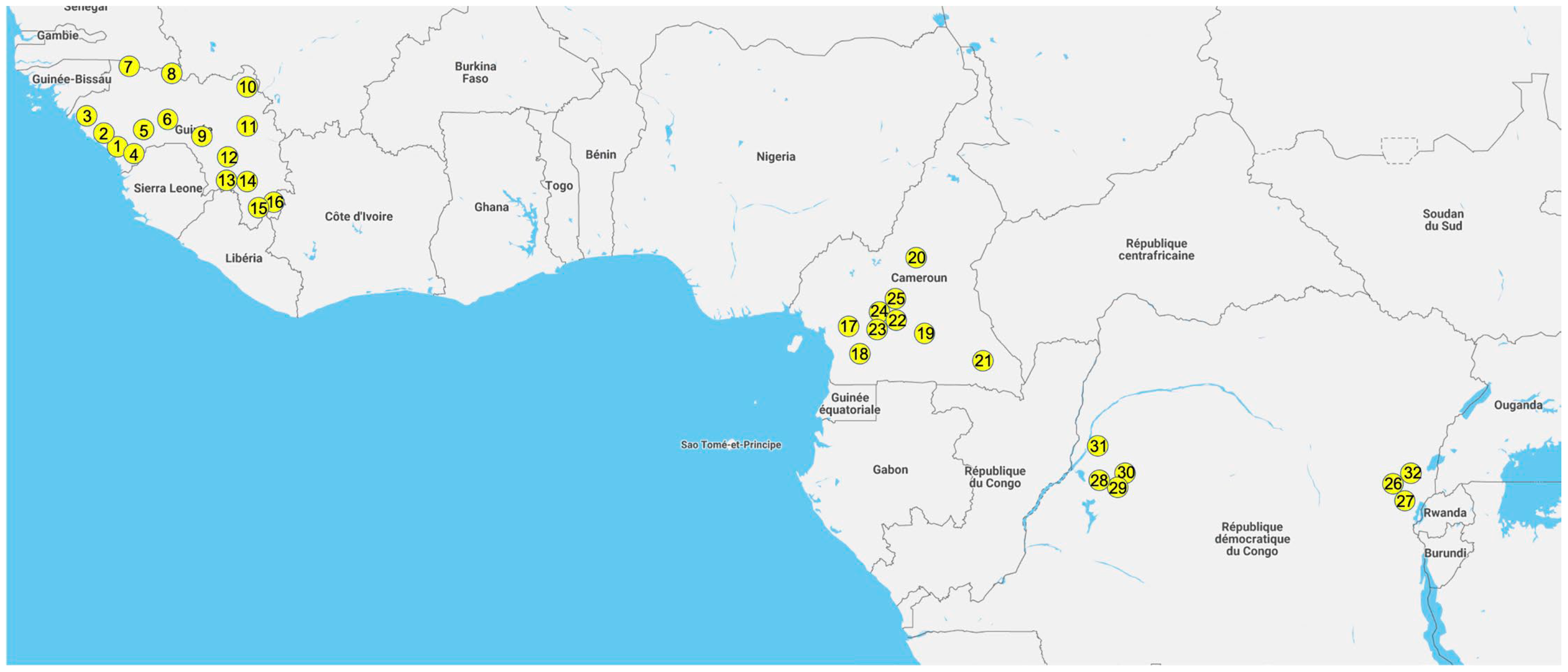
2.1. Study Sites and Sample Collection

Samples were collected from free-ranging frugivorous and insectivorous bats in Guinea, Cameroon and the Democratic Republic of Congo (DRC) between November 2015 and February 2022, as previously described [27]. Briefly, bats were captured using mist nets or harp traps in roosting and foraging sites and released immediately after sampling. Venipuncture was done of the propatagial or brachial vein and blood drops were directly transferred onto Whatman 903 filter paper (GE-Healthcare, Feasterville-Trevose, PA, USA), which were air-dried and preserved individually in plastic bags containing silica desiccant and stored in a hermetic box as dried blood spots (DBS). DBS (a maximum of 2–3 weeks after collection) were subsequently stored frozen (−20 °C) until analysis. Rectal and/or oral swabs were collected and stored in 500 µL RNA-later (Ambion, Austin, TX, USA) in the field at ambient temperature for a maximum of two weeks and subsequently frozen in the laboratory. For each bat that was sampled, information on capture sites (GPS coordinates, ecological environment), capture method, morphology (body measurements, weight, color), sex, age class (adult, juvenile) and visual species identification were recorded. In Cameroon, adults were also classified into subadults and mature adults based on the presence of enlarged testes and/or distended cauda epididymis for males and the development and morphology of the mammary glands and thoracic (axillary) and pubic nipples for females.

2.2. Nucleic Acid Extraction and Screening for the Presence of Coronavirus RNA

Total DNA and RNA were extracted from swabs and feces using the NucliSENS EasyMAG platform (BioMérieux, Marcy-l’Etoile, France) or the Qiagen Viral RNA mini kit (Les Ulis, France). Briefly, for the Nuclisens method, 250 µL of sample was incubated with 2 mL of lysis buffer for 15 min and extraction was performed using the manufacturer’s instructions. The total nucleic acids were resuspended in 60 µL elution buffer. For the Qiagen method, nucleic acids were extracted from 250 µL of sample as per the manufacturer’s instructions. RNA was eluted in 60 µL elution buffer. Extracted RNAs were stored at −80 °C until use. For the detection of coronaviruses, cDNA was first synthesized from denatured RNA (70 °C for 10 min) using a Reverse Transcription System kit with random primers (Promega, Madison, WI, USA), following the manufacturer’s instructions. Broadly reactive degenerate primers were used to amplify a 440 base pair (bp) fragment in the highly conserved RdRp region by nested PCR [28]. Amplification conditions were slightly adapted, as previously described [29]. cDNA was amplified with the GoTaq Hot Start Master Mix PCR kit (Promega, Madison, WI, USA) to obtain a 620 bp fragment in the first round with 40 cycles of 92 °C for 30 s, 48 °C for 30 s and 72 °C for 50 s. Cycling conditions for the second round used a touch-down technique to reduce non-specific amplifications (10 cycles of 92 °C for 30 s, 53 °C for 30 s with −0.5 °C/cycle and 72 °C for 30 s) followed by 35 cycles of 92 °C for 30 s, 53 °C for 30 s and 72 °C for 30 s). The PCR products were visualized by agarose gel electrophoresis. PCR products were directly sequenced with a BigDye Terminator version 3.1 sequencing kit (Life Technologies, Courtaboeuf, France) on an Applied Biosystems 3500 Genetic Analyzer (Thermo Fisher Scientific, Foster City, CA, USA). Sequences from both strands were reconstituted using the SeqMan Pro tool from the package DNAStar v17.0.2 (Lasergene, Madison, WI, USA). The newly obtained RdRp sequences of coronaviruses were then analyzed with the R package MyCoV for rapid classification in subgenera [30]. The MyCoV package is available at https://github.com/dw974/MyCoV (accessed on 19 May 2022).

2.3. Phylogenetic Analyses

The new RdRp sequences were aligned with representatives of the different subgenera of alpha and beta coronaviruses and with sequences reported from bats in Africa from the same regions, when possible. Multiple sequence alignment (MSA) was obtained using MAFFT v7 (https://mafft.cbrc.jp/alignment/server/, accessed on 23 August 2022). The alignment was manually checked and end-trimmed to match the newly obtained RdRp sequences and to remove the PCR primer sequences. The final alignment was used for maximum likelihood (ML) phylogenetic analysis with GTR + F + I + 4Γ as the best-fit model of nucleotide substitution according to BIC and 1000 bootstrap resampling using the IQ-Tree server (http://iqtree.cibiv.univie.aC.at, accessed on 23 August 2022) [31,32]. Consensus trees were edited with FigTree v1.4.4 (http://tree.bio.ed.ac.uk/software/figtree/, accessed on 23 August 2022).

2.4. Molecular Confirmation of Bat Species

Species identification recorded in the field was molecularly confirmed for the majority of bats that were infected with a coronavirus using the corresponding DBS sample, as in our previous studies on viruses in bats [27,33,34]. In addition, at least one sample per sampling date, per capture method and per morphologic description at each site was confirmed. A 800 bp fragment of the mitochondrial cytochrome b (CytB) region was amplified using previously described primers to identify mammal species, including bats, Cytb-L14724 (forward) and Cytb-H15506 (reverse) [35]. To increase PCR specificity for certain bat species, the forward primer was replaced by a newly designed primer Cytb-L1 5′-ATG ACC AAC ATC CGA AAA TCN CAC-3′ or Cytb-L2 5′-ATY TCY TCM TGA TGA AAY TTY GGM T-3′ [34]. For a subset of samples, species identification recorded in the field was confirmed by amplifying a 386-bp mitochondrial DNA fragment of the 12S rRNA gene with primers 12S-L1091 and 12S-H1478 [36]. The PCR products were directly sequenced and reconstructed, as described above. Sequences were pasted in the NCBI BLAST web interface (https://blast.ncbi.nlm.nih.gov/Blast.cgi, accessed on 12 May 2022) to identify the most similar bat species. For samples with no or low similarity (<97%) hits with species in Genbank, a phylogenetic tree was constructed using maximum likelihood methods implemented in PhyML with reference sequences in order to obtain genus identification. Species identification was extrapolated for the remaining samples by combining molecular and field data. For certain insectivorous bats (from Molossidae, Rhinolophidae, Hipposideridae and Nycteridae families), identification was only possible at the genus level, mostly due to the lack of reference sequences in Genbank. For Epomophorus gambiensis and Micropteropus pusillus, species discrimination cannot be done by CytB sequences only, and morphologic details on the forearm and weight measurements were also used to discriminate the species, as previously described [34,37].

2.5. Statistical Analyses

To explore the impact of age, sex and reproductive stage of the collected samples on the detection of coronavirus RNA, we performed Kruskal–Wallis or Chi-2 tests. Significant results were considered for a p−value of <0.05.

3. Results

3.1. Bat Samples

Swabs from a total of 4597 bats were analyzed, 897 from DRC, 2191 from Cameroon and 1509 from Guinea, including 319 bats from a previous pilot study [29]. The samples were collected at various sites across the 3 countries: 16 for Guinea, 9 for Cameroon, and 7 for DRC (Figure 1 and Supplementary Table S1). Bats were captured in different ecological environments: villages (15.5%), forest sites (20.2%), urban sites (17.3%), plantations (7.3%), caves (25.9%) and diverse other settings for the remainder. For a total of 1760 (38.2%) bats, species identification in the field was confirmed by sequence analysis. Details on bat families, genera and species analyzed in this study are shown in Supplementary Table S2. Overall, we analyzed a total of 2986 (65%) frugivorous bats from at least 12 species. Among them, the predominant species were Rousettus aegyptiacus (n = 791), Epomophorus sp. (n = 737) and Eidolon helvum (n = 733), followed by Epomops sp. (n = 258), Micropteropus pusillus (n = 165), Hypsignathus monstrosus (n = 103), Myonycteris torquata (n = 91) and Lissonycteris angolensis (n = 65). Among the 1611 (35%) insectivorous bats, at least 7 different families were represented; the highest number of bats were from the Rhinolophus genus (n = 899), followed by species from the Mollosidae (n = 399) and Hipposideridae (n = 291) families. Marginal numbers of samples, less than 25, were obtained for 4 frugivorous bat species (Casinycteris arginnis (n = 24), Megaloglossus woermanni (n = 15), Nanonycteris sp. (n = 3) and Scotonycteris sp. (n = 1)) and 5 insectivorous bat species (Coleura afra (n = 1), Miniopterus sp. (n = 9), Nycteris sp. (n = 5), Myotis sp. (n = 4) and Scotophilus sp. (n = 3)), which were often only captured in a single country. Among the predominant species, the majority were captured in the three countries, as is the case for E. helvum, Hipposideros genus and representatives of the Mollossidae family. R. aegyptiacus was only captured in Guinea and Cameroon. Epomophorus species were only captured in DRC and Guinea. In addition, in DRC in the East, E. labiatus was captured versus E. gambiensis in Western DRC and Guinea. Concerning the Epomops genus, two species were captured: E. franqueti in DRC and Cameroon and E. buettikoferi in Guinea. Rhinolophus bats were only captured in Cameroon and Guinea.
Overall, 2104 (45.8%) were male and 2480 (53.9%) were female bats; for 13 (0.3%) bats, the sex was not recorded. Only 391 (8.5%) were juvenile bats, 4139 (90.0%) were adults, and for 67 (1.5%), the age class was not available.

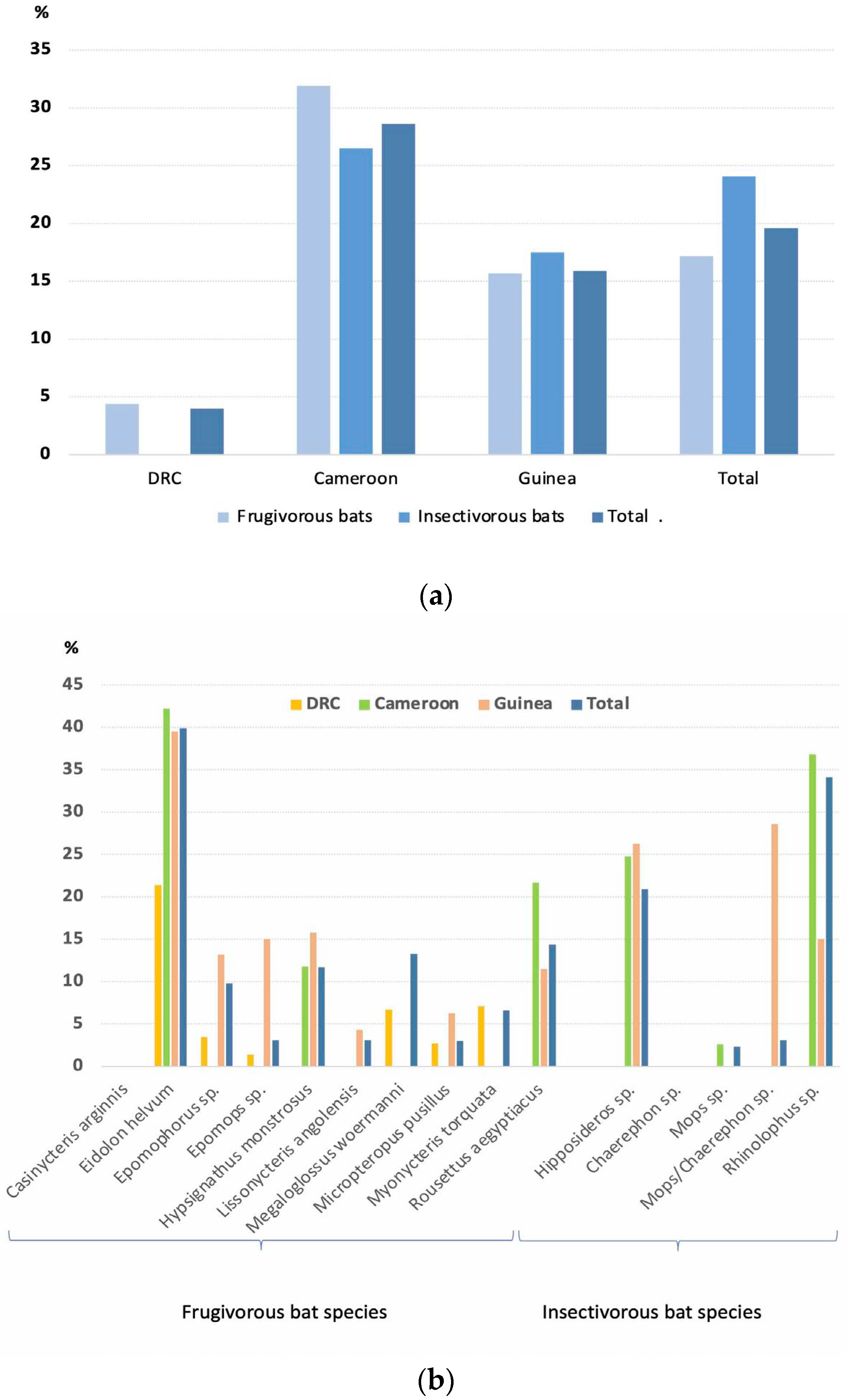
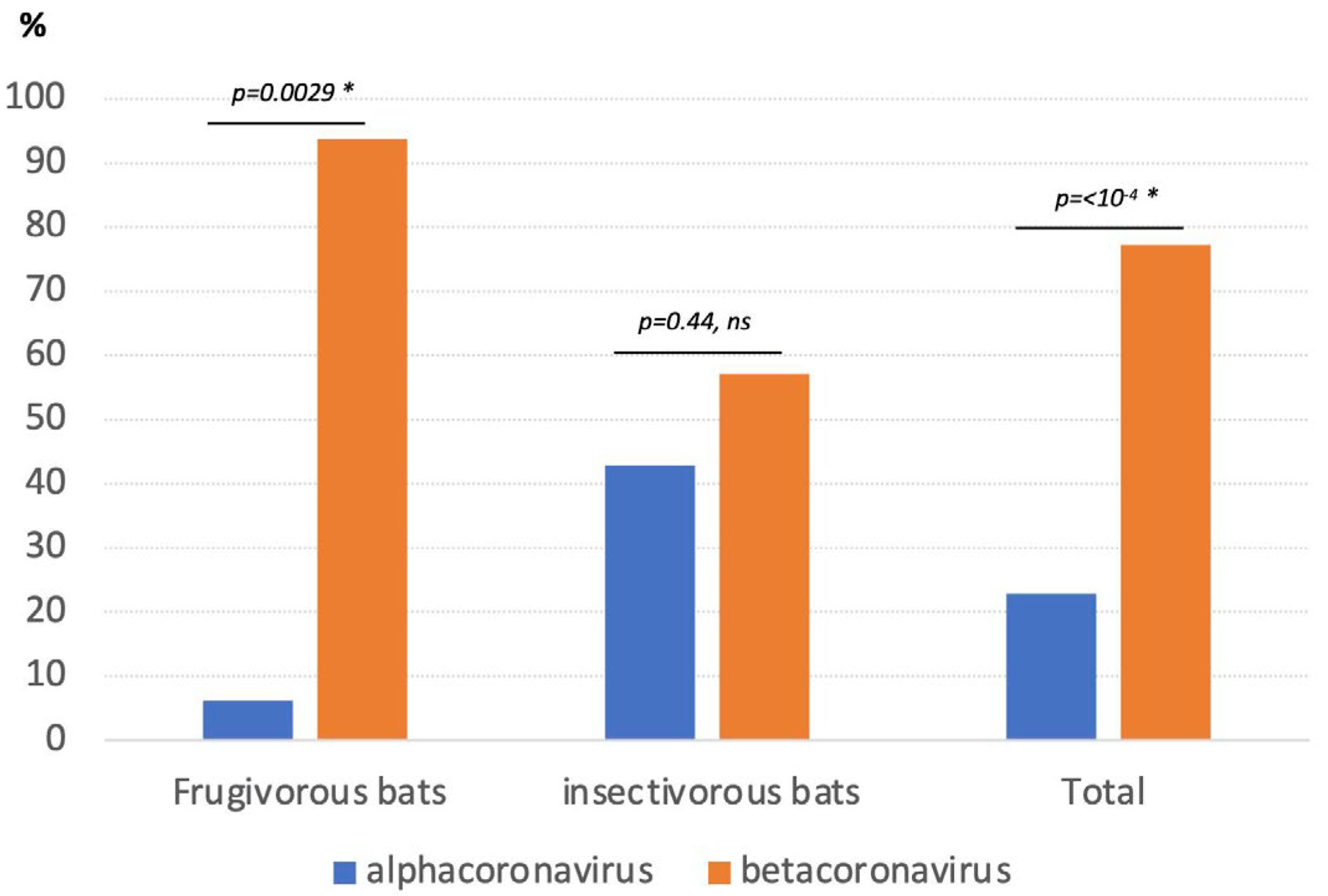
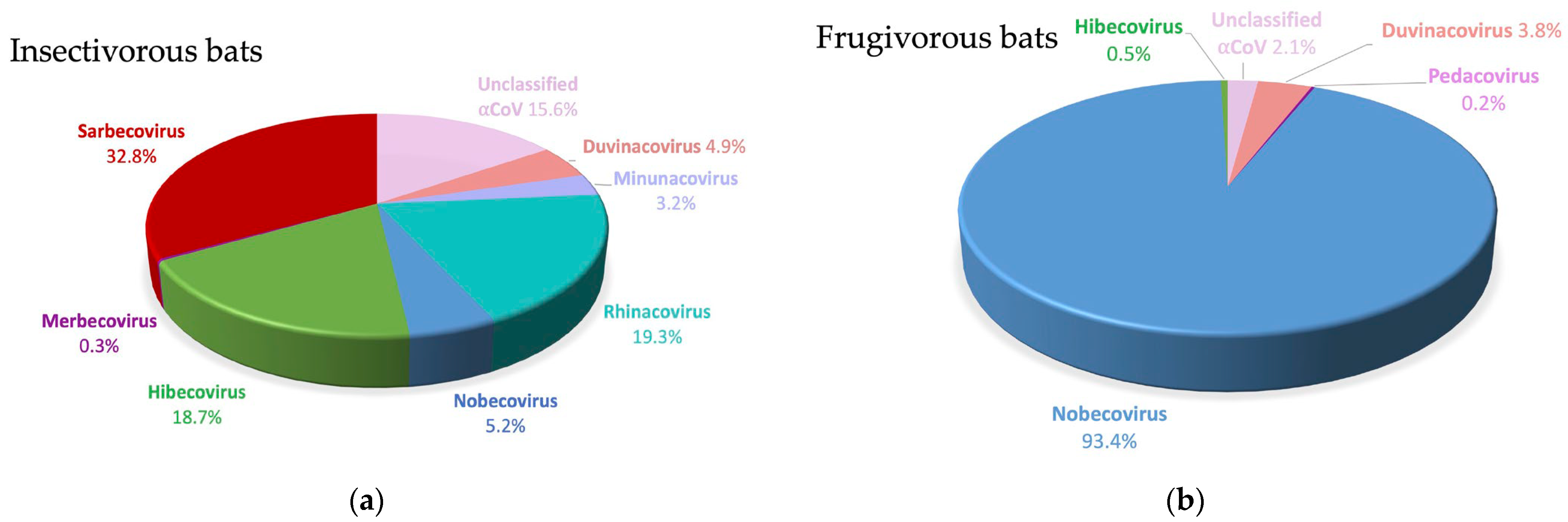
3.2. Detection and Genetic Diversity of Coronaviruses

Among the 4597 bats tested by RT–PCR for the presence of coronavirus RNA, a total of 903 (19.6%) tested positive: 515/2986 (17.2%) of the frugivorous bats and 388/1611 (24.1%) of the insectivorous bats. CoVs were identified in all species, except Casinycteris argynnis, E. buettikoferi and Scotonycteris bergmansi, for which low numbers were tested, 24, 3 and 1, respectively. Positivity rates varied per species and were highest in E. helvum (29/733 (39.9%)), Rhinolophus sp. (306/899 (34.1%) and Hipposideros sp. (61/291 (20.9%)) for which large numbers of samples were collected. Intermediate rates were observed in R. aegyptiacus (114/791 (14.4%), H. monstrosus (12/103 (11.7%)) and Epomophorus sp. (72/737 (9.8%)) and lowest rates were seen in M. torquata (6/91 (6.6%)), Epomops sp. (8/258 (3.1%)), L. angolensis (2/65 (3.1%)), M. pusillus (5/165 (3.0%)) and in bats from the insectivorous Mollossidae family (10/399 (2.5%)). For the other species, sample numbers were low and detection rates were most likely not representative. Figure 2 shows percentages of frugivorous and insectivorous bats that were positive per country (Figure 2a) and per species (Figure 2b), and details are shown in Supplementary Table S2.
Among the 903 PCR-positive samples, 768 (85.2%) were sequenced and revealed an overall predominance of beta-CoVs, 77.2% (593/768) (Supplementary Table S3). Frugivorous bats were predominantly infected with beta-CoVs, which represented 93.8% (395/421) of the sequences versus 57.1% (198/347) for insectivorous bats (Figure 3). Whereas alpha-CoVs were only marginally detected in the majority of frugivorous bat species tested, 21.6% (21/97) sequences from R. aegyptiacus were alpha-CoVs.
As shown in Table 1 and Figure 4, representatives of all subgenera, except Embecovirus, were observed among the beta-CoVs. The vast majority were Nobecoviruses (69.3%), followed by Sarbecoviruses (19.2%), Hibecoviruses (9.6%) and a single sequence from a Nycteris bat in Guinea was classified as a Merbecovirus. Sarbecoviruses were only detected in Rhinolophus species in Cameroon and Guinea. Hibecoviruses were predominantly detected in Hipposideros bats from Cameroon and Guinea, but also in Rhinolophus bats in Cameroon captured in the same cave. Nobecoviruses were identified in all positive frugivorous species in a minority of Hipposideros and Rhinolophus species. All beta-CoVs observed in the Molossidae family belonged to the Nobecovirus subgenus. A wide diversity of alpha-CoVs was also observed, with representatives of Duvinacoviruses (n = 17), Minunacoviruses (n = 11), Rhinacoviruses (n = 67), sequences that are close to Pedacoviruses (n = 1) and Decacoviruses (n = 16) and 63 sequences that do not belong to currently described subgenera. The Duvinacovirus subgenus also harbors the HCoV-229E strain, which causes mild symptoms in humans across the world [38]. Figure 4 illustrates the higher diversity of subgenera observed in insectivorous versus frugivorous bats, i.e., 10 versus 4, respectively.
Phylogenetic analysis of all newly identified sequences with references from the different subgenera and from other studies in Africa showed a high diversity in the Nobecovirus subgenus, with at least 6 different mostly species-specific subclades (Table 1, Figure 5). One major clade contained almost exclusively sequences amplified in Eidolon in Guinea, Cameroon and DRC and other previously published sequences from E. helvum bats from other countries in East, West and Central Africa. Among the 236 beta-CoVs identified in E. helvum bats in our study, 233 fell into this clade. A second clade contains mainly sequences from Epomophorus and Micropteropus bats, as well as from all regions confounded together with previously published sequences from these species. Sequences obtained from the two Epomophorus subspecies fell into the same clade. Sequences obtained from Epomops franqueti from DRC and Epomops buettikoferi bats from Guinea also fell within this clade. A third clade comprises sequences from Lissonycteris and Myonycteris bats from Guinea and DRC, respectively. Coronaviruses from Megaloglossus bats also formed a separate clade in the Nobecovirus subgenus. Rousettus aegyptiacus was infected with two species-specific Nobecovirus clades, one that included the HKU9 strain obtained from a Rousettus bat in Guangdong province in China [39] and a second clade with lower numbers of sequences from R. aegyptiacus bats from Guinea and Cameroon. Additionally, among the Hibecoviruses, a wide diversity was seen among the sequences obtained from the Hipposideros species. Sarbecoviruses were identified in Rhinolophus bats in Cameroon and Guinea.
Among the Sarbecoviruses, two major clades were identified: one with sequences from Asia and the other with sequences from Africa and Europe. The Asian clade was further subdivided into four different lineages (i.e. lineage 1, 2, 3 and 5). The new African Sarbecovirus sequences fell into the previously identified “lineage 4,” containing strains from African and European Rhinolophidae, but the sequences from Cameroon and Guinea each fell into separate clusters within the cluster of strains from African bats (Figure 6) [40]. Moreover, the Sarbecoviruses amplified in Rhinolophus bats from Cameroon formed two separate clusters related to the year of collection. All new Sarbecovirus sequences from West and West Central Africa were different from those observed in bats from East Africa.
In general, each bat species/genus was infected with a predominant alpha- and/or beta-CoV variant independent of their geographic origin, but co-circulation with variants from other CoV subgenera or subclades from the same subgenus was also observed. For example, Eidolon helvum bats from Guinea, Cameroon and DRC were infected with the same Nobecovirus subclade, but a small minority (3/236 sequences) fell in the subclade dominated by Epomophorus and Micropteropus species. On the other hand, Rhinolophus bats were infected with a wide diversity of alpha- and beta-CoVs; i.e., at least 3 different subgenera of alpha-CoVs and 3 subgenera of beta-CoVs, including 3 Nobecovirus subclades (Table 1, Figure 5). R. aegyptiacus bats were also infected with a wide diversity of CoVs, at least 3 Nobecovirus subclades, Hibecoviruses and two alpha CoV subgenera.

3.3. Factors Associated with Detection of Coronaviruses in Bats

We compared whether age or sex could play a role in coronavirus positivity using PCR. Overall, among all bat samples tested, 18.0% (16.5–19.5%; 447/2480) of female and 20.9% (19.1–22.6%; 439/2104) of male bats were positive for coronaviral RNA (Table 2). For frugivorous bats, there was a trend of lower positivity in female bats, 14.7% (13–16.4%) versus 19.4% (17.3–21.5%) (Chi-2; p = 0.0007). This was not homogenous among species; for example, equal proportions of positivity were seen for Epomophorus sp. and Rousettus aegyptiacus bats, and a higher prevalence was seen in male bats for Epomops sp. and Eidolon helvum (Chi-2; p = 0.0197) (Table 2). In insectivorous bats, no overall difference in positivity was seen between female and male bats (Chi-2; p = 0.57), but the results differed according to species and genus, as shown in Table 2.
Prevalence was higher in juvenile bats; i.e., 34% (29.3–38.7%) versus 18.7% (17.5–19.9%) in adult bats (Chi-2; p < 0.0001) (Table 3). This trend was clearly confirmed for frugivorous bats (Chi-2; p < 0.0001) and for the majority of species for which sufficient samples from both age categories were tested, i.e., Eidolon helvum (Chi-2; p < 0.0001), R. aegyptiacus (Chi-2; p = 0.0001) and Rhinolophus sp. (Chi-2; p < 0.0001), except for Epomophorus bats, but the number of juvenile bats tested for this species was low. For insectivorous bats, equal detection rates were observed for juvenile and adult bats, but overall, a low number of juvenile bats was tested. In Cameroon, age determination of bats differentiated between adults and subadults. Supplementary Table S4 shows the percentage of PCR-positive bats for the three different age categories in this country and clearly illustrates that among adults, the prevalence was higher in subadults, i.e., 40.6% (36.0–44.2%) versus 18.9% (16.6–21.3%) in mature adults. Positivity rates in subadults were close to those observed in juvenile bats. This trend was observed in frugivorous and insectivorous bats and for all species with sufficient samples tested in the different age categories, i.e., Eidolon helvum, Rousettus aegyptiacus and Rhinolophus sp.
We also looked more in detail at whether the prevalence of coronaviruses differed in adult females according to the reproductive cycle, i.e., gestation or lactation. We observed no significant difference for gestating bats, 23.4% versus 21.2% (Chi-2; p = 0.09), but the detection rate of coronaviruses was lower in lactating females (11.8% (4.9–18.6%)) than in non-lactating bats (22.7% (18.6–26.7%)) (Chi-2; p = 0.035) (Table 4).

4. Discussion

We conducted a large survey to study the prevalence and genetic diversity of coronaviruses in almost 4600 bats from West and West Central Africa and provide significant new information on coronaviruses in bats from Guinea, West Africa and on species that were under-sampled in previous surveys in Cameroon and DRC, such as Epomophorus species in DRC or Hypsignatus monstrosus, Mops and Rhinolophus species in Cameroon [38,41]. Overall, we report a high prevalence of coronaviruses, with 19.6% of the bats that were infected. Prevalence can vary among species, but coronaviruses have been detected in almost all species for which sufficient samples have been tested. A prevalence above 30% was observed in the widely present frugivorous Eidolon helvum but also in Rhinolophus bats, for which high numbers have been tested. We also identified a wide diversity of coronaviruses, with representatives of almost all known subgenera of alpha- and beta-CoVs and identified a subset of strains that cannot be classified in the alpha-CoV genus. We detected bat CoVs (n = 199) that belong to 3 subgenera that also harbor HCoV strains known to be pathogenic for humans, i.e., Sarbecoviruses, Merbecoviruses and Duvinacoviruses. This is also the first study that shows that Sarbecoviruses are widely present in Rhinolophus bats in Africa, representing 114 of the 275 sequences (41.4%) retrieved from the 306 positive Rhinolophus bats that have been detected in this bat genus in our study.
As previously reported, we observed host specificity at the genus and/or species levels [26]. Nobecoviruses are largely predominant in frugivorous bats, with six major subclades that are associated with species or genus, as previously reported [26]. Nevertheless, some of these frugivorous species are also sporadically infected with Nobecoviruses from another subclade. We also show for the first time that Hypsignathus monstrosus are infected with Nobecoviruses, and mainly with the Eidolon helvum-specific clade, most likely because they roost in the same fruit trees, as is the case in Yaoundé, Cameroon, where the majority of them have been sampled and where a large colony of Eidolon helvum bats is present. Representatives of the Hibecovirus subgenus have been identified mainly in Hipposideros bats but also in a significant proportion of Rhinolophus bats in Cameroon, especially in those that share habitats in a cave located in the southwest, thus confirming host switching of coronaviruses among bat species that share habitats, as previously reported [19].
Sarbecoviruses are widespread in Rhinolophus bats in Asia and Europe and phylogeographic clustering has been reported in Asian Rhinolophus bats, which have been largely studied because Chinese Rhinolophidae are the natural reservoir for Sarbecoviruses [42,43,44]. Five subclades or lineages have been described for Sarbecoviruses by Yu et al. [43], with lineage 1 including SARS-CoV-1. Bat Sarbecovirus strains from Europe and Africa have been classified as lineage 4 and SARS-CoV-2 and other related viruses from Rhinolophus bats and pangolins from Asia fall into lineage 5 [40,45,46,47,48,49,50]. The Sarbecoviruses that we observed in Rhinolophus bats from Cameroon and Guinea fall into lineage 4 but form separate clusters with the other Sarbecoviruses identified in Rhinolophus bats from East Africa (Rwanda, Kenya and Uganda), thus illustrating phylogeographic clustering of African Sarbecoviruses. Nevertheless, Sarbecoviruses from Asian lineages 1 and 5 co-circulate in the same geographic area and are at the origin of SARS-CoV-1 and 2, respectively. This highlights that more sampling has to be done in Rhinolophidae in Asia, but also in Africa to examine further genetic diversity, geographic clustering and co-circulation of Sarbecovirus lineages. More studies are also needed to confirm whether all African strains are unable to utilize human ACE2 and are not able to infect human cells, as shown by in vitro studies [40]. However, it cannot be excluded whether the Sarbecoviruses from Africa use a different receptor. The colony of Rhinolophus bats infected at high rates (34%) with Sarbecoviruses has been identified in Cameroon in a cave that is frequently used by hunters as shelter for protection during bad weather conditions (rain) and where Rousettus aegyptiacus bats are hunted during the whole year for consumption (Ndong Bass, personal observation), which implies thus that humans might be in contact with guano (bat feces) dropped on the cave’s floor or with air saturated with contaminated aerosols. Our exploratory study on knowledge, attitudes and practices in Southern Cameroon showed that surrounding villages did not exploit bat guano as fertilizer [22], in contrast to what was reported, for example, in Zimbabwe [51].
Additionally, for alpha-CoVs, species-specific clades are seen; for example, Rhinacovirus and a separate clade of unclassified viruses were predominantly observed in Rhinolophus bats and Duvinacoviruses in Hipposideros bats. Nevertheless, Rhinolophus, Hipposideros and Rousettus aegyptiacus bats are infected with at least 3 to 6 different subgenera of alpha- and beta-CoVs, illustrating host switching most likely because they roost together in the same caves. Among the frugivorous bats, only R. aegyptiacus is infected to some extent with alphacoronaviruses, representing around 20% of the infections in this species. The majority belong to the Decacovirus subgenus, which includes the HKU10 strain identified in a Rousettus bat from China [52].
We also confirmed that juvenile and subadult bats are infected at higher rates than adult bats, as previously reported [19,53]. This was observed in all species for which sufficient juvenile and/or subadults have been included. We observed a trend in which males are more infected than females, especially in frugivorous bats, but this was heterogeneous according to the different species and could be due to sample bias. In our study, the prevalence of coronavirus infection was not different between gestating and non-gestating adult females, but in contrast to other reports, we observed lower rates in lactating females [54,55,56]. This difference could be due to different sample numbers or the predominance of different species. Apparently, the impact of some factors such as sex, gestation or lactation are not universal for all bat species [26]. However, in our study, we did not have enough samples from lactating bats for each species to evaluate species-specific differences. Overall, our study and others clearly show that high-risk periods for coronavirus shedding are related to age of bats and are highest when juvenile and subadults are present in large numbers in the colonies. Importantly, this period is specific for each bat species and can differ according to geographic sites and must therefore be evaluated for each species separately and in different areas to estimate spillover risk.
It is clear that humans are exposed to a wide diversity of bat coronaviruses in Africa, either directly through hunting and consumption or indirectly via contact with feces, urine or saliva on fruit or via guano. It will be important to evaluate to what extent some of these viruses can cross the species barrier or already cross the species barrier and lead to mild or asymptomatic infections. Serological assays with antigens from the different subgenera of alpha- and beta-CoVs need to be developed to evaluate this. Pre-existing immunity to other coronaviruses can also be one of the explanations for the lower morbidity of SARS-CoV-2 infections in Africa. This could be the case for coronaviruses that circulate at high rates in Eidolon helvum, which are one of the most common bats in Africa and roost in fruit trees in gardens of houses in cities and villages. For MERS-CoV, it is clear that multiple spill-over events occurred, but with limited subsequent spread in humans [57]. Although difficult to realize in practice given the mild symptoms and short viremic period, but given the high frequency of HCoV-H229E-related strains in bats, as observed in our and previous studies [38,58], molecular characterization of these or other mild coronaviruses in humans would be of interest to evaluate whether spillover events are still ongoing or whether the mild viruses, such as HCoV-229E, are the result of a single cross-species event.
Overall, we documented the high prevalence and genetic diversity of coronaviruses in different bat species from West and West Central Africa, especially in juveniles and subadults. More studies are needed to identify the high-risk periods, i.e., presence of high proportions of juvenile and subadult bats, to evaluate risk for spillover events for the different bat species. Importantly, our study shows that certain Rhinolophus colonies are highly infected with Sarbecoviruses and it can thus not completely be excluded that a spillover event could occur and lead to another SARS-CoV variant in humans. More cross-sectional and longitudinal studies are needed to document the extent of Sarbecovirus infections in Rhinolophus colonies in Africa to identify periods and environments in which spillover to humans is at higher risk. More studies on human behavior and diversity of interactions with bats are also needed in addition to viral characterization, as well as the development of specific antibody assays to evaluate past spillover events or to evaluate risk for future events. Finally, our study used only a small fragment in the RdRp region, which has been shown to be coherent with the classification of whole genome sequences for the large majority of strains, as shown by Wilkinson and colleagues [30]. Nevertheless, this classification has some limits, such as the absence of detection of recombinant lineages and provides no information on co-receptor use or factors associated with the evolution of these viruses in general [40,59]. Therefore, it is important to increase efforts to obtain more whole genome sequences from coronaviruses that circulate in bats in order to better document the diversity and evolution of these viruses in their natural hosts and their potential to infect humans.

Supplementary Materials

The following supporting information can be downloaded at: https://www.mdpi.com/article/10.3390/v15020337/s1, Supplementary Tables S1–S6: Table S1: Number of samples collected (N) at the different geographic locations in each country. The location of the different sites is visualized in Figure 1; Table S2: Number (n) and percentages (%) of bat genus/species positive for coronaviruses per country and per species; Table S3: Number (n) and proportion (%) of alpha and beta coronaviruses per bat genus/species, and p−values resulting from comparison of the proportions between alpha and betacoronavirus per genus/species, when applicable (Chi-2 test); Table S4: Number of bats positive for coronaviruses (n pos) on total number tested (N) and percentages (%) of samples per bat genus/species in Cameroon per age category (adults (A), immature adults (imm) and juveniles (J) and per species). The p−values result from comparison of the proportions of positive per genus/species, when applicable; Table S5: Genbank accession numbers and information on bat hosts for the new sequences obtained in this study; Table S6: Accession numbers and details of reference strains from alpha and betacoronavirus subgenera used in the phylogenetic tree in Figure 5.

Author Contributions

Conceptualization, A.A., E.M.N., E.D., A.K.K., S.A.-M. and M.P.; methodology, A.L., E.M.N., A.K.K., S.A.-M., A.A. and M.P.; validation, A.L., A.A., A.K.K., S.A.-M. and M.P.; formal analysis, A.A., A.L. and M.P.; investigation, D.M.D., A.L., A.K.S., E.K.L., A.M., R.R., A.E., I.N.B., F.A.M.D., S.G., S.P.N.-K., G.L., P.M.K., D.M.B. and J.L.; data curation, M.P., A.L. and A.K.K.; writing—original draft preparation, M.P.; writing—review and editing, D.M.D., A.L., A.A., E.D. and M.P.; supervision, A.L., A.A., A.T., A.K.K., S.A.-M., C.K. and M.P., project administration, E.M.N., C.K., A.T., J.-J.M.T., S.A.-M., E.D. and M.P.; funding acquisition, E.D. and M.P. All authors have read and agreed to the published version of the manuscript.

Funding

This work was supported by grants of the Agence Nationale de Recherche sur le SIDA et les Maladies Infectieuses (ANRS-COV10), EBO-SURSY project funded by the European Union (FOOD/2016/379-660), and BIODIV-AFREID project (BiodivERsA ERA-Net COFUND programme), 2018–2019 joint call for proposals (grant number ANR-19-EBI3-0004). Eddy Lusamaki was supported by a doctoral fellowship from the French foreign office.

Institutional Review Board Statement

The study was conducted in compliance with the national ethics committee from the DRC (ESP/CE/009/2020), Guinea (approval reference 074/CNERS/15, 26 November 2015) and Cameroon (N°2018/09/1090/CE/CNERSH/SP).

Informed Consent Statement

Not applicable.

Data Availability Statement

Data are available upon request, and data on bat sampling are available on the EBO-SURSY website (https://rr-africa.woah.org/en/projects/ebo-sursy-en/, accessed on 20 November 2022). All new RdRp coronavirus sequences are available in GenBank with the following accession numbers: OP773878 to OP774616.

Acknowledgments

We thank the Ministries of Health, Environment and the National Ethic committees from Guinea, Cameroon and The Democratic Republic of Congo for permission to perform this study. We thank all field staff from DRC for the collection of bat samples, and we also thank the provincial authorities of Equateur, North Kivu and Ituri for the assistance in field missions. We thank all the field staff from Guinea for the collection of bat samples. We thank Mamady Doumbouya, National Director of Veterinary Services of Guinea for his involvement and support in the implementation of the project. We thank Seny Mane and Daouda Bangoura, former National Directors of Guinean Veterinary Services and OMSA focal point in Guinea and Colonel Bhoye Sow, former Deputy Director General of the Guinean Office of Parks and Reserves for their constant support to the project and the facilitation of field missions. We thank the staff of CREMER (Joseph Moudindo, Aime Mebanga, Thomas Atemkeng) for logistical support in the field in Cameroon.

Conflicts of Interest

The authors declare no conflict of interest.

References

  1. World Health Organization (WHO). WHO Coronavirus (COVID-19) Dashboard. Available online: https://covid19.who.int (accessed on 19 October 2022).
  2. Chisale, M.R.O.; Ramazanu, S.; Mwale, S.E.; Kumwenda, P.; Chipeta, M.; Kaminga, A.C.; Nkhata, O.; Nyambalo, B.; Chavura, E.; Mbakaya, B.C. Seroprevalence of anti-SARS-CoV-2 antibodies in Africa: A systematic review and meta-analysis. Rev. Med. Virol. 2022, 32, e2271. [Google Scholar] [CrossRef]
  3. Ndongo, F.A.; Guichet, E.; Mimbé, E.D.; Ndié, J.; Pelloquin, R.; Varloteaux, M.; Esemu, L.; Mpoudi-Etame, M.; Lamare, N.; Edoul, G.; et al. Rapid increase of community SARS-CoV-2 seroprevalence during second wave of COVID-19; Yaoundé; Cameroon. Emerg. Infect. Dis. 2022, 28, 1233–1236. [Google Scholar] [CrossRef]
  4. Nkuba, A.N.; Makiala, S.M.; Guichet, E.; Tshiminyi, P.M.; Bazitama, Y.M.; Yambayamba, M.K.; Kazenza, B.M.; Kabeya, T.M.; Matungulu, E.B.; Baketana, L.K.; et al. High prevalence of anti-Severe Acute Respiratory Syndrome Coronavirus 2 (Anti-SARS-CoV-2) antibodies after the first wave of Coronavirus Disease 2019 (COVID-19) in Kinshasa; Democratic Republic of the Congo: Results of a cross-sectional household-based survey. Clin. Infect. Dis. 2022, 74, 882–890. [Google Scholar] [CrossRef]
  5. Soumah, A.A.; Diallo, M.S.K.; Guichet, E.; Maxmlman, D.; Thaurignac, G.; Keita, A.K.; Bouillin, J.; Diallo, H.; Pelloquin, R.; Ayouba, A.; et al. High and rapid increase in seroprevalence for SARS-CoV-2 in Conakry; Guinea: Results from 3 successive cross-sectional surveys (ANRS COV16-ARIACOV). Open Forum Infect. Dis. 2022, 9, ofac152. [Google Scholar] [CrossRef]
  6. Worobey, M.; Levy, J.I.; Serrano, L.M.; Crits-Christoph, A.; Pekar, J.E.; Goldstein, S.A.; Rasmussen, A.L.; Kraemer, M.U.G.; Newman, C.; Koopmans, M.P.G.; et al. The Huanan seafood wholesale market in Wuhan was the early epicenter of the COVID-19 pandemic. Science 2022, 377, 951–959. [Google Scholar] [CrossRef]
  7. Su, S.; Wong, G.; Shi, W.; Liu, J.; Lai, A.C.K.; Zhou, J.; Liu, W.; Bi, Y.; Gao, G.F. Epidemiology; Genetic Recombination; and Pathogenesis of Coronaviruses. Trends Microbiol. 2016, 24, 490–502. [Google Scholar] [CrossRef] [Green Version]
  8. Ye, Z.W.; Yuan, S.; Yuen, K.S.; Fung, S.Y.; Chan, C.P.; Jin, D.Y. Zoonotic origins of human coronaviruses. Int. J. Biol. Sci. 2020, 16, 1686–1697. [Google Scholar] [CrossRef] [Green Version]
  9. Guan, Y.; Zheng, B.J.; He, Y.Q.; Liu, X.L.; Zhuang, Z.X.; Cheung, C.L.; Luo, S.W.; Li, P.H.; Zhang, L.J.; Guan, Y.J.; et al. Isolation and characterization of viruses related to the SARS coronavirus from animals in southern China. Science 2003, 302, 276–278. [Google Scholar] [CrossRef] [Green Version]
  10. Lau, S.K.; Woo, P.C.; Li, K.S.; Huang, Y.; Tsoi, H.W.; Wong, B.H.; Wong, S.S.; Leung, S.Y.; Chan, K.H.; Yuen, K.Y. Severe acute respiratory syndrome coronavirus-like virus in Chinese horseshoe bats. Proc. Natl. Acad. Sci. USA 2005, 102, 14040–14045. [Google Scholar] [CrossRef] [Green Version]
  11. Li, W.; Shi, Z.; Yu, M.; Ren, W.; Smith, C.; Epstein, J.H.; Wang, H.; Crameri, G.; Hu, Z.; Zhang, H.; et al. Bats are natural reservoirs of SARS-like coronaviruses. Science 2005, 310, 676–679. [Google Scholar] [CrossRef]
  12. Luo, C.M.; Wang, N.; Yang, X.L.; Liu, H.Z.; Zhang, W.; Li, B.; Hu, B.; Peng, C.; Geng, Q.B.; Zhu, G.J.; et al. Discovery of novel bat coronaviruses in South China that use the same receptor as Middle East Respiratory Syndrome Coronavirus. J. Virol. 2018, 92, e00116-18. [Google Scholar] [CrossRef] [Green Version]
  13. Chan, J.F.; Lau, S.K.; To, K.K.; Cheng, V.C.; Woo, P.C.; Yuen, K.Y. Middle East respiratory syndrome coronavirus: Another zoonotic betacoronavirus causing SARS-like disease. Clin. Microbiol. Rev. 2015, 28, 465–522. [Google Scholar] [CrossRef] [Green Version]
  14. Corman, V.M.; Ithete, N.L.; Richards, L.R.; Schoeman, M.C.; Preiser, W.; Drosten, C.; Drexler, J.F. Rooting the phylogenetic tree of Middle East Respiratory Syndrome Coronavirus by characterization of a conspecific virus from an African bat. J. Virol. 2014, 88, 11297–11303. [Google Scholar] [CrossRef] [Green Version]
  15. Cui, J.; Li, F.; Shi, Z.L. Origin and evolution of pathogenic coronaviruses. Nat. Rev. Microbiol. 2019, 17, 181–192. [Google Scholar] [CrossRef] [Green Version]
  16. Lam, T.T.; Jia, N.; Zhang, Y.W.; Shum, M.H.; Jiang, J.F.; Zhu, H.C.; Tong, Y.G.; Shi, Y.X.; Ni, X.B.; Liao, Y.S.; et al. Identifying SARS-CoV-2-related coronaviruses in Malayan pangolins. Nature 2020, 583, 282–285. [Google Scholar] [CrossRef] [Green Version]
  17. Zhou, P.; Yang, X.L.; Wang, X.G.; Hu, B.; Zhang, L.; Zhang, W.; Si, H.R.; Zhu, Y.; Li, B.; Huang, C.L.; et al. A pneumonia outbreak associated with a new coronavirus of probable bat origin. Nature 2020, 579, 270–273. [Google Scholar] [CrossRef] [Green Version]
  18. Pekar, J.E.; Magee, A.; Parker, E.; Moshiri, N.; Izhikevich, K.; Havens, J.L.; Gangavarapu, K.; Malpica Serrano, L.M.; Crits-Christoph, A.; Matteson, N.L.; et al. The molecular epidemiology of multiple zoonotic origins of SARS-CoV-2. Science 2022, 377, 960–966. [Google Scholar] [CrossRef]
  19. Anthony, S.J.; Johnson, C.K.; Greig, D.J.; Kramer, S.; Che, X.; Wells, H.; Hicks, A.L.; Joly, D.O.; Wolfe, N.D.; Daszak, P.; et al. Global patterns in coronavirus diversity. Virus Evol. 2017, 3, vex012. [Google Scholar] [CrossRef] [Green Version]
  20. Burgin, C.J.; Colella, J.P.; Kahn, P.L.; Upham, N.S. How many species of mammals are there? J. Mammal. 2018, 99, 1–14. [Google Scholar] [CrossRef] [Green Version]
  21. Frick, W.F.; Kingston, T.; Flanders, J. A review of the major threats and challenges to global bat conservation. Ann. N. Y. Acad. Sci. 2019, 1469, 5–25. [Google Scholar] [CrossRef]
  22. Baudel, H.; De Nys, H.; Mpoudi Ngole, E.; Peeters, M.; Desclaux, A. Understanding Ebola virus and other zoonotic transmission risks through human-bat contacts: Exploratory study on knowledge; attitudes and practices in Southern Cameroon. Zoonoses Public Health 2019, 66, 288–295. [Google Scholar] [CrossRef]
  23. Mickleburgh, S.; Waylen, K.; Racey, P. Bats as bushmeat: A global review. Oryx 2009, 43, 217–234. [Google Scholar] [CrossRef] [Green Version]
  24. Kamins, A.O.; Restif, O.; Ntiamoa-Baidu, Y.; Suu-Ire, R.; Hayman, D.T.; Cunningham, A.A.; Wood, J.L.; Rowcliffe, J.M. Uncovering the fruit bat bushmeat commodity chain and the true extent of fruit bat hunting in Ghana; West Africa. Biol. Conserv. 2011, 144, 3000–3008. [Google Scholar] [CrossRef] [Green Version]
  25. Anti, P.; Owusu, M.; Agbenyega, O.; Annan, A.; Badu, E.K.; Nkrumah, E.E.; Tschapka, M.; Oppong, S.; Adu-Sarkodie, Y.; Drosten, C. Human-bat interactions in rural West Africa. Emerg. Infect. Dis. 2015, 21, 1418–1421. [Google Scholar] [CrossRef] [Green Version]
  26. Geldenhuys, M.; Mortlock, M.; Epstein, J.H.; Pawęska, J.T.; Weyer, J.; Markotter, W. Overview of bat and wildlife coronavirus surveillance in Africa: A framework for global investigations. Viruses 2021, 13, 936. [Google Scholar] [CrossRef]
  27. De Nys, H.M.; Kingebeni, P.M.; Keita, A.K.; Butel, C.; Thaurignac, G.; Villabona-Arenas, C.J.; Lemarcis, T.; Geraerts, M.; Vidal, N.; Esteban, A.; et al. Survey of Ebola viruses in frugivorous and insectivorous bats in Guinea; Cameroon; and the Democratic Republic of the Congo; 2015-2017. Emerg. Infect. Dis. 2018, 24, 2228–2240. [Google Scholar] [CrossRef]
  28. Chu, D.K.; Leung, C.Y.; Gilbert, M.; Joyner, P.H.; Ng, E.M.; Tse, T.M.; Guan, Y.; Peiris, J.S.; Poon, L.L. Avian coronavirus in wild aquatic birds. J. Virol. 2011, 85, 12815–12820. [Google Scholar] [CrossRef] [Green Version]
  29. Lacroix, A.; Vidal, N.; Keita, A.K.; Thaurignac, G.; Esteban, A.; De Nys, H.; Diallo, R.; Toure, A.; Goumou, S.; Soumah, A.K.; et al. Wide diversity of coronaviruses in frugivorous and insectivorous bat species: A pilot study in Guinea; West Africa. Viruses 2020, 12, 855. [Google Scholar] [CrossRef]
  30. Wilkinson, D.A.; Joffrin, L.; Lebarbenchon, C.; Mavingui, P. Analysis of partial sequences of the RNA-dependent RNA polymerase gene as a tool for genus and subgenus classification of coronaviruses. J. Gen. Virol. 2020, 101, 1261–1269. [Google Scholar] [CrossRef]
  31. Trifinopoulos, J.; Nguyen, L.T.; von Haeseler, A.; Minh, B.Q. W-IQ-TREE: A fast online phylogenetic tool for maximum likelihood analysis. Nucleic Acids Res. 2016, 44, W232–W235. [Google Scholar] [CrossRef] [Green Version]
  32. Minh, B.Q.; Hahn, M.W.; Lanfear, R. New methods to calculate concordance factors for phylogenomic datasets. Mol. Biol. Evol. 2020, 37, 2727–2733. [Google Scholar] [CrossRef] [PubMed]
  33. Raulino, R.; Thaurignac, G.; Keita, A.K.; Esteban, A.; Goumou, S.; Diallo, R.; Ndimbo-Kumugo, S.P.; Ndong Bass, I.; Mbala Kingebeni, P.; Toure, A.; et al. Seroprevalence of IgG antibodies against multiple arboviruses in bats from Cameroon; Guinea; and the Democratic Republic of Congo. Vector Borne Zoonotic Dis. 2022, 22, 252–262. [Google Scholar] [CrossRef] [PubMed]
  34. Lacroix, A.; Mbala Kingebeni, P.; Ndimbo Kumugo, S.P.; Lempu, G.; Butel, C.; Serrano, L.; Vidal, N.; Thaurignac, G.; Esteban, A.; Mukadi Bamuleka, D.; et al. Investigating the circulation of Ebola viruses in bats during the Ebola virus disease outbreaks in the Equateur and North Kivu provinces of the Democratic Republic of Congo from 2018. Pathogens 2021, 10, 557. [Google Scholar] [CrossRef] [PubMed]
  35. Irwin, D.M.; Kocher, T.D.; Wilson, A.C. Evolution of the Cytochrome b Gene of Mammals. J. Mol. Evol. 1991, 32, 128–144. [Google Scholar] [CrossRef] [PubMed]
  36. van der Kuyl, A.C.; Kuiken, C.L.; Dekker, J.T.; Goudsmit, J. Phylogeny of African monkeys based upon mitochondrial 12S rRNA sequences. J. Mol. Evol. 1995, 40, 173–180. [Google Scholar] [CrossRef]
  37. Nesi, N.; Nakouné, E.; Cruaud, C.; Hassanin, A. DNA barcoding of African fruit bats (Mammalia; Pteropodidae). The mitochondrial genome does not provide a reliable discrimination between Epomophorus gambianus and Micropteropus pusillus. Comptes R. Biol. 2011, 334, 544–554. [Google Scholar] [CrossRef]
  38. Ntumvi, N.F.; Ndze, V.N.; Gillis, A.; Le Doux Diffo, J.; Tamoufe, U.; Takuo, J.M.; Mouiche, M.M.M.; Nwobegahay, J.; LeBreton, M.; Rimoin, A.W.; et al. Wildlife in Cameroon harbor diverse coronaviruses; including many closely related to human coronavirus 229E. Virus Evol. 2022, 8, veab110. [Google Scholar] [CrossRef] [PubMed]
  39. Woo, P.C.; Wang, M.; Lau, S.K.; Xu, H.; Poon, R.W.; Guo, R.; Wong, B.H.; Gao, K.; Tsoi, H.W.; Huang, Y.; et al. Comparative analysis of twelve genomes of three novel group 2c and group 2d coronaviruses reveals unique group and subgroup features. J. Virol. 2007, 81, 1574–1585. [Google Scholar] [CrossRef] [PubMed] [Green Version]
  40. Wells, H.L.; Letko, M.; Lasso, G.; Ssebide, B.; Nziza, J.; Byarugaba, D.K.; Navarrete-Macias, I.; Liang, E.; Cranfield, M.; Han, B.A.; et al. The evolutionary history of ACE2 usage within the coronavirus subgenus Sarbecovirus. Virus Evol. 2021, 7, veab007. [Google Scholar] [CrossRef]
  41. Kumakamba, C.; Niama, F.R.; Muyembe, F.; Mombouli, J.V.; Kingebeni, P.M.; Nina, R.A.; Lukusa, I.N.; Bounga, G.; N’Kawa, F.; Nkoua, C.G.; et al. Coronavirus surveillance in wildlife from two Congo basin countries detects RNA of multiple species circulating in bats and rodents. PLoS ONE 2021, 16, e0236971. [Google Scholar] [CrossRef]
  42. Hu, B.; Zeng, L.P.; Yang, X.L.; Ge, X.Y.; Zhang, W.; Li, B.; Xie, J.Z.; Shen, X.R.; Zhang, Y.Z.; Wang, N.; et al. Discovery of a rich gene pool of bat SARS-related coronaviruses provides new insights into the origin of SARS coronavirus. PLoS Pathog. 2017, 13, e1006698. [Google Scholar] [CrossRef]
  43. Yu, P.; Hu, B.; Shi, Z.L.; Cui, J. Geographical structure of bat SARS-related coronaviruses. Infect. Genet. Evol. 2019, 69, 224–229. [Google Scholar] [CrossRef] [PubMed]
  44. Wong, A.C.P.; Li, X.; Lau, S.K.P.; Woo, P.C.Y. Global epidemiology of bat coronaviruses. Viruses 2019, 11, 174. [Google Scholar] [CrossRef] [PubMed] [Green Version]
  45. Ar Gouilh, M.; Puechmaille, S.J.; Diancourt, L.; Vandenbogaert, M.; Serra-Cobo, J.; Lopez Roïg, M.; Brown, P.; Moutou, F.; Caro, V.; Vabret, A.; et al. SARS-CoV related Betacoronavirus and diverse Alphacoronavirus members found in western old-world. Virology 2018, 517, 88–97. [Google Scholar] [CrossRef]
  46. Lecis, R.; Mucedda, M.; Pidinchedda, E.; Pittau, M.; Alberti, A. Molecular identification of Betacoronavirus in bats from Sardinia (Italy): First detection and phylogeny. Virus Genes 2019, 55, 60–67. [Google Scholar] [CrossRef] [PubMed] [Green Version]
  47. Drexler, J.F.; Gloza-Rausch, F.; Glende, J.; Corman, V.M.; Muth, D.; Goettsche, M.; Seebens, A.; Niedrig, M.; Pfefferle, S.; Yordanov, S.; et al. Genomic characterization of severe acute respiratory syndrome-related coronavirus in European bats and classification of coronaviruses based on partial RNA-dependent RNA polymerase gene sequences. J. Virol. 2010, 84, 11336–11349. [Google Scholar] [CrossRef] [PubMed] [Green Version]
  48. Rihtaric, D.; Hostnik, P.; Steyer, A.; Grom, J.; Toplak, I. Identification of SARS-like coronaviruses in horseshoe bats (Rhinolophus hipposideros) in Slovenia. Arch. Virol. 2010, 155, 507–514. [Google Scholar] [CrossRef] [PubMed]
  49. Lelli, D.; Papetti, A.; Sabelli, C.; Rosti, E.; Moreno, A.; Boniotti, M.B. Detection of coronaviruses in bats of various species in Italy. Viruses 2013, 5, 2679–2689. [Google Scholar] [CrossRef] [PubMed] [Green Version]
  50. Markotter, W.; Geldenhuys, M.; Jansen van Vuren, P.; Kemp, A.; Mortlock, M.; Mudakikwa, A.; Nel, L.; Nziza, J.; Paweska, J.; Weyer, J. Paramyxo- and coronaviruses in Rwandan bats. Trop. Med. Infect. Dis. 2019, 4, 99. [Google Scholar] [CrossRef] [Green Version]
  51. Chidoti, V.; De Nys, H.; Pinarello, V.; Mashura, G.; Missé, D.; Guerrini, L.; Pfukenyi, D.; Cappelle, J.; Chiweshe, N.; Ayouba, A.; et al. Longitudinal survey of coronavirus circulation and diversity in insectivorous bat colonies in Zimbabwe. Viruses 2022, 14, 781. [Google Scholar] [CrossRef]
  52. Lau, S.K.; Li, K.S.; Tsang, A.K.; Shek, C.T.; Wang, M.; Choi, G.K.; Guo, R.; Wong, B.H.; Poon, R.W.; Lam, C.S.; et al. Recent transmission of a novel alphacoronavirus; bat coronavirus HKU10; from Leschenault’s rousettes to pomona leaf-nosed bats: First evidence of interspecies transmission of coronavirus between bats of different suborders. J. Virol. 2012, 86, 11906–11918. [Google Scholar] [CrossRef] [PubMed] [Green Version]
  53. Montecino-Latorre, D.; Goldstein, T.; Kelly, T.R.; Wolking, D.J.; Kindunda, A.; Kongo, G.; Bel-Nono, S.O.; Kazwala, R.R.; Suu-Ire, R.D.; Barker, C.M.; et al. Seasonal shedding of coronavirus by straw-colored fruit bats at urban roosts in Africa. PLoS ONE 2022, 17, e0274490. [Google Scholar] [CrossRef] [PubMed]
  54. Gloza-Rausch, F.; Ipsen, A.; Seebens, A.; Göttsche, M.; Panning, M.; Drexler, J.F.; Petersen, N.; Annan, A.; Grywna, K.; Müller, M.; et al. Detection and prevalence patterns of group I coronaviruses in bats; northern Germany. Emerg. Infect. Dis. 2008, 14, 626–631. [Google Scholar] [CrossRef] [PubMed]
  55. Drexler, J.F.; Corman, V.M.; Wegner, T.; Tateno, A.F.; Zerbinati, R.M.; Gloza-Rausch, F.; Seebens, A.; Müller, M.A.; Drosten, C. Amplification of emerging viruses in a bat colony. Emerg. Infect. Dis. 2011, 17, 449–456. [Google Scholar] [CrossRef]
  56. Annan, A.; Baldwin, H.J.; Corman, V.M.; Klose, S.M.; Owusu, M.; Nkrumah, E.E.; Badu, E.K.; Anti, P.; Agbenyega, O.; Meyer, B.; et al. Human betacoronavirus 2c EMC/2012-related viruses in bats; Ghana and Europe. Emerg. Infect. Dis. 2013, 19, 456–459. [Google Scholar] [CrossRef]
  57. Sabir, J.S.; Lam, T.T.; Ahmed, M.M.; Li, L.; Shen, Y.; Abo-Aba, S.E.; Qureshi, M.I.; Abu-Zeid, M.; Zhang, Y.; Khiyami, M.A.; et al. Co-circulation of three camel coronavirus species and recombination of MERS-CoVs in Saudi Arabia. Science 2016, 351, 81–84. [Google Scholar] [CrossRef] [Green Version]
  58. Maganga, G.D.; Pinto, A.; Mombo, I.M.; Madjitobaye, M.; Mbeang Beyeme, A.M.; Boundenga, L.; Ar Gouilh, M.; N’Dilimabaka, N.; Drexler, J.F.; Drosten, C.; et al. Genetic diversity and ecology of coronaviruses hosted by cave-dwelling bats in Gabon. Sci. Rep. 2020, 10, 7314. [Google Scholar] [CrossRef]
  59. Kumar, N.; Kaushik, R.; Tennakoon, C.; Uversky, V.N.; Mishra, A.; Sood, R.; Srivastava, P.; Tripathi, M.; Zhang, K.Y.J.; Bhatia, S. Evolutionary signatures governing the codon usage bias in coronaviruses and their implications for viruses infecting various bat species. Viruses 2021, 13, 1847. [Google Scholar] [CrossRef]
Figure 1. Collection sites of bat samples. Sites where bat samples were collected are highlighted with yellow circles. Sites are indicated with numbers as follows in the three different countries: Guinea (1, Conakry; 2, Boffa; 3, Boke; 4, Forecariah; 5, Kindia; 6, Mamou; 7, Koundara; 8, Mali; 9, Faranah; 10, Siguri; 11, Kankan; 12, Kissidougou; 13, Gueckedou; 14, Macenta; 15, Nzerekore; 16; Lola), Cameroon (17, Bipindi; 18, Campo; 19, Doumou Pierre; 20, Tibati; 21, Mambele; 22, Yaounde; 23, Mbankomo; 24, Nkolbisson; 25, Obala) and DRC (26, Beni; 27, Butembo; 28, Bikoro; 29, Iboko; 30, Ingende; 31, Mbandaka; 32, Mangina). Details on the number of samples per site are shown in Supplementary Table S1.
Viruses 15 00337 g001
Figure 2. Number and percentages of bat genus/species positive for coronaviruses per country (a) and per species for which sufficient samples have been tested (b). Details are shown in Supplementary Table S2.
Viruses 15 00337 g002
Figure 3. Proportion (%) of alpha and beta coronaviruses per type of bat. Details are shown in Supplementary Table S3. ns: non-significant χ2 test (p−value > 0.05); * significant χ2 test (p−value < 0.05).
Viruses 15 00337 g003
Figure 4. Proportion (%) of coronavirus subgenus sequences detected in positive insectivorous bats (a) and in frugivorous bats (b). The colors are consistent with the subgenus colors in Figure 5. Details are shown in Table 1.
Viruses 15 00337 g004
Figure 5. Phylogenetic tree with all new CoV sequences in the RNA-dependent RNA-polymerase (RdRp) partial nucleotide sequences (768 unambiguously aligned base pairs from bats obtained in this study). Phylogenetic analysis was performed as described in the methods and edited with increasing nodes and midpoint rooting in FigTree. Sequences in black and gray refer to reference sequences from the different CoV genera and sub-genera. Subgenera, including new CoV sequences from this study, are highlighted in color. The number of new sequences included in the different subgenera corresponds to the numbers indicated in Table 1 as follows: 255 Nobecoviruses (Eidolon cluster), 68 Nobecoviruses (Epomophorus cluster), 8 Nobecoviruses (Lissonycteris cluster), 69 Nobecoviruses (Rousettus cluster with HKU9), 9 Nobecoviruses (Rousettus cluster), 2 Nobecoviruses (Megaloglossus cluster), 67 Hibecoviruses, 1 Merbecovirus, 114, Sarbecoviruses, 63 unclassified alpha-CoVs, 16 Decacoviruses, 17 Duvinacoviruses, 11 Minunacoviruses, 1 Pedacovirus, 67 Rhinacoviruses. Details on reference sequences and new sequences are provided in Supplementary Tables S5 and S6, respectively.
Viruses 15 00337 g005
Figure 6. Phylogenetic tree with all new Sarbecovirus sequences detected from bats in this study and reference strains representing the 5 Sarbecovirus lineages previously described [40]. The new African Sarbecovirus sequences fall into the previously identified “lineage 4” containing strains from African and European Rhinolophidae. The sequences from Cameroon are highlighted in red and the sequences from Guinea in blue.
Viruses 15 00337 g006
Table 1. Number of coronaviruses (CoVs) from the different subgenera of alpha- and beta-coronaviruses per bat genus/species. For the Nobecovirus (NobeCoV) subgenus, the different clades observed by phylogenetic analysis (Figure 5) are also indicated.
AlphaCoronavirusesBetaCoronaviruses
Unclass.
CoV
DecaCoVDuvinaCoVMinunaCoVPedaCoVRhinaCoVNobeCoV EidolonNobeCoV EpomophorusNobeCoV LissonycterisNobeCoV Rousettus (HKU9)NobeCoV RousettusNobeCoV MegaloglossusHibeCoVMerbeCoVSarbeCoV
Eidolon helvum2-----2333-------
Epomophorus sp. a 2-----250-1-----
Epomops sp. b-------5-------
Hypsignathus monstrosus------62-1--1--
Lissonycteris angolensis----1---1------
Megaloglossus woermanni-----------2---
Micropteropus pusillus-------5-------
Myonycteris torquata--------6------
Nanonycteris sp. c-------1-------
Rousettus aegyptiacus516----3--648-1--
Total frugivorous916--1-24466766822--
Coleura afra--1------------
Hipposideros sp. c--16---1-11--34--
Miniopterus sp. c---7-----------
Mops sp. c---3--3--------
Mops/Chaerephon sp. c------22-------
Nycteris sp. c-------------1-
Rhinolophus sp. c54--1-675--21-31-114
Total insectivorous54-1711-67112131-651114
TOTAL631617111672556886992671114
a Two Epomophorus species were observed: E. gambianus in Guinea, Cameroon and Western DRC and E. labiatus in Eastern DRC. b Two Epomops species were observed: E. franqueti in Cameroon and DRC and E. buettikoferi. c Identification at the species level was not possible for a significant proportion of samples tested and were therefore grouped at the genus level.
Table 2. Number of bats positive for coronaviruses (n+) on the total number of bats tested (N) and percentages of samples per sex and per bat genus/species.
FFMMp−Values
n+/N% Posn+/N% Posχ2 Test
Frugivorous bats
Family PTEROPODIDAE
Casinycteris arginnis0/130.00/110.0na d
Eidolon helvum122/33536.4 170/40442.10.0197 *
Epomophorus sp. a43/4479.627/2869.40.96, ns e
Epomops sp. b3/1551.95/1024.9na
Hypsignathus monstrosus10/6515.43/387.9na
Lissonycteris angolensis1/392.60/250.0na
Megaloglossus woermanni1/520.01/1010.0na
Micropteropus pusillus2/802.53/853.5na
Myonycteris torquata2/385.34/537.5na
Nanonycteris sp. c1/11000/20.0na
Rousettus aegyptiacus68/47214.446/31914.40.99, ns
Scotonycteris bergmansi0/10.0--na
Subtotal frugivorous bats243/165114.7259/133519.40.0007 *
Insectivorous bats
Family EMBALLONURIDAE
Coleura afra1/1100--na
Family HIPPOSIDERIDAE
Hipposideros sp. c19/12515.241/16425.00.04 *
Family MINIOPTERIDAE
Miniopterus sp. c2/21006/6100na
Family MOLOSSIDAE
Chaerephon sp. c0/650.00/510.0na
Mops sp.4/1313.12/1181.7na
Mops/Chaerephon sp. c4/137.70/140.0na
Family NYCTERIDAE
Nycteris sp. c0/20.01/333.3na
Family RHINOLOPHIDAE
Rhinolophus sp. c174/48535.9130/41131.60.2052, ns
Family VESPERTILIONIDAE
Myotis sp. c0/20.00/20.0na
Scotophilus sp. c0/30.0--na
Subtotal insectivorous bats204/82924.6180/76923.40.57, ns
TOTAL447/248018.0439/210420.90.0152 *
a Two Epomophorus species were observed: E. gambianus in Guinea, Cameroon and Western DRC and E. labiatus in Eastern DRC. b Two Epomops species were observed: E. franqueti in Cameroon and DRC and E. buettikoferi. c Identification at the species level was not possible for a significant proportion of samples tested and were therefore grouped at the genus level. d na: not applicable (sample number is too low or does not exist). e ns non-significant χ2 test (p−value < 0.05). * significant χ2 test (p−value < 0.05).
Table 3. Number of bats positive for coronaviruses (n+) on total number (N) tested and percentages of positive (% pos) samples per bat genus/species positive per age category (adults and juveniles) and per species.
AdultAdultJuvenileJuvenilep−Values
n+/N% Posn+/N % Posχ2 Test
Frugivorous bats
Family PTEROPODIDAE
Casinycteris arginnis0/240.0na dnana
Eidolon helvum211/57436.481/15884.2<10−4 *
Epomophorus sp. a76/69310.92/375.4na
Epomops sp. b8/250 3.20/60.0na
Hypsignathus monstrosus14/9514.72/825na
Lissonycteris angolensis2/623.20/20.0na
Megaloglossus woermanni2/1513.3nanana
Micropteropus pusillus5/1603.10/20.0na
Myonycteris torquata6/866.7nanana
Nanonycteris sp. c1/333.3nanana
Rousettus aegyptiacus88/70012.626/9128.60.0001 *
Scotonycteris bergmansi0/10.0nanana
Subtotal frugivorous bats413/266715.5111/30436.5<10−4 *
Insectivorous bats
Family EMBALLONURIDAE
Coleura afra1/1100nanana
Family HIPPOSIDERIDAE
Hipposideros sp. c59/28620.61/333.3na
Family MINIOPTERIDAE
Miniopterus sp. c8/8100nanana
Family MOLOSSIDAE
Chaerephon sp. c0/890.00/250.0na
Mops sp. c4/1812.20/310.0na
Mops/Chaerephon sp. c4/2714.8nanana
Family NYCTERIDAE
Nycteris sp. c1/520.0nanana
Family RHINOLOPHIDAE
Rhinolophus sp. c283/86832.621/2875.0<10−4 *
Family VESPERTILIONIDAE
Myotis sp. c0/40.0nanana
Scotophilus sp. c0/30.0nanana
Subtotal insectivorous bats360/147224.522/8725.30.9627, ns e
Total773/413918.7133/39134.0<10−4
a Two Epomophorus species were observed: E. gambianus in Guinea, Cameroon and Western DRC and E. labiatus in Eastern DRC. b Two Epomops species were observed, E. franqueti in Cameroon and DRC and E. buettikoferi. c Identification at the species level was not possible for a significant proportion of samples tested and were therefore grouped at the genus level. d na: not applicable (sample number is too low or does not exist). e ns non-significant χ2 test (p−value < 0.05). * significant χ2 test (p−value < 0.05).
Table 4. Reproductive stage of female bats and coronavirus detection in bats from Cameroon. Number of bats positive (n+) on total number (N) tested and percentages of positive (%) for coronavirus per reproductive status.
Statusn+/N Tested% Posp−Value
Adult females, gestation11/4723.4%0.0882, ns
Adult females, no gestation57/26921.2%
Adult females, lactation10/8511.8%0.0349 *
Adult females, no lactation93/41022.7%
Subadult females150/38938.6%0.8833, ns
Juvenile females58/14639.7%
* significant χ2 test (p−value < 0.05), ns: non-significant χ2 test (p−value < 0.05).
Disclaimer/Publisher’s Note: The statements, opinions and data contained in all publications are solely those of the individual author(s) and contributor(s) and not of MDPI and/or the editor(s). MDPI and/or the editor(s) disclaim responsibility for any injury to people or property resulting from any ideas, methods, instructions or products referred to in the content.

