Article

Rapid and Non-Invasive Detection of Aedes aegypti Co-Infected with Zika and Dengue Viruses Using Near Infrared Spectroscopy

by
Gabriela A. Garcia
1,†,
Anton R. Lord
2,3,†,
Lilha M. B. Santos
1,
Tharanga N. Kariyawasam
2,
Mariana R. David
1,
Dinair Couto-Lima
1,
Aline Tátila-Ferreira
1,
Márcio G. Pavan
1,
Maggy T. Sikulu-Lord
2,*,† and
Rafael Maciel-de-Freitas
1,4,*,†
1
Laboratório de Mosquitos Transmissores de Hematozoários, Oswaldo Cruz Institute, Oswaldo Cruz Foundation, Rio de Janeiro 21040-360, Rio de Janeiro, Brazil
2
School of Biological Sciences, University of Queensland, Brisbane, QLD 4072, Australia
3
Spectroscopy and Data Consultants Pty Ltd., Brisbane, QLD 4035, Australia
4
Department of Arbovirology, Bernhard Nocht Institute of Tropical Medicine, 20359 Hamburg, Germany
*
Authors to whom correspondence should be addressed.
These authors contributed equally to this work.
Viruses 2023, 15(1), 11; https://doi.org/10.3390/v15010011
Submission received: 24 October 2022 / Revised: 3 December 2022 / Accepted: 14 December 2022 / Published: 20 December 2022

Abstract

:
The transmission of dengue (DENV) and Zika (ZIKV) has been continuously increasing worldwide. An efficient arbovirus surveillance system is critical to designing early-warning systems to increase preparedness of future outbreaks in endemic countries. The Near Infrared Spectroscopy (NIRS) is a promising high throughput technique to detect arbovirus infection in Ae. aegypti with remarkable advantages such as cost and time effectiveness, reagent-free, and non-invasive nature over existing molecular tools for similar purposes, enabling timely decision making through rapid detection of potential disease. Our aim was to determine whether NIRS can differentiate Ae. aegypti females infected with either ZIKV or DENV single infection, and those coinfected with ZIKV/DENV from uninfected ones. Using 200 Ae. aegypti females reared and infected in laboratory conditions, the training model differentiated mosquitoes into the four treatments with 100% accuracy. DENV-, ZIKV-, and ZIKV/DENV-coinfected mosquitoes that were used to validate the model could be correctly classified into their actual infection group with a predictive accuracy of 100%, 84%, and 80%, respectively. When compared with mosquitoes from the uninfected group, the three infected groups were predicted as belonging to the infected group with 100%, 97%, and 100% accuracy for DENV-infected, ZIKV-infected, and the co-infected group, respectively. Preliminary lab-based results are encouraging and indicate that NIRS should be tested in field settings to evaluate its potential role to monitor natural infection in field-caught mosquitoes.

1. Introduction

The transmission of arbovirus such as dengue (DENV), Zika (ZIKV), and chikungunya (CHIKV) has increased in over 50 countries in the last decade. It is estimated that 3.9 billion people from 128 countries are now at risk of DENV epidemic, whilst ZIKV and CHIKV have rapidly spread to a global public health menace [1,2,3,4]. The transmission of these arboviruses is highly associated with the distribution of their primary vector, the mosquito Aedes aegypti. This species dominates urban settings and is closely associated with human dwellings, laying eggs mostly in man-made breeding sites and blood-feeding preferentially in humans [5,6,7,8]. A secondary species for arboviruses transmission is Ae. albopictus, a species more abundant in areas with high vegetation coverage, more diverse host preferences and lower vectorial capacity than Ae. aegypti [9,10,11,12,13].
Arbovirus surveillance is a key element in early warning systems for epidemics as rapidly identifying and responding to outbreaks is critical for vector control strategies [14,15,16,17,18]. However, current surveillance systems most commonly rely on notifications of human infections following diagnosis by molecular assays such as Reverse Transcriptase-Polymerase Chain Reaction (RT-PCR), which can be performed on only a small fraction of suspected cases [19,20]. Rapid and cost-effective tools that can detect infections in disease vectors would be helpful to provide alert signals of arbovirus circulation before its detection in humans seeking for local health units, which lately could increase preparedness and reduce disease outbreaks among susceptible populations [21]. Traditionally, screening of natural arbovirus infection in mosquitoes is done by RT-PCR. Although accurate, RT-PCR requires skilled technicians, expensive consumables, and involves time consuming and technically demanding experiments. This limits the number of samples that can be processed to determine the level of risk to a population in a timely manner, making RT-PCR unsuitable for large-scale surveillance and early warning system for arbovirus outbreaks [18,22,23,24,25].
The Near-Infrared spectroscopy (NIRS) technique has recently been proposed as an alternative tool to improve surveillance of arbovirus vectors [26]. The NIRS technique, whose wavelength ranges from 750 to 2500 nm, measures specific frequencies of light absorbed by functional groups such as C-H, O-H, S-H, and N-H present in a biological sample. A spectrum is generated for each sample, and it is used to classify it based on the type and the concentration of these compounds. The technique is (a) non-destructive, as it preserves the biological material, (b) low cost, as it does not require reagents to operate, (c) rapid, as it can analyze a sample in just 3 s, and (d) eco-friendly, since does not produce laboratory waste.
For mosquitoes, NIRS was first applied to simultaneously predict age and cryptic species identification [26,27,28,29,30,31,32,33,34]. More recently, it has been used to screen mosquito vectors for pathogens like Wolbachia [35,36,37], Plasmodium [38,39,40], ZIKV [32], and CHIKV [37]. For instance, NIRS non-invasively detected ZIKV in laboratory reared Ae. aegypti with a prediction accuracy ranging from 88.8% to 99.3% [32]. NIRS can also predict age and infections in preserved [30,41,42] and fresh samples [37]. While the ability for NIRS to detect ZIKV and CHIKV has been explored, no available data reported the accuracy of predictive models for DENV, the most widespread arbovirus. Likewise, NIRS ability to detect which of two arbovirus infection is present or coinfections has not been assessed. A significant step toward evaluating the potential of NIRS to improve surveillance relies on its capacity to differentiate arbovirus infection in disease vectors. Here, we further demonstrate the accuracy of NIRS for differentiating Ae. aegypti females infected with either DENV or ZIKV single infection and coinfection DENV/ZIKV.

2. Materials and Methods

2.1. Mosquito Rearing

Eggs were collected through 80 ovitraps placed roughly every 25 m from each other in the Urca (22°56′43″ S; 43°09′42″ W) neighborhood, Rio de Janeiro, Brazil. Ovitraps were installed over an extensive geographic area to ensure the genetic variability of local Ae. aegypti was captured. The trapping period lasted four consecutive weeks. Captured mosquitoes were reared in the insectary at Laboratório de Transmissores de Hematozoários, Fiocruz, Brazil, at 27 ± 2 °C, 70 ± 5% humidity, and 12:12 h light/dark cycle. Aedes aegypti eggs (F2 generation, Urca, Rio de Janeiro, Brazil, lab colony) were hatched, larvae were fed on TetraMin tropical flakes (Tetra, Melle, Germany) and pupae were transferred into cages (40 cm × 40 cm × 30 cm) for adult emergence. Adults were allowed to mate for 3 to 4 days and were fed on a 10% sugar solution ad libitum until 36 h before conducting experimental infections.

2.2. Experimental Infections

Five to seven days-old, mated Ae. aegypti females were separated into four different groups for infection with either DENV, ZIKV, coinfection of both viruses, and negative controls. Both viral strains have a history of low passage in C6/36 cells. We used the currently circulating strain of ZIKV [BRPE243/2015 (BRPE)] in Brazil, which was isolated from a ZIKV-infected patient in late 2015 and maintained in cell culture [43], and a DENV serotype 1 strain MV17 (DENV-1) isolated from a human case at Minas Gerais State, Brazil in 2015 (DENV1/H. sapiens/Brazil Contagem-MG/MV17/2015) [44]. Viral titers were quantified via plaque-forming assay PFU (plaque forming units)/mL before experimental infection. For single infections, 1 mL of ZIKV 4.3 × 10⁶ PFU/mL or DENV 7.9 × 10⁵ PFU/mL was mixed with 1 mL of human blood (1:1). For co-infection, 1 mL of ZIKV, 1 mL of DENV (both viruses at the same titer as mentioned above) and 2 mL of human blood (1:1:2) were mixed and used to orally infect females. The control group were fed with the same blood and virus-free culture medium.
Following infection, visually engorged mosquitoes were maintained in incubators at 27 °C and 70% relative humidity. At 14-days post-infection (dpi) between 50–100 Ae. aegypti females from control and infected groups were randomly selected for NIR spectrum collection. Experimental infection was replicated twice to produce two distinct cohorts for the development and validation of machine learning algorithms.

2.3. Scanning of Mosquitoes Using NIR

Prior to scanning, mosquitoes were killed by placing them in a closed jar with an acetate-soaked cotton ball for 1 min. Whole mosquitoes were arranged on their sides on a Spectralon diffuse reflectance stage (Malvern Panalytical, Malvern, United Kingdom). The head/thorax were placed under an optic illumination fiber and scanned using a Labspec 4i NIR instrument with wavelengths ranging from 350–2500 nm (Malvern Panalytical, Malvern, United Kingdom). A reflectance spectrum was collected with an external fiber optic probe using a previously described protocol [27]. An average spectrum of 15 scans was collected for each head/thorax scanned. For each infection group and for both cohorts at least 50 mosquitoes were scanned at 14 days post incubation. The mean spectra of all treatment groups are shown in Figure 1.

2.4. Confirmation of ZIKV and DENV Infections by RT-qPCR

Viral RNA was extracted from individual mosquitoes randomly selected at 14 dpi, which is the estimated extrinsic incubation period for DENV in Ae. aegypti females [45] using a QIAamp Viral RNA Mini kit (Qiagen, Hilden, Germany). Viral RNA detection and quantification in each specimen was performed through RT-qPCR with the SuperScript III Platinum One-Step qRT-PCR Kit (Invitrogen, Waltham, MA, USA) in QuantStudio 6 Flex Real-Time PCR System (Applied Biosystems, Waltham, MA, USA) using previously published primers and amplification conditions [23,32]. At least 3–4 uninfected mosquitoes from the lab controls were added as negative controls in every 96-well plate. Virus copy numbers were calculated through absolute quantification in each run using a standard curve of a seven-point dilution series (102 to 108 copies) of in vitro transcribed ZIKV or DENV RNA [43]. Such an approach allowed us to correlate the accuracy of NIRS with a range of DENV and ZIKV titers.

2.5. Data Analysis

Raw spectra were organized in excel and exported into JMP (Version 16 software, SAS Institute Inc., Cary, NC, USA) for analysis. To assess the capacity of NIRS to differentiate DENV, ZIKV single infection, and ZIKV/DENV coinfected from uninfected Ae. aegypti, mosquito samples were divided into a training set comprising 60% of the data set (n = 359) and a test set. The training set was further split into training (75%) and validation sets, i.e., samples seen by the model but not used to train the model (25% of the data). The remaining data (n = 159) was used as a test set i.e., samples not seen by the model to validate the accuracy of the model.
Predictive analytics were developed using artificial neural networks (ANN). These networks were fully connected, consisting of an input layer, an output layer and 1 hidden layer and a TanH activation function. A single model was generated to differentiate mosquitoes in the 4 groups.
The number of viral copies in mosquito samples was not normally distributed (Shapiro-Wilk W = 0.28, p < 0.001) and thus virus quantities observed for the different conditions (ZIKV and DENV copies in mono infections, and in co-infection) were compared with Kruskal–Wallis rank sum test (H), followed by pairwise comparisons using Wilcoxon rank sum test (U-test) in the R environment [46].

2.6. Ethical Considerations

Aedes aegypti colonies were maintained in the lab by blood-feeding of anonymous donors acquired from the Rio de Janeiro State University blood bank. The blood bags were rejected from the bank due to small blood volume. No information on the donors (including sex, age, and clinical condition) was disclosed. The use of human blood was approved by the Fiocruz Ethical Committee (CAAE 53419815.9.0000.5248).

3. Results

3.1. Infection Results

A subset of 96 mosquitoes for either ZIKV-infected and DENV-infected groups and 88 insects for ZIKV/DENV coinfected were individually screened for both viruses at 14 dpi with RT-qPCR. The average infection rate was ~90% for all groups: 87, 87, and 85 Ae. aegypti females were confirmed positive by RT-PCR for ZIKV and DENV single infection and for ZIKV/DENV coinfection, respectively (Table 1). Remarkably, the number of DENV copies were similar between single infection and ZIKV/DENV coinfection (U-test, p = 0.35), whereas a statistically significant higher number of ZIKV copies were observed in co-infection than in single ZIKV infection (H = 162, df = 3, p < 0.001; U-test, p < 0.001). In ZIKV/DENV co-infected mosquitoes, the number of ZIKV copies was higher than the number of DENV copies at 14 dpi (U-test, p < 0.001) (Figure 2).

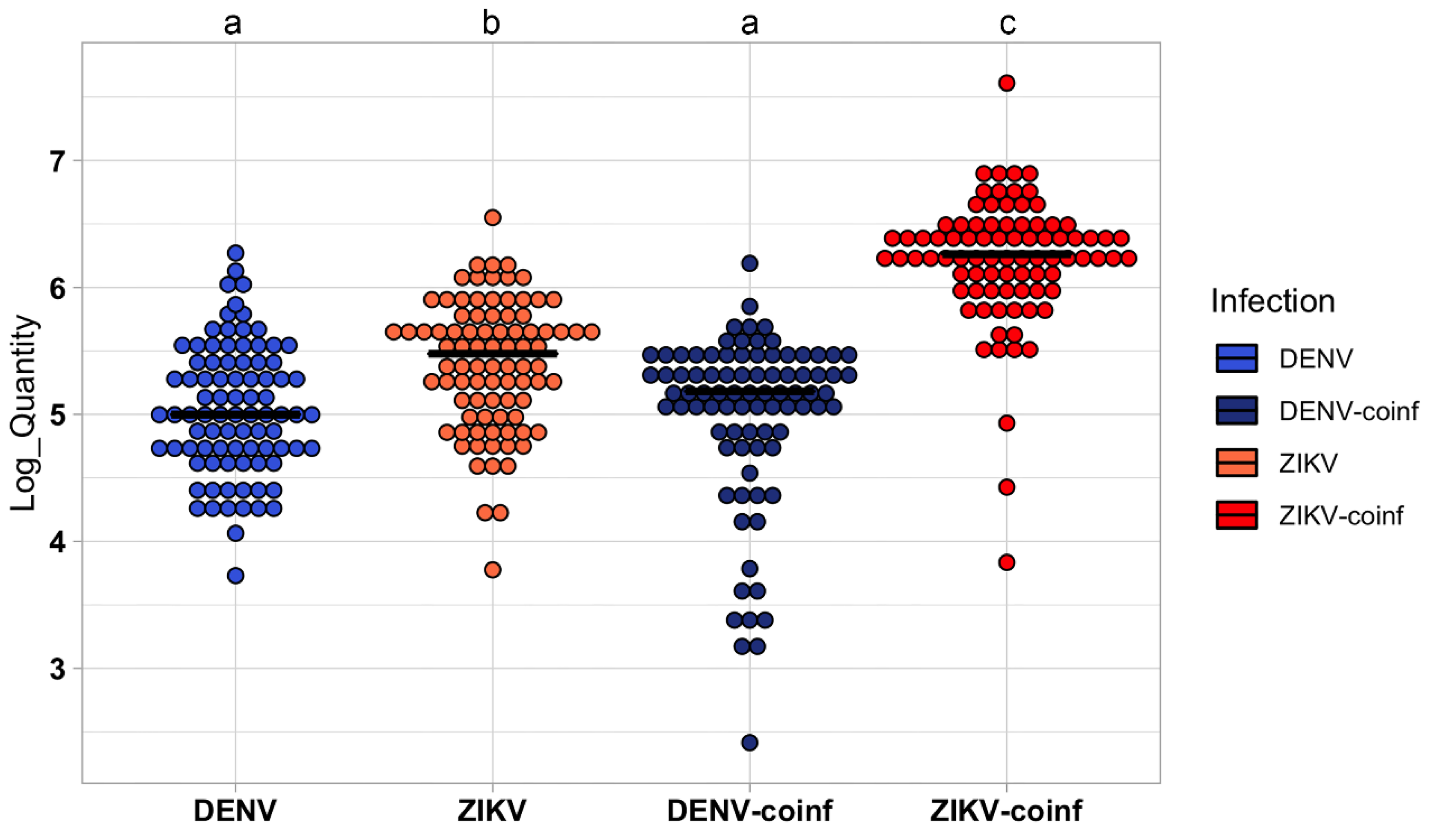
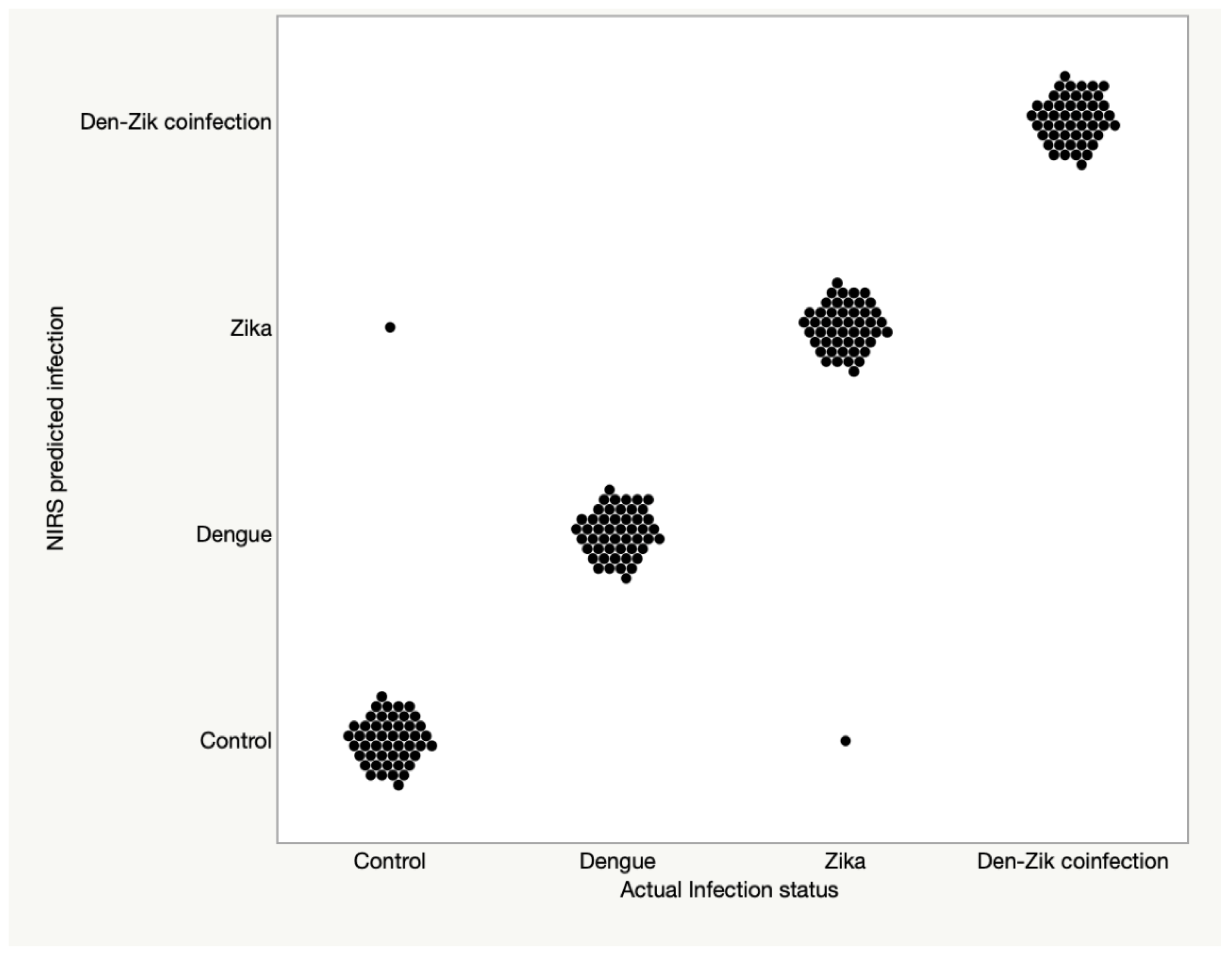
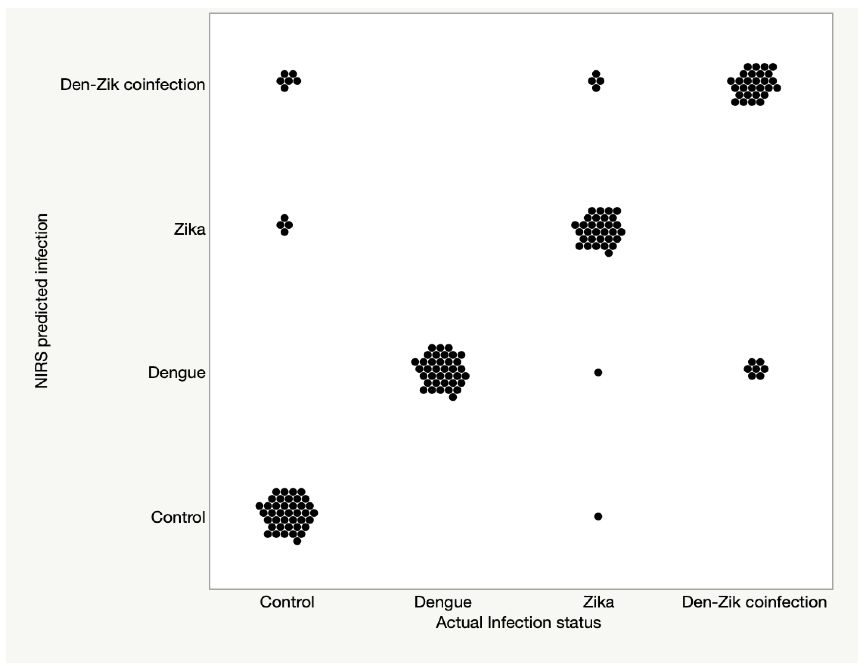
3.2. Differentiation between DENV, ZIKV, and ZIKV + DENV Coinfections in Ae. aegypti

The training model was built using a total of 200 Ae. aegypti females, with between 50–60 individuals belonging to each of the four treatments we used: DENV-infected, ZIKV-infected, ZIKV/DENV coinfected and uninfected mosquitoes. The training model was able to differentiate Ae. aegypti mosquitoes into the four treatments of samples with 100% accuracy. In the training model the accuracy for correctly identifying the uninfected individuals was of 98% (n = 50) (Table 2).
When the model was applied to predict the test set, DENV-infected, ZIKV-infected, and ZIKV/DENV coinfected mosquitoes could be correctly classified into their actual infection group with a predictive accuracy of 100% (n = 37), 84% (n = 37) and 80% (n = 35), respectively. Furthermore, when compared with insects from the uninfected group, the three infected groups were predicted as belonging into the infected group with 100%, 97%, and 100% accuracy for DENV-infected, ZIKV-infected and the coinfected group, respectively. Uninfected mosquitoes were predicted with 80% accuracy (n = 50).
In summary, NIRS was highly sensitivity for detecting any infection type and could distinguish ZIKV and DENV infected mosquitoes from uninfected samples and samples coinfected with both viruses. Prediction accuracy for the training set and the validation set is shown in Figure 3 and Table 2, whereas test set data is shown in Figure 4 and Table 2.

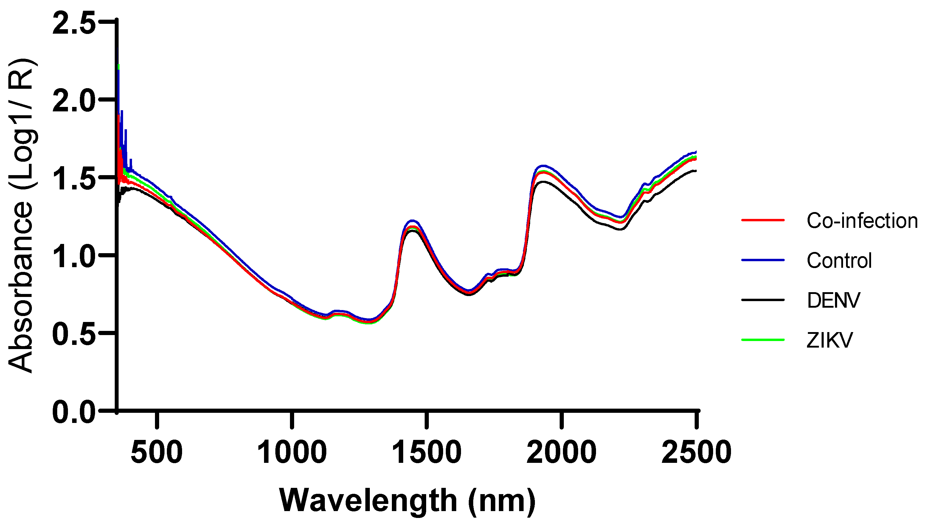
3.3. Second Derivative Figure of Infected and Uninfected Mosquitoes

Figure 5 shows the second derivative average spectra of all ZIKV-infected, DENV-infected, ZIK-DENV- coinfected, and uninfected mosquitoes with distinct bands from the first overtone region at 1685 and 1741 and the combination band region at 1868, 1890, 2234, 2256, 2301, and 2320 nm. Bands from the first overtone region are dominated by C-H and O-H, while bands within the combination region are dominated by C-H, N-H, and O-H bonds representing proteins. A general change in absorbance is observed from the raw spectra as well as the second derivative spectra. Reduced bands among infected mosquitoes in particular those infected with ZIKV are observed at 1685, 2256, and 2301 nm whereas bands at 1741 and 1868 nm are increased in uninfected mosquitoes. Bands at 2234 and 2320 nm are increased in ZIKV infected mosquitos relative to other mosquitoes.

4. Discussion

This study sought to test the capacity of the NIRS technique to differentiate Ae. aegypti female mosquitoes single infected with either ZIKV or DENV, coinfected with both ZIKV and DENV and uninfected samples. NIRS has previously been used to detect single infections of ZIKV [32], CHIKV [37], and DENV [47] in laboratory-reared Ae. aegypti mosquitoes. Here, our results show that NIRS can also differentiate DENV-infected Ae. aegypti females from their uninfected counterparts, with an accuracy of 100%. Additionally, this manuscript also shows that NIRS can detect and differentiate DENV and ZIKV infected mosquitoes from ZIKV/DENV coinfections and uninfected Ae. aegypti with high predictive sensitivities > 96%. Preliminary results obtained so far have shown that predictive models using Ae. aegypti mosquitoes experimentally infected under laboratory conditions have accuracies above 92% in detecting DENV, ZIKV, and CHIKV, three of the most widespread arboviruses. Taken together, our results are encouraging to continue evaluating the potential role of NIR to improve arboviruses surveillance in endemic settings. Thus, next steps should involve collection of semi-field or field data to move further with estimating the potential role that NIRS might have in arboviruses surveillance systems [17,18].
As far as we know, the results presented here are the first to show that NIR can detect and differentiate Ae. aegypti infected with single ZIKV or DENV infection and ZIKV/DENV co-infection of uninfected insects. The use of spectroscopy techniques at the infrared light for arboviruses diagnosis is recent. The overall high predictive ability observed for DENV are consistent with previous findings where ZIKV infected Ae. aegypti could be detected in fresh samples with a sensitivity > 95% in head and thoraces [32] and in samples left in BG sentinel traps for 7 days with sensitivity of 93.2% [37]. These results are encouraging to applying this technique in further tests under more realistic scenarios like semi-field or field testing. Actual evidence shows NIRS might become a relevant tool for surveillance of arbovirus programs in areas where both ZIKV and DENV occur simultaneously like in several tropical regions worldwide [48,49,50].
The occurrence of natural coinfection of arboviruses in Ae. aegypti is still poorly known, although several studies have demonstrated both in the laboratory and under field settings that mosquitoes are able to be simultaneously infected by two different arboviruses [51,52,53,54,55,56]. It has also been reported that the presence of a coinfection does not affect Ae. aegypti vector competence for each pathogen [53,54], but the presence of one virus can enhance the replication of the other virus in the salivary gland. For example, a co-infection of DENV and CHIKV enhanced the replication of DENV in the salivary glands [55]. Our data shows that the number of ZIKV copies were increased in Ae. aegypti when insects were co-infected with DENV. Ruckert and colleagues also demonstrated that both ZIKV infection rates and DENV dissemination rates were reduced for Ae. aegypti with a coinfection of CHIKV whereas CHIKV transmission was reduced with DENV infection [54]. The sensitivity for ZIKV infected mosquitoes was 84% which was lower than the sensitivity for DENV prediction (100%). While the sensitivity of ZIKV was lower, samples incorrectly predicted as ZIKV positive were predicted as infected with DENV instead, suggesting infection was still identified, albeit the incorrect one. As we only collected NIR spectra from the heads and thoraces of mosquitoes at 14 dpi, it is possible that the presence of DENV particularly in the salivary glands might be higher than ZIKV, even though the overall quantity of ZIKV from mosquito whole body was higher than DENV as determined by RT-PCR (Figure 2). We hypothesized that these factors may have resulted in a more accurate training model for DENV-infected mosquitoes, which eventually misclassified ZIKV-infected mosquitoes as DENV-infected. ZIKV and DENV are closely related viruses within the Flaviviridae family sharing 55.6% amino acid sequence identity [57], with 79 genes that were regulated in the same direction (up or downregulation) during Ae. aegypti single infections with both viruses [58]. Such similarities between DENV and ZIKV are also evident from the overlapping raw spectra of single-infected Ae. aegypti mosquitoes (Figure 1). Regardless of the infection type, NIR was 84% sensitive for predicting presence of infection in ZIKV infected mosquitoes. This sensitivity is consistent with what was previously reported by the last two studies [32,37]. Another possible explanation for the misclassification of ZIKV into DENV-infected mosquitoes might be related to the time in dpi the qPCR screening was conducted. The duration of the extrinsic incubation period (EIP) varies between these viruses. The EIP for DENV is estimated to last 14 days [45], whereas the EIP for ZIKV is shorter and estimated as approximately 10 days [59]. Therefore, by selecting to scan mosquitoes at 14 dpi, we could unintentionally favor a misclassification toward DENV because a peak on its viral load at mosquito body is expected for 14 dpi, whereas the shorter EIP for ZIKV could make that on 14 dpi, its viral load might not be on its peak anymore [45,59].
The NIR measures specific frequencies of light absorbed between 750–2500 nm and is capable to differentiate infected from uninfected disease vectors most likely by assessing the physiological and biochemical changes due to pathogen presence. For example, the detection of the endosymbiont Wolbachia pipientis in both D. simulans and D. melanogaster has been attributed to spectral signatures related to either the presence and the concentration of lipopolysaccharide molecules or the physiological changes caused by this bacterium in infected flies [60]. In regards of ZIKV and DENV infection in Ae. aegypti, physiological changes due to arbovirus infection have been reported using different approaches. Aedes aegypti females when infected with DENV or ZIKV have a reduction in life-history traits such as survivorship, fecundity, and fertility [61,62,63,64,65]. On a molecular and biochemical level, there are abundant data using gene expression, transcriptomics, and proteomics approaches revealing that most of the altered genes are involved in metabolic processes, cellular processes, and proteolysis [58,66,67,68]. Thus, most likely the NIR detects those physiological changes caused by infection and then is able to differentiate between infected and uninfected individuals.
NIRS represents an ideal arbovirus surveillance tool as it can be implemented for real time screening of field mosquitoes, with remarkable advantages such as cost and time effectiveness, reagent-free, and non-invasive nature over existing molecular tools for similar purposes. Previously, it was demonstrated that NIRS is 18 times faster and 110 times cheaper than RT-qPCR for detecting ZIKV in Ae. aegypti mosquitoes [32]. Its high throughput nature allows hundreds of samples to be screened in a day by unskilled technicians ultimately enabling timely decision making through rapid detection of potential disease. This is particularly important when considering the silent circulation of arboviruses before an outbreak within the human population is detected, leading to the possibility of early detection of the virus within the mosquito [69,70,71,72].
Future work should focus on establishing identity of the chemical signatures preferably by scanning pure viruses. Identification of virus specific spectral peaks will speed up the implementation of the tool for field collected mosquitoes. This study and the previous two studies that used NIRS to predict ZIKV in fresh Ae aegypti [32] and ZIKV, Wolbachia, and CHIKV in mosquitoes left in BG sentinel trap for 7 days [37] provide strong ground to progress the application of NIRS to assist with the assessments of arbovirus transmission studies that require processing of hundreds of samples in the laboratory. However, the use of NIRS in the field to detect mosquitoes with arboviruses will require further validation including training of fresh models built directly from field mosquitoes infected with these viruses.

Author Contributions

Conceptualization, G.A.G., M.T.S.-L. and R.M.-d.-F.; methodology, G.A.G., A.R.L., L.M.B.S., T.N.K., M.R.D., D.C.-L., A.T.-F., M.G.P., M.T.S.-L. and R.M.-d.-F.; validation, A.R.L., T.N.K. and M.T.S.-L.; formal analysis, G.A.G., A.R.L., T.N.K., M.R.D., D.C.-L., M.G.P., M.T.S.-L. and R.M.-d.-F.; investigation, G.A.G., A.R.L., T.N.K., M.G.P., M.T.S.-L. and R.M.-d.-F.; resources, M.T.S.-L. and R.M.-d.-F.; data curation, G.A.G., A.R.L., T.N.K., M.T.S.-L. and R.M.-d.-F.; writing—original draft preparation, G.A.G., M.T.S.-L. and R.M.-d.-F.; writing—review and editing, G.A.G., A.R.L., L.M.B.S., T.N.K., M.R.D., D.C.-L., A.T.-F., M.G.P., M.T.S.-L. and R.M.-d.-F.; visualization, G.A.G., A.R.L., L.M.B.S., T.N.K., M.R.D., D.C.-L., A.T.-F., M.G.P., M.T.S.-L. and R.M.-d.-F.; supervision, M.T.S.-L. and R.M.-d.-F.; project administration, M.T.S.-L. and R.M.-d.-F.; funding acquisition, M.T.S.-L. and R.M.-d.-F. All authors have read and agreed to the published version of the manuscript.

Funding

This research was funded by the funding agencies FAPERJ [E-26/201.075/2022], CNPq [306702/2021-5] and INOVA-Fiocruz [16030509].

Institutional Review Board Statement

Not applicable.

Informed Consent Statement

Not applicable.

Data Availability Statement

Not applicable.

Acknowledgments

We thank Marcelo Celestino dos Santos, Mauro Menezes Muniz and Renato Carvalho de Andrade for their tireless support in field collections.

Conflicts of Interest

The authors declare no conflict of interest.

References

  1. Brady, O.J.; Hay, S.I. The Global Expansion of Dengue: How Aedes Aegypti Mosquitoes Enabled the First Pandemic Arbovirus. Annu. Rev. Entomol. 2020, 65, 191–208. [Google Scholar] [CrossRef] [PubMed] [Green Version]
  2. Bhatt, S.; Gething, P.W.; Brady, O.J.; Messina, J.P.; Farlow, A.W.; Moyes, C.L.; Drake, J.M.; Brownstein, J.S.; Hoen, A.G.; Sankoh, O.; et al. The Global Distribution and Burden of Dengue. Nature 2013, 496, 504–507. [Google Scholar] [CrossRef] [PubMed] [Green Version]
  3. Sukhralia, S.; Verma, M.; Gopirajan, S.; Dhanaraj, P.S.; Lal, R.; Mehla, N.; Kant, C.R. From Dengue to Zika: The Wide Spread of Mosquito-Borne Arboviruses. Eur. J. Clin. Microbiol. Infect. Dis. 2019, 38, 3–14. [Google Scholar] [CrossRef] [PubMed]
  4. Chala, B.; Hamde, F. Emerging and Re-Emerging Vector-Borne Infectious Diseases and the Challenges for Control: A Review. Front. Public Health 2021, 9, 715759. [Google Scholar] [CrossRef]
  5. Edman, J.D.; Strickman, D.; Kittayapong, P.; Scott, T.W. Female Aedes Aegypti (Diptera: Culicidae) in Thailand Rarely Feed on Sugar. J. Med. Entomol. 1992, 29, 1035–1038. [Google Scholar] [CrossRef]
  6. David, M.R.; Dantas, E.S.; Maciel-de-Freitas, R.; Codeço, C.T.; Prast, A.E.; Lourenço-de-Oliveira, R. Influence of Larval Habitat Environmental Characteristics on Culicidae Immature Abundance and Body Size of Adult Aedes Aegypti. Front. Ecol. Evol. 2021, 9, 626757. [Google Scholar] [CrossRef]
  7. Maciel-de-Freitas, R.; Marques, W.A.; Peres, R.C.; Cunha, S.P.; Lourenço-de-Oliveira, R. Variation in Aedes Aegypti(Diptera: Culicidae) Container Productivity in a Slum and a Suburban District of Rio de Janeiro during Dry and Wet Seasons. Mem. Inst. Oswaldo Cruz 2007, 102, 489–496. [Google Scholar] [CrossRef] [Green Version]
  8. Abílio, A.P.; Abudasse, G.; Kampango, A.; Candrinho, B.; Sitoi, S.; Luciano, J.; Tembisse, D.; Sibindy, S.; de Almeida, A.P.G.; Garcia, G.A.; et al. Distribution and Breeding Sites of Aedes Aegypti and Aedes Albopictus in 32 Urban/Peri-Urban Districts of Mozambique: Implication for Assessing the Risk of Arbovirus Outbreaks. PLoS Negl. Trop. Dis. 2018, 12, e0006692. [Google Scholar] [CrossRef] [Green Version]
  9. Zouache, K.; Fontaine, A.; Vega-Rua, A.; Mousson, L.; Thiberge, J.-M.; Lourenco-De-Oliveira, R.; Caro, V.; Lambrechts, L.; Failloux, A.-B. Three-Way Interactions between Mosquito Population, Viral Strain and Temperature Underlying Chikungunya Virus Transmission Potential. Proc. R. Soc. B Biol. Sci. 2014, 281, 20141078. [Google Scholar] [CrossRef]
  10. González, M.A.; Pavan, M.G.; Fernandes, R.S.; Busquets, N.; David, M.R.; Lourenço-Oliveira, R.; García-Pérez, A.L.; Maciel-de-Freitas, R. Limited Risk of Zika Virus Transmission by Five Aedes Albopictus Populations from Spain. Parasit. Vectors 2019, 12, 150. [Google Scholar] [CrossRef]
  11. Chouin-Carneiro, T.; Vega-Rua, A.; Vazeille, M.; Yebakima, A.; Girod, R.; Goindin, D.; Dupont-Rouzeyrol, M.; Lourenço-de-Oliveira, R.; Failloux, A.-B. Differential Susceptibilities of Aedes Aegypti and Aedes Albopictus from the Americas to Zika Virus. PLoS Negl. Trop. Dis. 2016, 10, e0004543. [Google Scholar] [CrossRef] [PubMed]
  12. De Filipps, A.M.B.; Vazeille, M.; Lourenço-de-Oliveira, R.; Failloux, A.-B. Large Genetic Differentiation and Low Variation in Vector Competence for Dengue and Yellow Fever Viruses of Aedes Albopictus from Brazil, the United States, and the Cayman Islands. Am. J. Trop. Med. Hyg. 2003, 69, 105–114. [Google Scholar] [CrossRef] [Green Version]
  13. Pereira dos Santos, T.; Roiz, D.; Santos de Abreu, F.V.; Luz, S.L.B.; Santalucia, M.; Jiolle, D.; Santos Neves, M.S.A.; Simard, F.; Lourenço-de-Oliveira, R.; Paupy, C. Potential of Aedes Albopictus as a Bridge Vector for Enzootic Pathogens at the Urban-Forest Interface in Brazil. Emerg. Microbes Infect. 2018, 7, 191. [Google Scholar] [CrossRef] [PubMed] [Green Version]
  14. Roiz, D.; Wilson, A.L.; Scott, T.W.; Fonseca, D.M.; Jourdain, F.; Müller, P.; Velayudhan, R.; Corbel, V. Integrated Aedes Management for the Control of Aedes-Borne Diseases. PLoS Negl. Trop. Dis. 2018, 12, e0006845. [Google Scholar] [CrossRef] [Green Version]
  15. Olliaro, P.; Fouque, F.; Kroeger, A.; Bowman, L.; Velayudhan, R.; Santelli, A.C.; Garcia, D.; Skewes Ramm, R.; Sulaiman, L.H.; Tejeda, G.S.; et al. Improved Tools and Strategies for the Prevention and Control of Arboviral Diseases: A Research-to-Policy Forum. PLoS Negl. Trop. Dis. 2018, 12, e0005967. [Google Scholar] [CrossRef]
  16. Ramírez, A.L.; van den Hurk, A.F.; Meyer, D.B.; Ritchie, S.A. Searching for the Proverbial Needle in a Haystack: Advances in Mosquito-Borne Arbovirus Surveillance. Parasit. Vectors 2018, 11, 320. [Google Scholar] [CrossRef] [PubMed] [Green Version]
  17. de Leandro, A.S.; Lopes, R.D.; Martins, C.A.; Rivas, A.V.; da Silva, I.; Galvão, S.R.; Maciel-de-Freitas, R. The Adoption of the One Health Approach to Improve Surveillance of Venomous Animal Injury, Vector-Borne and Zoonotic Diseases in Foz Do Iguaçu, Brazil. PLoS Negl. Trop. Dis. 2021, 15, e0009109. [Google Scholar] [CrossRef] [PubMed]
  18. Leandro, A.S.; de Castro, W.A.C.; Lopes, R.D.; Delai, R.M.; Villela, D.A.M.; De-Freitas, R.M. Citywide Integrated Aedes Aegypti Mosquito Surveillance as Early Warning System for Arbovirus Transmission, Brazil. Emerg. Infect. Dis. 2022, 28, 701–706. [Google Scholar] [CrossRef]
  19. Zanluca, C.; De Melo, V.C.A.; Mosimann, A.L.P.; Dos Santos, G.I.V.; dos Santos, C.N.D.; Luz, K. First Report of Autochthonous Transmission of Zika Virus in Brazil. Mem. Inst. Oswaldo Cruz 2015, 110, 569–572. [Google Scholar] [CrossRef]
  20. Nunes, M.R.T.; Faria, N.R.; de Vasconcelos, J.M.; Golding, N.; Kraemer, M.U.G.; de Oliveira, L.F.; Azevedo, R.d.S.d.S.; da Silva, D.E.A.; da Silva, E.V.P.; da Silva, S.P.; et al. Emergence and Potential for Spread of Chikungunya Virus in Brazil. BMC Med. 2015, 13, 102. [Google Scholar] [CrossRef] [PubMed]
  21. Gu, W.; Novak, R.J. Short Report: Detection Probability of Arbovirus Infection in Mosquitopopulations. Am. J. Trop. Med. Hyg. 2004, 71, 636–638. [Google Scholar] [CrossRef]
  22. Juliano, S.A.; Ribeiro, G.S.; Maciel-de-Freitas, R.; Castro, M.G.; Codeço, C.; Lourenço-de-Oliveira, R.; Lounibos, L.P. She’s a Femme Fatale: Low-Density Larval Development Produces Good Disease Vectors. Mem. Inst. Oswaldo Cruz 2014, 109, 1070–1077. [Google Scholar] [CrossRef] [PubMed]
  23. Ferreira-de-Brito, A.; Ribeiro, I.P.; de Miranda, R.M.; Fernandes, R.S.; Campos, S.S.; da Silva, K.A.B.; de Castro, M.G.; Bonaldo, M.C.; Brasil, P.; Lourenço-de-Oliveira, R. First Detection of Natural Infection of Aedes Aegypti with Zika Virus in Brazil and throughout South America. Mem. Inst. Oswaldo Cruz 2016, 111, 655–658. [Google Scholar] [CrossRef] [PubMed] [Green Version]
  24. Maciel-de-Freitas, R.; Avendanho, F.C.; Santos, R.; Sylvestre, G.; Araújo, S.C.; Lima, J.B.P.; Martins, A.J.; Coelho, G.E.; Valle, D. Undesirable Consequences of Insecticide Resistance Following Aedes Aegypti Control Activities Due to a Dengue Outbreak. PLoS ONE 2014, 9, e92424. [Google Scholar] [CrossRef]
  25. Garcia-Rejon, J.; Loroño-Pino, M.A.; Farfan-Ale, J.A.; Flores-Flores, L.; Rosado-Paredes, E.D.P.; Rivero-Cardenas, N.; Najera-Vazquez, R.; Gomez-Carro, S.; Lira-Zumbardo, V.; Gonzalez-Martinez, P.; et al. Dengue Virus–Infected Aedes Aegypti in the Home Environment. Am. J. Trop. Med. Hyg. 2008, 79, 940–950. [Google Scholar] [CrossRef] [PubMed]
  26. Goh, B.; Ching, K.; Soares Magalhães, R.J.; Ciocchetta, S.; Edstein, M.D.; Maciel-de-Freitas, R.; Sikulu-Lord, M.T. The Application of Spectroscopy Techniques for Diagnosis of Malaria Parasites and Arboviruses and Surveillance of Mosquito Vectors: A Systematic Review and Critical Appraisal of Evidence. PLoS Negl. Trop. Dis. 2021, 15, e0009218. [Google Scholar] [CrossRef]
  27. Mayagaya, V.S.; Michel, K.; Benedict, M.Q.; Killeen, G.F.; Wirtz, R.A.; Ferguson, H.M.; Dowell, F.E. Non-Destructive Determination of Age and Species of Anopheles Gambiae s.l. Using Near-Infrared Spectroscopy. Am. J. Trop. Med. Hyg. 2009, 81, 622–630. [Google Scholar] [CrossRef]
  28. Sikulu, M.; Killeen, G.F.; Hugo, L.E.; Ryan, P.A.; Dowell, K.M.; Wirtz, R.A.; Moore, S.J.; Dowell, F.E. Near-Infrared Spectroscopy as a Complementary Age Grading and Species Identification Tool for African Malaria Vectors. Parasit. Vectors 2010, 3, 49. [Google Scholar] [CrossRef] [Green Version]
  29. Ntamatungiro, A.J.; Mayagaya, V.S.; Rieben, S.; Moore, S.J.; Dowell, F.E.; Maia, M.F. The Influence of Physiological Status on Age Prediction of Anopheles Arabiensis Using near Infra-Red Spectroscopy. Parasit. Vectors 2013, 6, 298. [Google Scholar] [CrossRef] [Green Version]
  30. Sikulu, M.; Dowell, K.M.; Hugo, L.E.; Wirtz, R.A.; Michel, K.; Peiris, K.H.; Moore, S.; Killeen, G.F.; Dowell, F.E. Evaluating RNAlater® as a Preservative for Using Near-Infrared Spectroscopy to Predict Anopheles Gambiae Age and Species. Malar. J. 2011, 10, 186. [Google Scholar] [CrossRef]
  31. Sikulu, M.T.; Majambere, S.; Khatib, B.O.; Ali, A.S.; Hugo, L.E.; Dowell, F.E. Using a Near-Infrared Spectrometer to Estimate the Age of Anopheles Mosquitoes Exposed to Pyrethroids. PLoS ONE 2014, 9, e90657. [Google Scholar] [CrossRef] [PubMed] [Green Version]
  32. Fernandes, J.N.; dos Santos, L.M.B.; Chouin-Carneiro, T.; Pavan, M.G.; Garcia, G.A.; David, M.R.; Beier, J.C.; Dowell, F.E.; Maciel-de-Freitas, R.; Sikulu-Lord, M.T. Rapid, Noninvasive Detection of Zika Virus in Aedes Aegypti Mosquitoes by near-Infrared Spectroscopy. Sci. Adv. 2018, 4, eaat0496. [Google Scholar] [CrossRef] [PubMed] [Green Version]
  33. Milali, M.P.; Sikulu-Lord, M.T.; Kiware, S.S.; Dowell, F.E.; Povinelli, R.J.; Corliss, G.F. Do NIR Spectra Collected from Laboratory-Reared Mosquitoes Differ from Those Collected from Wild Mosquitoes? PLoS ONE 2018, 13, e0198245. [Google Scholar] [CrossRef] [PubMed] [Green Version]
  34. Milali, M.P.; Sikulu-Lord, M.T.; Kiware, S.S.; Dowell, F.E.; Corliss, G.F.; Povinelli, R.J. Age Grading An. Gambiae and An. Arabiensis Using near Infrared Spectra and Artificial Neural Networks. PLoS ONE 2019, 14, e0209451. [Google Scholar] [CrossRef] [PubMed] [Green Version]
  35. Sikulu-Lord, M.T.; Milali, M.P.; Henry, M.; Wirtz, R.A.; Hugo, L.E.; Dowell, F.E.; Devine, G.J. Near-Infrared Spectroscopy, a Rapid Method for Predicting the Age of Male and Female Wild-Type and Wolbachia Infected Aedes Aegypti. PLoS Negl. Trop. Dis. 2016, 10, e0005040. [Google Scholar] [CrossRef] [PubMed] [Green Version]
  36. Sikulu-Lord, M.T.; Maia, M.F.; Milali, M.P.; Henry, M.; Mkandawile, G.; Kho, E.A.; Wirtz, R.A.; Hugo, L.E.; Dowell, F.E.; Devine, G.J. Rapid and Non-Destructive Detection and Identification of Two Strains of Wolbachia in Aedes Aegypti by Near-Infrared Spectroscopy. PLoS Negl. Trop. Dis. 2016, 10, e0004759. [Google Scholar] [CrossRef] [Green Version]
  37. Santos, L.M.B.; Mutsaers, M.; Garcia, G.A.; David, M.R.; Pavan, M.G.; Petersen, M.T.; Corrêa-Antônio, J.; Couto-Lima, D.; Maes, L.; Dowell, F.; et al. High Throughput Estimates of Wolbachia, Zika and Chikungunya Infection in Aedes Aegypti by near-Infrared Spectroscopy to Improve Arbovirus Surveillance. Commun. Biol. 2021, 4, 67. [Google Scholar] [CrossRef]
  38. Esperança, P.M.; Blagborough, A.M.; Da, D.F.; Dowell, F.E.; Churcher, T.S. Detection of Plasmodium Berghei Infected Anopheles Stephensi Using Near-Infrared Spectroscopy. Parasit. Vectors 2018, 11, 377. [Google Scholar] [CrossRef] [Green Version]
  39. Da, D.F.; McCabe, R.; Somé, B.M.; Esperança, P.M.; Sala, K.A.; Blight, J.; Blagborough, A.M.; Dowell, F.; Yerbanga, S.R.; Lefèvre, T.; et al. Detection of Plasmodium Falciparum in Laboratory-Reared and Naturally Infected Wild Mosquitoes Using near-Infrared Spectroscopy. Sci. Rep. 2021, 11, 10289. [Google Scholar] [CrossRef]
  40. Maia, M.F.; Kapulu, M.; Muthui, M.; Wagah, M.G.; Ferguson, H.M.; Dowell, F.E.; Baldini, F.; Ranford-Cartwright, L. Detection of Plasmodium Falciparum Infected Anopheles Gambiae Using Near-Infrared Spectroscopy. Malar. J. 2019, 18, 85. [Google Scholar] [CrossRef]
  41. Dowell, F.E.; Noutcha, A.E.M.; Michel, K. The Effect of Preservation Methods on Predicting Mosquito Age by Near Infrared Spectroscopy. Am. J. Trop. Med. Hyg. 2011, 85, 1093–1096. [Google Scholar] [CrossRef] [PubMed] [Green Version]
  42. Mayagaya, V.; Ntamatungiro, A.; Moore, S.; Wirtz, R.; Dowell, F.; Maia, M. Evaluating Preservation Methods for Identifying Anopheles Gambiae s.s. and Anopheles Arabiensis Complex Mosquitoes Species Using near Infra-Red Spectroscopy. Parasit. Vectors 2015, 8, 60. [Google Scholar] [CrossRef] [PubMed] [Green Version]
  43. Bonaldo, M.C.; Ribeiro, I.P.; Lima, N.S.; dos Santos, A.A.C.; Menezes, L.S.R.; da Cruz, S.O.D.; de Mello, I.S.; Furtado, N.D.; de Moura, E.E.; Damasceno, L.; et al. Isolation of Infective Zika Virus from Urine and Saliva of Patients in Brazil. PLoS Negl. Trop. Dis. 2016, 10, e0004816. [Google Scholar] [CrossRef] [PubMed] [Green Version]
  44. Caragata, E.P.; Rocha, M.N.; Pereira, T.N.; Mansur, S.B.; Dutra, H.L.C.; Moreira, L.A. Pathogen Blocking in Wolbachia-Infected Aedes Aegypti Is Not Affected by Zika and Dengue Virus Co-Infection. PLoS Negl. Trop. Dis. 2019, 13, e0007443. [Google Scholar] [CrossRef] [PubMed] [Green Version]
  45. Watts, D.M.; Burke, D.S.; Harrison, B.A.; Whitmire, R.E.; Nisalak, A. Effect of Temperature on the Vector Efficiency of Aedes Aegypti for Dengue 2 Virus. Am. J. Trop. Med. Hyg. 1987, 36, 143–152. [Google Scholar] [CrossRef] [PubMed]
  46. R Development Core Team. R: A Language and Environment for Statistical Computing; R Foundation for Statistical Computing: Vienna, Austria, 2020. [Google Scholar]
  47. Santos, M.C.D.; Viana, J.L.S.; Monteiro, J.D.; Freire, R.C.M.; Freitas, D.L.D.; Câmara, I.M.; Silva, G.J.S.; Gama, R.A.; Araújo, J.M.G.; Lima, K.M.G. Infrared spectroscopy (NIRS and ATR-FTIR) together with multivariate classification for non-destructive differentiation between female mosquitoes of Aedes aegypti recently infected with dengue vs. uninfected females. Acta Trop 2022, 235, 106633. [Google Scholar] [CrossRef]
  48. Lowe, R.; Barcellos, C.; Brasil, P.; Cruz, O.; Honório, N.; Kuper, H.; Carvalho, M. The Zika Virus Epidemic in Brazil: From Discovery to Future Implications. Int. J. Environ. Res. Public Health 2018, 15, 96. [Google Scholar] [CrossRef] [Green Version]
  49. Kazazian, L.; Lima Neto, A.S.; Sousa, G.S.; do Nascimento, O.J.; Castro, M.C. Spatiotemporal Transmission Dynamics of Co-Circulating Dengue, Zika, and Chikungunya Viruses in Fortaleza, Brazil: 2011–2017. PLoS Negl. Trop. Dis. 2020, 14, e0008760. [Google Scholar] [CrossRef]
  50. Nunes, P.C.G.; Daumas, R.P.; Sánchez-Arcila, J.C.; Nogueira, R.M.R.; Horta, M.A.P.; dos Santos, F.B. 30 Years of Fatal Dengue Cases in Brazil: A Review. BMC Public Health 2019, 19, 329. [Google Scholar] [CrossRef]
  51. Ciota, A.T. The Role of Co-Infection and Swarm Dynamics in Arbovirus Transmission. Virus Res. 2019, 265, 88–93. [Google Scholar] [CrossRef] [PubMed]
  52. Caron, M.; Paupy, C.; Grard, G.; Becquart, P.; Mombo, I.; Nso, B.B.B.; Kassa Kassa, F.; Nkoghe, D.; Leroy, E.M. Recent Introduction and Rapid Dissemination of Chikungunya Virus and Dengue Virus Serotype 2 Associated with Human and Mosquito Coinfections in Gabon, Central Africa. Clin. Infect. Dis. 2012, 55, e45–e53. [Google Scholar] [CrossRef] [PubMed] [Green Version]
  53. Göertz, G.P.; Vogels, C.B.F.; Geertsema, C.; Koenraadt, C.J.M.; Pijlman, G.P. Mosquito Co-Infection with Zika and Chikungunya Virus Allows Simultaneous Transmission without Affecting Vector Competence of Aedes Aegypti. PLoS Negl. Trop. Dis. 2017, 11, e0005654. [Google Scholar] [CrossRef] [PubMed] [Green Version]
  54. Rückert, C.; Weger-Lucarelli, J.; Garcia-Luna, S.M.; Young, M.C.; Byas, A.D.; Murrieta, R.A.; Fauver, J.R.; Ebel, G.D. Impact of Simultaneous Exposure to Arboviruses on Infection and Transmission by Aedes Aegypti Mosquitoes. Nat. Commun. 2017, 8, 15412. [Google Scholar] [CrossRef] [Green Version]
  55. Kantor, A.M.; Lin, J.; Wang, A.; Thompson, D.C.; Franz, A.W.E. Infection Pattern of Mayaro Virus in Aedes Aegypti (Diptera: Culicidae) and Transmission Potential of the Virus in Mixed Infections with Chikungunya Virus. J. Med. Entomol. 2019, 56, 832–843. [Google Scholar] [CrossRef]
  56. Le Coupanec, A.; Tchankouo-Nguetcheu, S.; Roux, P.; Khun, H.; Huerre, M.; Morales-Vargas, R.; Enguehard, M.; Lavillette, D.; Missé, D.; Choumet, V. Co-Infection of Mosquitoes with Chikungunya and Dengue Viruses Reveals Modulation of the Replication of Both Viruses in Midguts and Salivary Glands of Aedes Aegypti Mosquitoes. Int. J. Mol. Sci. 2017, 18, 1708. [Google Scholar] [CrossRef] [Green Version]
  57. Chang, H.-H.; Huber, R.G.; Bond, P.J.; Grad, Y.H.; Camerini, D.; Maurer-Stroh, S.; Lipsitch, M. Systematic Analysis of Protein Identity between Zika Virus and Other Arthropod-Borne Viruses. Bull. World Health Organ. 2017, 95, 517–525I. [Google Scholar] [CrossRef]
  58. Angleró-Rodríguez, Y.I.; MacLeod, H.J.; Kang, S.; Carlson, J.S.; Jupatanakul, N.; Dimopoulos, G. Aedes Aegypti Molecular Responses to Zika Virus: Modulation of Infection by the Toll and Jak/Stat Immune Pathways and Virus Host Factors. Front. Microbiol. 2017, 8, 2050. [Google Scholar] [CrossRef] [Green Version]
  59. Murrieta, R.A.; Garcia-Luna, S.M.; Murrieta, D.J.; Halladay, G.; Young, M.C.; Fauver, J.R.; Gendernalik, A.; Weger-Lucarelli, J.; Rückert, C.; Ebel, G.D. Impact of Extrinsic Incubation Temperature on Natural Selection during Zika Virus Infection of Aedes Aegypti and Aedes Albopictus. PLOS Pathog. 2021, 17, e1009433. [Google Scholar] [CrossRef]
  60. Aw, W.C.; Dowell, F.E.; Ballard, J.W.O. Using Near-Infrared Spectroscopy to Resolve the Species, Gender, Age, and the Presence of Wolbachia Infection in Laboratory-Reared Drosophila. G3 Genes|Genomes|Genet. 2012, 2, 1057–1065. [Google Scholar] [CrossRef] [Green Version]
  61. Petersen, M.T.; da Silveira, I.D.; Tátila-Ferreira, A.; David, M.R.; Chouin-Carneiro, T.; Van den Wouwer, L.; Maes, L.; Maciel-de-Freitas, R. The Impact of the Age of First Blood Meal and Zika Virus Infection on Aedes Aegypti Egg Production and Longevity. PLoS ONE 2018, 13, e0200766. [Google Scholar] [CrossRef] [PubMed]
  62. Maciel-de-Freitas, R.; Sylvestre, G.; Gandini, M.; Koella, J.C. The Influence of Dengue Virus Serotype-2 Infection on Aedes Aegypti (Diptera: Culicidae) Motivation and Avidity to Blood Feed. PLoS ONE 2013, 8, e65252. [Google Scholar] [CrossRef] [PubMed]
  63. Maciel-de-Freitas, R.; Koella, J.C.; Lourenço-de-Oliveira, R. Lower Survival Rate, Longevity and Fecundity of Aedes Aegypti (Diptera: Culicidae) Females Orally Challenged with Dengue Virus Serotype 2. Trans. R. Soc. Trop. Med. Hyg. 2011, 105, 452–458. [Google Scholar] [CrossRef] [PubMed]
  64. da Silveira, I.D.; Petersen, M.T.; Sylvestre, G.; Garcia, G.A.; David, M.R.; Pavan, M.G.; Maciel-de-Freitas, R. Zika Virus Infection Produces a Reduction on Aedes Aegypti Lifespan but No Effects on Mosquito Fecundity and Oviposition Success. Front. Microbiol. 2018, 9, 3011. [Google Scholar] [CrossRef] [PubMed] [Green Version]
  65. Javed, N.; Bhatti, A.; Paradkar, P.N. Advances in Understanding Vector Behavioural Traits after Infection. Pathogens 2021, 10, 1376. [Google Scholar] [CrossRef]
  66. Etebari, K.; Hegde, S.; Saldaña, M.A.; Widen, S.G.; Wood, T.G.; Asgari, S.; Hughes, G.L. Global Transcriptome Analysis of Aedes Aegypti Mosquitoes in Response to Zika Virus Infection. mSphere 2017, 2, 1648–1659. [Google Scholar] [CrossRef] [Green Version]
  67. Ramos, L.F.C.; Martins, M.; Murillo, J.R.; Domont, G.B.; de Oliveira, D.M.P.; Nogueira, F.C.S.; Maciel-de-Freitas, R.; Junqueira, M. Interspecies Isobaric Labeling-Based Quantitative Proteomics Reveals Protein Changes in the Ovary of Aedes Aegypti Coinfected with ZIKV and Wolbachia. Front. Cell. Infect. Microbiol. 2022, 12, 900608. [Google Scholar] [CrossRef]
  68. Martins, M.; Ramos, L.F.C.; Murillo, J.R.; Torres, A.; de Carvalho, S.S.; Domont, G.B.; de Oliveira, D.M.P.; Mesquita, R.D.; Nogueira, F.C.S.; Maciel-de-Freitas, R.; et al. Comprehensive Quantitative Proteome Analysis of Aedes Aegypti Identifies Proteins and Pathways Involved in Wolbachia Pipientis and Zika Virus Interference Phenomenon. Front. Physiol. 2021, 12, 642237. [Google Scholar] [CrossRef]
  69. Ayllón, T.; Campos, R.D.M.; Brasil, P.; Morone, F.C.; Câmara, D.C.P.; Meira, G.L.S.; Tannich, E.; Yamamoto, K.A.; Carvalho, M.S.; Pedro, R.S.; et al. Early Evidence for Zika Virus Circulation among Aedes Aegypti Mosquitoes, Rio de Janeiro, Brazil. Emerg. Infect. Dis. 2017, 23, 1411–1412. [Google Scholar] [CrossRef] [Green Version]
  70. dos Reis, I.C.; Gibson, G.; Ayllón, T.; de Medeiros Tavares, A.; de Araújo, J.M.G.; da Silva Monteiro, E.; Rodrigues Aguiar, A.; de Oliveira, J.V.; de Paiva, A.A.P.; Wana Bezerra Pereira, H.; et al. Entomo-Virological Surveillance Strategy for Dengue, Zika and Chikungunya Arboviruses in Field-Caught Aedes Mosquitoes in an Endemic Urban Area of the Northeast of Brazil. Acta Trop. 2019, 197, 105061. [Google Scholar] [CrossRef]
  71. Singh, J.; Balakrishnan, N.; Bhardwaj, M.; Amuthadevi, P.; George, E.G.; Subramani, K.; Soundararajan, K.; Appavoo, N.C.; Jain, D.C.; Ichhpujani, R.L.; et al. Silent Spread of Dengue and Dengue Haemorrhagic Fever to Coimbatore and Erode Districts in Tamil Nadu, India, 1998: Need for Effective Surveillance to Monitor and Control the Disease. Epidemiol. Infect. 2000, 125, 195–200. [Google Scholar] [CrossRef] [PubMed]
  72. Leandro, A.S.; Lopes, R.D.; Amaral Martins, C.; Delai, R.M.; Villela, D.A.M.; Maciel-de-Freitas, R. Entomo-virological surveillance followed by serological active survey of symptomatic individuals is helpful to identify hotspots of early arbovirus transmission. Front. Public Health 2022, 10, 1024187. [Google Scholar] [CrossRef] [PubMed]
Figure 1. Average NIR spectra of Co-infected, ZIKV, DENV and uninfected mosquitoes (control) collected at 14 days post-infection by the Labspec 4i NIR spectrometer. In particular the absorbance value of uninfected mosquitoes (Blue line) was slightly higher than those of infected mosquitoes.
Viruses 15 00011 g001
Figure 2. The number of viral copies in Aedes aegypti females at 14 dpi when mosquitoes were either single infected with ZIKV or DENV, or simultaneously co-infected with both ZIKV/DENV. Different letters indicate statistically significant differences (Wilcoxon rank sum test (U-test), p < 0.001). Each circle represents an individual mosquito; negative control samples (uninfected Ae. aegypti from laboratory colony) are not shown because they did not produce any amplification signal.
Viruses 15 00011 g002
Figure 3. The predictive accuracy of NIRS for samples that were used for training and validation of the ANN model.
Viruses 15 00011 g003
Figure 4. The predictive accuracy of NIRS for samples that were used to test the ANN model.
Viruses 15 00011 g004
Figure 5. The second derivative spectra of Aedes aegypti mosquitoes showing overtones of infected and uninfected groups. The black arrows in the figure indicate the wavelength (nm) band regions that are important for differentiating the groups.
Viruses 15 00011 g005
Table 1. Infection rate for DENV or ZIKV single infection and DENV/ZIKV coinfection.
Group
ZIKV-Infected DENV-Infected ZIKV/DENV Coinfected
Infection rate90.62% (n = 87/96)90.62% (n = 87/96)96.6% (n = 85/88)
Table 2. Prediction accuracy for Ae. aegypti infection status in training/validation set and test set. Mosquitoes could be infected with either DENV or ZIKV single infection, coinfected with DENV and ZIKV or uninfected (control).
Training and Validation SetTest Set
% Spec (n)% Sensitivity (n)% Spec (n)% Sensitivity (n)
Predicted GroupControlDENVZIKVCo-Infection ZIKV/DENVControlDENVZIKVCo-Infection ZIKV/DENV
Predicted into actual infection group98 (n = 50) 100 (n = 50)98 (n = 50)100 (n = 50)80 (n = 50)100 (n = 37)84 (n = 37)80 (n = 35)
Predicted as infected2 (n = 50)100 (n = 50)98 (n = 50)100 (n = 50)20 (n = 50)100 (n = 37)97 (n = 37)100 (n = 35)
Disclaimer/Publisher’s Note: The statements, opinions and data contained in all publications are solely those of the individual author(s) and contributor(s) and not of MDPI and/or the editor(s). MDPI and/or the editor(s) disclaim responsibility for any injury to people or property resulting from any ideas, methods, instructions or products referred to in the content.

