Next Issue
Volume 15, January
Previous Issue
Volume 14, November


Viruses, Volume 14, Issue 12 (December 2022) – 240 articles

Cover Story (view full-size image): 
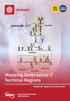
Upon cell entry, plus-strand RNA viruses undergo translation by the host translational machinery. After the viral replicase proteins are produced, plus-strand RNA viruses then undergo replication. Members of the genus Umbravirus, whose genomes are uncapped and nonpolyadenylated, have abnormally long 3′ untranslated regions (≈700 nt) that form highly complex structures. Despite much prior work on umbravirus translation, little is known about umbravirus replication. Our phylogenetic analysis reveals novel, umbravirus-specific 3′ terminal structures critical for viral replication, including an element that functions in vivo but does not form in transcripts in vitro. We have also developed a trans-replication system for umbravirus genomes in planta and shed light on the evolutionary history of umbraviruses. View this paper
  • Issues are regarded as officially published after their release is announced to the table of contents alert mailing list.
  • You may sign up for e-mail alerts to receive table of contents of newly released issues.
  • PDF is the official format for papers published in both, html and pdf forms. To view the papers in pdf format, click on the "PDF Full-text" link, and use the free Adobe Reader to open them.
Order results
Result details
Section
Select all
Export citation of selected articles as:
14 pages, 1702 KiB  
Article
Evaluation of Non-Invasive Gargle Lavage Sampling for the Detection of SARS-CoV-2 Using rRT-PCR or Antigen Assay
by Ondrej Bouska, Hana Jaworek, Vladimira Koudelakova, Katerina Kubanova, Petr Dzubak, Rastislav Slavkovsky, Branislav Siska, Petr Pavlis, Jana Vrbkova and Marian Hajduch
Viruses 2022, 14(12), 2829; https://doi.org/10.3390/v14122829 - 19 Dec 2022
Cited by 2 | Viewed by 1418
Abstract
Severe acute respiratory syndrome coronavirus 2 (SARS-CoV-2) has caused considerable disruption worldwide. For efficient SARS-CoV-2 detection, new methods of rapid, non-invasive sampling are needed. This study aimed to investigate the stability of SARS-CoV-2 in a novel medium for gargle-lavage (GL) self-sampling and to [...] Read more.
Severe acute respiratory syndrome coronavirus 2 (SARS-CoV-2) has caused considerable disruption worldwide. For efficient SARS-CoV-2 detection, new methods of rapid, non-invasive sampling are needed. This study aimed to investigate the stability of SARS-CoV-2 in a novel medium for gargle-lavage (GL) self-sampling and to compare the performance of SARS-CoV-2 detection in paired self-collected GL and clinician-obtained nasopharyngeal swab (NPS) samples. The stability study for SARS-CoV-2 preservation in a novel medium was performed over 14 days (4 °C, 24–27 °C, and 37 °C). In total, 494 paired GL and NPS samples were obtained at the University Hospital in Olomouc in April 2021. SARS-CoV-2 detection in paired samples was performed with a SARS-CoV-2 Nucleic Acid Detection Kit (Zybio, Chongqing Municipality, Chongqing, China), an Elecsys® SARS-CoV-2 Antigen assay (Roche Diagnostics, Mannheim, Germany), and a SARS-CoV-2 Antigen ELISA (EUROIMMUN, Lübeck, Germany). The stability study demonstrated excellent SARS-CoV-2 preservation in the novel medium for 14 days. SARS-CoV-2 was detected in 55.7% of NPS samples and 55.7% of GL samples using rRT-PCR, with an overall agreement of 91.9%. The positive percent agreement (PPA) of the rRT-PCR in the GL samples was 92.7%, and the negative percent agreement (NPA) was 90.9%, compared with the NPS samples. The PPA of the rRT-PCR in the NPS and GL samples was 93.2% when all positive tests were used as the reference standard. Both antigen detection assays showed poor sensitivity compared to rRT-PCR (33.2% and 36.0%). rRT-PCR SARS-CoV-2 detection in self-collected GL samples had a similar PPA and NPA to that of NPSs. GL self-sampling offers a suitable and more comfortable alternative for SARS-CoV-2 detection. Full article
(This article belongs to the Section Human Virology and Viral Diseases)
Show Figures

Figure 1

17 pages, 5511 KiB  
Article
Insights from the Infection Cycle of VSV-ΔG-Spike Virus
by Elad Milrot, Shlomi Lazar, Ofir Schuster, Efi Makdasi, Shlomo Shmaya, Yfat Yahalom-Ronen, Hadas Tamir and Orly Laskar
Viruses 2022, 14(12), 2828; https://doi.org/10.3390/v14122828 - 19 Dec 2022
Viewed by 1640
Abstract
Fundamental key processes in viral infection cycles generally occur in distinct cellular sites where both viral and host factors accumulate and interact. These sites are usually termed viral replication organelles, or viral factories (VF). The generation of VF is accompanied by the synthesis [...] Read more.
Fundamental key processes in viral infection cycles generally occur in distinct cellular sites where both viral and host factors accumulate and interact. These sites are usually termed viral replication organelles, or viral factories (VF). The generation of VF is accompanied by the synthesis of viral proteins and genomes and involves the reorganization of cellular structure. Recently, rVSV-ΔG-spike (VSV-S), a recombinant VSV expressing the SARS-CoV-2 spike protein, was developed as a vaccine candidate against SARS-CoV-2. By combining transmission electron microscopy (TEM) tomography studies and immuno-labeling techniques, we investigated the infection cycle of VSV-S in Vero E6 cells. RT-real-time-PCR results show that viral RNA synthesis occurs 3–4 h post infection (PI), and accumulates as the infection proceeds. By 10–24 h PI, TEM electron tomography results show that VSV-S generates VF in multi-lamellar bodies located in the cytoplasm. The VF consists of virus particles with various morphologies. We demonstrate that VSV-S infection is associated with accumulation of cytoplasmatic viral proteins co-localized with dsRNA (marker for RNA replication) but not with ER membranes. Newly formed virus particles released from the multi-lamellar bodies containing VF, concentrate in a vacuole membrane, and the infection ends with the budding of particles after the fusion of the vacuole membrane with the plasma membrane. In summary, the current study describes detailed 3D imaging of key processes during the VSV-S infection cycle. Full article
(This article belongs to the Section Viral Immunology, Vaccines, and Antivirals)
Show Figures

Figure 1

12 pages, 1191 KiB  
Article
Point-of-Care Testing for Sensitive Detection of the African Swine Fever Virus Genome
by Ahmed Elnagar, Sandra Blome, Martin Beer and Bernd Hoffmann
Viruses 2022, 14(12), 2827; https://doi.org/10.3390/v14122827 - 19 Dec 2022
Cited by 4 | Viewed by 2257
Abstract
African swine fever (ASF) is a contagious viral hemorrhagic disease that affects domestic pigs and wild boar. The disease is notifiable to the World Organization of Animal Health (WOAH), and causes significant deaths and economic losses. There is currently no fully licensed vaccine [...] Read more.
African swine fever (ASF) is a contagious viral hemorrhagic disease that affects domestic pigs and wild boar. The disease is notifiable to the World Organization of Animal Health (WOAH), and causes significant deaths and economic losses. There is currently no fully licensed vaccine available. As a result, early identification of the causative agent, ASF virus (ASFV), is crucial for the implementation of control measures. PCR and real-time PCR are the WOAH-recommended standard methods for the direct detection of ASFV. However, under special field conditions or in simple or remote field laboratories, there may be no sophisticated equipment or even stable electricity available. Under these circumstances, point-of-care systems can be put in place. Along these lines, a previously published, rapid, reliable, and electricity-free extraction method (TripleE) was used to isolate viral nucleic acid from diagnostic specimens. With this tool, nucleic acid extraction from up to eight diagnostic samples can be realized in one run in less than 10 min. In addition, the possibility of completely omitting viral DNA extraction was analyzed with so-called direct real-time PCR protocols using ASFV original samples diluted to 1:40 in RNase-free water. Furthermore, three real-time PCR cyclers, developed for use under field conditions (IndiField, Liberty16 and UF-300 GenecheckerTM), were comparatively applied for the sensitive high-speed detection of ASFV genomes, with overall PCR run times between 20 and 54 min. Depending on the viral DNA extraction/releasing method used and the point-of-care cycler applied, a total time for detection of 30 to 60 min for up to eight samples was feasible. As expected, the limitations in analytical sensitivity were positively correlated to the analysis time. These limitations are acceptable for ASFV diagnostics due to the expected high ASFV genome loads in diseased animals or carcasses. Full article
(This article belongs to the Special Issue African Swine Fever Virus 3.0)
Show Figures

Figure 1

12 pages, 4327 KiB  
Article
Global Infection Rate of Rotavirus C during 1980–2022 and Analysis of Critical Factors in the Host Range Restriction of Virus VP4
by Simiao Zhao, Xinshun Jin, Lingling Zang, Ziwei Liu, Xiaobo Wen and Xuhua Ran
Viruses 2022, 14(12), 2826; https://doi.org/10.3390/v14122826 - 19 Dec 2022
Cited by 6 | Viewed by 1704
Abstract
Information on rotavirus C (RVC) infection is lacking, partly because the prevalence of RVC among humans and animals worldwide is undefined. Data on the characteristics of the P genotype among RVC strains are also required. We performed systematic searches on the infection rates [...] Read more.
Information on rotavirus C (RVC) infection is lacking, partly because the prevalence of RVC among humans and animals worldwide is undefined. Data on the characteristics of the P genotype among RVC strains are also required. We performed systematic searches on the infection rates of RVC since 1980 based on the literature and gene sequences of the PubMed and GenBank databases. A phylogenetic tree of VP4 genes was constructed to evaluate the distribution of the P genotype of RVC from various hosts. The specific mutation motifs in VP8* with P [2]/P [4]/P [5] specificity were analyzed to elucidate their roles in host range restriction. The rate of RVC infection in humans has fallen from 3% before 2009 to 1%, whereas in animals it has risen from 10% to 25%. The P genotype of RVC showed strict host species specificity, and current human RVC infections are exclusively caused by genotype P [2]. In the VP8* hemagglutinin domain of the P [4]/P [5] genotype of swine RVC, specific insertion or deletion were found relative to the human P [2] genotype, and these motifs are a possible critical factor for host range restriction. Our findings highlight the need for further epidemiological surveillance, preventive strategies, and elucidation of the factors involved in the specific host range restriction of RVC-circulating strains. Full article
(This article belongs to the Section General Virology)
Show Figures

Figure 1

15 pages, 7118 KiB  
Article
Development of Fluorescence-Tagged SARS-CoV-2 Virus-like Particles by a Tri-Cistronic Vector Expression System for Investigating the Cellular Entry of SARS-CoV-2
by Young-Sheng Chang, Li-Wei Chu, Zan-Yu Chen, Joh-Sin Wu, Wen-Chi Su, Chia-Jui Yang, Yueh-Hsin Ping and Cheng-Wen Lin
Viruses 2022, 14(12), 2825; https://doi.org/10.3390/v14122825 - 19 Dec 2022
Cited by 4 | Viewed by 2162
Abstract
Severe acute respiratory syndrome-related coronavirus-2 (SARS-CoV-2) has caused the pandemic that began late December 2019. The co-expression of SARS-CoV-2 structural proteins in cells could assemble into several types of virus-like particles (VLPs) without a viral RNA genome. VLPs containing S proteins with the [...] Read more.
Severe acute respiratory syndrome-related coronavirus-2 (SARS-CoV-2) has caused the pandemic that began late December 2019. The co-expression of SARS-CoV-2 structural proteins in cells could assemble into several types of virus-like particles (VLPs) without a viral RNA genome. VLPs containing S proteins with the structural and functional properties of authentic virions are safe materials to exploit for virus-cell entry and vaccine development. In this study, to generate SARS-CoV-2 VLPs (SCoV2-SEM VLPs) composed of three structural proteins including spike (S), envelop (E) protein and membrane (M) protein, a tri-cistronic vector expression system was established in a cell line co-expressing SARS-CoV-2 S, E and M proteins. The SCoV2-SEM VLPs were harvested from the cultured medium, and three structure proteins were confirmed by Western blot assay. A negative-stain TEM assay demonstrated the size of the SCoV2-SEM VLPs with a diameter of about 90 nm. To further characterize the infectious properties of SCoV2-SEM VLPs, the VLPs (atto647N-SCoV2-SEM VLPs) were fluorescence-labeled by conjugation with atto-647N and visualized under confocal microscopy at a single-particle resolution. The results of the infection assay revealed that atto647N-SCoV2-SEM VLPs attached to the surface of the HEK293T cells at the pre-binding phase in a ACE2-dependent manner. At the post-infection phase, atto647N-SCoV2-SEM VLPs either fused with the cellular membrane or internalized into the cytoplasm with mCherry-rab5-positive early endosomes. Moreover, fusion with the cellular membrane and the internalization with early endosomes could be inhibited by the treatment of camostat (a pharmacological inhibitor of TMPRSS2) and chlorpromazine (an endocytosis inhibitor), respectively. These results elucidated that SCoV2-SEM VLPs behave similarly to the authentic live SARS-CoV-2 virus, suggesting that the development of SCoV2-SEM VLPs provide a realistic and safe experimental model for studying the infectious mechanism of SARS-CoV-2. Full article
(This article belongs to the Special Issue Synthesis, Assembly and Processing of Viral Proteins)
Show Figures

Figure 1

20 pages, 1136 KiB  
Review
Advanced Nanomedicine for High-Risk HPV-Driven Head and Neck Cancer
by Qiang Xu, Ye Chen, Yuan Jin, Zhiyu Wang, Haoru Dong, Andreas M. Kaufmann, Andreas E. Albers and Xu Qian
Viruses 2022, 14(12), 2824; https://doi.org/10.3390/v14122824 - 19 Dec 2022
Cited by 3 | Viewed by 2597
Abstract
The incidence of high-risk Human Papillomavirus (HR-HPV)-driven head and neck squamous cell carcinoma (HNSCC) is on the rise globally. HR-HPV-driven HNSCC displays molecular and clinical characteristics distinct from HPV-uninvolved cases. Therapeutic strategies for HR-HPV-driven HNSCC are under investigation. HR-HPVs encode the oncogenes E6 [...] Read more.
The incidence of high-risk Human Papillomavirus (HR-HPV)-driven head and neck squamous cell carcinoma (HNSCC) is on the rise globally. HR-HPV-driven HNSCC displays molecular and clinical characteristics distinct from HPV-uninvolved cases. Therapeutic strategies for HR-HPV-driven HNSCC are under investigation. HR-HPVs encode the oncogenes E6 and E7, which are essential in tumorigenesis. Meanwhile, involvement of E6 and E7 provides attractive targets for developing new therapeutic regimen. Here we will review some of the recent advancements observed in preclinical studies and clinical trials on HR-HPV-driven HNSCC, focusing on nanotechnology related methods. Materials science innovation leads to great improvement for cancer therapeutics including HNSCC. This article discusses HPV-E6 or -E7- based vaccines, based on plasmid, messenger RNA or peptide, at their current stage of development and testing as well as how nanoparticles can be designed to target and access cancer cells and activate certain immunology pathways besides serving as a delivery vehicle. Nanotechnology was also used for chemotherapy and photothermal treatment. Short interference RNA targeting E6/E7 showed some potential in animal models. Gene editing by CRISPR-CAS9 combined with other treatments has also been assessed. These advancements have the potential to improve the outcome in HR-HPV-driven HNSCC, however breakthroughs are still to be awaited with nanomedicine playing an important role. Full article
(This article belongs to the Special Issue HPV in the Head and Neck Region 2.0)
Show Figures

Figure 1

13 pages, 1447 KiB  
Article
In Vitro Diagnostic Assay to Detect SARS-CoV-2-Neutralizing Antibody in Patient Sera Using Engineered ACE-2 Mini-Protein
by Bruna Andersen Pereira de Jesus, Anderson Albino Gomes, Alex E. Clark, Tayse Andrade Rodrigues, Melissa Ledgerwood-Lee, Westley Van Zant, Howard Brickner, Meiqiao Wang, David L. Blum, Maria B. Cassera, Aaron F. Carlin, Eliah S. Aronoff-Spencer, Gustavo Felippe da Silva, Maria de Lourdes Borba Magalhães and Partha Ray
Viruses 2022, 14(12), 2823; https://doi.org/10.3390/v14122823 - 18 Dec 2022
Viewed by 2768
Abstract
The recent development and mass administration of Severe Acute Respiratory Syndrome Coronavirus 2 (SARS-CoV-2) vaccines allowed for disease control, reducing hospitalizations and mortality. Most of these vaccines target the SARS-CoV-2 Spike (S) protein antigens, culminating with the production of neutralizing antibodies (NAbs) that [...] Read more.
The recent development and mass administration of Severe Acute Respiratory Syndrome Coronavirus 2 (SARS-CoV-2) vaccines allowed for disease control, reducing hospitalizations and mortality. Most of these vaccines target the SARS-CoV-2 Spike (S) protein antigens, culminating with the production of neutralizing antibodies (NAbs) that disrupt the attachment of the virus to ACE2 receptors on the host cells. However, several studies demonstrated that the NAbs typically rise within a few weeks after vaccination but quickly reduce months later. Thus, multiple booster administration is recommended, leading to vaccination hesitancy in many populations. Detecting serum anti-SARS-CoV-2 NAbs can instruct patients and healthcare providers on correct booster strategies. Several in vitro diagnostics kits are available; however, their high cost impairs the mass NAbs diagnostic testing. Recently, we engineered an ACE2 mimetic that interacts with the Receptor Binding Domain (RBD) of the SARS-2 S protein. Here we present the use of this engineered mini-protein (p-deface2 mut) to develop a detection assay to measure NAbs in patient sera using a competitive ELISA assay. Serum samples from twenty-one patients were tested. Nine samples (42.8%) tested positive, and twelve (57.1%) tested negative for neutralizing sera. The data correlated with the result from the standard commercial assay that uses human ACE2 protein. This confirmed that p-deface2 mut could replace human ACE2 in ELISA assays. Using bacterially expressed p-deface2 mut protein is cost-effective and may allow mass SARS-CoV-2 NAbs detection, especially in low-income countries where economical diagnostic testing is crucial. Such information will help providers decide when a booster is required, reducing risks of reinfection and preventing the administration before it is medically necessary. Full article
Show Figures

Graphical abstract

22 pages, 4872 KiB  
Article
A Single Amino Acid Substitution in Porcine Reproductive and Respiratory Syndrome Virus Glycoprotein 2 Significantly Impairs Its Infectivity in Macrophages
by Jayeshbhai Chaudhari, Raquel Arruda Leme, Kassandra Durazo-Martinez, Sarah Sillman, Aspen M. Workman and Hiep L. X. Vu
Viruses 2022, 14(12), 2822; https://doi.org/10.3390/v14122822 - 18 Dec 2022
Viewed by 1782
Abstract
Porcine reproductive and respiratory syndrome virus (PRRSV) has a restricted tropism for macrophages and CD163 is a key receptor for infection. In this study, the PRRSV strain NCV1 was passaged on MARC-145 cells for 95 passages, and two plaque-clones (C1 and C2) were [...] Read more.
Porcine reproductive and respiratory syndrome virus (PRRSV) has a restricted tropism for macrophages and CD163 is a key receptor for infection. In this study, the PRRSV strain NCV1 was passaged on MARC-145 cells for 95 passages, and two plaque-clones (C1 and C2) were randomly selected for further analysis. The C1 virus nearly lost the ability to infect porcine alveolar macrophages (PAMs), as well as porcine kidney cells expressing porcine CD163 (PK15-pCD163), while the C2 virus replicates well in these two cell types. Pretreatment of MARC-145 cells with an anti-CD163 antibody nearly blocked C1 virus infection, indicating that the virus still required CD163 to infect cells. The C1 virus carried four unique amino acid substitutions: three in the nonstructural proteins and a K160I in GP2. The introduction of an I160K substitution in GP2 of the C1 virus restored its infectivity in PAMs and PK15-pCD163 cells, while the introduction of a K160I substitution in GP2 of the low-passaged, virulent PRRSV strain NCV13 significantly impaired its infectivity. Importantly, pigs inoculated with the rNCV13-K160I mutant exhibited lower viremia levels and lung lesions than those infected with the parental rNCV13. These results demonstrated that the K160 residue in GP2 is one of the key determinants of PRRSV tropism. Full article
(This article belongs to the Special Issue Viral-Host Cell Interactions of Animal Viruses)
Show Figures

Figure 1

11 pages, 2127 KiB  
Article
Nanomechanical and Vibrational Signature of Chikungunya Viral Particles
by Ruana Cardoso-Lima, Joel Félix Silva Diniz Filho, Maria Luiza de Araujo Dorneles, Renato Simões Gaspar, Pedro Filho Noronha Souza, Clenilton Costa dos Santos, Daniela Santoro Rosa, Ralph Santos-Oliveira and Luciana Magalhães Rebelo Alencar
Viruses 2022, 14(12), 2821; https://doi.org/10.3390/v14122821 - 17 Dec 2022
Cited by 3 | Viewed by 1543
Abstract
Chikungunya virus (CHIKV) belongs to the genus Alphaviridae, with a single-stranded positive-sense RNA genome of 11.8 kbp encoding a polyprotein that generates both non-structural proteins and structural proteins. The virus is transmitted by the Aedes aegypti and A. albopictus mosquitoes, depending on the [...] Read more.
Chikungunya virus (CHIKV) belongs to the genus Alphaviridae, with a single-stranded positive-sense RNA genome of 11.8 kbp encoding a polyprotein that generates both non-structural proteins and structural proteins. The virus is transmitted by the Aedes aegypti and A. albopictus mosquitoes, depending on the location. CHIKV infection leads to dengue-like musculoskeletal symptoms and has been responsible for several outbreaks worldwide since its discovery in 1952. Patients often experience fever, headache, muscle pain, joint swelling, and skin rashes. However, the ultrastructural and mechanical properties of CHIKV have not been fully characterized. Thus, this study aims to apply a physical approach to investigate CHIKV′s ultrastructural morphology and mechanical properties, using atomic force microscopy and Raman spectroscopy as the main tools. Using nanomechanical assays of AFM and a gold nanoparticles substrate for Raman signal enhancement, we explored the conformational plasticity, morphology, vibrational signature, and nanomechanical properties of the chikungunya virus, providing new information on its ultrastructure at the nanoscale and offering a novel understanding of the virus’ behavior upon mechanical disruptions besides its molecular composition. Full article
(This article belongs to the Special Issue Chikungunya Virus and Emerging Alphaviruses)
Show Figures

Figure 1

14 pages, 1099 KiB  
Article
A Novel Coronavirus and a Broad Range of Viruses in Kenyan Cave Bats
by Joseph Kamau, Koray Ergunay, Paul W. Webala, Silvia A. Justi, Brian P. Bourke, Maureen W. Kamau, James Hassell, Mary N. Chege, David K. Mwaura, Cynthia Simiyu, Sospeter Kibiwot, Samson Onyuok, Laura Caicedo-Quiroga, Tao Li, Dawn M. Zimmerman and Yvonne-Marie Linton
Viruses 2022, 14(12), 2820; https://doi.org/10.3390/v14122820 - 17 Dec 2022
Cited by 5 | Viewed by 2316
Abstract
Background and Methods: To investigate virus diversity in hot zones of probable pathogen spillover, 54 oral-fecal swabs were processed from five bat species collected from three cave systems in Kenya, using metagenome sequencing. Results: Viruses belonging to the Astroviridae, Circoviridae, Coronaviridae [...] Read more.
Background and Methods: To investigate virus diversity in hot zones of probable pathogen spillover, 54 oral-fecal swabs were processed from five bat species collected from three cave systems in Kenya, using metagenome sequencing. Results: Viruses belonging to the Astroviridae, Circoviridae, Coronaviridae, Dicistroviridae, Herpesviridae and Retroviridae were detected, with unclassified viruses. Retroviral sequences were prevalent; 74.1% of all samples were positive, with distinct correlations between virus, site and host bat species. Detected retroviruses comprised Myotis myotis, Myotis ricketti, Myotis daubentonii and Galidia endogenous retroviruses, murine leukemia virus-related virus and Rhinolophus ferrumequinum retrovirus (RFRV). A near-complete genome of a local RFRV strain with identical genome organization and 2.8% nucleotide divergence from the prototype isolate was characterized. Bat coronavirus sequences were detected with a prevalence of 24.1%, where analyses on the ORF1ab region revealed a novel alphacoronavirus lineage. Astrovirus sequences were detected in 25.9%of all samples, with considerable diversity. In 9.2% of the samples, other viruses including Actinidia yellowing virus 2, bat betaherpesvirus, Bole tick virus 4, Cyclovirus and Rhopalosiphum padi virus were identified. Conclusions: Further monitoring of bats across Kenya is essential to facilitate early recognition of possibly emergent zoonotic viruses. Full article
(This article belongs to the Section Animal Viruses)
Show Figures

Figure 1

27 pages, 4803 KiB  
Article
Recombinant Simian Varicella Virus-Simian Immunodeficiency Virus Vaccine Induces T and B Cell Functions and Provides Partial Protection against Repeated Mucosal SIV Challenges in Rhesus Macaques
by Bapi Pahar, Wayne Gray, Marissa Fahlberg, Brooke Grasperge, Meredith Hunter, Arpita Das, Christopher Mabee, Pyone Pyone Aye, Faith Schiro, Krystle Hensley, Aneeka Ratnayake, Kelly Goff, Celia LaBranche, Xiaoying Shen, Georgia D. Tomaras, C. Todd DeMarco, David Montefiori, Patricia Kissinger, Preston A. Marx and Vicki Traina-Dorge
Viruses 2022, 14(12), 2819; https://doi.org/10.3390/v14122819 - 17 Dec 2022
Cited by 2 | Viewed by 2687
Abstract
HIV vaccine mediated efficacy, using an expanded live attenuated recombinant varicella virus-vectored SIV rSVV-SIVgag/env vaccine prime with adjuvanted SIV-Env and SIV-Gag protein boosts, was evaluated in a female rhesus macaques (RM) model against repeated intravaginal SIV challenges. Vaccination induced anti-SIV IgG responses and [...] Read more.
HIV vaccine mediated efficacy, using an expanded live attenuated recombinant varicella virus-vectored SIV rSVV-SIVgag/env vaccine prime with adjuvanted SIV-Env and SIV-Gag protein boosts, was evaluated in a female rhesus macaques (RM) model against repeated intravaginal SIV challenges. Vaccination induced anti-SIV IgG responses and neutralizing antibodies were found in all vaccinated RMs. Three of the eight vaccinated RM remained uninfected (vaccinated and protected, VP) after 13 repeated challenges with the pathogenic SIVmac251-CX-1. The remaining five vaccinated and infected (VI) macaques had significantly reduced plasma viral loads compared with the infected controls (IC). A significant increase in systemic central memory CD4+ T cells and mucosal CD8+ effector memory T-cell responses was detected in vaccinated RMs compared to controls. Variability in lymph node SIV-Gag and Env specific CD4+ and CD8+ T cell cytokine responses were detected in the VI RMs while all three VP RMs had more durable cytokine responses following vaccination and prior to challenge. VI RMs demonstrated predominately SIV-specific monofunctional cytokine responses while the VP RMs generated polyfunctional cytokine responses. This study demonstrates that varicella virus-vectored SIV vaccination with protein boosts induces a 37.5% efficacy rate against pathogenic SIV challenge by generating mucosal memory, virus specific neutralizing antibodies, binding antibodies, and polyfunctional T-cell responses. Full article
(This article belongs to the Section Viral Immunology, Vaccines, and Antivirals)
Show Figures

Figure 1

15 pages, 18923 KiB  
Article
Characterization of Nervous Necrosis Virus (NNV) Nonstructural Protein B2 and Its Enhancement on Virus Proliferation
by Yuqi Zhang, Fujing Dong, Jing Xing, Xiaoqian Tang, Xiuzhen Sheng, Heng Chi and Wenbin Zhan
Viruses 2022, 14(12), 2818; https://doi.org/10.3390/v14122818 - 17 Dec 2022
Cited by 1 | Viewed by 1661
Abstract
The nerve necrosis virus (NNV), a pathogen of viral nervous necrosis disease in several important mariculture economic fish species, causes economic loss. Its nonstructural protein B2 encoded by the sub-genomic RNA3 affects the amplification of the virus. In this study, the B2 protein [...] Read more.
The nerve necrosis virus (NNV), a pathogen of viral nervous necrosis disease in several important mariculture economic fish species, causes economic loss. Its nonstructural protein B2 encoded by the sub-genomic RNA3 affects the amplification of the virus. In this study, the B2 protein was recombinantly expressed, the polyclonal antibodies were produced and the dynamics of the B2 protein and genomes were measured in vivo and in vitro after NNV infection. Then, the effects of the overexpressed B2 protein on virus proliferation were investigated. The results showed that the polyclonal antibodies can recognize the B2 protein in both SSN-1 cells and the brain/eye of the grouper. The RNA3 expression significantly increased at 12 h and kept rising till the end of the experiment; it was 106.9 copies/μL at 120 h. The B2 protein could be first detected at 3 h post-infection, which was earlier than the capsid protein was first detected (12 h post-infection). The B2 protein can be detected in the brain, eye and heart on day 3 and the copy number of genomes reached a maximum at 6 d post-infection. There was a low expression of NNV genomes in the liver, spleen and kidney, and no virus was detected in the gill, stomach and intestine. In the meantime, the B2 protein was successfully expressed in GF-1 cells and significantly enhanced virus proliferation, which produced an earlier cytopathic effect and higher cell death rates after 3 d post-infection than the control. In conclusion, the B2 protein acts as an early expressed protein during virus replication and proliferation and is involved in the early infection of NNV. The results may provide insight into the early stage of virus infection and prevention of the disease. Full article
(This article belongs to the Special Issue Fish Antiviral Immunity)
Show Figures

Figure 1

15 pages, 694 KiB  
Review
Thromboembolic Events in Patients with Influenza: A Scoping Review
by Raffaella Rubino, Claudia Imburgia, Silvia Bonura, Marcello Trizzino, Chiara Iaria and Antonio Cascio
Viruses 2022, 14(12), 2817; https://doi.org/10.3390/v14122817 - 17 Dec 2022
Cited by 6 | Viewed by 2071
Abstract
Introduction: Influenza is an acute respiratory infection that usually causes a short-term and self-limiting illness. However, in high-risk populations, this can lead to several complications, with an increase in mortality. Aside from the well-known extrapulmonary complications, several studies have investigated the relationship between [...] Read more.
Introduction: Influenza is an acute respiratory infection that usually causes a short-term and self-limiting illness. However, in high-risk populations, this can lead to several complications, with an increase in mortality. Aside from the well-known extrapulmonary complications, several studies have investigated the relationship between influenza and acute cardio and cerebrovascular events. Reviews of the thromboembolic complications associated with influenza are lacking. Objectives: the study aims to conduct a scoping review to analyze the epidemiological and clinical characteristics of patients suffering from influenza and thromboembolic complications. Materials and methods: A computerized search of historical published cases using PubMed and the terms “influenza” or “flu” and “thrombosis”, “embolism”, “thromboembolism”, “stroke”, or “infarct” for the last twenty-five years was conducted. Only articles reporting detailed data on patients with thromboembolic complications of laboratory-confirmed influenza were considered eligible for inclusion in the scoping review. Results: Fifty-eight cases with laboratory documented influenza A or B and a related intravascular thrombosis were retrieved. Their characteristics were analyzed along with those of a patient who motivated our search. The localizations of thromboembolic events were pulmonary embolism 21/58 (36.2%), DVT 12/58 (20.6%), DVT and pulmonary embolism 3/58 (5.1%), acute ischemic stroke 11/58 (18.9%), arterial thrombosis 4/58 (6.8%), and acute myocardial infarction 5/58 (8.6%). Discussion: Our findings are important in clarifying which thromboembolic complications are more frequent in adults and children with influenza. Symptoms of pulmonary embolism and influenza can be very similar, so a careful clinical evaluation is required for proper patient management, possible instrumental deepening, and appropriate pharmacological interventions, especially for patients with respiratory failure. Full article
(This article belongs to the Special Issue Acute Lung Injury Induced by Viral Infection)
Show Figures

Figure 1

14 pages, 1323 KiB  
Article
Comparative Analysis of Tomato Brown Rugose Fruit Virus Isolates Shows Limited Genetic Diversity
by Peter Abrahamian, Weili Cai, Schyler O. Nunziata, Kai-Shu Ling, Namrata Jaiswal, Vessela A. Mavrodieva, Yazmín Rivera and Mark K. Nakhla
Viruses 2022, 14(12), 2816; https://doi.org/10.3390/v14122816 - 17 Dec 2022
Cited by 9 | Viewed by 2305
Abstract
Tomato is an important vegetable in the United States and around the world. Recently, tomato brown rugose fruit virus (ToBRFV), an emerging tobamovirus, has impacted tomato crops worldwide and can result in fruit loss. ToBRFV causes severe symptoms, such as mosaic, puckering, and [...] Read more.
Tomato is an important vegetable in the United States and around the world. Recently, tomato brown rugose fruit virus (ToBRFV), an emerging tobamovirus, has impacted tomato crops worldwide and can result in fruit loss. ToBRFV causes severe symptoms, such as mosaic, puckering, and necrotic lesions on leaves; other symptoms include brown rugose and marbling on fruits. More importantly, ToBRFV can overcome resistance in tomato cultivars carrying the Tm-22 locus. In this study, we recovered ToBRFV sequences from tomato seeds, leaves, and fruits from the U.S., Mexico, and Peru. Samples were pre-screened using a real-time RT-PCR assay prior to high-throughput sequencing. Virus draft genomes from 22 samples were assembled and analyzed against more than 120 publicly available genomes. Overall, most sequenced isolates were similar to each other and did not form a distinct population. Phylogenetic analysis revealed three clades within the ToBRFV population. Most of the isolates (95%) clustered in clade 3. Genetic analysis revealed differentiation between the three clades indicating minor divergence occurring. Overall, pairwise identity showed limited genetic diversity among the isolates in this study with worldwide isolates, with a pairwise identity ranging from 99.36% and 99.97%. The overall population is undergoing high gene flow and population expansion with strong negative selection pressure at all ToBRFV genes. Based on the results of this study, it is likely that the limited ToBRFV diversity is associated with the rapid movement and eradication of ToBRFV-infected material between countries. Full article
(This article belongs to the Special Issue Emerging Fruit and Vegetable Viruses)
Show Figures

Figure 1

13 pages, 3030 KiB  
Article
Influence of Physicochemical Factors on Adsorption of Ten Shigella flexneri Phages
by David Tomat, Virginia Aquili, Cecilia Casabonne and Andrea Quiberoni
Viruses 2022, 14(12), 2815; https://doi.org/10.3390/v14122815 - 16 Dec 2022
Cited by 1 | Viewed by 1466
Abstract
Bacterial viruses known as bacteriophages have been demonstrated to be effective in killing foodborne pathogens such as Shigella flexneri. Adsorption is the first step in the phage–host interaction. In the present work, 10 Shigella phages were used to characterize the adsorption process [...] Read more.
Bacterial viruses known as bacteriophages have been demonstrated to be effective in killing foodborne pathogens such as Shigella flexneri. Adsorption is the first step in the phage–host interaction. In the present work, 10 Shigella phages were used to characterize the adsorption process on Shigella flexneri ATCC12022 in several physicochemical conditions related to food and in a food matrix. One-step growth curves were drawn for all the Shigella-phages evaluated. Furthermore, the adsorption rate for each of the 10 phages was determined. In addition, the influence of temperature, Na+, Mg2+, pH, sucrose and glycerol on phage adsorption was investigated. Two phages (Shi22 and Shi30) showed higher burst sizes values (67 and 64 PFU cell−1, respectively) and burst times of 25 min to 30 min, while the other eight phages exhibited burst sizes ranging from 14 to 17 PFU cell−1 with slower burst times. Furthermore, most phages achieved a high adsorption rate, and the adsorption constants (k) ranged from ~10−9 to 10−10 mL min−1. Regarding the influence of temperature, cations and pH, a high or moderate percentage of adsorption was observed for most of the phages evaluated. The adsorption decreased at increasing concentrations of Na+, sucrose and glycerol, although at different levels, since adsorption was more affected by sucrose than by glycerol and Na+ for most phages. The adsorption obtained in Triptein soy broth (TSB) for most of the phages/strain systems evaluated was moderate or high, as well as those observed in a food matrix. Thus, our phages could potentially be used to improve food safety under a wide range of environmental conditions against foodborne pathogens. Full article
(This article belongs to the Special Issue Roles and Applications of Phages in the Food Industry and Agriculture)
Show Figures

Graphical abstract

14 pages, 968 KiB  
Review
Defective RNA Particles of Plant Viruses—Origin, Structure and Role in Pathogenesis
by Daria Budzyńska, Mark P. Zwart and Beata Hasiów-Jaroszewska
Viruses 2022, 14(12), 2814; https://doi.org/10.3390/v14122814 - 16 Dec 2022
Cited by 1 | Viewed by 1490
Abstract
The genomes of RNA viruses may be monopartite or multipartite, and sub-genomic particles such as defective RNAs (D RNAs) or satellite RNAs (satRNAs) can be associated with some of them. D RNAs are small, deletion mutants of a virus that have lost essential [...] Read more.
The genomes of RNA viruses may be monopartite or multipartite, and sub-genomic particles such as defective RNAs (D RNAs) or satellite RNAs (satRNAs) can be associated with some of them. D RNAs are small, deletion mutants of a virus that have lost essential functions for independent replication, encapsidation and/or movement. D RNAs are common elements associated with human and animal viruses, and they have been described for numerous plant viruses so far. Over 30 years of studies on D RNAs allow for some general conclusions to be drawn. First, the essential condition for D RNA formation is prolonged passaging of the virus at a high cellular multiplicity of infection (MOI) in one host. Second, recombination plays crucial roles in D RNA formation. Moreover, during virus propagation, D RNAs evolve, and the composition of the particle depends on, e.g., host plant, virus isolate or number of passages. Defective RNAs are often engaged in transient interactions with full-length viruses—they can modulate accumulation, infection dynamics and virulence, and are widely used, i.e., as a tool for research on cis-acting elements crucial for viral replication. Nevertheless, many questions regarding the generation and role of D RNAs in pathogenesis remain open. In this review, we summarise the knowledge about D RNAs of plant viruses obtained so far. Full article
(This article belongs to the Special Issue Plant Virus Epidemiology and Control 2022)
Show Figures

Figure 1

27 pages, 7112 KiB  
Article
Structure and Dynamics of the Unassembled Nucleoprotein of Rabies Virus in Complex with Its Phosphoprotein Chaperone Module
by Francine C. A. Gérard, Jean-Marie Bourhis, Caroline Mas, Anaïs Branchard, Duc Duy Vu, Sylvia Varhoshkova, Cédric Leyrat and Marc Jamin
Viruses 2022, 14(12), 2813; https://doi.org/10.3390/v14122813 - 16 Dec 2022
Cited by 3 | Viewed by 2006
Abstract
As for all non-segmented negative RNA viruses, rabies virus has its genome packaged in a linear assembly of nucleoprotein (N), named nucleocapsid. The formation of new nucleocapsids during virus replication in cells requires the production of soluble N protein in complex with its [...] Read more.
As for all non-segmented negative RNA viruses, rabies virus has its genome packaged in a linear assembly of nucleoprotein (N), named nucleocapsid. The formation of new nucleocapsids during virus replication in cells requires the production of soluble N protein in complex with its phosphoprotein (P) chaperone. In this study, we reconstituted a soluble heterodimeric complex between an armless N protein of rabies virus (RABV), lacking its N-terminal subdomain (NNT-ARM), and a peptide encompassing the N0 chaperon module of the P protein. We showed that the chaperone module undergoes a disordered−order transition when it assembles with N0 and measured an affinity in the low nanomolar range using a competition assay. We solved the crystal structure of the complex at a resolution of 2.3 Å, unveiling the details of the conserved interfaces. MD simulations showed that both the chaperon module of P and RNA-mediated polymerization reduced the ability of the RNA binding cavity to open and close. Finally, by reconstituting a complex with full-length P protein, we demonstrated that each P dimer could independently chaperon two N0 molecules. Full article
(This article belongs to the Special Issue Transcription and Replication of the Negative-Strand RNA Viruses)
Show Figures

Figure 1

8 pages, 977 KiB  
Article
Overweight and Obesity as Risk Factors for Recurrent Herpetic Stromal Keratitis during Long-Term Antiviral Prophylaxis
by Chiung-Ju Hsu, Jia-Horung Hung, I-Huang Lin, Sung-Huei Tseng, Sheng-Hsiang Lin and Yi-Hsun Huang
Viruses 2022, 14(12), 2812; https://doi.org/10.3390/v14122812 - 16 Dec 2022
Cited by 2 | Viewed by 1173
Abstract
Although past research has shown an association between obesity and herpes simplex virus infection, the relationship between body mass index (BMI) and herpetic stromal keratitis (HSK) recurrence has never been investigated. In this study, we included HSK patients who received oral valacyclovir as [...] Read more.
Although past research has shown an association between obesity and herpes simplex virus infection, the relationship between body mass index (BMI) and herpetic stromal keratitis (HSK) recurrence has never been investigated. In this study, we included HSK patients who received oral valacyclovir as prophylactic treatment between January 2016 and January 2021. Recurrence, possible risk factors, and the time to recurrence were recorded during follow-ups. Among the 56 patients included in this study, recurrence was reported in 21 (37.5%) patients. The age at disease onset and mean follow-up time were not significantly different in the recurrence and non-recurrence groups. However, in the Cox regression analysis, BMI ≥ 24 kg/m2 was noted as the variable having significant correlation with recurrence (p = 0.01 in univariate analysis and p = 0.001 in multivariate analysis). In conclusion, overweight and obesity were revealed as risk factors for HSK recurrence in patients receiving long-term antiviral prophylaxis. Further studies are needed to determine the appropriate acyclovir concentrations in the blood or aqueous humour in order to achieve desirable prophylactic effects, especially in the overweight and obese patients. Full article
(This article belongs to the Special Issue Ocular Viral Infections)
Show Figures

Figure 1

21 pages, 3351 KiB  
Review
Norovirus: An Overview of Virology and Preventative Measures
by Natalie Winder, Sara Gohar and Munitta Muthana
Viruses 2022, 14(12), 2811; https://doi.org/10.3390/v14122811 - 16 Dec 2022
Cited by 17 | Viewed by 8054
Abstract
Norovirus (NoV) is an enteric non-enveloped virus which is the leading cause of gastroenteritis across all age groups. It is responsible for around 200,000 deaths annually and outbreaks are common in small communities such as educational and care facilities. 40% of all NoV [...] Read more.
Norovirus (NoV) is an enteric non-enveloped virus which is the leading cause of gastroenteritis across all age groups. It is responsible for around 200,000 deaths annually and outbreaks are common in small communities such as educational and care facilities. 40% of all NoV outbreaks occur in long-term and acute-care facilities, forming the majority of outbreaks. Nosocomial settings set ideal environments for ease of transmission, especially due to the presence of immunocompromised groups. It is estimated to cost global economies around £48 billion a year, making it a global issue. NoV is transmitted via the faecal-oral route and infection with it results in asymptomatic cases or gastrointestinal disease. It has high mutational rates and this allows for new variants to emerge and be more resistant. The classification system available divides NoV into 10 genogroups and 49 genotypes based on whole amino acid sequencing of VP1 capsid protein and partial sequencing of RdRp, respectively. The most predominant genotypes which cause gastroenteritis in humans include GI.1 and GII.4, where GII.4 is responsible for more extreme clinical implications such as hospitalisation. In addition, GII.4 has been responsible for 6 pandemic strains, the last of which is the GII.4 Sydney (2012) variant. In recent years, the successful cultivation of HuNoV was reported in stem cell-derived human intestinal enteroids (HIEs), which promises to assist in giving a deeper understanding of its underlying mechanisms of infection and the development of more personalized control measures. There are no specific control measures against NoV, therefore common practices are used against it such as hand washing. No vaccine is available, but the HIL-214 candidate passed clinical phase 2b and shows promise. Full article
(This article belongs to the Special Issue Norovirus and Foodborne Diseases)
Show Figures

Figure 1

11 pages, 2289 KiB  
Article
Characterization of a Novel Pathogenic Reovirus in Grasshoppers
by Yao Xu, Jingyi Jiang, Zhen Wang, Jingjing Tian, Wangpeng Shi and Chuan Cao
Viruses 2022, 14(12), 2810; https://doi.org/10.3390/v14122810 - 16 Dec 2022
Cited by 1 | Viewed by 1465
Abstract
Grasshoppers can swarm in the millions and destroy crops over wide areas, posing a major economic threat to agriculture. A wide range of insect-related viruses has recently been reported in the metagenomics of grasshoppers. Here, we identified and isolated a novel reovirus from [...] Read more.
Grasshoppers can swarm in the millions and destroy crops over wide areas, posing a major economic threat to agriculture. A wide range of insect-related viruses has recently been reported in the metagenomics of grasshoppers. Here, we identified and isolated a novel reovirus from grasshoppers, named Acrididae reovirus (ARV). The complete genome of ARV was composed of nine dsRNA segments. Phylogenetic analysis revealed that ARV formed a monophyletic lineage with unclassified insect-associated reoviruses and was sufficiently distinct from known genera of Reoviridae. ARV could replicate in its host Locusta migratoria and result in host death. Lower-dose ARV infection affected ovary development and resulted in a significant reduction in fecundity. The identification and characterization of a novel pathogenic reovirus could potentially promote the development of new biological control agents. Full article
(This article belongs to the Section Insect Viruses)
Show Figures

Figure 1

6 pages, 214 KiB  
Brief Report
Experimental Senecavirus A Infection of Bovine Cell Lines and Colostrum-Deprived Calves
by Alexandra Buckley, Lauren Crawford, Kyle Hoffman and Shollie Falkenberg
Viruses 2022, 14(12), 2809; https://doi.org/10.3390/v14122809 - 16 Dec 2022
Cited by 1 | Viewed by 1396
Abstract
Senecavirus A (SVA) is a causative agent for vesicular disease in swine, which is clinically indistinguishable from other vesicular diseases of swine including foot-and-mouth disease (FMD). Recently, it was reported that buffalo in Guangdong, China were experiencing clinical symptoms similar to FMD including [...] Read more.
Senecavirus A (SVA) is a causative agent for vesicular disease in swine, which is clinically indistinguishable from other vesicular diseases of swine including foot-and-mouth disease (FMD). Recently, it was reported that buffalo in Guangdong, China were experiencing clinical symptoms similar to FMD including mouth ulcers and lameness tested positive for SVA. The objective of this study was to determine the susceptibility of cattle (Bos taurus) to SVA infection. Initial in vitro work using the PrimeFlow assay demonstrated that bovine cell lines and peripheral blood mononuclear cells from cattle were susceptible to SVA infection. Subsequently, six colostrum-deprived Holstein calves were challenged with SVA intranasally. No vesicular lesions were observed after challenge. Serum, oral, nasal, and rectal swabs tested for SVA nucleic acid did not support significant viral replication and there was no evidence of seroconversion. Therefore, demonstrating cattle from this study were not susceptible to experimental SVA infection. Full article
20 pages, 1614 KiB  
Review
Human T-Cell Leukemia Virus Type 1-Related Diseases May Constitute a Threat to the Elimination of Human Immunodeficiency Virus, by 2030, in Gabon, Central Africa
by Eldridge Fedricksen Oloumbou, Jéordy Dimitri Engone-Ondo, Issakou Mamimandjiami Idam, Pamela Moussavou-Boudzanga, Ivan Mfouo-Tynga and Augustin Mouinga-Ondeme
Viruses 2022, 14(12), 2808; https://doi.org/10.3390/v14122808 - 16 Dec 2022
Cited by 1 | Viewed by 1835
Abstract
The Joint United Nations Program on HIV/AIDS (UNAIDS) has adopted the Sustainable Development Goals (SDGs) to end the HIV/AIDS epidemic by 2030. Several factors related to the non-suppression of HIV, including interruptions of antiretroviral therapy (ART) and opportunistic infections could affect and delay [...] Read more.
The Joint United Nations Program on HIV/AIDS (UNAIDS) has adopted the Sustainable Development Goals (SDGs) to end the HIV/AIDS epidemic by 2030. Several factors related to the non-suppression of HIV, including interruptions of antiretroviral therapy (ART) and opportunistic infections could affect and delay this projected epidemic goal. Human T-Cell leukemia virus type 1 (HTLV-1) appears to be consistently associated with a high risk of opportunistic infections, an early onset of HTLV-1 and its associated pathologies, as well as a fast progression to the AIDS phase in co-infected individuals, when compared to HIV-1 or HTLV-1 mono-infected individuals. In Gabon, the prevalence of these two retroviruses is very high and little is known about HTLV-1 and the associated pathologies, leaving most of them underdiagnosed. Hence, HTLV-1/HIV-1 co-infections could simultaneously imply a non-diagnosis of HIV-1 positive individuals having developed pathologies associated with HTLV-1, but also a high mortality rate among the co-infected individuals. All of these constitute potential obstacles to pursue targeted objectives. A systematic review was conducted to assess the negative impacts of HTLV-1/HIV-1 co-infections and related factors on the elimination of HIV/AIDS by 2030 in Gabon. Full article
(This article belongs to the Special Issue HIV Elimination as the Goal by 2030)
Show Figures

Figure 1

13 pages, 4735 KiB  
Article
Tropism and Infectivity of Pandemic Influenza A H1N1/09 Virus in the Human Placenta
by Yan-Na Xiao, Fei-Yuan Yu, Qian Xu and Jiang Gu
Viruses 2022, 14(12), 2807; https://doi.org/10.3390/v14122807 - 15 Dec 2022
Cited by 2 | Viewed by 1322
Abstract
Influenza virus infection in pregnant women may put the fetus at higher risk; however, to date, there has been no detailed research about the expression of influenza virus receptors in the human placenta. We employed the lectin staining technique, which is a classic [...] Read more.
Influenza virus infection in pregnant women may put the fetus at higher risk; however, to date, there has been no detailed research about the expression of influenza virus receptors in the human placenta. We employed the lectin staining technique, which is a classic influenza virus receptor research method for studying the distribution of viral receptors in the human placenta. In addition, we examined the susceptibility of the human placenta to H1N1/09, by detecting viral proteins and RNA at different time points post-infection. We found that the human placenta expressed both avian and human influenza A virus receptors (α-2, 3-linked sialic acid and α-2, 6-linked sialic acid). In addition, H1N1/09 did not only infect the human placenta, but also replicated and was released into the culture media. We concluded that the human placenta is susceptible to the 2009 influenza A virus (H1N1/09) infection, and that particular attention should be paid to shielding pregnant women from infection during influenza season. Full article
(This article belongs to the Special Issue State-of-the-Art Influenza Virus Research in China)
Show Figures

Figure 1

16 pages, 4202 KiB  
Article
Isolation and Pathogenicity of a Novel Goose Astrovirus from Overfed Adult Landaise Geese in China
by Yinchu Zhu, Hongyu Wang, Jionggang Hua, Weicheng Ye, Liu Chen, Zheng Ni, Tao Yun, Jiale Ma, Huochun Yao, Endong Bao and Cun Zhang
Viruses 2022, 14(12), 2806; https://doi.org/10.3390/v14122806 - 15 Dec 2022
Cited by 2 | Viewed by 1471
Abstract
Goose astrovirus (GAstV) is an important pathogen causing visceral gout and high mortality in goslings, which has broken out and spread across China. In 2021, a disease characterized by urate deposition on the visceral surface and 30% mortality occurred in commercial adult Landaise [...] Read more.
Goose astrovirus (GAstV) is an important pathogen causing visceral gout and high mortality in goslings, which has broken out and spread across China. In 2021, a disease characterized by urate deposition on the visceral surface and 30% mortality occurred in commercial adult Landaise geese in Zhejiang Province, China. A systematic study identified an infecting astrovirus, designated ZJCX, that was efficiently isolated from a diseased goose with a chicken hepatocellular carcinoma cell line (LMH). In contrast to other GAstVs originating from goslings, ZJCX caused cytopathogenic effects in LMH cells, and the crystalline arrangement of viral particles was observed through transmission electron microscopy. Indeed, phylogenetic analysis and nucleotide homology comparison revealed that ZJCX isolate belongs to the genotype II cluster of GAstVs and displays 97.8–98.4% identity with other GAstV II strains. However, several specific mutations occurred in the polyprotein and capsid protein regions. Moreover, a pathogenicity assessment of ZJCX with a gosling model was conducted, and typical visceral gout was reproduced and led to 18% mortality. The viral loads of ZJCX in the blood, kidney, and liver were detected with specific primers after inoculation, which demonstrated that the kidney and liver presented viral loads peaking at seven days post-inoculation (dpi). Biochemical parameter examination showed that AST, ALT, γ-GT, UA, and BUN levels were significantly increased by GAstV, whereas body weight was reduced. Overall, this study indicated that the GAstV isolate could infect adult geese, and the results regarding the viral loads and biochemical parameters induced by ZJCX provide insight into GAstV pathogenicity. Full article
(This article belongs to the Special Issue Advances in Veterinary Virology)
Show Figures

Figure 1

18 pages, 3919 KiB  
Article
Back to Where It Was First Described: Vectors of Sylvatic Yellow Fever Transmission in the 2017 Outbreak in Espírito Santo, Brazil
by Luciana Matos de Abreu Stanzani, Monique de Albuquerque Motta, Rafael Santos Erbisti, Filipe Vieira Santos de Abreu, Agostinho Cardoso Nascimento-Pereira, Anielly Ferreira-de-Brito, Maycon Sebastião Alberto Santos Neves, Gláucio Rocha Pereira, Glauber Rocha Pereira, Claudiney Biral dos Santos, Israel de Souza Pinto, Creuza Rachel Vicente, Álvaro Adolfo Faccini-Martínez, Karina Ribeiro Leite Jardim Cavalcante, Aloísio Falqueto and Ricardo Lourenço-de-Oliveira
Viruses 2022, 14(12), 2805; https://doi.org/10.3390/v14122805 - 15 Dec 2022
Cited by 4 | Viewed by 1668
Abstract
Evidence of sylvatic yellow fever was first reported in Atlantic Forest areas in Espírito Santo, Brazil, during a yellow fever virus (YFV) outbreak in 1931. An entomological survey was conducted in six forest sites during and after an outbreak reported ~80 years after [...] Read more.
Evidence of sylvatic yellow fever was first reported in Atlantic Forest areas in Espírito Santo, Brazil, during a yellow fever virus (YFV) outbreak in 1931. An entomological survey was conducted in six forest sites during and after an outbreak reported ~80 years after the last case in the area. Among 10,658 mosquitoes of 78 species, Haemagogus leucocelaenus, and Hg. janthinomys/capricornii were considered the main vectors as they had a relatively high abundance, co-occurred in essentially all areas, and showed high YFV infection rates. Sabethes chloropterus, Sa. soperi, Sa. identicus, Aedes aureolineatus, and Shannoniana fluviatilis may have a secondary role in transmission. This is the first report of Sa. identicus, Ae. aureolineatus, and Sh. fluviatilis infected with YFV. Our study emphasizes the importance of entomological monitoring and maintenance of high vaccination coverage in receptive areas to YFV transmission. Full article
(This article belongs to the Special Issue Arboviral Lifecycle)
Show Figures

Figure 1

12 pages, 3783 KiB  
Communication
The V2 Protein from the Geminivirus Tomato Yellow Leaf Curl Virus Largely Associates to the Endoplasmic Reticulum and Promotes the Accumulation of the Viral C4 Protein in a Silencing Suppression-Independent Manner
by Liping Wang, Pengfei Fan, Tamara Jimenez-Gongora, Dan Zhang, Xue Ding, Laura Medina-Puche and Rosa Lozano-Durán
Viruses 2022, 14(12), 2804; https://doi.org/10.3390/v14122804 - 15 Dec 2022
Cited by 5 | Viewed by 1879
Abstract
Viruses are strict intracellular parasites that rely on the proteins encoded in their genomes for the effective manipulation of the infected cell that ultimately enables a successful infection. Viral proteins have to be produced during the cell invasion and takeover in sufficient amounts [...] Read more.
Viruses are strict intracellular parasites that rely on the proteins encoded in their genomes for the effective manipulation of the infected cell that ultimately enables a successful infection. Viral proteins have to be produced during the cell invasion and takeover in sufficient amounts and in a timely manner. Silencing suppressor proteins evolved by plant viruses can boost the production of viral proteins; although, additional mechanisms for the regulation of viral protein production likely exist. The strongest silencing suppressor encoded by the geminivirus tomato yellow leaf curl virus (TYLCV) is V2: V2 suppresses both post-transcriptional and transcriptional gene silencing (PTGS and TGS), activities that are associated with its localization in punctate cytoplasmic structures and in the nucleus, respectively. However, V2 has been previously described to largely localize in the endoplasmic reticulum (ER), although the biological relevance of this distribution remains mysterious. Here, we confirm the association of V2 to the ER in Nicotiana benthamiana and assess the silencing suppression activity-independent impact of V2 on protein accumulation. Our results indicate that V2 has no obvious influence on the localization of ER-synthesized receptor-like kinases (RLKs) or ER quality control (ERQC)/ER-associated degradation (ERAD), but dramatically enhances the accumulation of the viral C4 protein, which is co-translationally myristoylated, possibly in proximity to the ER. By using the previously described V2C84S/86S mutant, in which the silencing suppression activity is abolished, we uncouple RNA silencing from the observed effect. Therefore, this work uncovers a novel function of V2, independent of its capacity to suppress silencing, in the promotion of the accumulation of another crucial viral protein. Full article
(This article belongs to the Section Viruses of Plants, Fungi and Protozoa)
Show Figures

Figure 1

11 pages, 2810 KiB  
Brief Report
A New Gnotobiotic Pig Model of P[6] Human Rotavirus Infection and Disease for Preclinical Evaluation of Rotavirus Vaccines
by Charlotte Nyblade, Casey Hensley, Viviana Parreño, Peng Zhou, Maggie Frazier, Annie Frazier, Ashwin Ramesh, Shaohua Lei, Juan Ignacio Degiuseppe, Ming Tan and Lijuan Yuan
Viruses 2022, 14(12), 2803; https://doi.org/10.3390/v14122803 - 15 Dec 2022
Cited by 2 | Viewed by 1204
Abstract
Human rotavirus (HRV) is a leading cause of gastroenteritis in children under 5 years of age. Licensed vaccines containing G1P[8] and G1-4P[8] strains are less efficacious against newly emerging P[6] strains, indicating an urgent need for better cross protective vaccines. Here, we report [...] Read more.
Human rotavirus (HRV) is a leading cause of gastroenteritis in children under 5 years of age. Licensed vaccines containing G1P[8] and G1-4P[8] strains are less efficacious against newly emerging P[6] strains, indicating an urgent need for better cross protective vaccines. Here, we report our development of a new gnotobiotic (Gn) pig model of P[6] HRV infection and disease as a tool for evaluating potential vaccine candidates. The Arg HRV (G4P[6]) strain was derived from a diarrheic human infant stool sample and determined to be free of other viruses by metagenomic sequencing. Neonatal Gn pigs were orally inoculated with the stool suspension containing 5.6 × 10⁵ fluorescent focus units (FFU) of the virus. Small and large intestinal contents were collected at post inoculation day 2 or 3. The virus was passaged 6 times in neonatal Gn pigs to generate a large inoculum pool. Next, 33–34 day old Gn pigs were orally inoculated with 10−2, 103, 104, and 105 FFU of Arg HRV to determine the optimal challenge dose. All pigs developed clinical signs of infection, regardless of the inoculum dose. The optimal challenge dose was determined to be 105 FFU. This new Gn pig model is ready to be used to assess the protective efficacy of candidate monovalent and multivalent vaccines against P[6] HRV. Full article
(This article belongs to the Section Human Virology and Viral Diseases)
Show Figures

Figure 1

14 pages, 2390 KiB  
Article
Comparative HIV-1 Proviral Dynamics in Two Individuals That Maintained Viral Replication Control with or without Antiretroviral Therapy following Superinfection
by Suwellen Sardinha Dias de Azevedo, Fernanda H. Côrtes, Larissa M. Villela, Brenda Hoagland, Beatriz Grinsztejn, Valdilea G. Veloso, Mariza G. Morgado and Gonzalo Bello
Viruses 2022, 14(12), 2802; https://doi.org/10.3390/v14122802 - 15 Dec 2022
Viewed by 1071
Abstract
The analysis of the HIV-1 proviral dynamics after superinfection in the context of both natural and antiretroviral therapy (ART)-mediated suppression could yield unique insights into understanding the persistence of viral variants that seeded the infected cells at different times. In this study, we [...] Read more.
The analysis of the HIV-1 proviral dynamics after superinfection in the context of both natural and antiretroviral therapy (ART)-mediated suppression could yield unique insights into understanding the persistence of viral variants that seeded the infected cells at different times. In this study, we performed a longitudinal analysis of the env diversity of PBMC-associated HIV DNA quasispecies in two HIV controllers (EEC09 and VC32) that were superinfected with subtype F1 viruses several years after primoinfection with subtype B viruses. Patient EEC09 started ART soon after superinfection, while patient VC32 maintained a natural control of virus replication for at least six years following the superinfection. Our analysis revealed no significant temporal changes in the overall proportion of primo-infecting and superinfecting proviral variants over 2–3 years after superinfection in both HIV controllers. Upon the introduction of ART, individual EEC09 displayed no evidence of HIV-infected cell turnover or viral evolution, while subject VC32 displayed some level of HIV-infected cell reseeding and detectable evolution (divergence) of both viral variants. These results confirm that proviral variants that seeded the reservoir at different times throughout infection could persist for long periods under fully suppressive ART or natural viremic control, but the HIV-1 proviral dynamics could be different in both settings. Full article
(This article belongs to the Special Issue State-of-the-Art HIV and HTLV Research in Latin America)
Show Figures

Figure 1

17 pages, 2265 KiB  
Article
Chimeric Peptidoglycan Hydrolases Kill Staphylococcal Mastitis Isolates in Raw Milk and within Bovine Mammary Gland Epithelial Cells
by Anja P. Keller, Shera Ly, Steven Daetwyler, Fritz Eichenseher, Martin J. Loessner and Mathias Schmelcher
Viruses 2022, 14(12), 2801; https://doi.org/10.3390/v14122801 - 15 Dec 2022
Cited by 2 | Viewed by 1394
Abstract
Staphylococcus aureus is a major causative agent of bovine mastitis, a disease considered one of the most economically devastating in the dairy sector. Considering the increasing prevalence of antibiotic-resistant strains, novel therapeutic approaches efficiently targeting extra- and intracellular bacteria and featuring high activity [...] Read more.
Staphylococcus aureus is a major causative agent of bovine mastitis, a disease considered one of the most economically devastating in the dairy sector. Considering the increasing prevalence of antibiotic-resistant strains, novel therapeutic approaches efficiently targeting extra- and intracellular bacteria and featuring high activity in the presence of raw milk components are needed. Here, we have screened a library of eighty peptidoglycan hydrolases (PGHs) for high activity against S. aureus in raw bovine milk, twelve of which were selected for further characterization and comparison in time-kill assays. The bacteriocins lysostaphin and ALE-1, and the chimeric PGH M23LST(L)_SH3b2638 reduced bacterial numbers in raw milk to the detection limit within 10 min. Three CHAP-based PGHs (CHAPGH15_SH3bAle1, CHAPK_SH3bLST_H, CHAPH5_LST_H) showed gradually improving activity with increasing dilution of the raw milk. Furthermore, we demonstrated synergistic activity of CHAPGH15_SH3bAle1 and LST when used in combination. Finally, modification of four PGHs (LST, M23LST(L)_SH3b2638, CHAPK_SH3bLST, CHAPGH15_SH3bAle1) with the cell-penetrating peptide TAT significantly enhanced the eradication of intracellular S. aureus in bovine mammary alveolar cells compared to the unmodified parentals in a concentration-dependent manner. Full article
(This article belongs to the Special Issue State-of-the-Art Phage Therapy Development in Europe 2022)
Show Figures

Figure 1

8 pages, 753 KiB  
Review
Reassessment of Evidence about Coinfection of Chickenpox and Monkeypox (Mpox) in African Children
by Ola Khallafallah and Charles Grose
Viruses 2022, 14(12), 2800; https://doi.org/10.3390/v14122800 - 15 Dec 2022
Cited by 10 | Viewed by 3630
Abstract
In west and central Africa, monkeypox occurs mainly in older children, adolescents and young adults. In two large epidemiology studies of monkeypox outbreaks, the investigators observed a sizable number of coinfections of chickenpox (varicella) and monkeypox. Based on a review of the literature, [...] Read more.
In west and central Africa, monkeypox occurs mainly in older children, adolescents and young adults. In two large epidemiology studies of monkeypox outbreaks, the investigators observed a sizable number of coinfections of chickenpox (varicella) and monkeypox. Based on a review of the literature, we propose that chickenpox (human herpesvirus-3 infection) is a risk factor for acquisition of monkeypox infection. Our hypothesis states that the chickenpox skin lesion provides an entry site for the monkeypox virus, which is harbored on a fomite in the environment of the patient. The fact that monkeypox can enter via a scratch or abrasion is a known mechanism of spread for three other poxviruses, including mousepox (ectromelia), orf and molluscum contagiosum. There are many similarities in pathogenesis between certain poxviruses and chickenpox, including a viremia with a cellular stress response leading to high levels of the IL-6 cytokine. One very revealing observation in the two epidemiology studies was that the number of pox as well as the severity of disease in children with chickenpox and monkeypox coinfection was not greater than found in children with monkeypox alone. Based on the above observations, we conclude that, when chickenpox precedes monkeypox, priming of the immune system by the earlier chickenpox infection moderates the severity of the secondary infection with monkeypox. This conclusion also has important public health implications about chickenpox surveillance. Full article
(This article belongs to the Special Issue Monkeypox Virus (Mpox))
Show Figures

Figure 1

Previous Issue
Next Issue
Back to TopTop