Next Issue
Volume 13, September
Previous Issue
Volume 13, July


Viruses, Volume 13, Issue 8 (August 2021) – 256 articles

Cover Story (view full-size image): 
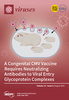
Guinea pig CMV (GPCMV) is the only small animal model for congenital disease. Knockout of GPCMV tegument protein innate immune evasion factor pp65 (GP83) attenuated the virus as a live vaccine strain, which induced an immune response in vaccinated animals. Studies demonstrated a robust neutralizing antibody response to viral glycoprotein complexes, including the viral pentamer, to prevent infection of both fibroblast and non-fibroblast cell types. Vaccination protected pregnant animals against challenges by wild-type viruses and provided complete in utero protection of pups against congenital CMV. View this paper
  • Issues are regarded as officially published after their release is announced to the table of contents alert mailing list.
  • You may sign up for e-mail alerts to receive table of contents of newly released issues.
  • PDF is the official format for papers published in both, html and pdf forms. To view the papers in pdf format, click on the "PDF Full-text" link, and use the free Adobe Reader to open them.
Order results
Result details
Section
Select all
Export citation of selected articles as:
23 pages, 7272 KiB  
Article
The Post-Acute Phase of SARS-CoV-2 Infection in Two Macaque Species Is Associated with Signs of Ongoing Virus Replication and Pathology in Pulmonary and Extrapulmonary Tissues
by Kinga P. Böszörményi, Marieke A. Stammes, Zahra C. Fagrouch, Gwendoline Kiemenyi-Kayere, Henk Niphuis, Daniella Mortier, Nikki van Driel, Ivonne Nieuwenhuis, Richard A. W. Vervenne, Tom Haaksma, Boudewijn Ouwerling, Deborah Adema, Roja Fidel Acar, Ella Zuiderwijk-Sick, Lisette Meijer, Petra Mooij, Ed J. Remarque, Herman Oostermeijer, Gerrit Koopman, Alexis C. R. Hoste, Patricia Sastre, Bart L. Haagmans, Ronald E. Bontrop, Jan A. M. Langermans, Willy M. Bogers, Ivanela Kondova, Ernst J. Verschoor and Babs E. Verstrepenadd Show full author list remove Hide full author list
Viruses 2021, 13(8), 1673; https://doi.org/10.3390/v13081673 - 23 Aug 2021
Cited by 22 | Viewed by 7580
Abstract
The post-acute phase of SARS-CoV-2 infection was investigated in rhesus (Macaca mulatta) and cynomolgus macaques (Macaca fascicularis). During the acute phase of infection, SARS-CoV-2 was shed via the nose and throat, and viral RNA was occasionally detected in feces. [...] Read more.
The post-acute phase of SARS-CoV-2 infection was investigated in rhesus (Macaca mulatta) and cynomolgus macaques (Macaca fascicularis). During the acute phase of infection, SARS-CoV-2 was shed via the nose and throat, and viral RNA was occasionally detected in feces. This phase coincided with a transient change in systemic immune activation. Even after the alleged resolution of the infection, computed tomography (CT) and positron emission tomography (PET)-CT revealed pulmonary lesions and activated tracheobronchial lymph nodes in all animals. Post-mortem histological examination of the lung tissue revealed mostly marginal or resolving minimal lesions that were indicative of SARS-CoV-2 infection. Evidence for SARS-CoV-2-induced histopathology was also found in extrapulmonary tissue samples, such as conjunctiva, cervical, and mesenteric lymph nodes. However, 5–6 weeks after SARS-CoV-2 exposure, upon necropsy, viral RNA was still detectable in a wide range of tissue samples in 50% of the macaques and included amongst others the heart, the respiratory tract and surrounding lymph nodes, salivary gland, and conjunctiva. Subgenomic messenger RNA was detected in the lungs and tracheobronchial lymph nodes, indicative of ongoing virus replication during the post-acute phase. These results could be relevant for understanding the long-term consequences of COVID-19 in humans. Full article
(This article belongs to the Special Issue SARS-CoV-2 and Animal Models)
Show Figures

Figure 1

14 pages, 2513 KiB  
Article
Characteristics of Classical Swine Fever Virus Variants Derived from Live Attenuated GPE Vaccine Seed
by Taksoo Kim, Loc Tan Huynh, Shizuka Hirose, Manabu Igarashi, Takahiro Hiono, Norikazu Isoda and Yoshihiro Sakoda
Viruses 2021, 13(8), 1672; https://doi.org/10.3390/v13081672 - 23 Aug 2021
Cited by 5 | Viewed by 2987
Abstract
The GPE strain is a live attenuated vaccine for classical swine fever (CSF) developed in Japan. In the context of increasing attention for the differentiating infected from vaccinated animals (DIVA) concept, the achievement of CSF eradication with the GPE proposes it [...] Read more.
The GPE strain is a live attenuated vaccine for classical swine fever (CSF) developed in Japan. In the context of increasing attention for the differentiating infected from vaccinated animals (DIVA) concept, the achievement of CSF eradication with the GPE proposes it as a preferable backbone for a recombinant CSF marker vaccine. While its infectious cDNA clone, vGPE, is well characterized, 10 amino acid substitutions were recognized in the genome, compared to the original GPE vaccine seed. To clarify the GPE seed availability, this study aimed to generate and characterize a clone possessing the identical amino acid sequence to the GPE seed. The attempt resulted in the loss of the infectious GPE seed clone production due to the impaired replication by an amino acid substitution in the viral polymerase NS5B. Accordingly, replication-competent GPE seed variant clones were produced. Although they were mostly restricted to propagate in the tonsils of pigs, similarly to vGPE, their type I interferon-inducing capacity was significantly lower than that of vGPE. Taken together, vGPE mainly retains ideal properties for the CSF vaccine, compared with the seed variants, and is probably useful in the development of a CSF marker vaccine. Full article
(This article belongs to the Special Issue Enteric and Respiratory Viruses in Animals)
Show Figures

Figure 1

12 pages, 1555 KiB  
Systematic Review
Congenital Zika Infection and the Risk of Neurodevelopmental, Neurological, and Urinary Track Disorders in Early Childhood. A Systematic Review
by Evangelia Antoniou, Eirini Orovou, Paraskevi Eva Andronikidi, Christos Orovas, Nikolaos Rigas, Ermioni Palaska, Angeliki Sarella, Georgios Iatrakis and Chrysa Voyiatzaki
Viruses 2021, 13(8), 1671; https://doi.org/10.3390/v13081671 - 23 Aug 2021
Cited by 5 | Viewed by 2359
Abstract
It was late 2015 when Northeast Brazil noticed a worrying increase in neonates born with microcephaly and other congenital malformations. These abnormalities, characterized by an abnormally small head and often neurological impairment and later termed Congenital Zika Syndrome, describe the severity of neurodevelopmental [...] Read more.
It was late 2015 when Northeast Brazil noticed a worrying increase in neonates born with microcephaly and other congenital malformations. These abnormalities, characterized by an abnormally small head and often neurological impairment and later termed Congenital Zika Syndrome, describe the severity of neurodevelopmental and nephrological outcomes in early childhood, and the implication of microcephaly at birth. The purpose of the study was to describe the neurodevelopmental outcomes in children exposed to Zika virus during fetal life, with and without microcephaly at birth. The systematic review included research studies about the neurodevelopmental outcomes with and without microcephaly, as well as nephrological outcomes in early childhood. We searched PubMed, Crossref, PsycINFO, Scopus, and Google Scholar publications and selected 19 research articles published from 2018 to 2021. Most studies have linked the severity of microcephaly in childbirth to the neurodevelopmental and urinary outcomes in early childhood. However, most children without microcephaly at birth develop typically, while others may be at risk for language impairment. Full article
(This article belongs to the Special Issue Zika Virus: Unanswered Questions)
Show Figures

Figure 1

14 pages, 606 KiB  
Article
Unique Severe COVID-19 Placental Signature Independent of Severity of Clinical Maternal Symptoms
by Marjolein F. Husen, Lotte E. van der Meeren, Robert M. Verdijk, Pieter L. A. Fraaij, Annemiek A. van der Eijk, Marion P. G. Koopmans, Liv Freeman, Hein Bogers, Marjolijn D. Trietsch, Irwin K. M. Reiss, Philip L. J. DeKoninck and Sam Schoenmakers
Viruses 2021, 13(8), 1670; https://doi.org/10.3390/v13081670 - 23 Aug 2021
Cited by 32 | Viewed by 16144
Abstract
Background: Although the risk for transplacental transmission of SARS-CoV-2 is rare, placental infections with adverse functional consequences have been reported. This study aims to analyse histological placental findings in pregnancies complicated by SARS-CoV-2 infection and investigate its correlation with clinical symptoms and perinatal [...] Read more.
Background: Although the risk for transplacental transmission of SARS-CoV-2 is rare, placental infections with adverse functional consequences have been reported. This study aims to analyse histological placental findings in pregnancies complicated by SARS-CoV-2 infection and investigate its correlation with clinical symptoms and perinatal outcomes. We want to determine which pregnancies are at-risk to prevent adverse pregnancy outcomes related to COVID-19 in the future. Methods: A prospective, longitudinal, multicentre, cohort study. All pregnant women presenting between April 2020 and March 2021 with a nasopharyngeal RT-PCR-confirmed SARS-CoV-2 infection were included. Around delivery, maternal, foetal and placental PCR samples were collected. Placental pathology was correlated with clinical maternal characteristics of COVID-19. Results: Thirty-six patients were included, 33 singleton pregnancies (n = 33, 92%) and three twin pregnancies (n = 3, 8%). Twenty-four (62%) placentas showed at least one abnormality. Four placentas (4/39, 10%) showed placental staining positive for the presence of SARS-CoV-2 accompanied by a unique combination of diffuse, severe inflammatory placental changes with massive perivillous fibrin depositions, necrosis of syncytiotrophoblast, diffuse chronic intervillositis, and a specific, unprecedented CD20+ B-cell infiltration. This SARS-CoV-2 placental signature seems to correlate with foetal distress (75% vs. 15.6%, p = 0.007) but not with the severity of maternal COVID-19 disease. Conclusion: We describe a unique placental signature in pregnant patients with COVID-19, which has not been reported in a historical cohort. We show that the foetal environment can be seriously compromised by disruption of placental function due to local, devastating SARS-CoV-2 infection. Maternal clinical symptoms did not predict the severity of the SARS-CoV-2-related placental signature, resulting in a lack of adequate identification of maternal criteria for pregnancies at risk. Close foetal monitoring and pregnancy termination in case of foetal distress can prevent adverse pregnancy outcomes due to COVID-19 related placental disease. Full article
Show Figures

Figure 1

13 pages, 1875 KiB  
Article
All-Trans Retinoic Acid Exhibits Antiviral Effect against SARS-CoV-2 by Inhibiting 3CLpro Activity
by Takeshi Morita, Kei Miyakawa, Sundararaj Stanleyraj Jeremiah, Yutaro Yamaoka, Mitsuru Sada, Tomoko Kuniyoshi, Jinwei Yang, Hirokazu Kimura and Akihide Ryo
Viruses 2021, 13(8), 1669; https://doi.org/10.3390/v13081669 - 23 Aug 2021
Cited by 19 | Viewed by 3317
Abstract
The pandemic of COVID-19 caused by SARS-CoV-2 continues to spread despite the global efforts taken to control it. The 3C-like protease (3CLpro), the major protease of SARS-CoV-2, is one of the most interesting targets for antiviral drug development because it is highly conserved [...] Read more.
The pandemic of COVID-19 caused by SARS-CoV-2 continues to spread despite the global efforts taken to control it. The 3C-like protease (3CLpro), the major protease of SARS-CoV-2, is one of the most interesting targets for antiviral drug development because it is highly conserved among SARS-CoVs and plays an important role in viral replication. Herein, we developed high throughput screening for SARS-CoV-2 3CLpro inhibitor based on AlphaScreen. We screened 91 natural product compounds and found that all-trans retinoic acid (ATRA), an FDA-approved drug, inhibited 3CLpro activity. The 3CLpro inhibitory effect of ATRA was confirmed in vitro by both immunoblotting and AlphaScreen with a 50% inhibition concentration (IC50) of 24.7 ± 1.65 µM. ATRA inhibited the replication of SARS-CoV-2 in VeroE6/TMPRSS2 and Calu-3 cells, with IC50 = 2.69 ± 0.09 µM in the former and 0.82 ± 0.01 µM in the latter. Further, we showed the anti-SARS-CoV-2 effect of ATRA on the currently circulating variants of concern (VOC); alpha, beta, gamma, and delta. These results suggest that ATRA may be considered as a potential therapeutic agent against SARS-CoV-2. Full article
(This article belongs to the Special Issue Vaccines and Therapeutics against Coronaviruses)
Show Figures

Figure 1

13 pages, 1857 KiB  
Article
Molecular Characterization of Human Papillomavirus Type 159 (HPV159)
by Iva Marković, Lea Hošnjak, Katja Seme and Mario Poljak
Viruses 2021, 13(8), 1668; https://doi.org/10.3390/v13081668 - 23 Aug 2021
Cited by 1 | Viewed by 2127
Abstract
Human papillomavirus type 159 (HPV159) was identified in an anal swab sample and preliminarily genetically characterized by our group in 2012. Here we present a detailed molecular in silico analysis that showed that the HPV159 viral genome is 7443 bp in length and [...] Read more.
Human papillomavirus type 159 (HPV159) was identified in an anal swab sample and preliminarily genetically characterized by our group in 2012. Here we present a detailed molecular in silico analysis that showed that the HPV159 viral genome is 7443 bp in length and divided into five early and two late genes, with conserved functional domains and motifs, and a non-coding long control region (LCR) with significant regulatory sequences that allow the virus to complete its life cycle and infect novel host cells. HPV159, clustering into the cutaneotropic Betapapillomavirus (Beta-PV) genus, is phylogenetically most similar to HPV9, forming an individual phylogenetic group in the viral species Beta-2. After testing a large representative collection of clinical samples with HPV159 type-specific RT-PCR, in addition to the anal canal from which the first HPV159 isolate was obtained, HPV159 was further detected in other muco-cutaneous (4/181, 2.2%), mucosal (22/764, 2.9%), and cutaneous (14/554, 2.5%) clinical samples, suggesting its extensive tissue tropism. However, because very low HPV159 viral loads were estimated in the majority of positive samples, it seemed that HPV159 mainly caused clinically insignificant infections of the skin and mucosa. Using newly developed, highly sensitive HPV159-specific nested PCRs, two additional HPV159 LCR viral variants were identified. Nevertheless, all HPV159 mutations were demonstrated outside important functional domains of the LCR, suggesting that the HPV159 viral variants were most probably not pathogenically different. This complete molecular characterization of HPV159 enhances our knowledge of the genome characteristics, tissue tropism, and phylogenetic diversity of Beta-PVs that infect humans. Full article
(This article belongs to the Special Issue State-of-the-Art Virology Research in Croatia)
Show Figures

Figure 1

21 pages, 8225 KiB  
Article
Peptide Platform as a Powerful Tool in the Fight against COVID-19
by Michela Murdocca, Gennaro Citro, Isabella Romeo, Antonio Lupia, Shane Miersch, Bruno Amadio, Alessia Bonomo, Antonio Rossi, Sachdev S. Sidhu, Pier Paolo Pandolfi, Stefano Alcaro, Federica Carla Sangiuolo and Giuseppe Novelli
Viruses 2021, 13(8), 1667; https://doi.org/10.3390/v13081667 - 23 Aug 2021
Cited by 9 | Viewed by 4889
Abstract
Severe acute respiratory syndrome coronavirus 2 (SARS-CoV-2) has resulted in a global pandemic causing over 195 million infections and more than 4 million fatalities as of July 2021.To date, it has been demonstrated that a number of mutations in the spike glycoprotein (S [...] Read more.
Severe acute respiratory syndrome coronavirus 2 (SARS-CoV-2) has resulted in a global pandemic causing over 195 million infections and more than 4 million fatalities as of July 2021.To date, it has been demonstrated that a number of mutations in the spike glycoprotein (S protein) of SARS-CoV-2 variants of concern abrogate or reduce the neutralization potency of several therapeutic antibodies and vaccine-elicited antibodies. Therefore, the development of additional vaccine platforms with improved supply and logistic profile remains a pressing need. In this work, we have validated the applicability of a peptide-based strategy focused on a preventive as well as a therapeutic purpose. On the basis of the involvement of the dipeptidyl peptidase 4 (DPP4), in addition to the angiotensin converting enzyme 2 (ACE2) receptor in the mechanism of virus entry, we analyzed peptides bearing DPP4 sequences by protein–protein docking and assessed their ability to block pseudovirus infection in vitro. In parallel, we have selected and synthetized peptide sequences located within the highly conserved receptor-binding domain (RBD) of the S protein, and we found that RBD-based vaccines could better promote elicitation of high titers of neutralizing antibodies specific against the regions of interest, as confirmed by immunoinformatic methodologies and in vivo studies. These findings unveil a key antigenic site targeted by broadly neutralizing antibodies and pave the way to the design of pan-coronavirus vaccines. Full article
(This article belongs to the Section SARS-CoV-2 and COVID-19)
Show Figures

Figure 1

11 pages, 12157 KiB  
Article
APOBEC Mutagenesis Is Concordant between Tumor and Viral Genomes in HPV-Positive Head and Neck Squamous Cell Carcinoma
by Daniel L. Faden, Krystle A. Lang Kuhs, Maoxuan Lin, Adam Langenbucher, Maisa Pinheiro, Meredith Yeager, Michael Cullen, Joseph F. Boland, Mia Steinberg, Sara Bass, James S. Lewis, Michael S. Lawrence, Robert L. Ferris and Lisa Mirabello
Viruses 2021, 13(8), 1666; https://doi.org/10.3390/v13081666 - 23 Aug 2021
Cited by 18 | Viewed by 3450
Abstract
APOBEC is a mutagenic source in human papillomavirus (HPV)-mediated malignancies, including HPV+ oropharyngeal squamous cell carcinoma (HPV + OPSCC), and in HPV genomes. It is unknown why APOBEC mutations predominate in HPV + OPSCC, or if the APOBEC-induced mutations observed in both human [...] Read more.
APOBEC is a mutagenic source in human papillomavirus (HPV)-mediated malignancies, including HPV+ oropharyngeal squamous cell carcinoma (HPV + OPSCC), and in HPV genomes. It is unknown why APOBEC mutations predominate in HPV + OPSCC, or if the APOBEC-induced mutations observed in both human cancers and HPV genomes are directly linked. We performed sequencing of host somatic exomes, transcriptomes, and HPV16 genomes from 79 HPV + OPSCC samples, quantifying APOBEC mutational burden and activity in both host and virus. APOBEC was the dominant mutational signature in somatic exomes. In viral genomes, there was a mean of five (range 0–29) mutations per genome. The mean of APOBEC mutations in viral genomes was one (range 0–5). Viral APOBEC mutations, compared to non-APOBEC mutations, were more likely to be low-variant allele fraction mutations, suggesting that APOBEC mutagenesis actively occurrs in viral genomes during infection. HPV16 APOBEC-induced mutation patterns in OPSCC were similar to those previously observed in cervical samples. Paired host and viral analyses revealed that APOBEC-enriched tumor samples had higher viral APOBEC mutation rates (p = 0.028), and APOBEC-associated RNA editing (p = 0.008), supporting the concept that APOBEC mutagenesis in host and viral genomes is directly linked and occurrs during infection. Using paired sequencing of host somatic exomes, transcriptomes, and viral genomes, we demonstrated for the first-time definitive evidence of concordance between tumor and viral APOBEC mutagenesis. This finding provides a missing link connecting APOBEC mutagenesis in host and virus and supports a common mechanism driving APOBEC dysregulation. Full article
(This article belongs to the Special Issue HPV Genomics)
Show Figures

Figure 1

14 pages, 3012 KiB  
Article
Antiviral Activity of Umifenovir In Vitro against a Broad Spectrum of Coronaviruses, Including the Novel SARS-CoV-2 Virus
by Irina Leneva, Nadezhda Kartashova, Artem Poromov, Anastasiia Gracheva, Ekaterina Korchevaya, Ekaterina Glubokova, Olga Borisova, Anna Shtro, Svetlana Loginova, Veronika Shchukina, Ravil Khamitov and Evgeny Faizuloev
Viruses 2021, 13(8), 1665; https://doi.org/10.3390/v13081665 - 23 Aug 2021
Cited by 17 | Viewed by 5417
Abstract
An escalating pandemic of the novel SARS-CoV-2 virus is impacting global health, and effective antivirals are needed. Umifenovir (Arbidol) is an indole-derivative molecule, licensed in Russia and China for prophylaxis and treatment of influenza and other respiratory viral infections. It has been shown [...] Read more.
An escalating pandemic of the novel SARS-CoV-2 virus is impacting global health, and effective antivirals are needed. Umifenovir (Arbidol) is an indole-derivative molecule, licensed in Russia and China for prophylaxis and treatment of influenza and other respiratory viral infections. It has been shown that umifenovir has broad spectrum activity against different viruses. We evaluated the sensitivity of different coronaviruses, including the novel SARS-CoV-2 virus, to umifenovir using in vitro assays. Using a plaque assay, we revealed an antiviral effect of umifenovir against seasonal HCoV-229E and HCoV-OC43 coronaviruses in Vero E6 cells, with estimated 50% effective concentrations (EC50) of 10.0 ± 0.5 µM and 9.0 ± 0.4 µM, respectively. Umifenovir at 90 µM significantly suppressed plaque formation in CMK-AH-1 cells infected with SARS-CoV. Umifenovir also inhibited the replication of SARS-CoV-2 virus, with EC50 values ranging from 15.37 ± 3.6 to 28.0 ± 1.0 µM. In addition, 21–36 µM of umifenovir significantly suppressed SARS-CoV-2 virus titers (≥2 log TCID50/mL) in the first 24 h after infection. Repurposing of antiviral drugs is very helpful in fighting COVID-19. A safe, pan-antiviral drug such as umifenovir could be extremely beneficial in combating the early stages of a viral pandemic. Full article
(This article belongs to the Special Issue State-of-the-Art Respiratory Viruses Research in Russia)
Show Figures

Figure 1

18 pages, 2276 KiB  
Review
Papillomaviruses in Domestic Cats
by John S. Munday and Neroli A. Thomson
Viruses 2021, 13(8), 1664; https://doi.org/10.3390/v13081664 - 22 Aug 2021
Cited by 19 | Viewed by 3445
Abstract
Papillomaviruses (PVs) are well established to cause hyperplastic papillomas (warts) in humans and animals. In addition, due to their ability to alter cell regulation, PVs are also recognized to cause approximately 5% of human cancers and these viruses have been associated with neoplasia [...] Read more.
Papillomaviruses (PVs) are well established to cause hyperplastic papillomas (warts) in humans and animals. In addition, due to their ability to alter cell regulation, PVs are also recognized to cause approximately 5% of human cancers and these viruses have been associated with neoplasia in a number of animal species. In contrast to other domestic species, cats have traditionally been thought to less frequently develop disease due to PV infection. However, in the last 15 years, the number of viruses and the different lesions associated with PVs in cats have greatly expanded. In this review, the PV life cycle and the subsequent immune response is briefly discussed along with methods used to investigate a PV etiology of a lesion. The seven PV types that are currently known to infect cats are reviewed. The lesions that have been associated with PV infections in cats are then discussed and the review finishes with a brief discussion on the use of vaccines to prevent PV-induced disease in domestic cats. Full article
(This article belongs to the Special Issue Feline Viruses and Viral Diseases 2.0)
Show Figures

Figure 1

9 pages, 1282 KiB  
Article
Efficacy of Unsupervised Self-Collected Mid-Turbinate FLOQSwabs for the Diagnosis of Coronavirus Disease 2019 (COVID-19)
by Egildo Luca D’Andrea, Alessia Maria Cossu, Marianna Scrima, Vincenzo Messina, Pasquale Iuliano, Felice Di Perna, Marco Pizza, Fabio Pizza, Nicola Coppola, Luca Rinaldi, Anna Maria Bellizzi, Chiara Pelosi, Carmen Cocca, Angelo Frieri, Fabio Lo Calzo, Giovambattista Capasso, Santina Castriciano, Paolo Maggi, Alessandra Fucci and Michele Caraglia
Viruses 2021, 13(8), 1663; https://doi.org/10.3390/v13081663 - 22 Aug 2021
Cited by 4 | Viewed by 3328
Abstract
Context: The Global Severe Acute Respiratory Syndrome Coronavirus-2 (SARS-CoV-2) pandemic has resulted in explosive patterns of transmission in most countries. Nasopharyngeal swabs were the specimen’s collection tools recommended for the diagnosis of SARS-CoV-2 infection, and for monitoring infection outbreaks in communities. Our objective [...] Read more.
Context: The Global Severe Acute Respiratory Syndrome Coronavirus-2 (SARS-CoV-2) pandemic has resulted in explosive patterns of transmission in most countries. Nasopharyngeal swabs were the specimen’s collection tools recommended for the diagnosis of SARS-CoV-2 infection, and for monitoring infection outbreaks in communities. Our objective was to report the quality and efficacy of unsupervised self-collected mid turbinate “dry FLOQSwabs” (MT FLOQSwabs) (56380CS01, Copan). There were 111 specimens collected for the study: 36 by health care personnel, from themselves, to verify the quality and efficacy of mid-turbinate swabs; 75 to compare and assess the diagnostic performance, among health care personnel, of nasopharyngeal swabs and self-collected mid-turbinate FLOQSwabs. A collection of 51 specimens was enrolled to define the efficacy of the Testami program (validation). Our analyses demonstrate that self-collected mid-turbinate dry swabs ensure an accuracy of 97.3%, as compared to the standard nasopharyngeal swabs collected by health care workers. Furthermore, the mid-turbinate FLOQSwabs can be stored without medium for six days at room temperature without affecting the molecular diagnosis of the SARS-CoV-2 virus infection. Self-collection of diagnostic specimens at home could offer an avenue to increase testing availability for SARS-CoV-2 infection without asking people to travel to a clinic or a laboratory, thus reducing people’s exposure to infection. Our findings demonstrate that unsupervised self-collection swabs, transported dry, are sensitive, practical and easy-to-use tools and should be considered for diagnosis of SARS-COV-2 and coronavirus disease 2019 (COVID-19) surveillance. Full article
(This article belongs to the Special Issue Viral Markers and the Diagnosis of COVID-19)
Show Figures

Figure 1

15 pages, 1563 KiB  
Review
Beta-Genus Human Papillomavirus 8 E6 Destabilizes the Host Genome by Promoting p300 Degradation
by Dalton Dacus and Nicholas A. Wallace
Viruses 2021, 13(8), 1662; https://doi.org/10.3390/v13081662 - 21 Aug 2021
Cited by 10 | Viewed by 2378
Abstract
The beta genus of human papillomaviruses infects cutaneous keratinocytes. Their replication depends on actively proliferating cells and, thus, they conflict with the cellular response to the DNA damage frequently encountered by these cells. This review focus on one of these viruses (HPV8) that [...] Read more.
The beta genus of human papillomaviruses infects cutaneous keratinocytes. Their replication depends on actively proliferating cells and, thus, they conflict with the cellular response to the DNA damage frequently encountered by these cells. This review focus on one of these viruses (HPV8) that counters the cellular response to damaged DNA and mitotic errors by expressing a protein (HPV8 E6) that destabilizes a histone acetyltransferase, p300. The loss of p300 results in broad dysregulation of cell signaling that decreases genome stability. In addition to discussing phenotypes caused by p300 destabilization, the review contains a discussion of the extent to which E6 from other β-HPVs destabilizes p300, and provides a discussion on dissecting HPV8 E6 biology using mutants. Full article
(This article belongs to the Special Issue Viral Infection: A Threat for Genomic Stability in Host Cells)
Show Figures

Figure 1

14 pages, 1554 KiB  
Review
Adaptation and Virulence of Enterovirus-A71
by Kyousuke Kobayashi and Satoshi Koike
Viruses 2021, 13(8), 1661; https://doi.org/10.3390/v13081661 - 21 Aug 2021
Cited by 9 | Viewed by 3220
Abstract
Outbreaks of hand, foot, and mouth disease caused by enterovirus-A71 (EV-A71) can result in many deaths, due to central nervous system complications. Outbreaks with many fatalities have occurred sporadically in the Asia-Pacific region and have become a serious public health concern. It is [...] Read more.
Outbreaks of hand, foot, and mouth disease caused by enterovirus-A71 (EV-A71) can result in many deaths, due to central nervous system complications. Outbreaks with many fatalities have occurred sporadically in the Asia-Pacific region and have become a serious public health concern. It is hypothesized that virulent mutations in the EV-A71 genome cause these occasional outbreaks. Analysis of EV-A71 neurovirulence determinants is important, but there are no virulence determinants that are widely accepted among researchers. This is because most studies have been done in artificially infected mouse models and because EV-A71 mutates very quickly to adapt to the artificial host environment. Although EV-A71 uses multiple receptors for infection, it is clear that adaptation-related mutations alter the binding specificity of the receptors and allow the virus to adopt the best entry route for each environment. Such mutations have confused interpretations of virulence in animal models. This article will discuss how environment-adapted mutations in EV-A71 occur, how they affect virulence, and how such mutations can be avoided. We also discuss future perspectives for EV-A71 virulence research. Full article
(This article belongs to the Special Issue RNA Viruses: Structure, Adaptation, and Evolution)
Show Figures

Figure 1

11 pages, 1482 KiB  
Article
Co-Circulation of Phleboviruses and Leishmania Parasites in Sand Flies from a Single Site in Italy Monitored between 2017 and 2020
by Mattia Calzolari, Giuseppe Romeo, Emanuele Callegari, Paolo Bonilauri, Chiara Chiapponi, Elena Carra, Gianluca Rugna, Roberta Taddei, Davide Lelli and Michele Dottori
Viruses 2021, 13(8), 1660; https://doi.org/10.3390/v13081660 - 21 Aug 2021
Cited by 11 | Viewed by 2491
Abstract
Sand flies transmit Leishmania infantum, which is responsible for causing leishmaniasis, as well as many phleboviruses, including the human pathogenic Toscana virus. We screened sand flies collected from a single site between 2017 and 2020 for the presence of both phleboviruses and [...] Read more.
Sand flies transmit Leishmania infantum, which is responsible for causing leishmaniasis, as well as many phleboviruses, including the human pathogenic Toscana virus. We screened sand flies collected from a single site between 2017 and 2020 for the presence of both phleboviruses and Leishmania. The sand flies were sampled with attractive carbon dioxide traps and CDC light traps between May and October. We collected more than 50,000 sand flies; 2826 were identified at the species level as Phlebotomus perfiliewi (98%) or Phlebotomus perniciosus (2%). A total of 16,789 sand flies were tested in 355 pools, and phleboviruses were found in 61 pools (6 Toscana virus positive pools, 2 Corfou virus positive pools, 42 Fermo virus positive pools, and 7 Ponticelli virus positive pools, and 4 unidentified phlebovirus positive pools). Leishmania was found in 75 pools and both microorganisms were detected in 16 pools. We isolated nine phleboviruses from another 2960 sand flies (five Ponticelli viruses and for Fermo viruses), not tested for Leishmania; the complete genome of a Fermo virus isolate was sequenced. The simultaneous detection in space and time of the Fermo virus and L. infantum is evidence that supports the co-circulation of both microorganisms in the same location and partial overlap of their cycles. A detailed characterization of the epidemiology of these microorganisms will support measures to limit their transmission. Full article
(This article belongs to the Special Issue Sand Fly-Borne Phleboviruses)
Show Figures

Figure 1

12 pages, 2278 KiB  
Article
Persistence of Multiple Paramyxoviruses in a Closed Captive Colony of Fruit Bats (Eidolon helvum)
by Louise Gibson, Maria Puig Ribas, James Kemp, Olivier Restif, Richard D. Suu-Ire, James L. N. Wood and Andrew A. Cunningham
Viruses 2021, 13(8), 1659; https://doi.org/10.3390/v13081659 - 20 Aug 2021
Cited by 7 | Viewed by 3423
Abstract
Bats have been identified as the natural hosts of several emerging zoonotic viruses, including paramyxoviruses, such as Hendra and Nipah viruses, that can cause fatal disease in humans. Recently, African fruit bats with populations that roost in or near urban areas have been [...] Read more.
Bats have been identified as the natural hosts of several emerging zoonotic viruses, including paramyxoviruses, such as Hendra and Nipah viruses, that can cause fatal disease in humans. Recently, African fruit bats with populations that roost in or near urban areas have been shown to harbour a great diversity of paramyxoviruses, posing potential spillover risks to public health. Understanding the circulation of these viruses in their reservoir populations is essential to predict and prevent future emerging diseases. Here, we identify a high incidence of multiple paramyxoviruses in urine samples collected from a closed captive colony of circa 115 straw-coloured fruit bats (Eidolon helvum). The sequences detected have high nucleotide identities with those derived from free ranging African fruit bats and form phylogenetic clusters with the Henipavirus genus, Pararubulavirus genus and other unclassified paramyxoviruses. As this colony had been closed for 5 years prior to this study, these results indicate that within-host paramyxoviral persistence underlies the role of bats as reservoirs of these viruses. Full article
(This article belongs to the Special Issue Ecology of Virus Emergence from Wildlife)
Show Figures

Figure 1

15 pages, 1426 KiB  
Review
Cross Talk between Viruses and Insect Cells Cytoskeleton
by Ayda Khorramnejad, Hugo D. Perdomo, Umberto Palatini, Mariangela Bonizzoni and Laila Gasmi
Viruses 2021, 13(8), 1658; https://doi.org/10.3390/v13081658 - 20 Aug 2021
Cited by 10 | Viewed by 3736
Abstract
Viruses are excellent manipulators of host cellular machinery, behavior, and life cycle, with the host cell cytoskeleton being a primordial viral target. Viruses infecting insects generally enter host cells through clathrin-mediated endocytosis or membrane fusion mechanisms followed by transport of the viral particles [...] Read more.
Viruses are excellent manipulators of host cellular machinery, behavior, and life cycle, with the host cell cytoskeleton being a primordial viral target. Viruses infecting insects generally enter host cells through clathrin-mediated endocytosis or membrane fusion mechanisms followed by transport of the viral particles to the corresponding replication sites. After viral replication, the viral progeny egresses toward adjacent cells and reaches the different target tissues. Throughout all these steps, actin and tubulin re-arrangements are driven by viruses. The mechanisms used by viruses to manipulate the insect host cytoskeleton are well documented in the case of alphabaculoviruses infecting Lepidoptera hosts and plant viruses infecting Hemiptera vectors, but they are not well studied in case of other insect–virus systems such as arboviruses–mosquito vectors. Here, we summarize the available knowledge on how viruses manipulate the insect host cell cytoskeleton, and we emphasize the primordial role of cytoskeleton components in insect virus motility and the need to expand the study of this interaction. Full article
(This article belongs to the Special Issue Viral Manipulation of Host Cytoskeletal Networks)
Show Figures

Figure 1

36 pages, 3134 KiB  
Review
Equine Influenza Virus and Vaccines
by Fatai S. Oladunni, Saheed Oluwasina Oseni, Luis Martinez-Sobrido and Thomas M. Chambers
Viruses 2021, 13(8), 1657; https://doi.org/10.3390/v13081657 - 20 Aug 2021
Cited by 21 | Viewed by 6851
Abstract
Equine influenza virus (EIV) is a constantly evolving viral pathogen that is responsible for yearly outbreaks of respiratory disease in horses termed equine influenza (EI). There is currently no evidence of circulation of the original H7N7 strain of EIV worldwide; however, the EIV [...] Read more.
Equine influenza virus (EIV) is a constantly evolving viral pathogen that is responsible for yearly outbreaks of respiratory disease in horses termed equine influenza (EI). There is currently no evidence of circulation of the original H7N7 strain of EIV worldwide; however, the EIV H3N8 strain, which was first isolated in the early 1960s, remains a major threat to most of the world’s horse populations. It can also infect dogs. The ability of EIV to constantly accumulate mutations in its antibody-binding sites enables it to evade host protective immunity, making it a successful viral pathogen. Clinical and virological protection against EIV is achieved by stimulation of strong cellular and humoral immunity in vaccinated horses. However, despite EI vaccine updates over the years, EIV remains relevant, because the protective effects of vaccines decay and permit subclinical infections that facilitate transmission into susceptible populations. In this review, we describe how the evolution of EIV drives repeated EI outbreaks even in horse populations with supposedly high vaccination coverage. Next, we discuss the approaches employed to develop efficacious EI vaccines for commercial use and the existing system for recommendations on updating vaccines based on available clinical and virological data to improve protective immunity in vaccinated horse populations. Understanding how EIV biology can be better harnessed to improve EI vaccines is central to controlling EI. Full article
(This article belongs to the Special Issue Equine Influenza Viruses)
Show Figures

Figure 1

15 pages, 2290 KiB  
Article
Protective Effects of Astodrimer Sodium 1% Nasal Spray Formulation against SARS-CoV-2 Nasal Challenge in K18-hACE2 Mice
by Jeremy R. A. Paull, Carolyn A. Luscombe, Alex Castellarnau, Graham P. Heery, Michael D. Bobardt and Philippe A. Gallay
Viruses 2021, 13(8), 1656; https://doi.org/10.3390/v13081656 - 20 Aug 2021
Cited by 14 | Viewed by 10748
Abstract
Strategies to combat COVID-19 require multiple ways to protect vulnerable people from infection. SARS-CoV-2 is an airborne pathogen and the nasal cavity is a primary target of infection. The K18-hACE2 mouse model was used to investigate the anti-SARS-CoV-2 efficacy of astodrimer sodium formulated [...] Read more.
Strategies to combat COVID-19 require multiple ways to protect vulnerable people from infection. SARS-CoV-2 is an airborne pathogen and the nasal cavity is a primary target of infection. The K18-hACE2 mouse model was used to investigate the anti-SARS-CoV-2 efficacy of astodrimer sodium formulated in a mucoadhesive nasal spray. Animals received astodrimer sodium 1% nasal spray or PBS intranasally, or intranasally and intratracheally, for 7 days, and they were infected intranasally with SARS-CoV-2 after the first product administration on Day 0. Another group was infected intranasally with SARS-CoV-2 that had been pre-incubated with astodrimer sodium 1% nasal spray or PBS for 60 min before the neutralisation of test product activity. Astodrimer sodium 1% significantly reduced the viral genome copies (>99.9%) and the infectious virus (~95%) in the lung and trachea vs. PBS. The pre-incubation of SARS-CoV-2 with astodrimer sodium 1% resulted in a significant reduction in the viral genome copies (>99.9%) and the infectious virus (>99%) in the lung and trachea, and the infectious virus was not detected in the brain or liver. Astodrimer sodium 1% resulted in a significant reduction of viral genome copies in nasal secretions vs. PBS on Day 7 post-infection. A reduction in the viral shedding from the nasal cavity may result in lower virus transmission rates. Viraemia was low or undetectable in animals treated with astodrimer sodium 1% or infected with treated virus, correlating with the lack of detectable viral replication in the liver. Similarly, low virus replication in the nasal cavity after treatment with astodrimer sodium 1% potentially protected the brain from infection. Astodrimer sodium 1% significantly reduced the pro-inflammatory cytokines IL-6, IL-1α, IL-1β, TNFα and TGFβ and the chemokine MCP-1 in the serum, lung and trachea vs. PBS. Astodrimer sodium 1% nasal spray blocked or reduced SARS-CoV-2 replication and its sequelae in K18-hACE2 mice. These data indicate a potential role for the product in preventing SARS-CoV-2 infection or for reducing the severity of COVID-19. Full article
Show Figures

Figure 1

18 pages, 1968 KiB  
Article
Identification of a Common Conformational Epitope on the Glycoprotein E2 of Classical Swine Fever Virus and Border Disease Virus
by Yu-Liang Huang, Denise Meyer, Alexander Postel, Kuo-Jung Tsai, Hsin-Meng Liu, Chia-Huei Yang, Yu-Chun Huang, Nicholas Berkley, Ming-Chung Deng, Fun-In Wang, Paul Becher, Helen Crooke and Chia-Yi Chang
Viruses 2021, 13(8), 1655; https://doi.org/10.3390/v13081655 - 20 Aug 2021
Cited by 9 | Viewed by 2946
Abstract
Classical swine fever virus (CSFV) shares high structural and antigenic homology with bovine viral diarrhea virus (BVDV) and border disease virus (BDV). Because all three viruses can infect swine and elicit cross-reactive antibodies, it is necessary to differentiate among them with regard to [...] Read more.
Classical swine fever virus (CSFV) shares high structural and antigenic homology with bovine viral diarrhea virus (BVDV) and border disease virus (BDV). Because all three viruses can infect swine and elicit cross-reactive antibodies, it is necessary to differentiate among them with regard to serological diagnosis of classical swine fever. To understand the mechanism of cross-reactivity, it is important to define common or specific epitopes of these viruses. For this purpose, epitope mapping of six monoclonal antibodies (mAbs) was performed using recombinant expressed antigenic domains of CSFV and BDV E2 proteins. One CSFV-specific conformational epitope and one CSFV and BDV common epitope within domain B/C of E2 were identified. Site-directed mutagenesis confirmed that residues G725 and V738/I738 of the CSFV-specific epitope and P709/L709 and E713 of the second epitope are important for mAbs binding. Infection of CSFV in porcine cells was significantly reduced after pre-incubation of the cells with the domain B/C of E2 or after pre-incubation of CSFV with the mAbs detecting domain B/C. 3D structural modeling suggested that both epitopes are exposed on the surface of E2. Based on this, the identified epitopes represent a potential target for virus neutralization and might be involved in the early steps of CSFV infection. Full article
(This article belongs to the Special Issue Advances in Pestivirus Research)
Show Figures

Figure 1

13 pages, 1584 KiB  
Article
Longitudinal Secretion of Paramyxovirus RNA in the Urine of Straw-Coloured Fruit Bats (Eidolon helvum)
by Elli Rosa Jolma, Louise Gibson, Richard D. Suu-Ire, Grace Fleischer, Samuel Asumah, Sylvester Languon, Olivier Restif, James L. N. Wood and Andrew A. Cunningham
Viruses 2021, 13(8), 1654; https://doi.org/10.3390/v13081654 - 20 Aug 2021
Cited by 2 | Viewed by 3052
Abstract
The straw-coloured fruit bat (Eidolon helvum) is widespread in sub-Saharan Africa and is widely hunted for bushmeat. It is known to harbour a range of paramyxoviruses, including rubuloviruses and henipaviruses, but the zoonotic potential of these is unknown. We previously found [...] Read more.
The straw-coloured fruit bat (Eidolon helvum) is widespread in sub-Saharan Africa and is widely hunted for bushmeat. It is known to harbour a range of paramyxoviruses, including rubuloviruses and henipaviruses, but the zoonotic potential of these is unknown. We previously found a diversity of paramyxoviruses within a small, captive colony of E. helvum after it had been closed to contact with other bats for 5 years. In this study, we used under-roost urine collection to further investigate the paramyxovirus diversity and ecology in this colony, which had been closed to the outside for 10 years at the time of sampling. By sampling urine weekly throughout an entire year, we investigated possible seasonal patterns of shedding of virus or viral RNA. Using a generic paramyxovirus L-gene PCR, we detected eight distinct paramyxovirus RNA sequences. Six distinct sequences were detected using a Henipavirus-specific PCR that targeted a different region of the L-gene. Sequence detection had a bi-annual pattern, with the greatest peak in July, although different RNA sequences appeared to have different shedding patterns. No significant associations were detected between sequence detection and birthing season, environmental temperature or humidity, and no signs of illness were detected in any of the bats in the colony during the period of sample collection. Full article
(This article belongs to the Special Issue Ecology of Virus Emergence from Wildlife)
Show Figures

Figure 1

19 pages, 10426 KiB  
Article
The Microvillar and Solitary Chemosensory Cells as the Novel Targets of Infection of SARS-CoV-2 in Syrian Golden Hamsters
by Jin-Seok Seo, Sun-Woo Yoon, Seung-Hyeon Hwang, Sung-Min Nam, Sang-Soep Nahm, Jei-Hyun Jeong, Jiho Lee, Ha-Na Youn, Jun-Beom Kim and Woosuk Kim
Viruses 2021, 13(8), 1653; https://doi.org/10.3390/v13081653 - 20 Aug 2021
Cited by 7 | Viewed by 2579
Abstract
Patients infected with severe acute respiratory syndrome coronavirus 2 (SARS-CoV-2), the causative agent of coronavirus disease 2019, suffer from respiratory and non-respiratory symptoms. Among these symptoms, the loss of smell has attracted considerable attention. The objectives of this study were to determine which [...] Read more.
Patients infected with severe acute respiratory syndrome coronavirus 2 (SARS-CoV-2), the causative agent of coronavirus disease 2019, suffer from respiratory and non-respiratory symptoms. Among these symptoms, the loss of smell has attracted considerable attention. The objectives of this study were to determine which cells are infected, what happens in the olfactory system after viral infection, and how these pathologic changes contribute to olfactory loss. For this purpose, Syrian golden hamsters were used. First, we verified the olfactory structures in the nasal cavity of Syrian golden hamsters, namely the main olfactory epithelium, the vomeronasal organ, and their cellular components. Second, we found angiotensin-converting enzyme 2 expression, a receptor protein of SARS-CoV-2, in both structures and infections of supporting, microvillar, and solitary chemosensory cells. Third, we observed pathological changes in the infected epithelium, including reduced thickness of the mucus layer, detached epithelia, indistinct layers of epithelia, infiltration of inflammatory cells, and apoptotic cells in the overall layers. We concluded that a structurally and functionally altered microenvironment influences olfactory function. We observed the regeneration of the damaged epithelium, and found multilayers of basal cells, indicating that they were activated and proliferating to reconstitute the injured epithelium. Full article
(This article belongs to the Section SARS-CoV-2 and COVID-19)
Show Figures

Figure 1

19 pages, 6024 KiB  
Article
RNA and Sugars, Unique Properties of Bacteriophages Infecting Multidrug Resistant Acinetobacter radioresistens Strain LH6
by Clay S. Crippen, Bibi Zhou, Silke Andresen, Robert T. Patry, Artur Muszyński, Craig T. Parker, Kerry K. Cooper and Christine M. Szymanski
Viruses 2021, 13(8), 1652; https://doi.org/10.3390/v13081652 - 20 Aug 2021
Cited by 2 | Viewed by 2696
Abstract
Bacteriophages (phages) are predicted to be the most ubiquitous biological entity on earth, and yet, there are still vast knowledge gaps in our understanding of phage diversity and phage–host interactions. Approximately one hundred Acinetobacter-infecting DNA viruses have been identified, and in this [...] Read more.
Bacteriophages (phages) are predicted to be the most ubiquitous biological entity on earth, and yet, there are still vast knowledge gaps in our understanding of phage diversity and phage–host interactions. Approximately one hundred Acinetobacter-infecting DNA viruses have been identified, and in this report, we describe eight more. We isolated two typical dsDNA lytic podoviruses (CAP1–2), five unique dsRNA lytic cystoviruses (CAP3–7), and one dsDNA lysogenic siphovirus (SLAP1), all capable of infecting the multidrug resistant isolate Acinetobacter radioresistens LH6. Using transmission electron microscopy, bacterial mutagenesis, phage infectivity assays, carbohydrate staining, mass-spectrometry, genomic sequencing, and comparative studies, we further characterized these phages. Mutation of the LH6 initiating glycosyltransferase homolog, PglC, necessary for both O-linked glycoprotein and capsular polysaccharide (CPS) biosynthesis, prevented infection by the lytic podovirus CAP1, while mutation of the pilin protein, PilA, prevented infection by CAP3, representing the lytic cystoviruses. Genome sequencing of the three dsRNA segments of the isolated cystoviruses revealed low levels of homology, but conserved synteny with the only other reported cystoviruses that infect Pseudomonas species. In Pseudomonas, the cystoviruses are known to be enveloped phages surrounding their capsids with the inner membrane from the infected host. To characterize any membrane-associated glycoconjugates in the CAP3 cystovirus, carbohydrate staining was used to identify a low molecular weight lipid-linked glycoconjugate subsequently identified by mutagenesis and mass-spectrometry as bacterial lipooligosaccharide. Together, this study demonstrates the isolation of new Acinetobacter-infecting phages and the determination of their cell receptors. Further, we describe the genomes of a new genus of Cystoviruses and perform an initial characterization of membrane-associated glycoconjugates. Full article
(This article belongs to the Special Issue Advances in Bacteriophage Biology)
Show Figures

Graphical abstract

22 pages, 7206 KiB  
Article
Large-Scale International Validation of an Indirect ELISA Based on Recombinant Nucleocapsid Protein of Rift Valley Fever Virus for the Detection of IgG Antibody in Domestic Ruminants
by Janusz T. Pawęska, Petrus Jansen van Vuren, Veerle Msimang, Modu Moustapha Lô, Yaya Thiongane, Leopold K. Mulumba-Mfumu, Alqadasi Mansoor, José M. Fafetine, Joseph W. Magona, Hiver Boussini, Barbara Bażanow, William C. Wilson, Michel Pepin, Hermann Unger and Gerrit Viljoen
Viruses 2021, 13(8), 1651; https://doi.org/10.3390/v13081651 - 19 Aug 2021
Cited by 1 | Viewed by 2514
Abstract
Diagnostic performance of an indirect enzyme-linked immunosorbent assay (I-ELISA) based on a recombinant nucleocapsid protein (rNP) of the Rift Valley fever virus (RVFV) was validated for the detection of the IgG antibody in sheep (n = 3367), goat (n = 2632), [...] Read more.
Diagnostic performance of an indirect enzyme-linked immunosorbent assay (I-ELISA) based on a recombinant nucleocapsid protein (rNP) of the Rift Valley fever virus (RVFV) was validated for the detection of the IgG antibody in sheep (n = 3367), goat (n = 2632), and cattle (n = 3819) sera. Validation data sets were dichotomized according to the results of a virus neutralization test in sera obtained from RVF-endemic (Burkina Faso, Democratic Republic of Congo, Mozambique, Senegal, Uganda, and Yemen) and RVF-free countries (France, Poland, and the USA). Cut-off values were defined using the two-graph receiver operating characteristic analysis. Estimates of the diagnostic specificity of the RVFV rNP I-ELISA in animals from RVF-endemic countries ranged from 98.6% (cattle) to 99.5% (sheep) while in those originating from RVF-free countries, they ranged from 97.7% (sheep) to 98.1% (goats). Estimates of the diagnostic sensitivity in ruminants from RVF-endemic countries ranged from 90.7% (cattle) to 100% (goats). The results of this large-scale international validation study demonstrate the high diagnostic accuracy of the RVFV rNP I-ELISA. Standard incubation and inactivation procedures evaluated did not have an adverse effect on the detectable levels of the anti-RVFV IgG in ruminant sera and thus, together with recombinant antigen-based I-ELISA, provide a simple, safe, and robust diagnostic platform that can be automated and carried out outside expensive bio-containment facilities. These advantages are particularly important for less-resourced countries where there is a need to accelerate and improve RVF surveillance and research on epidemiology as well as to advance disease control measures. Full article
(This article belongs to the Special Issue Arboviruses: Molecular Biology, Evolution and Control)
Show Figures

Figure 1

13 pages, 2255 KiB  
Brief Report
The Ebola Virus Interferon Antagonist VP24 Undergoes Active Nucleocytoplasmic Trafficking
by Angela R. Harrison, Cassandra T. David, Stephen M. Rawlinson and Gregory W. Moseley
Viruses 2021, 13(8), 1650; https://doi.org/10.3390/v13081650 - 19 Aug 2021
Cited by 6 | Viewed by 2210
Abstract
Viral interferon (IFN) antagonist proteins mediate evasion of IFN-mediated innate immunity and are often multifunctional, with distinct roles in viral replication. The Ebola virus IFN antagonist VP24 mediates nucleocapsid assembly, and inhibits IFN-activated signaling by preventing nuclear import of STAT1 via competitive binding [...] Read more.
Viral interferon (IFN) antagonist proteins mediate evasion of IFN-mediated innate immunity and are often multifunctional, with distinct roles in viral replication. The Ebola virus IFN antagonist VP24 mediates nucleocapsid assembly, and inhibits IFN-activated signaling by preventing nuclear import of STAT1 via competitive binding to nuclear import receptors (karyopherins). Proteins of many viruses, including viruses with cytoplasmic replication cycles, interact with nuclear trafficking machinery to undergo nucleocytoplasmic transport, with key roles in pathogenesis; however, despite established karyopherin interaction, potential nuclear trafficking of VP24 has not been investigated. We find that inhibition of nuclear export pathways or overexpression of VP24-binding karyopherin results in nuclear localization of VP24. Molecular mapping indicates that cytoplasmic localization of VP24 depends on a CRM1-dependent nuclear export sequence at the VP24 C-terminus. Nuclear export is not required for STAT1 antagonism, consistent with competitive karyopherin binding being the principal antagonistic mechanism, while export mediates return of nuclear VP24 to the cytoplasm where replication/nucleocapsid assembly occurs. Full article
(This article belongs to the Section Animal Viruses)
Show Figures

Figure 1

12 pages, 1649 KiB  
Brief Report
Serological Detection of SARS-CoV-2 Antibodies in Naturally-Infected Mink and Other Experimentally-Infected Animals
by Francisco J. Berguido, Peter D. Burbelo, Alessio Bortolami, Francesco Bonfante, Kerstin Wernike, Donata Hoffmann, Anne Balkema-Buschmann, Martin Beer, William G. Dundon, Charles E. Lamien and Giovanni Cattoli
Viruses 2021, 13(8), 1649; https://doi.org/10.3390/v13081649 - 19 Aug 2021
Cited by 6 | Viewed by 2695
Abstract
The recent emergence of SARS-CoV-2 in humans from a yet unidentified animal reservoir and the capacity of the virus to naturally infect pets, farmed animals and potentially wild animals has highlighted the need for serological surveillance tools. In this study, the luciferase immunoprecipitation [...] Read more.
The recent emergence of SARS-CoV-2 in humans from a yet unidentified animal reservoir and the capacity of the virus to naturally infect pets, farmed animals and potentially wild animals has highlighted the need for serological surveillance tools. In this study, the luciferase immunoprecipitation systems (LIPS), employing the spike (S) and nucleocapsid proteins (N) of SARS-CoV-2, was used to examine the suitability of the assay for antibody detection in different animal species. Sera from SARS-CoV-2 naturally-infected mink (n = 77), SARS-CoV-2 experimentally-infected ferrets, fruit bats and hamsters and a rabbit vaccinated with a purified spike protein were examined for antibodies using the SARS-CoV-2 N and/or S proteins. From comparison with the known neutralization status of the serum samples, statistical analyses including calculation of the Spearman rank-order-correlation coefficient and Cohen’s kappa agreement were used to interpret the antibody results and diagnostic performance. The LIPS immunoassay robustly detected the presence of viral antibodies in naturally infected SARS-CoV-2 mink, experimentally infected ferrets, fruit bats and hamsters as well as in an immunized rabbit. For the SARS-CoV-2-LIPS-S assay, there was a good level of discrimination between the positive and negative samples for each of the five species tested with 100% agreement with the virus neutralization results. In contrast, the SARS-CoV-2-LIPS-N assay did not consistently differentiate between SARS-CoV-2 positive and negative sera. This study demonstrates the suitability of the SARS-CoV-2-LIPS-S assay for the sero-surveillance of SARS-CoV-2 infection in a range of animal species. Full article
(This article belongs to the Topic Veterinary Infectious Diseases)
Show Figures

Figure 1

24 pages, 5568 KiB  
Article
SARS-CoV-2 Seroprevalence Structure of the Russian Population during the COVID-19 Pandemic
by Anna Y. Popova, Viacheslav S. Smirnov, Elena E. Andreeva, Elena A. Babura, Sergey V. Balakhonov, Natalia S. Bashketova, Svetlana A. Bugorkova, Maxim V. Bulanov, Natalia. N. Valeullina, Viacheslav. V. Vetrov, Dmitriy. V. Goryaev, Tatyana N. Detkovskaya, Elena B. Ezhlova, Natalia N. Zaitseva, Olga A. Istorik, Irina. V. Kovalchuk, Dmitriy N. Kozlovskikh, Svetlana Y. Kombarova, Olga. P. Kurganova, Alexander. E. Lomovtsev, Lena A. Lukicheva, Ludmila V. Lyalina, Albina. A. Melnikova, Olga M. Mikailova, Alexei K. Noskov, Ludmila N. Noskova, Elena E. Oglezneva, Tatyana P. Osmolovskaya, Marina A. Patyashina, Natalia A. Penkovskaya, Lada V. Samoilova, Tatyana F. Stepanova, Olga E. Trotsenko and Areg A. Totolianadd Show full author list remove Hide full author list
Viruses 2021, 13(8), 1648; https://doi.org/10.3390/v13081648 - 19 Aug 2021
Cited by 20 | Viewed by 3028
Abstract
The SARS-CoV-2 pandemic, which came to Russia in March 2020, is accompanied by morbidity level changes and can be tracked using serological monitoring of a representative population sample from Federal Districts (FDs) and individual regions. In a longitudinal cohort study conducted in 26 [...] Read more.
The SARS-CoV-2 pandemic, which came to Russia in March 2020, is accompanied by morbidity level changes and can be tracked using serological monitoring of a representative population sample from Federal Districts (FDs) and individual regions. In a longitudinal cohort study conducted in 26 model regions of Russia, distributed across all FDs, we investigated the distribution and cumulative proportions of individuals with antibodies (Abs) to the SARS-CoV-2 nucleocapsid antigen (Ag), in the period from June to December 2020, using a three-phase monitoring process. In addition, during the formation of the cohort of volunteers, the number of seropositive convalescents, persons who had contact with patients or COVID-19 convalescents, and the prevalence of asymptomatic forms of infection among seropositive volunteers were determined. According to a uniform methodology, 3 mL of blood was taken from the examined individuals, and plasma was separated, from which the presence of Abs to nucleocapsid Ag was determined on a Thermo Scientific Multiascan FC device using the “ELISA anti-SARS-CoV-2 IgG” reagent set (prod. Scientific Center for Applied Microbiology and Biotechnology), in accordance with the developer’s instructions. Volunteers (74,158) were surveyed and divided into seven age groups (1–17, 18–29, 30–39, 40–49, 59–59, 60–69, and 70+ years old), among whom 14,275 were identified as having antibodies to SARS-CoV-2. The average percent seropositive in Russia was 17.8% (IQR: 8.8–23.2). The largest proportion was found among children under 17 years old (21.6% (IQR: 13.1–31.7). In the remaining groups, seroprevalence ranged from 15.6% (IQR: 8–21.1) to 18.0% (IQR: 13.4–22.6). During monitoring, three (immune) response groups were found: (A) groups with a continuous increase in the proportion of seropositive; (B) those with a slow rate of increase in seroprevalence; and (C) those with a two-phase curve, wherein the initial increase was replaced by a decrease in the percentage of seropositive individuals. A significant correlation was revealed between the number of COVID-19 convalescents and contact persons, and between the number of contacts and healthy seropositive volunteers. Among the seropositive volunteers, more than 93.6% (IQR: 87.1–94.9) were asymptomatic. The results show that the COVID-19 pandemic is accompanied by an increase in seroprevalence, which may be important for the formation of herd immunity. Full article
(This article belongs to the Special Issue State-of-the-Art Respiratory Viruses Research in Russia)
Show Figures

Figure 1

14 pages, 2336 KiB  
Article
Monitoring SARS-CoV-2 Populations in Wastewater by Amplicon Sequencing and Using the Novel Program SAM Refiner
by Devon A. Gregory, Chris G. Wieberg, Jeff Wenzel, Chung-Ho Lin and Marc C. Johnson
Viruses 2021, 13(8), 1647; https://doi.org/10.3390/v13081647 - 19 Aug 2021
Cited by 22 | Viewed by 4233
Abstract
Sequencing Severe Acute Respiratory Syndrome Coronavirus 2 (SARS-CoV-2) from wastewater has become a useful tool in monitoring the spread of viral variants. Approaches to this task have been varied, relying on differing sequencing methods and computational analyses. We used a novel computation workflow [...] Read more.
Sequencing Severe Acute Respiratory Syndrome Coronavirus 2 (SARS-CoV-2) from wastewater has become a useful tool in monitoring the spread of viral variants. Approaches to this task have been varied, relying on differing sequencing methods and computational analyses. We used a novel computation workflow based on amplicon sequencing of SARS-CoV-2 spike domains in order to track viral populations in wastewater. As part of this workflow, we developed a program, SAM Refiner, that has a variety of outputs, including novel variant reporting as well as functions designed to remove polymerase chain reaction (PCR) generated chimeric sequences. With these methods, we were able to track viral population dynamics over time. We report here on the emergence of two variants of concern, B.1.1.7 (Alpha) and P.1 (Gamma), and their displacement of the D614G B.1 variant in a Missouri sewershed. Full article
(This article belongs to the Section SARS-CoV-2 and COVID-19)
Show Figures

Figure 1

4 pages, 348 KiB  
Editorial
1984—Discovery of the First Anti-HIV Drug, Suramin
by Erik De Clercq
Viruses 2021, 13(8), 1646; https://doi.org/10.3390/v13081646 - 19 Aug 2021
Cited by 3 | Viewed by 2504
Abstract
In 2021, we commemorate the 40th anniversary of the identification of the disease AIDS, the acquired immune deficiency syndrome, a name that for the first time in history was launched in 1981 [...] Full article
(This article belongs to the Special Issue The Fight between Human Beings and HIV: 40th Anniversary and Beyond)
Show Figures

Figure 1

7 pages, 536 KiB  
Article
Immunogenicity of Low-Dose Prime-Boost Vaccination of mRNA Vaccine CV07050101 in Non-Human Primates
by Neeltje van Doremalen, Robert J. Fischer, Jonathan E. Schulz, Myndi G. Holbrook, Brian J. Smith, Jamie Lovaglio, Benjamin Petsch and Vincent J. Munster
Viruses 2021, 13(8), 1645; https://doi.org/10.3390/v13081645 - 19 Aug 2021
Cited by 8 | Viewed by 2645
Abstract
Many different vaccine candidates against severe acute respiratory syndrome coronavirus 2 (SARS-CoV-2), the etiological agent of COVID-19, are currently approved and under development. Vaccine platforms vary from mRNA vaccines to viral-vectored vaccines, and several candidates have been shown to produce humoral and cellular [...] Read more.
Many different vaccine candidates against severe acute respiratory syndrome coronavirus 2 (SARS-CoV-2), the etiological agent of COVID-19, are currently approved and under development. Vaccine platforms vary from mRNA vaccines to viral-vectored vaccines, and several candidates have been shown to produce humoral and cellular responses in small animal models, non-human primates, and human volunteers. In this study, six non-human primates received a prime-boost intramuscular vaccination with 4 µg of mRNA vaccine candidate CV07050101, which encodes a pre-fusion stabilized spike (S) protein of SARS-CoV-2. Boost vaccination was performed 28 days post prime vaccination. As a control, six animals were similarly injected with PBS. Humoral and cellular immune responses were investigated at time of vaccination, and two weeks afterwards. No antibodies could be detected at two and four weeks after prime vaccination. Two weeks after boost vaccination, binding but no neutralizing antibodies were detected in four out of six non-human primates. SARS-CoV-2 S protein-specific T cell responses were detected in these four animals. In conclusion, prime-boost vaccination with 4 µg of vaccine candidate CV07050101 resulted in limited immune responses in four out of six non-human primates. Full article
(This article belongs to the Section Coronaviruses)
Show Figures

Figure 1

12 pages, 330 KiB  
Review
Barriers towards HPV Vaccinations for Boys and Young Men: A Narrative Review
by Maria Grandahl and Tryggve Nevéus
Viruses 2021, 13(8), 1644; https://doi.org/10.3390/v13081644 - 19 Aug 2021
Cited by 31 | Viewed by 6537
Abstract
Background: HPV vaccination of both girls and boys can protect against infection and eliminate the risk for HPV-associated cancer. Due to a common misconception that the virus only poses risks to women, vaccine coverage is suboptimal among men in many countries. It is [...] Read more.
Background: HPV vaccination of both girls and boys can protect against infection and eliminate the risk for HPV-associated cancer. Due to a common misconception that the virus only poses risks to women, vaccine coverage is suboptimal among men in many countries. It is urgent to identify barriers to vaccination of boys and men. Methods: We conducted a narrative review of publications examining attitudes and beliefs regarding HPV vaccination for boys and young men. The electronic databases searched were PubMed, PsychInfo and Scopus (December 2020; last update July 2021). A total of 103 original articles were included in the final analysis. Results: The central barriers against vaccination of boys and men are: (1) lack of knowledge, (2) vaccine hesitancy in general, (3) lack of recommendation from and/or discussions with healthcare providers, (4) cost and logistics, and (5) the idea that HPV vaccination may promote promiscuity. Men who have sex with men and families belonging to ethnic minorities express a need for information tailored to their situation. Conclusions: Boys should be included in national immunization programs and men should also be offered catch-up vaccinations. Future studies should focus on addressing vaccine hesitancy and developing interventions to promote pan-gender HPV vaccination. Full article
(This article belongs to the Special Issue HPV in the Head and Neck Region)
Previous Issue
Back to TopTop