Next Issue
Volume 13, December
Previous Issue
Volume 13, October


Viruses, Volume 13, Issue 11 (November 2021) – 228 articles

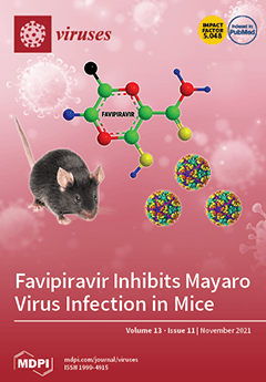
Cover Story (view full-size image): Mayaro virus (MAYV) infection, often associated with joint pain, is becoming a considerable health issue that unfortunately lacks a specific antiviral treatment. In the present study, the potential of Favipiravir, a broad-spectrum antiviral drug, to inhibit MAYV replication in an immunocompetent mouse model was evaluated. The results showed that orally administrated with the drug at pre- or concurrent-MAYV infection, Favipiravir strongly decreased footpad swelling and infectious viral particles in the blood and organs. In post-treated MAYV-infected mice, the drug significantly reduced viral replication at the virus inoculated site. View this paper.
  • Issues are regarded as officially published after their release is announced to the table of contents alert mailing list.
  • You may sign up for e-mail alerts to receive table of contents of newly released issues.
  • PDF is the official format for papers published in both, html and pdf forms. To view the papers in pdf format, click on the "PDF Full-text" link, and use the free Adobe Reader to open them.
Order results
Result details
Section
Select all
Export citation of selected articles as:
34 pages, 580 KiB  
Review
History of Arbovirus Research in the Czech Republic
by Zdenek Hubálek
Viruses 2021, 13(11), 2334; https://doi.org/10.3390/v13112334 - 22 Nov 2021
Cited by 6 | Viewed by 2919
Abstract
The aim of this review is to follow the history of studies on endemiv arboviruses and the diseases they cause which were detected in the Czech lands (Bohemia, Moravia and Silesia (i.e., the Czech Republic)). The viruses involve tick-borne encephalitis, West Nile and [...] Read more.
The aim of this review is to follow the history of studies on endemiv arboviruses and the diseases they cause which were detected in the Czech lands (Bohemia, Moravia and Silesia (i.e., the Czech Republic)). The viruses involve tick-borne encephalitis, West Nile and Usutu flaviviruses; the Sindbis alphavirus; Ťahyňa, Batai, Lednice and Sedlec bunyaviruses; the Uukuniemi phlebovirus; and the Tribeč orbivirus. Arboviruses temporarily imported from abroad to the Czech Republic have been omitted. This brief historical review includes a bibliography of all relevant papers. Full article
15 pages, 1401 KiB  
Article
Identification of African Swine Fever Virus Transcription within Peripheral Blood Mononuclear Cells of Acutely Infected Pigs
by Ann Sofie Olesen, Miyako Kodama, Louise Lohse, Francesc Accensi, Thomas Bruun Rasmussen, Christina M. Lazov, Morten T. Limborg, M. Thomas P. Gilbert, Anette Bøtner and Graham J. Belsham
Viruses 2021, 13(11), 2333; https://doi.org/10.3390/v13112333 - 22 Nov 2021
Cited by 12 | Viewed by 2231
Abstract
African swine fever virus (ASFV) has become widespread in Europe, Asia and elsewhere, thereby causing extensive economic losses. The viral genome includes nearly 200 genes, but their expression within infected pigs has not been well characterized previously. In this study, four pigs were [...] Read more.
African swine fever virus (ASFV) has become widespread in Europe, Asia and elsewhere, thereby causing extensive economic losses. The viral genome includes nearly 200 genes, but their expression within infected pigs has not been well characterized previously. In this study, four pigs were infected with a genotype II strain (ASFV POL/2015/Podlaskie); blood samples were collected before inoculation and at both 3 and 6 days later. During this period, a range of clinical signs of infection became apparent in the pigs. From the blood, peripheral blood mononuclear cells (PBMCs) were isolated. The transcription of the ASFV genes was determined using RNAseq on poly(A)+ mRNAs isolated from these cells. Only very low levels of virus transcription were detected in the PBMCs at 3 days post-inoculation (dpi) but, at 6 dpi, extensive transcription was apparent. This was co-incident with a large increase in the level of ASFV DNA within these cells. The pattern of the virus gene expression was very reproducible between the individual pigs. Many highly expressed genes have undefined roles. Surprisingly, some genes with key roles in virus replication were expressed at only low levels. As the functions of individual genes are identified, information about their expression becomes important for understanding their contribution to virus biology. Full article
(This article belongs to the Section Animal Viruses)
Show Figures

Figure 1

12 pages, 2237 KiB  
Article
Mass Spectrometry-Based System for Identifying and Typing Norovirus Major Capsid Protein VP1
by Pei-Yu Chu, Hui-Wen Huang, Michittra Boonchan, Yu-Chang Tyan, Kevin Leroy Louis, Kun-Mu Lee, Kazushi Motomura and Liang-Yin Ke
Viruses 2021, 13(11), 2332; https://doi.org/10.3390/v13112332 - 22 Nov 2021
Cited by 2 | Viewed by 2137
Abstract
Norovirus-associated diseases are the most common foodborne illnesses worldwide. Polymerase chain reaction-based methods are the primary diagnostics for clinical samples; however, the high mutation rate of norovirus makes viral amplification and genotyping challenging. Technological advances in mass spectrometry (MS) make it a promising [...] Read more.
Norovirus-associated diseases are the most common foodborne illnesses worldwide. Polymerase chain reaction-based methods are the primary diagnostics for clinical samples; however, the high mutation rate of norovirus makes viral amplification and genotyping challenging. Technological advances in mass spectrometry (MS) make it a promising tool for identifying disease markers. Besides, the superior sensitivity of MS and proteomic approaches may enable the detection of all variants. Thus, this study aimed to establish an MS-based system for identifying and typing norovirus. We constructed three plasmids containing the major capsid protein VP1 of the norovirus GII.4 2006b, 2006a, and 2009a strains to produce virus-like particles for use as standards. Digested peptide signals were collected using a nano-flow ultra-performance liquid chromatography mass spectrometry (nano-UPLC/MSE) system, and analyzed by ProteinLynx Global SERVER and TREE-PUZZLE software. Results revealed that the LC/MSE system had an excellent coverage rate: the system detected more than 94% of amino acids of 3.61 femtomole norovirus VP1 structural protein. In the likelihood-mapping analysis, the proportions of unresolved quartets were 2.9% and 4.9% in the VP1 and S domains, respectively, which is superior to the 15.1% unresolved quartets in current PCR-based methodology. In summary, the use of LC/MSE may efficiently monitor genotypes, and sensitively detect structural and functional mutations of noroviruses. Full article
(This article belongs to the Section Human Virology and Viral Diseases)
Show Figures

Figure 1

16 pages, 7995 KiB  
Article
A Combination of M50I and V151I Polymorphic Mutations in HIV-1 Subtype B Integrase Results in Defects in Autoprocessing
by Jun Yang, Ming Hao, Muhammad A. Khan, Muhammad T. Rehman, Helene C. Highbarger, Qian Chen, Suranjana Goswami, Brad T. Sherman, Catherine A. Rehm, Robin L. Dewar, Weizhong Chang and Tomozumi Imamichi
Viruses 2021, 13(11), 2331; https://doi.org/10.3390/v13112331 - 22 Nov 2021
Cited by 2 | Viewed by 1690
Abstract
We have recently reported that a recombinant HIV-1NL4.3 containing Met-to-Ile change at codon 50 of integrase (IN) (IN:M50I) exhibits suppression of the virus release below 0.5% of WT HIV, and the released viral particles are replication-incompetent due to defects in Gag/GagPol processing by [...] Read more.
We have recently reported that a recombinant HIV-1NL4.3 containing Met-to-Ile change at codon 50 of integrase (IN) (IN:M50I) exhibits suppression of the virus release below 0.5% of WT HIV, and the released viral particles are replication-incompetent due to defects in Gag/GagPol processing by inhibition of the initiation of autoprocessing of GagPol polyproteins in the virions and leads to replication-incompetent viruses. The coexisting Ser-to-Asn change at codon 17 of IN or Asn-to-Ser mutation at codon 79 of RNaseH (RH) compensated the defective IN:M50I phenotype, suggesting that both IN and RH regulate an HIV infectability. In the current study, to elucidate a distribution of the three mutations during anti-retroviral therapy among patients, we performed a population analysis using 529 plasma virus RNA sequences obtained through the MiSeq. The result demonstrated that 14 plasma HIVs contained IN:M50I without the compensatory mutations. Comparing the sequences of the 14 viruses with that of the defective virus illustrated that only Val-to-Ile change at codon 151 of IN (IN:V151I) existed in the recombinant virus. This IN:V151I is known as a polymorphic mutation and was derived from HIVNL4.3 backbone. A back-mutation at 151 from Ile-to-Val in the defective virus recovered HIV replication capability, and Western Blotting assay displayed that the back-mutation restored Gag/GagPol processing in viral particles. These results demonstrate that a combination of IN:M50I and IN:V151I mutations, but not IN:M50I alone, produces a defective virus. Full article
(This article belongs to the Special Issue Pathogenesis of Chronic Viral Infections)
Show Figures

Figure 1

14 pages, 1237 KiB  
Article
SARS-CoV-2 Infection and C-Section: A Prospective Observational Study
by Eva Morán Antolín, José Román Broullón Molanes, María Luisa de la Cruz Conty, María Begoña Encinas Pardilla, María del Pilar Guadix Martín, José Antonio Sainz Bueno, Laura Forcén Acebal, Pilar Pintado Recarte, Ana Álvarez Bartolomé, Juan Pedro Martínez Cendán, Óscar Martínez-Pérez and on behalf of the Spanish Obstetric Emergency Group
Viruses 2021, 13(11), 2330; https://doi.org/10.3390/v13112330 - 22 Nov 2021
Cited by 8 | Viewed by 2964
Abstract
Pregnant women are particularly vulnerable to the severe acute respiratory syndrome coronavirus 2 (SARS-CoV-2) pandemic. In addition to unfavorable perinatal outcomes, there has been an increase in obstetric interventions. With this study, we aimed to clarify the reasons, using Robson’s classification model, and [...] Read more.
Pregnant women are particularly vulnerable to the severe acute respiratory syndrome coronavirus 2 (SARS-CoV-2) pandemic. In addition to unfavorable perinatal outcomes, there has been an increase in obstetric interventions. With this study, we aimed to clarify the reasons, using Robson’s classification model, and risk factors for cesarean section (C-section) in SARS-CoV-2-infected mothers and their perinatal results. This was a prospective observational study that was carried out in 79 hospitals (Spanish Obstetric Emergency Group) with a cohort of 1704 SARS-CoV-2 PCR-positive pregnant women that were registered consecutively between 26 February and 5 November 2020. The data from 1248 pregnant women who delivered vaginally (vaginal + operative vaginal) was compared with those from 456 (26.8%) who underwent a C-section. C-section patients were older with higher rates of comorbidities, in vitro fertilization and multiple pregnancies (p < 0.05) compared with women who delivered vaginally. Moreover, C-section risk was associated with the presence of pneumonia (p < 0.001) and 41.1% of C-sections in patients with pneumonia were preterm (Robson’s 10th category). However, delivery care was similar between asymptomatic and mild–moderate symptomatic patients (p = 0.228) and their predisposing factors to C-section were the presence of uterine scarring (due to a previous C-section) and the induction of labor or programmed C-section for unspecified obstetric reasons. On the other hand, higher rates of hemorrhagic events, hypertensive disorders and thrombotic events were observed in the C-section group (p < 0.001 for all three outcomes), as well as for ICU admission. These findings suggest that this type of delivery was associated with the mother’s clinical conditions that required a rapid and early termination of pregnancy. Full article
(This article belongs to the Topic Burden of COVID-19 in Different Countries)
Show Figures

Figure 1

9 pages, 1372 KiB  
Communication
Dynamics of Neutralizing Antibody Responses Following Natural SARS-CoV-2 Infection and Correlation with Commercial Serologic Tests. A Reappraisal and Indirect Comparison with Vaccinated Subjects
by Constant Gillot, Julien Favresse, Vincent Maloteau, Jean-Michel Dogné and Jonathan Douxfils
Viruses 2021, 13(11), 2329; https://doi.org/10.3390/v13112329 - 22 Nov 2021
Cited by 10 | Viewed by 2098
Abstract
Neutralising antibodies (NAbs) represent the real source of protection against SARS-CoV-2 infections by preventing the virus from entering target cells. The gold standard in the detection of these antibodies is the plaque reduction neutralization test (PRNT). As these experiments must be done in [...] Read more.
Neutralising antibodies (NAbs) represent the real source of protection against SARS-CoV-2 infections by preventing the virus from entering target cells. The gold standard in the detection of these antibodies is the plaque reduction neutralization test (PRNT). As these experiments must be done in a very secure environment, other techniques based on pseudoviruses: pseudovirus neutralization test (pVNT) or surrogate virus neutralization test (sVNT) have been developed. Binding assays, on the other hand, measure total antibodies or IgG, IgM, and IgA directed against one epitope of the SARS-CoV-2, independently of their neutralizing capacity. The aim of this study is to compare the performance of six commercial binding assays to the pVNT and sVNT. In this study, we used blood samples from a cohort of 62 RT-PCR confirmed COVID-19 patients. Based on the results of the neutralizing assays, adapted cut-offs for the binding assays were calculated. The use of these adapted cut-offs does not permit to improve the accuracy of the serological assays and we did not find an adapted cut-off able to improve the capacity of these tests to detect NAbs. For a part of the population, a longitudinal follow-up with at least two samples for the same patient was performed. From day 14 to day 291, more than 75% of the samples were positive for NAbs (n = 87/110, 79.1%). Interestingly, 6 months post symptoms onset, the majority of the samples (N = 44/52, 84.6%) were still positive for NAbs. This is in sharp contrast with the results we obtained 6 months post-vaccination in our cohort of healthcare workers who have received the two-dose regimens of BNT162b2. In this cohort of vaccinated subjects, 43% (n = 25/58) of the participants no longer exhibit NAbs activity 180 days after the administration of the first dose of BNT162b2. Full article
(This article belongs to the Section SARS-CoV-2 and COVID-19)
Show Figures

Figure 1

14 pages, 1939 KiB  
Article
Analysis of the Physicochemical Properties, Replication and Pathophysiology of a Massively Glycosylated Hepatitis B Virus HBsAg Escape Mutant
by Md. Golzar Hossain, Yadarat Suwanmanee, Kaili Du and Keiji Ueda
Viruses 2021, 13(11), 2328; https://doi.org/10.3390/v13112328 - 22 Nov 2021
Cited by 2 | Viewed by 2345
Abstract
Mutations in HBsAg, the surface antigen of the hepatitis B virus (HBV), might affect the serum HBV DNA level of HBV-infected patients, since the reverse transcriptase (RT) domain of HBV polymerase overlaps with the HBsAg-coding region. We previously identified a diagnostic escape mutant [...] Read more.
Mutations in HBsAg, the surface antigen of the hepatitis B virus (HBV), might affect the serum HBV DNA level of HBV-infected patients, since the reverse transcriptase (RT) domain of HBV polymerase overlaps with the HBsAg-coding region. We previously identified a diagnostic escape mutant (W3S) HBV that produces massively glycosylated HBsAg. In this study, we constructed an HBV-producing vector that expresses W3S HBs (pHB-W3S) along with a wild-type HBV-producing plasmid (pHB-WT) in order to analyze the physicochemical properties, replication, and antiviral drug response of the mutant. Transfection of either pHB-WT or W3S into HepG2 cells yielded similar CsCl density profiles and eAg expression, as did transfection of a glycosylation defective mutant, pHB-W3S (N146G), in which a glycosylation site at the 146aa asparagine (N) site of HBs was mutated to glycine (G). Virion secretion, however, seemed to be severely impaired in cases of pHB-W3S and pHB-W3S (N146G), compared with pHB-WT, as determined by qPCR and Southern blot analysis. Furthermore, inhibition of glycosylation using tunicamycinTM on wild-type HBV production also reduced the virion secretion. These results suggested that the HBV core and Dane particle could be formed either by massively glycosylated or glycosylation-defective HBsAg, but reduced and/or almost completely blocked the virion secretion efficiency, indicating that balanced glycosylation of HBsAg is required for efficient release of HBV, and mutations inducing an imbalanced glycosylation of HBs would cause the virion to become stuck in the cells, which might be associated with various pathogeneses due to HBV infection. Full article
(This article belongs to the Special Issue Hepatitis B Virus: Its Life Cycle and the Therapeutic Targets)
Show Figures

Figure 1

6 pages, 220 KiB  
Article
The Practicability of the Xpert HCV Viral Load Fingerstick Point-of-Care Assay in Primary Care Settings
by David Petroff, Olaf Bätz, Katrin Jedrysiak, Jan Kramer, Thomas Berg and Johannes Wiegand
Viruses 2021, 13(11), 2327; https://doi.org/10.3390/v13112327 - 22 Nov 2021
Cited by 6 | Viewed by 1578
Abstract
Linkage to care presents one obstacle toward eliminating HCV, and the current two-step pathway (anti-HCV, followed by HCV-RNA testing) results in the loss of patients. HCV screening was tested in the primary care setting with the fingerstick Xpert HCV viral load point-of-care assay [...] Read more.
Linkage to care presents one obstacle toward eliminating HCV, and the current two-step pathway (anti-HCV, followed by HCV-RNA testing) results in the loss of patients. HCV screening was tested in the primary care setting with the fingerstick Xpert HCV viral load point-of-care assay to analyze the practicability of immediate diagnosis. Anti-HCV (Cobas) and HCV-RNA (Cobas Amplicor version 2.0, only performed if anti-HCV was positive) were analyzed centrally as the gold standard. The Xpert assay was performed by 10 primary care private practices. In total, 622 patients were recruited. Five individuals (0.8%) were anti-HCV positive, and one was HCV-RNA positive. The Xpert test was valid in 546/622 (87.8%) patients. It was negative in 544 and positive in 2 cases, both of whom were anti-HCV negative. The HCV-RNA PCR and the Xpert test were both negative in 4/5 anti-HCV-positive cases, and the individual with HCV-RNA 4.5 × 10⁶ IU/mL was not detected by the Xpert test. Primary care physicians rated the Xpert test practicability as bad, satisfactory, or good in 6%, 13%, and 81%, respectively, though 14/29 (48%) bad test ratings were assigned by a single practice. Despite adequate acceptance, interpretability and diagnostic performance in primary care settings should be further evaluated before its use in HCV screening can be recommended. Full article
(This article belongs to the Special Issue Ways to Eliminate Viral Hepatitis as a Global Health Threat)
8 pages, 576 KiB  
Article
Identification of Hepatitis E Virus Genotypes 3 and 7 in Israel: A Public Health Concern?
by Rachel Shirazi, Paolo Pozzi, Yael Gozlan, Marina Wax, Yaniv Lustig, Michal Linial, Ella Mendelson, Svetlana Bardenstein and Orna Mor
Viruses 2021, 13(11), 2326; https://doi.org/10.3390/v13112326 - 22 Nov 2021
Cited by 4 | Viewed by 1847
Abstract
Background: Hepatitis E (HEV) is an emerging cause of viral hepatitis worldwide. Swine carrying hepatitis E genotype 3 (HEV-3) are responsible for the majority of chronic viral hepatitis cases in developed countries. Recently, genotype 7 (HEV-7), isolated from a dromedary camel in the [...] Read more.
Background: Hepatitis E (HEV) is an emerging cause of viral hepatitis worldwide. Swine carrying hepatitis E genotype 3 (HEV-3) are responsible for the majority of chronic viral hepatitis cases in developed countries. Recently, genotype 7 (HEV-7), isolated from a dromedary camel in the United Arab Emirates, was also associated with chronic viral hepatitis in a transplant recipient. In Israel, chronic HEV infection has not yet been reported, although HEV seroprevalence in humans is ~10%. Camels and swine are >65% seropositive. Here we report on the isolation and characterization of HEV from local camels and swine. Methods: Sera from camels (n = 142), feces from swine (n = 18) and blood from patients suspected of hepatitis E (n = 101) were collected during 2017–2020 and used to detect and characterize HEV sequences. Results: HEV-3 isolated from local swine and the camel-derived HEV-7 sequence were highly similar to HEV-3f and HEV-7 sequences (88.2% and 86.4%, respectively) related to viral hepatitis. The deduced amino acid sequences of both isolates were also highly conserved (>98%). Two patients were HEV-RNA positive; acute HEV-1 infection could be confirmed in one of them. Discussion: The absence of any reported HEV-3 and HEV-7 infection in humans remains puzzling, especially considering the reported seroprevalence rates, the similarity between HEV sequences related to chronic hepatitis and the HEV genotypes identified in swine and camels in Israel. Full article
(This article belongs to the Special Issue State-of-the-Art Virology Research in Israel)
Show Figures

Figure 1

19 pages, 3750 KiB  
Article
Cross-Reactive Antibodies to SARS-CoV-2 and MERS-CoV in Pre-COVID-19 Blood Samples from Sierra Leoneans
by Rodrigo Borrega, Diana K. S. Nelson, Anatoliy P. Koval, Nell G. Bond, Megan L. Heinrich, Megan M. Rowland, Raju Lathigra, Duane J. Bush, Irina Aimukanova, Whitney N. Phinney, Sophia A. Koval, Andrew R. Hoffmann, Allison R. Smither, Antoinette R. Bell-Kareem, Lilia I. Melnik, Kaylynn J. Genemaras, Karissa Chao, Patricia Snarski, Alexandra B. Melton, Jaikin E. Harrell, Ashley A. Smira, Debra H. Elliott, Julie A. Rouelle, Gilberto Sabino-Santos, Jr., Arnaud C. Drouin, Mambu Momoh, John Demby Sandi, Augustine Goba, Robert J. Samuels, Lansana Kanneh, Michael Gbakie, Zoe L. Branco, Jeffrey G. Shaffer, John S. Schieffelin, James E. Robinson, Dahlene N. Fusco, Pardis C. Sabeti, Kristian G. Andersen, Donald S. Grant, Matthew L. Boisen, Luis M. Branco and Robert F. Garryadd Show full author list remove Hide full author list
Viruses 2021, 13(11), 2325; https://doi.org/10.3390/v13112325 - 21 Nov 2021
Cited by 20 | Viewed by 9545
Abstract
Many countries in sub-Saharan Africa have experienced lower COVID-19 caseloads and fewer deaths than countries in other regions worldwide. Under-reporting of cases and a younger population could partly account for these differences, but pre-existing immunity to coronaviruses is another potential factor. Blood samples [...] Read more.
Many countries in sub-Saharan Africa have experienced lower COVID-19 caseloads and fewer deaths than countries in other regions worldwide. Under-reporting of cases and a younger population could partly account for these differences, but pre-existing immunity to coronaviruses is another potential factor. Blood samples from Sierra Leonean Lassa fever and Ebola survivors and their contacts collected before the first reported COVID-19 cases were assessed using enzyme-linked immunosorbent assays for the presence of antibodies binding to proteins of coronaviruses that infect humans. Results were compared to COVID-19 subjects and healthy blood donors from the United States. Prior to the pandemic, Sierra Leoneans had more frequent exposures than Americans to coronaviruses with epitopes that cross-react with severe acute respiratory syndrome coronavirus-2 (SARS-CoV-2), SARS-CoV, and Middle Eastern respiratory syndrome coronavirus (MERS-CoV). The percentage of Sierra Leoneans with antibodies reacting to seasonal coronaviruses was also higher than for American blood donors. Serological responses to coronaviruses by Sierra Leoneans did not differ by age or sex. Approximately a quarter of Sierra Leonian pre-pandemic blood samples had neutralizing antibodies against SARS-CoV-2 pseudovirus, while about a third neutralized MERS-CoV pseudovirus. Prior exposures to coronaviruses that induce cross-protective immunity may contribute to reduced COVID-19 cases and deaths in Sierra Leone. Full article
(This article belongs to the Special Issue Cross-Reactivity in Virus Infection)
Show Figures

Figure 1

8 pages, 760 KiB  
Article
Indications of Persistent Glycocalyx Damage in Convalescent COVID-19 Patients: A Prospective Multicenter Study and Hypothesis
by Richard Vollenberg, Phil-Robin Tepasse, Kevin Ochs, Martin Floer, Markus Strauss, Florian Rennebaum, Iyad Kabar, Alexandros Rovas and Tobias Nowacki
Viruses 2021, 13(11), 2324; https://doi.org/10.3390/v13112324 - 21 Nov 2021
Cited by 22 | Viewed by 3671
Abstract
The COVID-19 pandemic is caused by the SARS CoV-2 virus and can lead to severe lung damage and hyperinflammation. In the context of COVID-19 infection, inflammation-induced degradation of the glycocalyx layer in endothelial cells has been demonstrated. Syndecan-1 (SDC-1) is an established parameter [...] Read more.
The COVID-19 pandemic is caused by the SARS CoV-2 virus and can lead to severe lung damage and hyperinflammation. In the context of COVID-19 infection, inflammation-induced degradation of the glycocalyx layer in endothelial cells has been demonstrated. Syndecan-1 (SDC-1) is an established parameter for measuring glycocalyx injury. This prospective, multicenter, observational, cross-sectional study analyzed SDC-1 levels in 24 convalescent patients that had been infected with SARS-CoV-2 with mild disease course without need of hospitalization. We included 13 age-matched healthy individuals and 10 age-matched hospitalized COVID-19 patients with acute mild disease course as controls. In convalescent COVID-19 patients, significantly elevated SDC-1 levels were detected after a median of 88 days after symptom onset compared to healthy controls, whereas no difference was found when compared to SDC-1 levels of hospitalized patients undergoing acute disease. This study is the first to demonstrate signs of endothelial damage in non-pre-diseased, convalescent COVID-19 patients after mild disease progression without hospitalization. The data are consistent with studies showing evidence of persistent endothelial damage after severe or critical disease progression. Further work to investigate endothelial damage in convalescent COVID-19 patients should follow. Full article
(This article belongs to the Special Issue COVID-19—Advances in Clinical and Epidemiological Aspects)
Show Figures

Figure 1

18 pages, 2603 KiB  
Article
Heterogeneity of Early Host Response to Infection with Four Low-Pathogenic H7 Viruses with a Different Evolutionary History in the Field
by Gianpiero Zamperin, Alice Bianco, Jacqueline Smith, Alessio Bortolami, Lonneke Vervelde, Alessia Schivo, Andrea Fortin, Sabrina Marciano, Valentina Panzarin, Eva Mazzetto, Adelaide Milani, Yohannes Berhane, Paul Digard, Francesco Bonfante and Isabella Monne
Viruses 2021, 13(11), 2323; https://doi.org/10.3390/v13112323 - 21 Nov 2021
Cited by 5 | Viewed by 10887
Abstract
Once low-pathogenic avian influenza viruses (LPAIVs) of the H5 and H7 subtypes from wild birds enter into poultry species, there is the possibility of them mutating into highly pathogenic avian influenza viruses (HPAIVs), resulting in severe epizootics with up to 100% mortality. This [...] Read more.
Once low-pathogenic avian influenza viruses (LPAIVs) of the H5 and H7 subtypes from wild birds enter into poultry species, there is the possibility of them mutating into highly pathogenic avian influenza viruses (HPAIVs), resulting in severe epizootics with up to 100% mortality. This mutation from a LPAIV to HPAIV strain is the main cause of an AIV’s major economic impact on poultry production. Although AIVs are inextricably linked to their hosts in their evolutionary history, the contribution of host-related factors in the emergence of HPAI viruses has only been marginally explored so far. In this study, transcriptomic sequencing of tracheal tissue from chickens infected with four distinct LP H7 viruses, characterized by a different history of pathogenicity evolution in the field, was implemented. Despite the inoculation of a normalized infectious dose of viruses belonging to the same subtype (H7) and pathotype (LPAI), the use of animals of the same age, sex and species as well as the identification of a comparable viral load in the target samples, the analyses revealed a heterogeneity in the gene expression profile in response to infection with each of the H7 viruses administered. Full article
(This article belongs to the Special Issue Drivers of Evolution of Animal RNA Viruses)
Show Figures

Figure 1

19 pages, 3245 KiB  
Article
Exploring the Diversity of the Human Blood Virome
by María Cebriá-Mendoza, María A. Bracho, Cristina Arbona, Luís Larrea, Wladimiro Díaz, Rafael Sanjuán and José M. Cuevas
Viruses 2021, 13(11), 2322; https://doi.org/10.3390/v13112322 - 21 Nov 2021
Cited by 15 | Viewed by 3324
Abstract
Metagenomics is greatly improving our ability to discover new viruses, as well as their possible associations with disease. However, metagenomics has also changed our understanding of viruses in general. The vast expansion of currently known viral diversity has revealed a large fraction of [...] Read more.
Metagenomics is greatly improving our ability to discover new viruses, as well as their possible associations with disease. However, metagenomics has also changed our understanding of viruses in general. The vast expansion of currently known viral diversity has revealed a large fraction of non-pathogenic viruses, and offers a new perspective in which viruses function as important components of many ecosystems. In this vein, studies of the human blood virome are often motivated by the search for new viral diseases, especially those associated with blood transfusions. However, these studies have revealed the common presence of apparently non-pathogenic viruses in blood, particularly human anelloviruses and, to a lower extent, human pegiviruses (HPgV). To shed light on the diversity of the human blood virome, we subjected pooled plasma samples from 587 healthy donors in Spain to a viral enrichment protocol, followed by massive parallel sequencing. This showed that anelloviruses were clearly the major component of the blood virome and showed remarkable diversity. In total, we assembled 332 complete or near-complete anellovirus genomes, 50 of which could be considered new species. HPgV was much less frequent, but we, nevertheless, recovered 17 different isolates that we subsequently used for characterizing the diversity of this virus. In-depth investigation of the human blood virome should help to elucidate the ecology of these viruses, and to unveil potentially associated diseases. Full article
(This article belongs to the Special Issue Virus Bioinformatics 2022)
Show Figures

Figure 1

14 pages, 2461 KiB  
Article
Cytomegalovirus Immunity, Inflammation and Cognitive Abilities in the Elderly
by Jacqueline Hesson, Neva Fudge and Michael Grant
Viruses 2021, 13(11), 2321; https://doi.org/10.3390/v13112321 - 21 Nov 2021
Viewed by 1840
Abstract
Reducing the socioeconomic toll from age-related physical and mental morbidities requires better understanding of factors affecting healthy aging. While many environmental, lifestyle, and genetic factors affect healthy aging, this study addressed the influence of cytomegalovirus (CMV) infection and immunity on age-related inflammation and [...] Read more.
Reducing the socioeconomic toll from age-related physical and mental morbidities requires better understanding of factors affecting healthy aging. While many environmental, lifestyle, and genetic factors affect healthy aging, this study addressed the influence of cytomegalovirus (CMV) infection and immunity on age-related inflammation and cognitive abilities. Healthy adults 70–90 years old were recruited into a prospective study investigating relationships between anti-CMV immunity, markers of inflammation, baseline measures of cognitive ability, and changes in cognitive ability over 18 months. Humoral and cellular responses against CMV, levels of inflammatory markers, and cognitive abilities were measured at study entry, with measurement of cognitive abilities repeated 18 months later. CMV-seropositive and -seronegative sub-groups were compared, and relationships between anti-CMV immunity, markers of inflammation, and cognitive ability were assessed. Twenty-eight of 39 participants were CMV-seropositive, and two had CMV-specific CD8+ T cell responses indicative of CMV immune memory inflation. No significant differences for markers of inflammation or measures of cognitive ability were observed between groups, and cognitive scores changed little over 18 months. Significant correlations between markers of inflammation and cognitive scores with interconnection between anti-CMV antibody levels, fractalkine, cognitive ability, and depression scores suggest areas of focus for future studies. Full article
(This article belongs to the Special Issue Cytomegalovirus Immunity 2021)
Show Figures

Figure 1

20 pages, 6966 KiB  
Article
Exchange of C-Terminal Variable Sequences within Morbillivirus Nucleocapsid Protein Are Tolerated: Development and Evaluation of Two Marker (DIVA) Vaccines (Sungri/96 DIVA, Nigeria/75/1 DIVA) against PPR
by Muneeswaran Selvaraj, Mana Mahapatra and Satya Parida
Viruses 2021, 13(11), 2320; https://doi.org/10.3390/v13112320 - 21 Nov 2021
Cited by 3 | Viewed by 2521
Abstract
Across Africa, the Middle East, and Asia, peste des petits ruminants virus (PPRV) places a huge disease burden on agriculture, affecting, in particular, small ruminant production. The recent PPR outbreaks in Northern Africa, the European part of Turkey, and Bulgaria represent a significant [...] Read more.
Across Africa, the Middle East, and Asia, peste des petits ruminants virus (PPRV) places a huge disease burden on agriculture, affecting, in particular, small ruminant production. The recent PPR outbreaks in Northern Africa, the European part of Turkey, and Bulgaria represent a significant threat to mainland Europe, as a source of disease. Although two safe and efficacious live attenuated vaccines (Sungri/96 and Nigeria/75/1) are available for the control of PPR, current serological tests do not enable the differentiation between naturally infected and vaccinated animals (DIVA). The vaccinated animals develop a full range of immune responses to viral proteins and, therefore, cannot be distinguished serologically from those that have recovered from a natural infection. This poses a serious problem for the post-vaccinal sero-surveillance during the ongoing PPR eradication program. Furthermore, during the latter stages of any eradication program, vaccination is only possible if the vaccine used is fully DIVA compliant. Using reverse genetics, we have developed two live attenuated PPR DIVA vaccines (Sungri/96 DIVA and Nigeria/75/1 DIVA), in which the C-terminal variable region of the PPRV N-protein has been replaced with dolphin morbillivirus (DMV). As a proof of principle, both the DIVA vaccines were evaluated in goats in pilot studies for safety and efficacy, and all the animals were clinically protected against the intranasal virulent virus challenge, similar to the parent vaccines. Furthermore, it is possible to differentiate between infected animals and vaccinated animals using two newly developed ELISAs. Therefore, these DIVA vaccines and associated tests can facilitate the sero-monitoring process and speed up the implementation of global PPR eradication through vaccination. Full article
Show Figures

Figure 1

15 pages, 3086 KiB  
Article
Characterization of Four Novel dsRNA Viruses Isolated from Mucor hiemalis Strains
by Tünde Kartali, Ildikó Nyilasi, Sándor Kocsubé, Roland Patai, Tamás F. Polgár, Nóra Zsindely, Gábor Nagy, László Bodai, Zoltán Lipinszki, Csaba Vágvölgyi and Tamás Papp
Viruses 2021, 13(11), 2319; https://doi.org/10.3390/v13112319 - 21 Nov 2021
Cited by 4 | Viewed by 2641
Abstract
We previously screened the total nucleic acid extracts of 123 Mucor strains for the presence of dsRNA molecules without further molecular analyses. Here, we characterized five novel dsRNA genomes isolated from four different Mucor hiemalis strains with next-generation sequencing (NGS), namely Mucor [...] Read more.
We previously screened the total nucleic acid extracts of 123 Mucor strains for the presence of dsRNA molecules without further molecular analyses. Here, we characterized five novel dsRNA genomes isolated from four different Mucor hiemalis strains with next-generation sequencing (NGS), namely Mucor hiemalis virus 1a (MhV1a) from WRL CN(M) 122; Mucor hiemalis virus 1b (MhV1b) from NRRL 3624; Mucor hiemalis virus 2 (MhV2) from NRRL 3616; and Mucor hiemalis virus 3 (MhV3) and Mucor hiemalis virus (MhV4) from NRRL 3617 strains. Genomes contain two open reading frames (ORF), which encode the coat protein (CP) and the RNA dependent RNA polymerase (RdRp), respectively. In MhV1a and MhV1b, it is predicted to be translated as a fusion protein via -1 ribosomal frameshift, while in MhV4 via a rare +1 (or−2) ribosomal frameshift. In MhV2 and MhV3, the presence of specific UAAUG pentanucleotide motif points to the fact for coupled translation termination and reinitialization. MhV1a, MhV2, and MhV3 are part of the clade representing the genus Victorivirus, while MhV4 is seated in Totivirus genus clade. The detected VLPs in Mucor strains were from 33 to 36 nm in diameter. Hybridization analysis revealed that the dsRNA molecules of MhV1a-MhV4 hybridized to the corresponding molecules. Full article
(This article belongs to the Collection Mycoviruses)
Show Figures

Figure 1

20 pages, 880 KiB  
Review
The Roles of Neutrophils in Cytokine Storms
by Lily Chan, Negar Karimi, Solmaz Morovati, Kasra Alizadeh, Julia E. Kakish, Sierra Vanderkamp, Fatemeh Fazel, Christina Napoleoni, Kimia Alizadeh, Yeganeh Mehrani, Jessica A. Minott, Byram W. Bridle and Khalil Karimi
Viruses 2021, 13(11), 2318; https://doi.org/10.3390/v13112318 - 21 Nov 2021
Cited by 29 | Viewed by 5300
Abstract
A cytokine storm is an abnormal discharge of soluble mediators following an inappropriate inflammatory response that leads to immunopathological events. Cytokine storms can occur after severe infections as well as in non-infectious situations where inflammatory cytokine responses are initiated, then exaggerated, but fail [...] Read more.
A cytokine storm is an abnormal discharge of soluble mediators following an inappropriate inflammatory response that leads to immunopathological events. Cytokine storms can occur after severe infections as well as in non-infectious situations where inflammatory cytokine responses are initiated, then exaggerated, but fail to return to homeostasis. Neutrophils, macrophages, mast cells, and natural killer cells are among the innate leukocytes that contribute to the pathogenesis of cytokine storms. Neutrophils participate as mediators of inflammation and have roles in promoting homeostatic conditions following pathological inflammation. This review highlights the advances in understanding the mechanisms governing neutrophilic inflammation against viral and bacterial pathogens, in cancers, and in autoimmune diseases, and how neutrophils could influence the development of cytokine storm syndromes. Evidence for the destructive potential of neutrophils in their capacity to contribute to the onset of cytokine storm syndromes is presented across a multitude of clinical scenarios. Further, a variety of potential therapeutic strategies that target neutrophils are discussed in the context of suppressing multiple inflammatory conditions. Full article
(This article belongs to the Special Issue Virus-Induced Cytokine Storms)
Show Figures

Figure 1

19 pages, 29559 KiB  
Communication
Drug-Screening Strategies for Inhibition of Virus-Induced Neuronal Cell Death
by Durbadal Ojha, Tyson A. Woods and Karin E. Peterson
Viruses 2021, 13(11), 2317; https://doi.org/10.3390/v13112317 - 20 Nov 2021
Cited by 4 | Viewed by 2998
Abstract
A number of viruses, including Herpes Simplex Virus (HSV), West Nile Virus (WNV), La Crosse Virus (LACV), Zika virus (ZIKV) and Tick-borne encephalitis virus (TBEV), have the ability to gain access to the central nervous system (CNS) and cause severe neurological disease or [...] Read more.
A number of viruses, including Herpes Simplex Virus (HSV), West Nile Virus (WNV), La Crosse Virus (LACV), Zika virus (ZIKV) and Tick-borne encephalitis virus (TBEV), have the ability to gain access to the central nervous system (CNS) and cause severe neurological disease or death. Although encephalitis cases caused by these viruses are generally rare, there are relatively few treatment options available for patients with viral encephalitis other than palliative care. Many of these viruses directly infect neurons and can cause neuronal death. Thus, there is the need for the identification of useful therapeutic compounds that can inhibit virus replication in neurons or inhibit virus-induced neuronal cell death. In this paper, we describe the methodology to test compounds for their ability to inhibit virus-induced neuronal cell death. These protocols include the isolation and culturing of primary neurons; the culturing of neuroblastoma and neuronal stem cell lines; infection of these cells with viruses; treatment of these cells with selected drugs; measuring virus-induced cell death using MTT or XTT reagents; analysis of virus production from these cells; as well as the basic understanding in mode of action. We further show direct evidence of the effectiveness of these protocols by utilizing them to test the effectiveness of the polyphenol drug, Rottlerin, at inhibiting Zika virus infection and death of neuronal cell lines. Full article
(This article belongs to the Special Issue Antiviral Therapeutics for Emerging Viruses)
Show Figures

Figure 1

13 pages, 2926 KiB  
Article
Functional Importance of Hydrophobic Patches on the Ebola Virus VP35 IFN-Inhibitory Domain
by Nodoka Kasajima, Keita Matsuno, Hiroko Miyamoto, Masahiro Kajihara, Manabu Igarashi and Ayato Takada
Viruses 2021, 13(11), 2316; https://doi.org/10.3390/v13112316 - 20 Nov 2021
Viewed by 1846
Abstract
Viral protein 35 (VP35) of Ebola virus (EBOV) is a multifunctional protein that mainly acts as a viral polymerase cofactor and an interferon antagonist. VP35 interacts with the viral nucleoprotein (NP) and double-stranded RNA for viral RNA transcription/replication and inhibition of type I [...] Read more.
Viral protein 35 (VP35) of Ebola virus (EBOV) is a multifunctional protein that mainly acts as a viral polymerase cofactor and an interferon antagonist. VP35 interacts with the viral nucleoprotein (NP) and double-stranded RNA for viral RNA transcription/replication and inhibition of type I interferon (IFN) production, respectively. The C-terminal portion of VP35, which is termed the IFN-inhibitory domain (IID), is important for both functions. To further identify critical regions in this domain, we analyzed the physical properties of the surface of VP35 IID, focusing on hydrophobic patches, which are expected to be functional sites that are involved in interactions with other molecules. Based on the known structural information of VP35 IID, three hydrophobic patches were identified on its surface and their biological importance was investigated using minigenome and IFN-β promoter-reporter assays. Site-directed mutagenesis revealed that some of the amino acid substitutions that were predicted to disrupt the hydrophobicity of the patches significantly decreased the efficiency of viral genome replication/transcription due to reduced interaction with NP, suggesting that the hydrophobic patches might be critical for the formation of a replication complex through the interaction with NP. It was also found that the hydrophobic patches were involved in the IFN-inhibitory function of VP35. These results highlight the importance of hydrophobic patches on the surface of EBOV VP35 IID and also indicate that patch analysis is useful for the identification of amino acid residues that directly contribute to protein functions. Full article
(This article belongs to the Section Animal Viruses)
Show Figures

Figure 1

16 pages, 3537 KiB  
Article
Susceptibility of Four Abalone Species, Haliotis gigantea, Haliotis discus discus, Haliotis discus hannai and Haliotis diversicolor, to Abalone asfa-like Virus
by Tomomasa Matsuyama, Ikunari Kiryu, Mari Inada, Tomokazu Takano, Yuta Matsuura and Takashi Kamaishi
Viruses 2021, 13(11), 2315; https://doi.org/10.3390/v13112315 - 20 Nov 2021
Cited by 4 | Viewed by 2524
Abstract
Abalone amyotrophia is a viral disease that causes mass mortality of juvenile Haliotis discus and H. madaka. Although the cause of this disease has yet to be identified, we had previously postulated a novel virus with partial genome sequence similarity to that [...] Read more.
Abalone amyotrophia is a viral disease that causes mass mortality of juvenile Haliotis discus and H. madaka. Although the cause of this disease has yet to be identified, we had previously postulated a novel virus with partial genome sequence similarity to that of African swine fever virus is the causative agent and proposed abalone asfa-like virus (AbALV) as a provisional name. In this study, three species of juvenile abalone (H. gigantea, H. discus discus, and H. diversicolor) and four species of adult abalone (the above three species plus H. discus hannai) were experimentally infected, and their susceptibility to AbALV was investigated by recording mortality, quantitatively determining viral load by PCR, and conducting immunohistological studies. In the infection test using 7-month-old animals, H. gigantea, which was previously reported to be insusceptible to the disease, showed multiplication of the virus to the same extent as in H. discus discus, resulting in mass mortality. H. discus discus at 7 months old showed abnormal cell masses, notches in the edge of the shell and brown pigmentation inside of the shell, which are histopathological and external features of this disease, while H. gigantea did not show any of these characteristics despite suffering high mortality. Adult abalones had low mortality and viral replication in all species; however, all three species, except H. diversicolor, became carriers of the virus. In immunohistological observations, cells positive for viral antigens were detected predominantly in the gills of juvenile H. discus discus and H. gigantea, and mass mortality was observed in these species. In H. diversicolor, neither juvenile nor adult mortality from infection occurred, and the AbALV genome was not increased by experimental infection through cohabitation or injection. Our results suggest that H. gigantea, H. discus discus and H. discus hannai are susceptible to AbALV, while H. diversicolor is not. These results confirmed that AbALV is the etiological agent of abalone amyotrophia. Full article
(This article belongs to the Topic Veterinary Infectious Diseases)
Show Figures

Figure 1

16 pages, 1508 KiB  
Article
Database and Statistical Analyses of Transcription Factor Binding Sites in the Non-Coding Control Region of JC Virus
by Kazuo Nakamichi and Toshio Shimokawa
Viruses 2021, 13(11), 2314; https://doi.org/10.3390/v13112314 - 19 Nov 2021
Cited by 3 | Viewed by 2080
Abstract
JC virus (JCV), as an archetype, establishes a lifelong latent or persistent infection in many healthy individuals. In immunocompromised patients, prototype JCV with variable mutations in the non-coding control region (NCCR) causes progressive multifocal leukoencephalopathy (PML), a severe demyelinating disease. This study was [...] Read more.
JC virus (JCV), as an archetype, establishes a lifelong latent or persistent infection in many healthy individuals. In immunocompromised patients, prototype JCV with variable mutations in the non-coding control region (NCCR) causes progressive multifocal leukoencephalopathy (PML), a severe demyelinating disease. This study was conducted to create a database of NCCR sequences annotated with transcription factor binding sites (TFBSs) and statistically analyze the mutational pattern of the JCV NCCR. JCV NCCRs were extracted from >1000 sequences registered in GenBank, and TFBSs within each NCCR were identified by computer simulation, followed by examination of their prevalence, multiplicity, and location by statistical analyses. In the NCCRs of the prototype JCV, the limited types of TFBSs, which are mainly present in regions D through F of archetype JCV, were significantly reduced. By contrast, modeling count data revealed that several TFBSs located in regions C and E tended to overlap in the prototype NCCRs. Based on data from the BioGPS database, genes encoding transcription factors that bind to these TFBSs were expressed not only in the brain but also in the peripheral sites. The database and NCCR patterns obtained in this study could be a suitable platform for analyzing JCV mutations and pathogenicity. Full article
(This article belongs to the Section Human Virology and Viral Diseases)
Show Figures

Figure 1

13 pages, 1099 KiB  
Article
Persistence of SARS-CoV-2-Specific Antibodies for 13 Months after Infection
by Indrė Kučinskaitė-Kodzė, Martynas Simanavičius, Aistis Šimaitis and Aurelija Žvirblienė
Viruses 2021, 13(11), 2313; https://doi.org/10.3390/v13112313 - 19 Nov 2021
Cited by 13 | Viewed by 2427
Abstract
Background: Dynamics of antibody responses were investigated after a SARS-CoV-2 outbreak in a private company during the first wave of the pandemic. Methods: Workers of a sewing company (Lithuania) with known SARS-CoV-2 RT-PCR result during the outbreak (April 2020) were invited to participate [...] Read more.
Background: Dynamics of antibody responses were investigated after a SARS-CoV-2 outbreak in a private company during the first wave of the pandemic. Methods: Workers of a sewing company (Lithuania) with known SARS-CoV-2 RT-PCR result during the outbreak (April 2020) were invited to participate in the study. Virus-specific IgG and IgM were monitored 2, 6 and 13 months after the outbreak via rapid IgG/IgM serological test and SARS-CoV-2 S protein-specific IgG ELISA. Results: Six months after the outbreak, 95% (CI 86–99%) of 59 previously infected individuals had virus-specific antibodies irrespective of the severity of infection. One-third of seropositive individuals had virus-specific IgM along with IgG indicating that IgM may persist for 6 months. Serological testing 13 months after the outbreak included 47 recovered individuals that remained non-vaccinated despite a wide accessibility of COVID-19 vaccines. The seropositivity rate was 83% (CI 69–91%) excluding one case of confirmed asymptomatic reinfection in this group. Between months 6 and 13, IgG levels either declined or remained stable in 31 individual and increased in 7 individuals possibly indicating an exposure to SARS-CoV-2 during the second wave of the pandemic. Conclusions: Detectable levels of SARS-CoV-2-specific antibodies persist up to 13 months after infection for the majority of the cases. Full article
(This article belongs to the Section Coronaviruses)
Show Figures

Figure 1

26 pages, 9096 KiB  
Article
The HIV-1 Nucleocapsid Regulates Its Own Condensation by Phase-Separated Activity-Enhancing Sequestration of the Viral Protease during Maturation
by Sébastien Lyonnais, S. Kashif Sadiq, Cristina Lorca-Oró, Laure Dufau, Sara Nieto-Marquez, Tuixent Escribà, Natalia Gabrielli, Xiao Tan, Mohamed Ouizougun-Oubari, Josephine Okoronkwo, Michèle Reboud-Ravaux, José Maria Gatell, Roland Marquet, Jean-Christophe Paillart, Andreas Meyerhans, Carine Tisné, Robert J. Gorelick and Gilles Mirambeau
Viruses 2021, 13(11), 2312; https://doi.org/10.3390/v13112312 - 19 Nov 2021
Cited by 7 | Viewed by 3906
Abstract
A growing number of studies indicate that mRNAs and long ncRNAs can affect protein populations by assembling dynamic ribonucleoprotein (RNP) granules. These phase-separated molecular ‘sponges’, stabilized by quinary (transient and weak) interactions, control proteins involved in numerous biological functions. Retroviruses such as HIV-1 [...] Read more.
A growing number of studies indicate that mRNAs and long ncRNAs can affect protein populations by assembling dynamic ribonucleoprotein (RNP) granules. These phase-separated molecular ‘sponges’, stabilized by quinary (transient and weak) interactions, control proteins involved in numerous biological functions. Retroviruses such as HIV-1 form by self-assembly when their genomic RNA (gRNA) traps Gag and GagPol polyprotein precursors. Infectivity requires extracellular budding of the particle followed by maturation, an ordered processing of ∼2400 Gag and ∼120 GagPol by the viral protease (PR). This leads to a condensed gRNA-NCp7 nucleocapsid and a CAp24-self-assembled capsid surrounding the RNP. The choreography by which all of these components dynamically interact during virus maturation is one of the missing milestones to fully depict the HIV life cycle. Here, we describe how HIV-1 has evolved a dynamic RNP granule with successive weak–strong–moderate quinary NC-gRNA networks during the sequential processing of the GagNC domain. We also reveal two palindromic RNA-binding triads on NC, KxxFxxQ and QxxFxxK, that provide quinary NC-gRNA interactions. Consequently, the nucleocapsid complex appears properly aggregated for capsid reassembly and reverse transcription, mandatory processes for viral infectivity. We show that PR is sequestered within this RNP and drives its maturation/condensation within minutes, this process being most effective at the end of budding. We anticipate such findings will stimulate further investigations of quinary interactions and emergent mechanisms in crowded environments throughout the wide and growing array of RNP granules. Full article
(This article belongs to the Special Issue Retroviral Nucleocapsid Proteins)
Show Figures

Figure 1

11 pages, 1303 KiB  
Article
Characterization of Single-Chain Fv Fragments of Neutralizing Antibodies to Rabies Virus Glycoprotein
by Kohei Yumoto, Tomoaki Arisaka, Kazuma Okada, Kyosuke Aoki, Toyoyuki Ose, Tatsunori Masatani, Makoto Sugiyama, Naoto Ito, Hideo Fukuhara and Katsumi Maenaka
Viruses 2021, 13(11), 2311; https://doi.org/10.3390/v13112311 - 19 Nov 2021
Cited by 2 | Viewed by 2346
Abstract
Rabies has almost a 100% case-fatality rate and kills more than 59,000 people annually around the world. There is no established treatment for rabies. The rabies virus (RABV) expresses only the glycoprotein (RABVG) at the viral surface, and it is the target for [...] Read more.
Rabies has almost a 100% case-fatality rate and kills more than 59,000 people annually around the world. There is no established treatment for rabies. The rabies virus (RABV) expresses only the glycoprotein (RABVG) at the viral surface, and it is the target for the neutralizing antibodies. We previously established mouse monoclonal antibodies, 15–13 and 12–22, which showed neutralizing activity against the RABV, targeting the sequential and conformational epitopes on the RABVG, respectively. However, the molecular basis for the neutralizing activity of these antibodies is not yet fully understood. In this study, we evaluated the binding characteristics of the Fab fragments of the 15–13 and 12–22 antibodies. The recombinant RABVG protein, in prefusion form for the binding analysis, was prepared by the silkworm–baculovirus expression system. Biolayer interferometry (BLI) analysis indicated that the 15–13 Fab interacts with the RABVG, with a KD value at the nM level, and that the 12–22 Fab has a weaker binding affinity (KD ~ μM) with the RABVG compared to the 15–13 Fab. Furthermore, we determined the amino acid sequences of both the antibodies and the designed single-chain Fv fragments (scFvs) of the 15–13 and 12–22 antibodies as another potential biopharmaceutical for targeting rabies. The 15–13 and 12–22 scFvs were successfully prepared by the refolding method and were shown to interact with the RABVG at the nM level and the μM level of the KD, respectively. These binding characteristics were similar to that of each Fab. On the other hand, differential scanning fluorometry (DSF) revealed that the thermal stability of these scFvs decreases compared to their Fabs. While the improvement of the stability of scFvs will still be required, these results provide insights into the neutralizing activity and the potential therapeutic use of antibody fragments for RABV infection. Full article
Show Figures

Figure 1

12 pages, 1060 KiB  
Article
Molecular and Serological Characterization of the SARS-CoV-2 Delta Variant in Bangladesh in 2021
by Asish Kumar Ghosh, Marco Kaiser, Md. Maruf Ahmed Molla, Tasnim Nafisa, Mahmuda Yeasmin, Rifat Hossain Ratul, Md. Mohiuddin Sharif, Arifa Akram, Nur Hosen, Rashid Mamunur, Md. Robed Amin, Alimul Islam, Md. Ehsanul Hoque, Olfert Landt and Simon D. Lytton
Viruses 2021, 13(11), 2310; https://doi.org/10.3390/v13112310 - 19 Nov 2021
Cited by 9 | Viewed by 2357
Abstract
Novel SARS-CoV-2 variants are emerging at an alarming rate. The delta variant and other variants of concern (VoC) carry spike (S)-protein mutations, which have the potential to evade protective immunity, to trigger break-through infections after COVID-19 vaccination, and to propagate future waves of [...] Read more.
Novel SARS-CoV-2 variants are emerging at an alarming rate. The delta variant and other variants of concern (VoC) carry spike (S)-protein mutations, which have the potential to evade protective immunity, to trigger break-through infections after COVID-19 vaccination, and to propagate future waves of COVID-19 pandemic. To identify SARS CoV-2 variants in Bangladesh, patients who are RT-PCR-positive for COVID-19 infections in Dhaka were screened by a RT-PCR melting curve analysis for spike protein mutations. To assess the anti-SARS CoV-2 antibody responses, the levels of the anti-S -proteins IgA and IgG and the anti-N-protein IgG were measured by ELISA. Of a total of 36 RT-PCR positive samples (75%), 27 were identified as delta variants, with one carrying an additional Q677H mutation and two with single nucleotide substitutions at position 23029 (compared to Wuhan-Hu-1 reference NC 045512) in the genome sequence. Three (8.3%) were identified as beta variants, two (5.5%) were identified as alpha variants, three (8.3%) were identified as having a B.1.1.318 lineage, and one sample was identified as an eta variant (B.1.525) carrying an additional V687L mutation. The trend of higher viral load (lower Cp values) among delta variants than in the alpha and beta variants was of borderline statistical significance (p = 0.045). Prospective studies with larger Bangladeshi cohorts are warranted to confirm the emergence of S-protein mutations and their association with antibody response in natural infection and potential breakthrough in vaccinated subjects. Full article
(This article belongs to the Topic Burden of COVID-19 in Different Countries)
Show Figures

Figure 1

26 pages, 1756 KiB  
Review
How Influenza A Virus NS1 Deals with the Ubiquitin System to Evade Innate Immunity
by Laurie-Anne Lamotte and Lionel Tafforeau
Viruses 2021, 13(11), 2309; https://doi.org/10.3390/v13112309 - 19 Nov 2021
Cited by 10 | Viewed by 4403
Abstract
Ubiquitination is a post-translational modification regulating critical cellular processes such as protein degradation, trafficking and signaling pathways, including activation of the innate immune response. Therefore, viruses, and particularly influenza A virus (IAV), have evolved different mechanisms to counteract this system to perform proper [...] Read more.
Ubiquitination is a post-translational modification regulating critical cellular processes such as protein degradation, trafficking and signaling pathways, including activation of the innate immune response. Therefore, viruses, and particularly influenza A virus (IAV), have evolved different mechanisms to counteract this system to perform proper infection. Among IAV proteins, the non-structural protein NS1 is shown to be one of the main virulence factors involved in these viral hijackings. NS1 is notably able to inhibit the host’s antiviral response through the perturbation of ubiquitination in different ways, as discussed in this review. Full article
(This article belongs to the Special Issue Viral Evasion of Innate Immunity and Drug Development)
Show Figures

Figure 1

14 pages, 1525 KiB  
Article
Huntingtin-Interacting Protein 1 Promotes Vpr-Induced G2 Arrest and HIV-1 Infection in Macrophages
by Tomoyuki Murakami, Ryosuke Matsuura, Nopporn Chutiwitoonchai, Masami Takei and Yoko Aida
Viruses 2021, 13(11), 2308; https://doi.org/10.3390/v13112308 - 19 Nov 2021
Cited by 3 | Viewed by 1828
Abstract
Human immunodeficiency virus type 1 (HIV-1) modulates the host cell cycle. The HIV-1 accessory protein Vpr arrests the cell cycle at the G2 phase in dividing cells, and the ability of Vpr to induce G2 arrest is well conserved among primate lentiviruses. Additionally, [...] Read more.
Human immunodeficiency virus type 1 (HIV-1) modulates the host cell cycle. The HIV-1 accessory protein Vpr arrests the cell cycle at the G2 phase in dividing cells, and the ability of Vpr to induce G2 arrest is well conserved among primate lentiviruses. Additionally, Vpr-mediated G2 arrest likely correlates with enhanced HIV-1 infection in monocyte-derived macrophages. Here, we screened small-interfering RNA to reveal candidates that suppress Vpr-induced G2 arrest and identified Huntingtin-interacting protein 1 (HIP1) required for efficient G2 arrest. Interestingly, HIP1 was not essential for Vpr-induced DNA double-strand breaks, which are required for activation of the DNA-damage checkpoint and G2 arrest. Furthermore, HIP1 knockdown suppressed HIV-1 infection in monocyte-derived macrophages. This study identifies HIP1 as a factor promoting Vpr-induced G2 arrest and HIV-1 infection in macrophages. Full article
(This article belongs to the Section Animal Viruses)
Show Figures

Figure 1

13 pages, 847 KiB  
Article
Full Viral Genome Sequencing and Phylogenomic Analysis of Feline Herpesvirus Type 1 (FHV-1) in Cheetahs (Acinonyx jubatus)
by Morgan E. Marino, Melanie A. Mironovich, Nikole E. Ineck, Scott B. Citino, Jessica A. Emerson, David J. Maggs, Lyndon M. Coghill, Edward J. Dubovi, Rachel C. Turner, Renee T. Carter and Andrew C. Lewin
Viruses 2021, 13(11), 2307; https://doi.org/10.3390/v13112307 - 19 Nov 2021
Cited by 9 | Viewed by 4734
Abstract
Feline herpesvirus type 1 (FHV-1) is endemic in captive cheetahs and sporadically causes devastating disease. Modified live vaccines (MLV), intended for use in domestic cats, are used in some captive cheetah populations and have been anecdotally linked to disease in certain subpopulations. Ten [...] Read more.
Feline herpesvirus type 1 (FHV-1) is endemic in captive cheetahs and sporadically causes devastating disease. Modified live vaccines (MLV), intended for use in domestic cats, are used in some captive cheetah populations and have been anecdotally linked to disease in certain subpopulations. Ten FHV-1 isolates from ten captive cheetahs and one isolate from an MLV used to inoculate four of the host animals were analyzed. Viral DNA was extracted for full-genome sequencing by Illumina MiSeq with viral genomes then used for phylogenomic and recombinational analyses. The FHV-1 shed by vaccinated cheetahs were almost identical to the MLV, with few variants among viral genomes. Eight cheetah FHV-1 isolates and the MLV were grouped in a clade along with FHV-1 isolates from domestic cats in the USA. The remaining two cheetah FHV-1 isolates (unknown host vaccine status) were not associated with a clade. The likely ancestral origin of these two isolates involves recombination events between Australian domestic cat and cheetah FHV-1 isolates. Collectively, these data suggest that the MLV is capable of causing clinical disease and viral shedding in some cheetahs and represents evidence of interspecies transmission of virus between domestic and wild cats. Full article
(This article belongs to the Topic Veterinary Infectious Diseases)
Show Figures

Figure 1

26 pages, 10872 KiB  
Article
Kite-Shaped Molecules Block SARS-CoV-2 Cell Entry at a Post-Attachment Step
by Shiu-Wan Chan, Talha Shafi and Robert C. Ford
Viruses 2021, 13(11), 2306; https://doi.org/10.3390/v13112306 - 19 Nov 2021
Cited by 5 | Viewed by 2277
Abstract
Anti-viral small molecules are currently lacking for treating coronavirus infection. The long development timescales for such drugs are a major problem, but could be shortened by repurposing existing drugs. We therefore screened a small library of FDA-approved compounds for potential severe acute respiratory [...] Read more.
Anti-viral small molecules are currently lacking for treating coronavirus infection. The long development timescales for such drugs are a major problem, but could be shortened by repurposing existing drugs. We therefore screened a small library of FDA-approved compounds for potential severe acute respiratory syndrome coronavirus-2 (SARS-CoV-2) antivirals using a pseudovirus system that allows a sensitive read-out of infectivity. A group of structurally-related compounds, showing moderate inhibitory activity with IC50 values in the 2–5 μM range, were identified. Further studies demonstrated that these “kite-shaped” molecules were surprisingly specific for SARS-CoV-1 and SARS-CoV-2 and that they acted early in the entry steps of the viral infectious cycle, but did not affect virus attachment to the cells. Moreover, the compounds were able to prevent infection in both kidney- and lung-derived human cell lines. The structural homology of the hits allowed the production of a well-defined pharmacophore that was found to be highly accurate in predicting the anti-viral activity of the compounds in the screen. We discuss the prospects of repurposing these existing drugs for treating current and future coronavirus outbreaks. Full article
Show Figures

Figure 1

15 pages, 3786 KiB  
Article
Analyses of Leishmania-LRV Co-Phylogenetic Patterns and Evolutionary Variability of Viral Proteins
by Alexei Y. Kostygov, Danyil Grybchuk, Yulia Kleschenko, Daniil S. Chistyakov, Alexander N. Lukashev, Evgeny S. Gerasimov and Vyacheslav Yurchenko
Viruses 2021, 13(11), 2305; https://doi.org/10.3390/v13112305 - 19 Nov 2021
Cited by 14 | Viewed by 2737
Abstract
Leishmania spp. are important pathogens causing a vector-borne disease with a broad range of clinical manifestations from self-healing ulcers to the life-threatening visceral forms. Presence of Leishmania RNA virus (LRV) confers survival advantage to these parasites by suppressing anti-leishmanial immunity in the vertebrate [...] Read more.
Leishmania spp. are important pathogens causing a vector-borne disease with a broad range of clinical manifestations from self-healing ulcers to the life-threatening visceral forms. Presence of Leishmania RNA virus (LRV) confers survival advantage to these parasites by suppressing anti-leishmanial immunity in the vertebrate host. The two viral species, LRV1 and LRV2 infect species of the subgenera Viannia and Leishmania, respectively. In this work we investigated co-phylogenetic patterns of leishmaniae and their viruses on a small scale (LRV2 in L. major) and demonstrated their predominant coevolution, occasionally broken by intraspecific host switches. Our analysis of the two viral genes, encoding the capsid and RNA-dependent RNA polymerase (RDRP), revealed them to be under the pressure of purifying selection, which was considerably stronger for the former gene across the whole tree. The selective pressure also differs between the LRV clades and correlates with the frequency of interspecific host switches. In addition, using experimental (capsid) and predicted (RDRP) models we demonstrated that the evolutionary variability across the structure is strikingly different in these two viral proteins. Full article
Show Figures

Figure 1

Previous Issue
Next Issue
Back to TopTop