Next Issue
Volume 11, October
Previous Issue
Volume 11, August


Viruses, Volume 11, Issue 9 (September 2019) – 113 articles

Cover Story (view full-size image): 
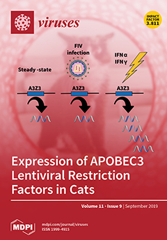
APOBEC3 (A3) proteins are key components of innate cellular defense against lentiviruses. In cats, the A3Z3 and A3Z2-Z3 proteins mediate restriction of feline immunodeficiency virus (FIV) infection. We quantified cat A3 mRNA expression in response to cytokines and FIV infection in vitro and in vivo. We found that cytokines upregulate A3 expression, but by and large, FIV infection did not appear to alter expression. A3Z3 mRNA abundance exceeded that of A3Z2-Z3 by 300-fold or more in all cells and tissues, suggesting that A3Z3 may be the primary anti-lentiviral A3 gene product in cats. View this paper.
  • Issues are regarded as officially published after their release is announced to the table of contents alert mailing list.
  • You may sign up for e-mail alerts to receive table of contents of newly released issues.
  • PDF is the official format for papers published in both, html and pdf forms. To view the papers in pdf format, click on the "PDF Full-text" link, and use the free Adobe Reader to open them.
Order results
Result details
Section
Select all
Export citation of selected articles as:
14 pages, 2592 KiB  
Article
An Avirulent Strain of Soybean Mosaic Virus Reverses the Defensive Effect of Abscisic Acid in a Susceptible Soybean Cultivar
by Mazen Alazem, Kristin Widyasari and Kook-Hyung Kim
Viruses 2019, 11(9), 879; https://doi.org/10.3390/v11090879 - 19 Sep 2019
Cited by 8 | Viewed by 3743
Abstract
In soybean cultivar L29, the Rsv3 gene is responsible for extreme resistance (ER) against the soybean mosaic virus avirulent strain G5H, but is ineffective against the virulent strain G7H. Part of this ER is attributed to the rapid increase in abscisic acid (ABA) [...] Read more.
In soybean cultivar L29, the Rsv3 gene is responsible for extreme resistance (ER) against the soybean mosaic virus avirulent strain G5H, but is ineffective against the virulent strain G7H. Part of this ER is attributed to the rapid increase in abscisic acid (ABA) and callose, and to the rapid induction of several genes in the RNA-silencing pathway. Whether these two defense mechanisms are correlated or separated in the ER is unknown. Here, we found that ABA treatment of L29 plants increased the expression of several antiviral RNA-silencing genes as well as the PP2C3a gene, which was previously shown to increase callose accumulation; as a consequence, ABA increased the resistance of L29 plants to G7H. The effect of ABA treatment on these genes was weaker in the rsv3-null cultivar (Somyungkong) than in L29. Besides, G5H-infection of Somyungkong plants subverted the effect of ABA leading to reduced callose accumulation and decreased expression of several RNA-silencing genes, which resulted in increased susceptibility to G5H infection. ABA treatment, however, still induced some resistance to G7H in Somyungkong, but only AGO7b was significantly induced. Our data suggest that Rsv3 modulates the effect of ABA on these two resistance mechanisms, i.e., callose accumulation and the antiviral RNA-silencing pathway, and that in the absence of Rsv3, some strains can reverse the effect of ABA and thereby facilitate their replication and spread. Full article
(This article belongs to the Special Issue Plant Virus Epidemiology and Control)
Show Figures

Figure 1

17 pages, 1184 KiB  
Review
Chasing Intracellular Zika Virus Using Proteomics
by Pietro Scaturro, Anna Lena Kastner and Andreas Pichlmair
Viruses 2019, 11(9), 878; https://doi.org/10.3390/v11090878 - 19 Sep 2019
Cited by 20 | Viewed by 6619
Abstract
Flaviviruses are the most medically relevant group of arboviruses causing a wide range of diseases in humans and are associated with high mortality and morbidity, as such posing a major health concern. Viruses belonging to this family can be endemic (e.g., dengue virus), [...] Read more.
Flaviviruses are the most medically relevant group of arboviruses causing a wide range of diseases in humans and are associated with high mortality and morbidity, as such posing a major health concern. Viruses belonging to this family can be endemic (e.g., dengue virus), but can also cause fulminant outbreaks (e.g., West Nile virus, Japanese encephalitis virus and Zika virus). Intense research efforts in the past decades uncovered shared fundamental strategies used by flaviviruses to successfully replicate in their respective hosts. However, the distinct features contributing to the specific host and tissue tropism as well as the pathological outcomes unique to each individual flavivirus are still largely elusive. The profound footprint of individual viruses on their respective hosts can be investigated using novel technologies in the field of proteomics that have rapidly developed over the last decade. An unprecedented sensitivity and throughput of mass spectrometers, combined with the development of new sample preparation and bioinformatics analysis methods, have made the systematic investigation of virus–host interactions possible. Furthermore, the ability to assess dynamic alterations in protein abundances, protein turnover rates and post-translational modifications occurring in infected cells now offer the unique possibility to unravel complex viral perturbations induced in the infected host. In this review, we discuss the most recent contributions of mass spectrometry–based proteomic approaches in flavivirus biology with a special focus on Zika virus, and their basic and translational potential and implications in understanding and characterizing host responses to arboviral infections. Full article
(This article belongs to the Section Animal Viruses)
Show Figures

Figure 1

12 pages, 631 KiB  
Article
Prevalence and Correlates of Pre-Treatment HIV Drug Resistance among HIV-Infected Children in Ethiopia
by Birkneh Tilahun Tadesse, Olivia Tsai, Adugna Chala, Tolossa Eticha Chaka, Temesgen Eromo, Hope R. Lapointe, Bemuluyigza Baraki, Aniqa Shahid, Sintayehu Tadesse, Eyasu Makonnen, Zabrina L. Brumme, Eleni Aklillu and Chanson J. Brumme
Viruses 2019, 11(9), 877; https://doi.org/10.3390/v11090877 - 19 Sep 2019
Cited by 8 | Viewed by 3735
Abstract
Pediatric human immunodeficiency virus (HIV) care in resource-limited settings remains a major challenge to achieving global HIV treatment and virologic suppression targets, in part because the administration of combination antiretroviral therapies (cART) is inherently complex in this population and because viral load and [...] Read more.
Pediatric human immunodeficiency virus (HIV) care in resource-limited settings remains a major challenge to achieving global HIV treatment and virologic suppression targets, in part because the administration of combination antiretroviral therapies (cART) is inherently complex in this population and because viral load and drug resistance genotyping are not routinely available in these settings. Children may also be at elevated risk of transmission of drug-resistant HIV as a result of suboptimal antiretroviral administration for prevention of mother-to-child transmission. We investigated the prevalence and the correlates of pretreatment HIV drug resistance (PDR) among HIV-infected, cART-naive children in Ethiopia. We observed an overall PDR rate of 14%, where all cases featured resistance to non-nucleoside reverse transcriptase inhibitors (NNRTIs): ~9% of participants harbored resistance solely to NNRTIs while ~5% harbored resistance to both NNRTIs and nucleoside reverse transcriptase inhibitors (NRTIs). No resistance to protease inhibitors was observed. No sociodemographic or clinical parameters were significantly associated with PDR, though limited statistical power is noted. The relatively high (14%) rate of NNRTI resistance in cART-naive children supports the use of non-NNRTI-based regimens in first-line pediatric treatment in Ethiopia and underscores the urgent need for access to additional antiretroviral classes in resource-limited settings. Full article
(This article belongs to the Special Issue Antiviral Agents)
Show Figures

Figure 1

15 pages, 2089 KiB  
Article
The 40 kDa Linear Polyethylenimine Inhibits Porcine Reproductive and Respiratory Syndrome Virus Infection by Blocking Its Attachment to Permissive Cells
by Jie Wang, Jie Li, Nana Wang, Qi Ji, Mingshuo Li, Yuchen Nan, En-Min Zhou, Yanjin Zhang and Chunyan Wu
Viruses 2019, 11(9), 876; https://doi.org/10.3390/v11090876 - 19 Sep 2019
Cited by 11 | Viewed by 4377
Abstract
Porcine reproductive and respiratory syndrome (PRRS) is one of the most economically devastating infectious diseases in pigs worldwide. The causative agent is the PRRS virus (PRRSV). In this study, we explored polyethylenimine (PEI), a cationic polymer with different forms (linear or branched), to [...] Read more.
Porcine reproductive and respiratory syndrome (PRRS) is one of the most economically devastating infectious diseases in pigs worldwide. The causative agent is the PRRS virus (PRRSV). In this study, we explored polyethylenimine (PEI), a cationic polymer with different forms (linear or branched), to inhibit the replication of PRRSV. Our results demonstrate that the linear but not the 40 kDa branched PEI, or the 25 kDa linear PEI, were well tolerated in cultured cells and exhibited a broad-spectrum inhibition of heterogeneous PRRSV-2 isolates in both MARC-145 cells and primary porcine pulmonary alveolar macrophages (PAMs). Further analysis suggests that PEI could prevent the attachment of PRRSV virions to the susceptible cells. Notably, PEI had a minimal effect on PRRSV internalization in MARC-145 cells, whereas PEI promoted the internalization of PRRSV virions in PAMs, which suggests that these two types of cells might have different internalization processes of PRRSV virions. In conclusion, our data demonstrate that PEI could be used as a novel inhibitor against PRRSV. Full article
(This article belongs to the Special Issue Antiviral Agents)
Show Figures

Figure 1

15 pages, 3064 KiB  
Article
Identification of Two Porcine Reproductive and Respiratory Syndrome Virus Variants Sharing High Genomic Homology but with Distinct Virulence
by Nanhua Chen, Mengxue Ye, Yucheng Huang, Shuai Li, Yanzhao Xiao, Xinshuai Li, Shubin Li, Xiangdong Li, Xiuling Yu, Kegong Tian and Jianzhong Zhu
Viruses 2019, 11(9), 875; https://doi.org/10.3390/v11090875 - 18 Sep 2019
Cited by 22 | Viewed by 3890
Abstract
Porcine reproductive and respiratory syndrome virus (PRRSV) causes huge economic loss to the global swine industry. Even though several control strategies have been applied, PRRS is still not effectively controlled due to the continuous emergence of new variants and limited cross-protection by current [...] Read more.
Porcine reproductive and respiratory syndrome virus (PRRSV) causes huge economic loss to the global swine industry. Even though several control strategies have been applied, PRRS is still not effectively controlled due to the continuous emergence of new variants and limited cross-protection by current vaccines. During the routine epidemiological investigation in 2017, two PRRSV variants were identified from a severe abortion farm and a clinically healthy farm, respectively. The viruses were isolated and denominated as XJ17-5 and JSTZ1712-12. Genomic sequencing indicated that their genomes are both 14,960 bp in length sharing 99.45% nucleotide identity. Sequence alignments identified a discontinuous 30-amino-acid deletion and a continuous 120-amino-acid deletion in nsp2 of both isolates. Genome-based phylogenetic analysis confirmed that XJ17-5 and JSTZ1712-12 belong to the HP-PRRSV subtype but form a new branch with other isolates containing the same 150-amino-acid deletion in nsp2. Pathogenic analysis showed that XJ17-5 is highly virulent causing 60% mortality, while JSTZ1712-12 is avirulent for piglets. Furthermore, fragment comparisons identified 34-amino-acid differences between XJ17-5 and JSTZ1712-12 that might be associated with the distinct virulence. The identification of highly homologous HP-PRRSV variants with new genetic feature and distinct virulence contributes to further analyze the pathogenesis and evolution of PRRSV in the field. Full article
(This article belongs to the Special Issue Emerging Viruses: Surveillance, Prevention, Evolution and Control)
Show Figures

Figure 1

19 pages, 2644 KiB  
Article
A Novel CXCR4 Targeting Protein SDF-1/54 as an HIV-1 Entry Inhibitor
by Suiyi Tan, Wenjuan Li, Zhaofeng Li, Yujing Li, Jiangyan Luo, Liangzhentian Yu, Jie Yang, Mengjie Qiu, Hongyan Cheng, Wei Xu, Shibo Jiang, Lu Lu, Shuwen Liu and Weifeng Ma
Viruses 2019, 11(9), 874; https://doi.org/10.3390/v11090874 - 18 Sep 2019
Cited by 8 | Viewed by 4142
Abstract
CXC chemokine receptor 4 (CXCR4) is a co-receptor for HIV-1 entry into target cells. Its natural ligand, the chemokine SDF-1, inhibits viral entry mediated by this receptor. However, the broad expression pattern of CXCR4 and its critical roles in various physiological and pathological [...] Read more.
CXC chemokine receptor 4 (CXCR4) is a co-receptor for HIV-1 entry into target cells. Its natural ligand, the chemokine SDF-1, inhibits viral entry mediated by this receptor. However, the broad expression pattern of CXCR4 and its critical roles in various physiological and pathological processes indicate that the direct application of SDF-1 as an entry inhibitor might have severe consequences. Previously, we constructed an effective SDF-1 mutant, SDF-1/54, by deleting the α-helix of the C-terminal functional region of SDF-1. Of note, SDF-1/54 shows remarkable decreased chemotoxic ability, but maintains a similar binding affinity to CXCR4, suggesting SDF-1/54 might better serve as a CXCR4 inhibitor. Here, we found that SDF-1/54 exhibited potent antiviral activity against various X4 HIV-1 strains, including the infectious clone HIV-1 NL4-3, laboratory-adapted strain HIV-1 IIIB, clinical isolates and even drug-resistant strains. By using time-of-addition assay, non-infectious and infectious cell–cell fusion assay and CXCR4 internalization assay, we demonstrated SDF-1/54 is an HIV-1 entry inhibitor. A combination of SDF-1/54 with several antiretroviral drugs exhibited potent synergistic anti-HIV-1 activity. Moreover, SDF-1/54 was stable and its anti-HIV-1 activity was not significantly affected by the presence of seminal fluid, vaginal fluid simulant and human serum albumin. SDF-1/54 showed limited in vitro cytotoxicity to lymphocytes and vaginal epithelial cells. Based on these findings, SDF-1/54 could have a therapeutic potential as an HIV-1 entry inhibitor. Full article
(This article belongs to the Special Issue Viral Entry Pathways)
Show Figures

Figure 1

26 pages, 4550 KiB  
Review
Mosquito-Specific Viruses—Transmission and Interaction
by Eric Agboli, Mayke Leggewie, Mine Altinli and Esther Schnettler
Viruses 2019, 11(9), 873; https://doi.org/10.3390/v11090873 - 17 Sep 2019
Cited by 61 | Viewed by 8006
Abstract
Mosquito-specific viruses (MSVs) are a subset of insect-specific viruses that are found to infect mosquitoes or mosquito derived cells. There has been an increase in discoveries of novel MSVs in recent years. This has expanded our understanding of viral diversity and evolution but [...] Read more.
Mosquito-specific viruses (MSVs) are a subset of insect-specific viruses that are found to infect mosquitoes or mosquito derived cells. There has been an increase in discoveries of novel MSVs in recent years. This has expanded our understanding of viral diversity and evolution but has also sparked questions concerning the transmission of these viruses and interactions with their hosts and its microbiome. In fact, there is already evidence that MSVs interact with the immune system of their host. This is especially interesting, since mosquitoes can be infected with both MSVs and arthropod-borne (arbo) viruses of public health concern. In this review, we give an update on the different MSVs discovered so far and describe current data on their transmission and interaction with the mosquito immune system as well as the effect MSVs could have on an arboviruses-co-infection. Lastly, we discuss potential uses of these viruses, including vector and transmission control. Full article
(This article belongs to the Special Issue Transmission Dynamics of Insect Viruses)
Show Figures

Figure 1

13 pages, 705 KiB  
Article
Identification of Loci Associated with Enhanced Virulence in Spodoptera litura Nucleopolyhedrovirus Isolates Using Deep Sequencing
by Mark P. Zwart, Ghulam Ali, Elisabeth A. van Strien, Elio G. W. M. Schijlen, Manli Wang, Wopke van der Werf and Just M. Vlak
Viruses 2019, 11(9), 872; https://doi.org/10.3390/v11090872 - 17 Sep 2019
Cited by 8 | Viewed by 3403
Abstract
Spodoptera litura is an emerging pest insect in cotton and arable crops in Central Asia. To explore the possibility of using baculoviruses as biological control agents instead of chemical pesticides, in a previous study we characterized a number of S. litura nucleopolyhedrovirus (SpltNPV) [...] Read more.
Spodoptera litura is an emerging pest insect in cotton and arable crops in Central Asia. To explore the possibility of using baculoviruses as biological control agents instead of chemical pesticides, in a previous study we characterized a number of S. litura nucleopolyhedrovirus (SpltNPV) isolates from Pakistan. We found significant differences in speed of kill, an important property of a biological control agent. Here we set out to understand the genetic basis of these differences in speed of kill, by comparing the genome of the fast-killing SpltNPV-Pak-TAX1 isolate with that of the slow-killing SpltNPV-Pak-BNG isolate. These two isolates and the SpltNPV-G2 reference strain from China were deep sequenced with Illumina. As expected, the two Pakistani isolates were closely related with >99% sequence identity, whereas the Chinese isolate was more distantly related. We identified two loci that may be associated with the fast action of the SpltNPV-Pak-TAX1 isolate. First, an analysis of rates of synonymous and non-synonymous mutations identified neutral to positive selection on open reading frame (ORF) 122, encoding a viral fibroblast growth factor (vFGF) that is known to affect virulence in other baculoviruses. Second, the homologous repeat region hr17, a putative enhancer of transcription and origin of replication, is absent in SpltNPV-Pak-TAX1 suggesting it may also affect virulence. Additionally, we found there is little genetic variation within both Pakistani isolates, and we identified four genes under positive selection in both isolates that may have played a role in adaptation of SpltNPV to conditions in Central Asia. Our results contribute to the understanding of the enhanced activity of SpltNPV-Pak-TAX1, and may help to select better SpltNPV isolates for the control of S. litura in Pakistan and elsewhere. Full article
(This article belongs to the Special Issue Insect Viruses and Pest Management)
Show Figures

Figure 1

15 pages, 3351 KiB  
Article
Novel Genetic Rearrangements Termed “Structural Variation Polymorphisms“ Contribute to the Genetic Diversity of Orthohepadnaviruses
by Kei Fujiwara, Kentaro Matsuura, Kayoko Matsunami, Etsuko Iio, Yoshihito Nagura, Shunsuke Nojiri and Hiromi Kataoka
Viruses 2019, 11(9), 871; https://doi.org/10.3390/v11090871 - 17 Sep 2019
Cited by 2 | Viewed by 5243
Abstract
The genetic diversity of orthohepadnaviruses is not yet fully understood. This study was conducted to investigate the role of structural variations (SVs) in their diversity. Genetic sequences of orthohepadnaviruses were retrieved from databases. The positions of sequence gaps were investigated, since they were [...] Read more.
The genetic diversity of orthohepadnaviruses is not yet fully understood. This study was conducted to investigate the role of structural variations (SVs) in their diversity. Genetic sequences of orthohepadnaviruses were retrieved from databases. The positions of sequence gaps were investigated, since they were found to be related to SVs, and they were further used to search for SVs. Then, a combination of pair-wise and multiple alignment analyses was performed to analyze the genomic structure. Unique patterns of SVs were observed; genetic sequences at certain genomic positions could be separated into multiple patterns, such as no SV, SV pattern 1, SV pattern 2, and SV pattern 3, which were observed as polymorphic changes. We provisionally referred to these genetic changes as SV polymorphisms. Our data showed that higher frequency of sequence gaps and lower genetic identity were observed in the pre-S1-S2 region of various types of HBVs. Detailed examination of the genetic structure in the pre-S region by a combination of pair-wise and multiple alignment analyses showed that the genetic diversity of orthohepadnaviruses in the pre-S1 region could have been also induced by SV polymorphisms. Our data showed that novel genetic rearrangements provisionally termed SV polymorphisms were observed in various orthohepadnaviruses. Full article
(This article belongs to the Section Animal Viruses)
Show Figures

Figure 1

21 pages, 1809 KiB  
Article
Interaction of a Densovirus with Glycans of the Peritrophic Matrix Mediates Oral Infection of the Lepidopteran Pest Spodoptera frugiperda
by Laetitia Pigeyre, Malvina Schatz, Marc Ravallec, Leila Gasmi, Nicolas Nègre, Cécile Clouet, Martial Seveno, Khadija El Koulali, Mathilde Decourcelle, Yann Guerardel, Didier Cot, Thierry Dupressoir, Anne-Sophie Gosselin-Grenet and Mylène Ogliastro
Viruses 2019, 11(9), 870; https://doi.org/10.3390/v11090870 - 17 Sep 2019
Cited by 3 | Viewed by 4269
Abstract
The success of oral infection by viruses depends on their capacity to overcome the gut epithelial barrier of their host to crossing over apical, mucous extracellular matrices. As orally transmitted viruses, densoviruses, are also challenged by the complexity of the insect gut barriers, [...] Read more.
The success of oral infection by viruses depends on their capacity to overcome the gut epithelial barrier of their host to crossing over apical, mucous extracellular matrices. As orally transmitted viruses, densoviruses, are also challenged by the complexity of the insect gut barriers, more specifically by the chitinous peritrophic matrix, that lines and protects the midgut epithelium; how capsids stick to and cross these barriers to reach their final cell destination where replication goes has been poorly studied in insects. Here, we analyzed the early interaction of the Junonia coenia densovirus (JcDV) with the midgut barriers of caterpillars from the pest Spodoptera frugiperda. Using combination of imaging, biochemical, proteomic and transcriptomic analyses, we examined in vitro, ex vivo and in vivo the early interaction of the capsids with the peritrophic matrix and the consequence of early oral infection on the overall gut function. We show that the JcDV particle rapidly adheres to the peritrophic matrix through interaction with different glycans including chitin and glycoproteins, and that these interactions are necessary for oral infection. Proteomic analyses of JcDV binding proteins of the peritrophic matrix revealed mucins and non-mucins proteins including enzymes already known to act as receptors for several insect pathogens. In addition, we show that JcDV early infection results in an arrest of N-Acetylglucosamine secretion and a disruption in the integrity of the peritrophic matrix, which may help viral particles to pass through. Finally, JcDV early infection induces changes in midgut genes expression favoring an increased metabolism including an increased translational activity. These dysregulations probably participate to the overall dysfunction of the gut barrier in the early steps of viral pathogenesis. A better understanding of early steps of densovirus infection process is crucial to build biocontrol strategies against major insect pests. Full article
(This article belongs to the Special Issue New Insights into Parvovirus Research)
Show Figures

Figure 1

17 pages, 4095 KiB  
Article
Bacterial Virus Lambda Gpd-Fusions to Cathelicidins, α- and β-Defensins, and Disease-Specific Epitopes Evaluated for Antimicrobial Toxicity and Ability to Support Phage Display
by Sidney Hayes
Viruses 2019, 11(9), 869; https://doi.org/10.3390/v11090869 - 17 Sep 2019
Cited by 4 | Viewed by 3987
Abstract
We showed that antimicrobial polypeptides, when translated as gene fusions to the bacteriophage lambda capsid decoration protein gpD, formed highly toxic molecules within E. coli, suggesting that they can retain their antimicrobial activity conformation when fused to gpD. These include gpD-fusions to [...] Read more.
We showed that antimicrobial polypeptides, when translated as gene fusions to the bacteriophage lambda capsid decoration protein gpD, formed highly toxic molecules within E. coli, suggesting that they can retain their antimicrobial activity conformation when fused to gpD. These include gpD-fusions to human and porcine cathelicidins LL37 and PR39, β-defensins HBD3 and DEFB126-Δ (deleted for its many COOH-terminal glycosylation sites), and α-defensin HD5. Antimicrobial toxicity was only observed when the peptides were displayed from the COOH-terminal, and not the NH2-terminal end, of gpD. This suggests that COOH-terminal displayed polypeptides of gpD-fusions can more readily form an active-state conformation than when they are displayed from the NH2-terminal end of gpD. The high toxicity of the COOH-displayed gpD-defensins suggests either that the fused defensin peptides can be oxidized, forming three correct intramolecular disulfide bonds within the cytosol of bacterial cells, or that the versions without disulfide bonds are highly toxigenic. We showed the high efficiency of displaying single epitope 17 amino-acid fusions to gpD on LDP (lambda display particles), even when the gpD-fusion protein was toxic. The efficient formation of high display density LDP, displaying a single disease specific epitope (DSE), suggests the utility of LDP-DSE constructs for use as single epitope vaccines (SEV). Full article
(This article belongs to the Section Bacterial Viruses)
Show Figures

Figure 1

10 pages, 936 KiB  
Article
Medical Outcomes in Women Who Became Pregnant after Vaccination with a Virus-Like Particle Experimental Vaccine against Influenza A (H1N1) 2009 Virus Tested during 2009 Pandemic Outbreak
by Arturo Cérbulo-Vázquez, Lourdes Arriaga-Pizano, Gabriela Cruz-Cureño, Ilka Boscó-Gárate, Eduardo Ferat-Osorio, Rodolfo Pastelin-Palacios, Ricardo Figueroa-Damian, Denisse Castro-Eguiluz, Javier Mancilla-Ramirez, Armando Isibasi and Constantino López-Macías
Viruses 2019, 11(9), 868; https://doi.org/10.3390/v11090868 - 17 Sep 2019
Cited by 2 | Viewed by 3461
Abstract
The clinical effects and immunological response to the influenza vaccine in women who later become pregnant remain to be thoroughly studied. Here, we report the medical outcomes of 40 women volunteers who became pregnant after vaccination with an experimental virus-like particle (VLP) vaccine [...] Read more.
The clinical effects and immunological response to the influenza vaccine in women who later become pregnant remain to be thoroughly studied. Here, we report the medical outcomes of 40 women volunteers who became pregnant after vaccination with an experimental virus-like particle (VLP) vaccine against pandemic influenza A(H1N1)2009 (influenza A(H1N1)pdm09) and their infants. When included in the VLP vaccine trial, none of the women were pregnant and were randomly assigned to one of the following groups: (1) placebo, (2) 15 μg dose of VLP vaccine, or (3) 45 μg dose of VLP vaccine. These 40 women reported becoming pregnant during the follow-up phase after receiving the placebo or VLP vaccine. Women were monitored throughout pregnancy and their infants were monitored until one year after birth. Antibody titers against VLP were measured in the mothers and infants at delivery and at six months and one year after birth. The incidence of preeclampsia, fetal death, preterm delivery, and premature rupture of membranes was similar among groups. All vaccinated women and their infants elicited antibody titers (≥1:40). Women vaccinated prior to pregnancy had no adverse events that were different from the nonvaccinated population. Even though this study is limited by the sample size, the results suggest that the anti-influenza A(H1N1)pdm09 VLP experimental vaccine applied before pregnancy is safe for both mothers and their infants. Full article
(This article belongs to the Special Issue Virus-Like Particle Vaccines)
Show Figures

Figure 1

24 pages, 937 KiB  
Review
Vector Competence: What Has Zika Virus Taught Us?
by Sasha R. Azar and Scott C. Weaver
Viruses 2019, 11(9), 867; https://doi.org/10.3390/v11090867 - 17 Sep 2019
Cited by 43 | Viewed by 8168
Abstract
The unprecedented outbreak of Zika virus (ZIKV) infection in the Americas from 2015 to 2017 prompted the publication of a large body of vector competence data in a relatively short period of time. Although differences in vector competence as a result of disparities [...] Read more.
The unprecedented outbreak of Zika virus (ZIKV) infection in the Americas from 2015 to 2017 prompted the publication of a large body of vector competence data in a relatively short period of time. Although differences in vector competence as a result of disparities in mosquito populations and viral strains are to be expected, the limited competence of many populations of the urban mosquito vector, Aedes aegypti, from the Americas (when its susceptibility is viewed relative to other circulating/reemerging mosquito-borne viruses such as dengue (DENV), yellow fever (YFV), and chikungunya viruses (CHIKV)) has proven a paradox for the field. This has been further complicated by the lack of standardization in the methodologies utilized in laboratory vector competence experiments, precluding meta-analyses of this large data set. As the calls for the standardization of such studies continue to grow in number, it is critical to examine the elements of vector competence experimental design. Herein, we review the various techniques and considerations intrinsic to vector competence studies, with respect to contemporary findings for ZIKV, as well as historical findings for other arboviruses, and discuss potential avenues of standardization going forward. Full article
(This article belongs to the Special Issue Transmission Dynamics of Insect Viruses)
Show Figures

Figure 1

16 pages, 572 KiB  
Review
African Swine Fever: Fast and Furious or Slow and Steady?
by Katja Schulz, Franz Josef Conraths, Sandra Blome, Christoph Staubach and Carola Sauter-Louis
Viruses 2019, 11(9), 866; https://doi.org/10.3390/v11090866 - 17 Sep 2019
Cited by 55 | Viewed by 7982
Abstract
Since the introduction of African swine fever (ASF) into Georgia in 2007, the disease has been spreading in an unprecedented way. Many countries that are still free from the disease fear the emergence of ASF in their territory either in domestic pigs or [...] Read more.
Since the introduction of African swine fever (ASF) into Georgia in 2007, the disease has been spreading in an unprecedented way. Many countries that are still free from the disease fear the emergence of ASF in their territory either in domestic pigs or in wild boar. In the past, ASF was often described as being a highly contagious disease with mortality often up to 100%. However, the belief that the disease might enter a naïve population and rapidly affect the entire susceptible population needs to be critically reviewed. The current ASF epidemic in wild boar, but also the course of ASF within outbreaks in domestic pig holdings, suggest a constant, but relatively slow spread. Moreover, the results of several experimental and field studies support the impression that the spread of ASF is not always fast. ASF spread and its speed depend on various factors concerning the host, the virus, and also the environment. Many of these factors and their effects are not fully understood. For this review, we collated published information regarding the spreading speed of ASF and the factors that are deemed to influence the speed of ASF spread and tried to clarify some issues and open questions in this respect. Full article
(This article belongs to the Section Animal Viruses)
Show Figures

Figure 1

16 pages, 2249 KiB  
Article
Metaviromics Reveals Unknown Viral Diversity in the Biting Midge Culicoides impunctatus
by Sejal Modha, Joseph Hughes, Giovanni Bianco, Heather M. Ferguson, Barbara Helm, Lily Tong, Gavin S. Wilkie, Alain Kohl and Esther Schnettler
Viruses 2019, 11(9), 865; https://doi.org/10.3390/v11090865 - 17 Sep 2019
Cited by 10 | Viewed by 14313
Abstract
Biting midges (Culicoides species) are vectors of arboviruses and were responsible for the emergence and spread of Schmallenberg virus (SBV) in Europe in 2011 and are likely to be involved in the emergence of other arboviruses in Europe. Improved surveillance and better [...] Read more.
Biting midges (Culicoides species) are vectors of arboviruses and were responsible for the emergence and spread of Schmallenberg virus (SBV) in Europe in 2011 and are likely to be involved in the emergence of other arboviruses in Europe. Improved surveillance and better understanding of risks require a better understanding of the circulating viral diversity in these biting insects. In this study, we expand the sequence space of RNA viruses by identifying a number of novel RNA viruses from Culicoides impunctatus (biting midge) using a meta-transcriptomic approach. A novel metaviromic pipeline called MetaViC was developed specifically to identify novel virus sequence signatures from high throughput sequencing (HTS) datasets in the absence of a known host genome. MetaViC is a protein centric pipeline that looks for specific protein signatures in the reads and contigs generated as part of the pipeline. Several novel viruses, including an alphanodavirus with both segments, a novel relative of the Hubei sobemo-like virus 49, two rhabdo-like viruses and a chuvirus, were identified in the Scottish midge samples. The newly identified viruses were found to be phylogenetically distinct to those previous known. These findings expand our current knowledge of viral diversity in arthropods and especially in these understudied disease vectors. Full article
(This article belongs to the Special Issue Viromics: Approaches, Advances, and Applications)
Show Figures

Figure 1

14 pages, 1930 KiB  
Review
An Update on African Swine Fever Virology
by Axel Karger, Daniel Pérez-Núñez, Jesús Urquiza, Patricia Hinojar, Covadonga Alonso, Ferdinando B. Freitas, Yolanda Revilla, Marie-Frédérique Le Potier and Maria Montoya
Viruses 2019, 11(9), 864; https://doi.org/10.3390/v11090864 - 17 Sep 2019
Cited by 86 | Viewed by 10414
Abstract
Animal diseases constitute a continuing threat to animal health, food safety, national economy, and the environment. Among those, African swine fever (ASF) is one of the most devastating viruses affecting pigs and wild suids due to the lack of vaccine or effective treatment. [...] Read more.
Animal diseases constitute a continuing threat to animal health, food safety, national economy, and the environment. Among those, African swine fever (ASF) is one of the most devastating viruses affecting pigs and wild suids due to the lack of vaccine or effective treatment. ASF is endemic in countries in sub-Saharan Africa, but since its introduction to the Caucasus region in 2007, a highly virulent strain of ASF virus (ASFV) has continued to circulate and spread into Eastern Europe and Russia, and most recently into Western Europe, China, and various countries of Southeast Asia. Given the importance of this disease, this review will highlight recent discoveries in basic virology with special focus on proteomic analysis, replication cycle, and some recent data on genes involved in cycle progression and viral–host interactions, such as I215L (E2 ubiquitin-conjugating enzyme), EP402R (CD2v), A104R (histone-like protein), QP509L, and Q706L (RNA helicases) or P1192R (Topoisomerase II). Taking into consideration the large DNA genome of ASFV and its complex interactions with the host, more studies and new approaches are to be taken to understand the basic virus–host interaction for ASFV. Proteomic studies are just paving the way for future research. Full article
(This article belongs to the Special Issue Porcine Viruses 2019)
Show Figures

Figure 1

14 pages, 1351 KiB  
Article
Clinical and Virological Aspects of HBV Reactivation: A Focus on Acute Liver Failure
by Olympia E. Anastasiou, Martin Theissen, Jens Verheyen, Barbara Bleekmann, Heiner Wedemeyer, Marek Widera and Sandra Ciesek
Viruses 2019, 11(9), 863; https://doi.org/10.3390/v11090863 - 16 Sep 2019
Cited by 5 | Viewed by 3642
Abstract
Hepatitis B virus (HBV) reactivation in immunosuppressed patients can cause considerable morbidity and mortality. The aim of our study was to evaluate factors associated with acute liver failure (ALF) in HBV reactivation. Clinical, laboratory, and virological data of 87 patients with HBV reactivation [...] Read more.
Hepatitis B virus (HBV) reactivation in immunosuppressed patients can cause considerable morbidity and mortality. The aim of our study was to evaluate factors associated with acute liver failure (ALF) in HBV reactivation. Clinical, laboratory, and virological data of 87 patients with HBV reactivation were analyzed retrospectively. Teno torque virus (TTV) plasma loads were measured as a measure of immune competence. HBV genomes isolated from 47 patients were analyzed by next-generation sequencing. A functional analysis of identified HBsAg mutants was performed. In patients with ALF the diagnosis was significantly later confirmed than in the non-ALF group. Patients diagnosed during immunosuppression had a milder clinical course compared to later diagnosed patients (p = 0.018, OR = 4.17). TTV viral loads did not differ significantly between the two groups. The HBV genomes isolated from ALF patients had higher viral complexity. A mutation in C-region of HBsAg (L216*), was associated with reduced HBsAg production and secretion. Patients diagnosed with HBV reactivation during immunosuppression had a milder clinical course compared to patients diagnosed during immune reconstitution. ALF was associated with higher viral complexity. An HBsAg mutation (L216*) was found to be more frequent in ALF patients and was associated with reduced HBsAg production and secretion. Full article
(This article belongs to the Special Issue Hepatitis B Virus Reactivation)
Show Figures

Figure 1

11 pages, 6230 KiB  
Article
Diverse Morphology and Structural Features of Old and New World Hantaviruses
by Amar Parvate, Evan P. Williams, Mariah K. Taylor, Yong-Kyu Chu, Jason Lanman, Erica Ollmann Saphire and Colleen B. Jonsson
Viruses 2019, 11(9), 862; https://doi.org/10.3390/v11090862 - 16 Sep 2019
Cited by 15 | Viewed by 5785
Abstract
To further understanding of the structure and morphology of the Orthohantavirus, family Hantaviridae, we have employed cryo-electron microscopy (cryo-EM) for three New World hantaviruses: Andes (ANDV), Sin Nombre (SNV), and Black Creek Canal (BCCV). Building upon our prior cryo-EM and cryo-tomography [...] Read more.
To further understanding of the structure and morphology of the Orthohantavirus, family Hantaviridae, we have employed cryo-electron microscopy (cryo-EM) for three New World hantaviruses: Andes (ANDV), Sin Nombre (SNV), and Black Creek Canal (BCCV). Building upon our prior cryo-EM and cryo-tomography study of the Old World hantavirus, Hantaan virus (HTNV), we have expanded our studies to examine the entire virion population present in cell culture supernatant. Hence, in contrast to the prior cryo-EM/ET studies in which we used a polyethylene precipitation, a sucrose gradient, and a sucrose cushion, we used two sucrose cushions. We inactivated the material after the first cushion. We tested the method using HTNV which has a known cryo-EM structure and observed equivalent results. Therefore, we used this method to assess the particle distribution of the New World hantaviruses by cryo-EM. Cryo-EM images showed a diverse range of sizes and morphologies for the New World viruses that we classified as round, tubular, and irregular. Strikingly, BCCV virions were mostly tubular. These first cryo-EM images of the New World Orthohantavirus confirm prior EM observations that noted tubular projections of SNV at the plasma membrane during virion morphogenesis but were not confirmed. These findings underscore the need for further investigation of virion morphogenesis of the Orthohantavirus. Full article
(This article belongs to the Special Issue Hantaviruses)
Show Figures

Figure 1

18 pages, 3371 KiB  
Article
A Metabolomics Approach to Unravel Cricket Paralysis Virus Infection in Silkworm Bm5 Cells
by Luo-Luo Wang, Luc Swevers, Caroline Rombouts, Ivan Meeus, Lieven Van Meulebroek, Lynn Vanhaecke and Guy Smagghe
Viruses 2019, 11(9), 861; https://doi.org/10.3390/v11090861 - 16 Sep 2019
Cited by 12 | Viewed by 3810
Abstract
How a host metabolism responds to infection with insect viruses and how it relates to pathogenesis, is little investigated. Our previous study observed that Cricket paralysis virus (CrPV, Dicistroviridae) causes short term persistence in silkworm Bm5 cells before proceeding to acute infection. [...] Read more.
How a host metabolism responds to infection with insect viruses and how it relates to pathogenesis, is little investigated. Our previous study observed that Cricket paralysis virus (CrPV, Dicistroviridae) causes short term persistence in silkworm Bm5 cells before proceeding to acute infection. In this study, a metabolomics approach based on high resolution mass spectrometry was applied to investigate how a host metabolism is altered during the course of CrPV infection in Bm5 cells and which changes are characteristic for the transition from persistence to pathogenicity. We observed that CrPV infection led to significant and stage-specific metabolic changes in Bm5 cells. Differential metabolites abundance and pathway analysis further identified specific metabolic features at different stages in the viral life cycle. Notably, both glucose and glutamine levels significantly increased during CrPV persistent infection followed by a steep decrease during the pathogenic stages, suggesting that the central carbon metabolism was significantly modified during CrPV infection in Bm5 cells. In addition, dynamic changes in levels of polyamines were detected. Taken together, this study characterized for the first time the metabolic dynamics of CrPV infection in insect cells, proposing a central role for the regulation of both amino acid and carbohydrate metabolism during the period of persistent infection of CrPV in Bm5 cells. Full article
(This article belongs to the Section Insect Viruses)
Show Figures

Figure 1

16 pages, 3755 KiB  
Article
Genetic Diversity of the Hepatitis B Virus Subgenotypes in Brazil
by Barbara V. Lago, Marcia P. do Espirito-Santo, Vanessa D. Costa, Vanessa A. Marques, Livia M. Villar, Lia L. Lewis-Ximenez, Elisabeth Lampe and Francisco C. A. Mello
Viruses 2019, 11(9), 860; https://doi.org/10.3390/v11090860 - 15 Sep 2019
Cited by 24 | Viewed by 3794
Abstract
Hepatitis B virus (HBV) subgenotypes may be related to clinical outcomes and response to antiviral therapy. Most Brazilian studies on HBV subgenotypes are restricted to some regions and to specific population groups. Here, we provide an insight about genetic diversity of HBV subgenotypes [...] Read more.
Hepatitis B virus (HBV) subgenotypes may be related to clinical outcomes and response to antiviral therapy. Most Brazilian studies on HBV subgenotypes are restricted to some regions and to specific population groups. Here, we provide an insight about genetic diversity of HBV subgenotypes in 321 serum samples from all five geographical regions, providing a representative overview of their circulation among chronic carriers. Overall, HBV/A1 was the most prevalent subgenotype, being found as the major one in all regions except in South Brazil. Among HBV/D samples, subgenotype D3 was the most prevalent, found in 51.5%, followed by D2 (27.3%) and D4 (21.2%). D2 and D3 were the most prevalent subgenotypes in South region, with high similarity with European strains. D4 was found in North and Northeast region and clustered with strains from Cape Verde and India. For HBV/F, the most frequent subgenotype was F2 (84.1%), followed by F4 (10.1%) and F1 (5.8%), closely related with strains from Venezuela, Argentina and Chile, respectively. Phylogeographic analyses were performed using an HBV full-length genome obtained from samples infected with genotypes rarely found in Brazil (B, C, and E). According to Bayesian inference, HBV/B2 and HBV/C2 were probably introduced in Brazil through China, and HBV/E from Guinea, all of them mostly linked to recent events of human migration. In conclusion, this study provided a comprehensive overview of the current circulation of HBV subgenotypes in Brazil. Our findings might contribute to a better understand of the dynamics of viral variants, to establish a permanent molecular surveillance on the introduction and dispersion patterns of new strains and, thus, to support public policies to control HBV dissemination in Brazil. Full article
(This article belongs to the Section Animal Viruses)
Show Figures

Figure 1

30 pages, 20991 KiB  
Review
Recombination in Enteroviruses, a Multi-Step Modular Evolutionary Process
by Claire Muslin, Alice Mac Kain, Maël Bessaud, Bruno Blondel and Francis Delpeyroux
Viruses 2019, 11(9), 859; https://doi.org/10.3390/v11090859 - 14 Sep 2019
Cited by 61 | Viewed by 11513
Abstract
RNA recombination is a major driving force in the evolution and genetic architecture shaping of enteroviruses. In particular, intertypic recombination is implicated in the emergence of most pathogenic circulating vaccine-derived polioviruses, which have caused numerous outbreaks of paralytic poliomyelitis worldwide. Recent experimental studies [...] Read more.
RNA recombination is a major driving force in the evolution and genetic architecture shaping of enteroviruses. In particular, intertypic recombination is implicated in the emergence of most pathogenic circulating vaccine-derived polioviruses, which have caused numerous outbreaks of paralytic poliomyelitis worldwide. Recent experimental studies that relied on recombination cellular systems mimicking natural genetic exchanges between enteroviruses provided new insights into the molecular mechanisms of enterovirus recombination and enabled to define a new model of genetic plasticity for enteroviruses. Homologous intertypic recombinant enteroviruses that were observed in nature would be the final products of a multi-step process, during which precursor nonhomologous recombinant genomes are generated through an initial inter-genomic RNA recombination event and can then evolve into a diversity of fitter homologous recombinant genomes over subsequent intra-genomic rearrangements. Moreover, these experimental studies demonstrated that the enterovirus genome could be defined as a combination of genomic modules that can be preferentially exchanged through recombination, and enabled defining the boundaries of these recombination modules. These results provided the first experimental evidence supporting the theoretical model of enterovirus modular evolution previously elaborated from phylogenetic studies of circulating enterovirus strains. This review summarizes our current knowledge regarding the mechanisms of recombination in enteroviruses and presents a new evolutionary process that may apply to other RNA viruses. Full article
(This article belongs to the Special Issue Human Picornaviruses)
Show Figures

Figure 1

15 pages, 595 KiB  
Review
Hematological Malignancies and HBV Reactivation Risk: Suggestions for Clinical Management
by Alessandra Zannella, Massimo Marignani and Paola Begini
Viruses 2019, 11(9), 858; https://doi.org/10.3390/v11090858 - 14 Sep 2019
Cited by 8 | Viewed by 3579
Abstract
It is well known that hepatitis B virus reactivation (HBVr) can occur among patients undergoing treatment for hematological malignancies (HM). The evaluation of HBVr risk in patients undergoing immunosuppressive treatments is a multidimensional process, which includes conducting an accurate clinical history and physical [...] Read more.
It is well known that hepatitis B virus reactivation (HBVr) can occur among patients undergoing treatment for hematological malignancies (HM). The evaluation of HBVr risk in patients undergoing immunosuppressive treatments is a multidimensional process, which includes conducting an accurate clinical history and physical examination, consideration of the virological categories, of the medication chosen to treat these hematological malignancies and the degree of immunosuppression induced. Once the risk of reactivation has been defined, it is crucial to adopt adequate management strategies (should reactivation occur). The purpose of treatment is to prevent dire clinical consequences of HBVr such as acute/fulminant hepatitis, and liver failure. Treatment will be instituted according to the indications and evidence provided by current international recommendations and to prevent interruption of lifesaving anti-neoplastic treatments. In this paper, we will present the available data regarding the risk of HBVr in this special population of immunosuppressed patients and explore the relevance of effective prevention and management of this potentially life-threatening event. A computerized literature search was performed using appropriate terms to discover relevant articles. Current evidence supports the policy of universal HBV testing of patients scheduled to undergo treatment for hematological malignancies, and clinicians should be aware of the inherent risk of viral reactivation among the different virological categories and classes of immunosuppressive drugs. Full article
(This article belongs to the Special Issue Hepatitis B Virus Reactivation)
Show Figures

Figure 1

15 pages, 1856 KiB  
Article
Highly Divergent Genetic Variants of Soricid-Borne Altai Virus (Hantaviridae) in Eurasia Suggest Ancient Host-Switching Events
by Hae Ji Kang, Se Hun Gu, Liudmila N. Yashina, Joseph A. Cook and Richard Yanagihara
Viruses 2019, 11(9), 857; https://doi.org/10.3390/v11090857 - 14 Sep 2019
Cited by 11 | Viewed by 3408
Abstract
With the recent discovery of genetically distinct hantaviruses (family Hantaviridae) in shrews (order Eulipotyphla, family Soricidae), the once-conventional view that rodents (order Rodentia) served as the primordial reservoir hosts now appears improbable. The newly identified soricid-borne hantaviruses generally demonstrate well-resolved lineages [...] Read more.
With the recent discovery of genetically distinct hantaviruses (family Hantaviridae) in shrews (order Eulipotyphla, family Soricidae), the once-conventional view that rodents (order Rodentia) served as the primordial reservoir hosts now appears improbable. The newly identified soricid-borne hantaviruses generally demonstrate well-resolved lineages organized according to host taxa and geographic origin. However, beginning in 2007, we detected sequences that did not conform to the prototypic hantaviruses associated with their soricid host species and/or geographic locations. That is, Eurasian common shrews (Sorex araneus), captured in Hungary and Russia, were found to harbor hantaviruses belonging to two separate and highly divergent lineages. We have since accumulated additional examples of these highly distinctive hantavirus sequences in the Laxmann’s shrew (Sorex caecutiens), flat-skulled shrew (Sorex roboratus) and Eurasian least shrew (Sorex minutissimus), captured at the same time and in the same location in the Sakha Republic in Far Eastern Russia. Pair-wise alignment and phylogenetic analysis of partial and full-length S-, M- and/or L-segment sequences indicate that a distinct hantavirus species related to Altai virus (ALTV), first reported in a Eurasian common shrew from Western Siberia, was being maintained in these closely related syntopic soricine shrew species. These findings suggest that genetic variants of ALTV might have resulted from ancient host-switching events with subsequent diversification within the Soricini tribe in Eurasia. Full article
(This article belongs to the Special Issue Hantaviruses)
Show Figures

Figure 1

18 pages, 5509 KiB  
Article
Vesiculopolins, a New Class of Anti-Vesiculoviral Compounds, Inhibit Transcription Initiation of Vesiculoviruses
by Minako Ogino, Yuriy Fedorov, Drew J. Adams, Kazuma Okada, Naoto Ito, Makoto Sugiyama and Tomoaki Ogino
Viruses 2019, 11(9), 856; https://doi.org/10.3390/v11090856 - 14 Sep 2019
Cited by 7 | Viewed by 3767
Abstract
Vesicular stomatitis virus (VSV) represents a promising platform for developing oncolytic viruses, as well as vaccines against significant human pathogens. To safely control VSV infection in humans, small-molecule drugs that selectively inhibit VSV infection may be needed. Here, using a cell-based high-throughput screening [...] Read more.
Vesicular stomatitis virus (VSV) represents a promising platform for developing oncolytic viruses, as well as vaccines against significant human pathogens. To safely control VSV infection in humans, small-molecule drugs that selectively inhibit VSV infection may be needed. Here, using a cell-based high-throughput screening assay followed by an in vitro transcription assay, compounds with a 7-hydroxy-6-methyl-3,4-dihydroquinolin-2(1H)-one structure and an aromatic group at position 4 (named vesiculopolins, VPIs) were identified as VSV RNA polymerase inhibitors. The most effective compound, VPI A, inhibited VSV-induced cytopathic effects and in vitro mRNA synthesis with micromolar to submicromolar 50% inhibitory concentrations. VPI A was found to inhibit terminal de novo initiation rather than elongation for leader RNA synthesis, but not mRNA capping, with the VSV L protein, suggesting that VPI A is targeted to the polymerase domain in the L protein. VPI A inhibited transcription of Chandipura virus, but not of human parainfluenza virus 3, suggesting that it specifically acts on vesiculoviral L proteins. These results suggest that VPIs may serve not only as molecular probes to elucidate the mechanisms of transcription of vesiculoviruses, but also as lead compounds to develop antiviral drugs against vesiculoviruses and other related rhabdoviruses. Full article
(This article belongs to the Section Viral Immunology, Vaccines, and Antivirals)
Show Figures

Figure 1

16 pages, 3089 KiB  
Article
Puumala and Tula Virus Differ in Replication Kinetics and Innate Immune Stimulation in Human Endothelial Cells and Macrophages
by Daniel Bourquain, Clemens Bodenstein, Stefanie Schürer and Lars Schaade
Viruses 2019, 11(9), 855; https://doi.org/10.3390/v11090855 - 14 Sep 2019
Cited by 8 | Viewed by 3967
Abstract
Old world hantaviruses cause hemorrhagic fever with renal syndrome (HFRS) upon zoonotic transmission to humans. In Europe, the Puumala virus (PUUV) is the main causative agent of HFRS. Tula virus (TULV) is also widely distributed in Europe, but there is little knowledge about [...] Read more.
Old world hantaviruses cause hemorrhagic fever with renal syndrome (HFRS) upon zoonotic transmission to humans. In Europe, the Puumala virus (PUUV) is the main causative agent of HFRS. Tula virus (TULV) is also widely distributed in Europe, but there is little knowledge about the pathogenicity of TULV for humans, as reported cases are rare. We studied the replication of TULV in different cell types in comparison to the pathogenic PUUV and analyzed differences in stimulation of innate immunity. While both viruses replicated to a similar extent in interferon (IFN)-deficient Vero E6 cells, TULV replication in human lung epithelial (A549) cells was slower and less efficient when compared to PUUV. In contrast to PUUV, no replication of TULV could be detected in human microvascular endothelial cells and in macrophages. While a strong innate immune response towards PUUV infection was evident at 48 h post infection, TULV infection triggered only a weak IFN response late after infection of A549 cells. Using appropriate in vitro cell culture models for the orthohantavirus infection, we could demonstrate major differences in host cell tropism, replication kinetics, and innate immune induction between pathogenic PUUV and the presumably non- or low-pathogenic TULV that are not observed in Vero E6 cells and may contribute to differences in virulence. Full article
(This article belongs to the Special Issue Hantaviruses)
Show Figures

Figure 1

19 pages, 1362 KiB  
Article
Diversity and Host Specificity Revealed by Biological Characterization and Whole Genome Sequencing of Bacteriophages Infecting Salmonella enterica
by Karen Fong, Denise M. Tremblay, Pascal Delaquis, Lawrence Goodridge, Roger C. Levesque, Sylvain Moineau, Curtis A. Suttle and Siyun Wang
Viruses 2019, 11(9), 854; https://doi.org/10.3390/v11090854 - 14 Sep 2019
Cited by 30 | Viewed by 7281
Abstract
Phages infecting members of the opportunistic human pathogen, Salmonella enterica, are widespread in natural environments and offer a potential source of agents that could be used for controlling populations of this bacterium; yet, relatively little is known about these phages. Here we [...] Read more.
Phages infecting members of the opportunistic human pathogen, Salmonella enterica, are widespread in natural environments and offer a potential source of agents that could be used for controlling populations of this bacterium; yet, relatively little is known about these phages. Here we describe the isolation and characterization of 45 phages of Salmonella enterica from disparate geographic locations within British Columbia, Canada. Host-range profiling revealed host-specific patterns of susceptibility and resistance, with several phages identified that have a broad-host range (i.e., able to lyse >40% of bacterial hosts tested). One phage in particular, SE13, is able to lyse 51 out of the 61 Salmonella strains tested. Comparative genomic analyses also revealed an abundance of sequence diversity in the sequenced phages. Alignment of the genomes grouped the phages into 12 clusters with three singletons. Phages within certain clusters exhibited extraordinarily high genome homology (>98% nucleotide identity), yet between clusters, genomes exhibited a span of diversity (<50% nucleotide identity). Alignment of the major capsid protein also supported the clustering pattern observed with alignment of the whole genomes. We further observed associations between genomic relatedness and the site of isolation, as well as genetic elements related to DNA metabolism and host virulence. Our data support the knowledge framework for phage diversity and phage–host interactions that are required for developing phage-based applications for various sectors, including biocontrol, detection and typing. Full article
(This article belongs to the Special Issue The Application of Viruses to Biotechnology)
Show Figures

Figure 1

20 pages, 5866 KiB  
Article
Functional Domains of the Herpes Simplex Virus Type 1 Tegument Protein pUL37: The Amino Terminus is Dispensable for Virus Replication in Tissue Culture
by Peter Grzesik, Erin N. Pryce, Akshay Bhalala, Mannika Vij, Ray Ahmed, Lyns Etienne, Patric Perez, J. Michael McCaffery and Prashant J. Desai
Viruses 2019, 11(9), 853; https://doi.org/10.3390/v11090853 - 14 Sep 2019
Cited by 7 | Viewed by 3548
Abstract
The herpes simplex virus type 1 (HSV-1) UL37 gene encodes for a multifunctional component of the virion tegument, which is necessary for secondary envelopment in the cytoplasm of infected cells, for motility of the viral particle, and for the first steps in the [...] Read more.
The herpes simplex virus type 1 (HSV-1) UL37 gene encodes for a multifunctional component of the virion tegument, which is necessary for secondary envelopment in the cytoplasm of infected cells, for motility of the viral particle, and for the first steps in the initiation of virus infection. This 120 kDa protein has several known viral interacting partners, including pUL36, gK/pUL20, pUS10, and VP26, and cellular interacting proteins which include TRAF6, RIG-I, and dystonin. These interactions are likely important for the functions of pUL37 at both early and late stages of infection. We employed a genetic approach to determine essential domains and amino acid residues of pUL37 and their associated functions in cellular localization and virion morphogenesis. Using marker-rescue/marker-transfer methods, we generated a library of GFP-tagged pUL37 mutations in the HSV-1 strain KOS genome. Through viral growth and ultra-structural analysis, we discovered that the C-terminus is essential for replication. The N-terminal 480 amino acids are dispensable for replication in cell culture, although serve some non-essential function as viral titers are reduced in the presence of this truncation. Furthermore, the C-terminal 133 amino acids are important in so much that their absence leads to a lethal phenotype. We further probed the carboxy terminal half of pUL37 by alanine scanning mutagenesis of conserved residues among alphaherpesviruses. Mutant viruses were screened for the inability to form plaques—or greatly reduced plaque size—on Vero cells, of which 22 mutations were chosen for additional analysis. Viruses discovered to have the greatest reduction in viral titers on Vero cells were examined by electron microscopy (EM) and by confocal light microscopy for pUL37–EGFP cellular localization. This genetic approach identified both essential and non-essential domains and residues of the HSV-1 UL37 gene product. The mutations identified in this study are recognized as significant candidates for further analysis of the pUL37 function and may unveil previously undiscovered roles and interactions of this essential tegument gene. Full article
(This article belongs to the Section Animal Viruses)
Show Figures

Figure 1

13 pages, 286 KiB  
Review
African Swine Fever: Disease Dynamics in Wild Boar Experimentally Infected with ASFV Isolates Belonging to Genotype I and II
by Pedro J. Sánchez-Cordón, Alejandro Nunez, Aleksija Neimanis, Emil Wikström-Lassa, María Montoya, Helen Crooke and Dolores Gavier-Widén
Viruses 2019, 11(9), 852; https://doi.org/10.3390/v11090852 - 13 Sep 2019
Cited by 44 | Viewed by 6561
Abstract
After the re-introduction of African swine fever virus (ASFV) genotype II isolates into Georgia in 2007, the disease spread from Eastern to Western Europe and then jumped first up to Mongolian borders and later into China in August 2018, spreading out of control [...] Read more.
After the re-introduction of African swine fever virus (ASFV) genotype II isolates into Georgia in 2007, the disease spread from Eastern to Western Europe and then jumped first up to Mongolian borders and later into China in August 2018, spreading out of control and reaching different countries of Southeast Asia in 2019. From the initial incursion, along with domestic pigs, wild boar displayed a high susceptibility to ASFV and disease development. The disease established self-sustaining cycles within the wild boar population, a key fact that helped its spread and that pointed to the wild boar population as a substantial reservoir in Europe and probably also in Asia, which may hinder eradication and serve as the source for further geographic expansion. The present review gathers the most relevant information available regarding infection dynamics, disease pathogenesis and immune response that experimental infections with different ASFV isolates belonging to genotype I and II in wild boar and feral pigs have generated. Knowledge gaps in areas such as disease pathogenesis and immune response highlights the importance of focusing future studies on unravelling the early mechanisms of virus-cell interaction and innate and/or adaptive immune responses, knowledge that will contribute to the development of efficacious treatments/vaccines against ASFV. Full article
(This article belongs to the Special Issue Porcine Viruses 2019)
14 pages, 583 KiB  
Communication
Equid alphaherpesvirus 1 from Italian Horses: Evaluation of the Variability of the ORF30, ORF33, ORF34 and ORF68 Genes
by Silvia Preziuso, Micaela Sgorbini, Paola Marmorini and Vincenzo Cuteri
Viruses 2019, 11(9), 851; https://doi.org/10.3390/v11090851 - 13 Sep 2019
Cited by 6 | Viewed by 3112
Abstract
Equid alphaherpesvirus 1 (EHV-1) is an important pathogen of horses. It is spread worldwide and causes significant economic losses. The ORF33 gene has a conserved region that is often used as target in diagnostic PCR protocols. Single nucleotide point (SNP) mutations in ORF30 [...] Read more.
Equid alphaherpesvirus 1 (EHV-1) is an important pathogen of horses. It is spread worldwide and causes significant economic losses. The ORF33 gene has a conserved region that is often used as target in diagnostic PCR protocols. Single nucleotide point (SNP) mutations in ORF30 are usually used to distinguish between neuropathogenic and non-neuropathogenic genotypes. An ORF68 SNP-based scheme has been used for grouping different isolates. Recently, the highest number of variable sites in EHV-1 from the UK has been found in ORF34. In this study, EHV-1 positive samples from Italian horses with a history of abortion were investigated by amplifying and sequencing the ORF30, ORF33, ORF34 and ORF68 genes. Most animals were infected by the neuropathogenic type A2254G. A 118 bp deletion was found at nucleotide positions 701–818 of the ORF68 gene, making impossible to assign the samples to a known group. Sequencing of the ORF34 gene with a newly designed nested PCR showed new SNPs. Analysis of these sequences and of those obtained from genetic databases allowed the identification of at least 12 groups. These data add depth to the knowledge of EHV-1 genotypes circulating in Italy. Full article
(This article belongs to the Special Issue Equine Viruses)
Show Figures

Figure 1

13 pages, 258 KiB  
Review
Hepatitis B Virus (HBV) Reactivation Following Pharmacological Eradication of Hepatitis C Virus (HCV)
by Mariantonietta Pisaturo, Margherita Macera, Loredana Alessio, Federica Calò and Nicola Coppola
Viruses 2019, 11(9), 850; https://doi.org/10.3390/v11090850 - 13 Sep 2019
Cited by 18 | Viewed by 4497
Abstract
The US Food and Drug Administration issued a black box warning related to the risk of reactivation of overt/occult hepatitis B virus (HBV) infection during direct acting-antivirals (DAA) treatment. This review evaluated the prevalence of HBV reactivation after hepatitis C virus (HCV) pharmacological [...] Read more.
The US Food and Drug Administration issued a black box warning related to the risk of reactivation of overt/occult hepatitis B virus (HBV) infection during direct acting-antivirals (DAA) treatment. This review evaluated the prevalence of HBV reactivation after hepatitis C virus (HCV) pharmacological suppression and hypothesized the management and prevention of this reactivation. During and after DAA-based treatment, reactivation of HBV infection is common in patients with detectable serum HBsAg (from 2% to 57%) and very low (less than 3%) in individuals with isolated anti-HBc antibodies. The severity of hepatic damage may range from HBV reactivation without hepatitis to fulminant hepatic failure requiring liver transplantation. Thus, HBsAg-positive patients should receive nucleo(s)tide analog (NA) treatment or prophylaxis at the same time as DAA therapy. For those patients with occult B infection, there are no sufficient recommendations to start prophylactic treatment. Reactivation of overt or occult HBV infection during or after eradication of HCV infection is an issue to consider, and additional studies would help to determine the best management of this virological and clinical event. Full article
(This article belongs to the Special Issue Hepatitis B Virus Reactivation)
Show Figures

Figure 1

Previous Issue
Next Issue
Back to TopTop