Next Issue
Volume 11, November
Previous Issue
Volume 11, September


Viruses, Volume 11, Issue 10 (October 2019) – 93 articles

Cover Story (view full-size image): 
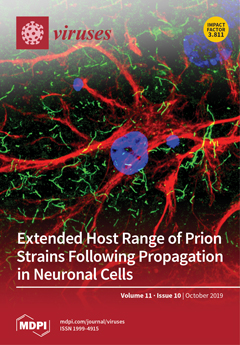
The phenomenon of “selective neuronal vulnerability” has been associated with conformational variants of rogue prion proteins, so-called prion strains, but their disease pathways remain unknown. To facilitate the search for molecular targets of prion strains, we established a novel neuroblastoma cell model with selectivity to distinct prion strains. Strikingly, the passage of prion strains in susceptible neuronal cells extends their host range to otherwise nonpermissive cell lines. View this paper.
  • Issues are regarded as officially published after their release is announced to the table of contents alert mailing list.
  • You may sign up for e-mail alerts to receive table of contents of newly released issues.
  • PDF is the official format for papers published in both, html and pdf forms. To view the papers in pdf format, click on the "PDF Full-text" link, and use the free Adobe Reader to open them.
Order results
Result details
Section
Select all
Export citation of selected articles as:
14 pages, 3071 KiB  
Article
The S2 Subunit of QX-type Infectious Bronchitis Coronavirus Spike Protein Is an Essential Determinant of Neurotropism
by Jinlong Cheng, Ye Zhao, Gang Xu, Keran Zhang, Wenfeng Jia, Yali Sun, Jing Zhao, Jia Xue, Yanxin Hu and Guozhong Zhang
Viruses 2019, 11(10), 972; https://doi.org/10.3390/v11100972 - 22 Oct 2019
Cited by 43 | Viewed by 11849
Abstract
Some coronaviruses (CoVs) have an extra furin cleavage site (RRKR/S, furin-S2′ site) upstream of the fusion peptide in the spike protein, which plays roles in virion adsorption and fusion. Mutation of the S2′ site of QX genotype (QX-type) infectious bronchitis virus (IBV) spike [...] Read more.
Some coronaviruses (CoVs) have an extra furin cleavage site (RRKR/S, furin-S2′ site) upstream of the fusion peptide in the spike protein, which plays roles in virion adsorption and fusion. Mutation of the S2′ site of QX genotype (QX-type) infectious bronchitis virus (IBV) spike protein (S) in a recombinant virus background results in higher pathogenicity, pronounced neural symptoms and neurotropism when compared with conditions in wild-type IBV (WT-IBV) infected chickens. In this study, we present evidence suggesting that recombinant IBV with a mutant S2′ site (furin-S2′ site) leads to higher mortality. Infection with mutant IBV induces severe encephalitis and breaks the blood–brain barrier. The results of a neutralization test and immunoprotection experiment show that an original serum and vaccine can still provide effective protection in vivo and in vitro. This is the first demonstration of IBV-induced neural symptoms in chickens with encephalitis and the furin-S2′ site as a determinant of neurotropism. Full article
(This article belongs to the Section Animal Viruses)
Show Figures

Figure 1

19 pages, 3066 KiB  
Article
High Diversity and Novel Enteric Viruses in Fecal Viromes of Healthy Wild and Captive Thai Cynomolgus Macaques (Macaca fascicularis)
by Vorthon Sawaswong, Elizabeth Fahsbender, Eda Altan, Taratorn Kemthong, Xutao Deng, Suchinda Malaivijitnond, Sunchai Payungporn and Eric Delwart
Viruses 2019, 11(10), 971; https://doi.org/10.3390/v11100971 - 22 Oct 2019
Cited by 16 | Viewed by 5196
Abstract
Cynomolgus macaques are common across South East Asian countries including Thailand. The National Primate Research Center of Thailand, Chulalongkorn University (NPRCT-CU) captures wild-borne cynomolgus macaque for research use. Limited information is available on the enteric viruses and possible zoonotic infections into or from [...] Read more.
Cynomolgus macaques are common across South East Asian countries including Thailand. The National Primate Research Center of Thailand, Chulalongkorn University (NPRCT-CU) captures wild-borne cynomolgus macaque for research use. Limited information is available on the enteric viruses and possible zoonotic infections into or from cynomolgus macaques. We characterized and compare the fecal virome of two populations; healthy wild-originated captive cynomolgus macaques (n = 43) reared in NPRCT-CU and healthy wild cynomolgus macaques (n = 35). Over 90% of recognized viral sequence reads amplified from feces were from bacterial viruses. Viruses from seven families of mammalian viruses were also detected (Parvoviridae, Anelloviridae, Picornaviridae, Adenoviridae, Papillomaviridae, Herpesviridae, and Caliciviridae). The genomes of a member of a new picornavirus genus we named Mafapivirus, a primate chapparvovirus, and a circular Rep-encoding single-strand (CRESS) DNA virus were also characterized. Higher abundance of CRESS DNA viruses of unknown tropism and invertebrate-tropic ambidensovirus were detected in wild versus captive macaques likely reflecting dietary differences. Short term rearing in captivity did not have a pronounced effect on the diversity of mammalian viruses of wild cynomolgus macaques. This study is the first report of the fecal virome of cynomolgus macaques, non-human primates frequently used in biomedical research and vaccination studies. Full article
(This article belongs to the Section Animal Viruses)
Show Figures

Figure 1

19 pages, 1394 KiB  
Review
Evasion of Innate and Intrinsic Antiviral Pathways by the Zika Virus
by Taryn M. Serman and Michaela U. Gack
Viruses 2019, 11(10), 970; https://doi.org/10.3390/v11100970 - 22 Oct 2019
Cited by 36 | Viewed by 7671
Abstract
The Zika virus (ZIKV) is a recently emerged mosquito-borne flavivirus that, while typically asymptomatic, can cause neurological symptoms in adults and birth defects in babies born to infected mothers. The interactions of ZIKV with many different pathways in the human host ultimately determine [...] Read more.
The Zika virus (ZIKV) is a recently emerged mosquito-borne flavivirus that, while typically asymptomatic, can cause neurological symptoms in adults and birth defects in babies born to infected mothers. The interactions of ZIKV with many different pathways in the human host ultimately determine successful virus replication and ZIKV-induced pathogenesis; however, the molecular mechanisms of such host-ZIKV interactions have just begun to be elucidated. Here, we summarize the recent advances that defined the mechanisms by which ZIKV antagonizes antiviral innate immune signaling pathways, with a particular focus on evasion of the type I interferon response in the human host. Furthermore, we describe emerging evidence that indicated the contribution of several cell-intrinsic mechanisms to an effective restriction of ZIKV infection, such as nonsense-mediated mRNA decay, stress granule formation, and “reticulophagy”, a type of selective autophagy. Finally, we summarize the recent work that identified strategies by which ZIKV modulated these intrinsic antiviral responses. Full article
(This article belongs to the Special Issue Viruses Ten-Year Anniversary)
Show Figures

Figure 1

8 pages, 3225 KiB  
Article
A Novel Hepadnavirus is Associated with Chronic Hepatitis and Hepatocellular Carcinoma in Cats
by Patricia A. Pesavento, Kenneth Jackson, Timothy Scase, Tiffany Tse, Bronte Hampson, John S. Munday, Vanessa R. Barrs and Julia A. Beatty
Viruses 2019, 11(10), 969; https://doi.org/10.3390/v11100969 - 21 Oct 2019
Cited by 39 | Viewed by 7463
Abstract
In 2015, over 850,000 people died from chronic hepatitis and hepatocellular carcinoma (HCC) caused by hepatitis B virus (HBV). A novel hepatitis B-like virus has recently been identified in domestic cats. The pathogenic potential of domestic cat hepadnavirus (DCH), for which 6.5% to [...] Read more.
In 2015, over 850,000 people died from chronic hepatitis and hepatocellular carcinoma (HCC) caused by hepatitis B virus (HBV). A novel hepatitis B-like virus has recently been identified in domestic cats. The pathogenic potential of domestic cat hepadnavirus (DCH), for which 6.5% to 10.8% of pet cats are viremic, is unknown. We evaluated stored formalin-fixed, paraffin-embedded biopsies of diseased and normal feline liver for the presence of DCH using PCR and in situ hybridization (ISH). DCH was detected in 43% (6/14) of chronic hepatitis cases and 28% (8/29) of HCCs, whereas cholangitis (n = 6), biliary carcinoma (n = 18) and normal liver (n = 15) all tested negative for DCH. Furthermore, in DCH-associated cases, the histologic features of inflammation and neoplasia, and the viral distribution on ISH were strikingly similar to those seen with HBV-associated disease. Several histological features common in human HBV-associated hepatitis, including piecemeal necrosis and apoptotic bodies, were identified in DCH-positive cases of chronic hepatitis. In two cases of HCC examined, the proliferation index in regions that were ISH-positive was higher than in ISH-negative regions. The intracellular distribution of virus in both hepatitis and HCC demonstrated that viral nucleic acid is present in both nuclear and cytoplasmic forms. Collectively, these findings demonstrate a compelling association between DCH and some cases of chronic hepatitis and hepatocellular carcinoma in the cat that mirrors features of HBV-associated hepatopathies. Future investigations of viral epidemiology and natural history are needed to establish the impact of DCH on feline health. Full article
(This article belongs to the Special Issue Feline Viruses and Viral Diseases)
Show Figures

Figure 1

23 pages, 5671 KiB  
Article
Gut DNA Virome Diversity and Its Association with Host Bacteria Regulate Inflammatory Phenotype and Neuronal Immunotoxicity in Experimental Gulf War Illness
by Ratanesh K. Seth, Rabia Maqsood, Ayan Mondal, Dipro Bose, Diana Kimono, LaRinda A. Holland, Patricia Janulewicz Lloyd, Nancy Klimas, Ronnie D. Horner, Kimberly Sullivan, Efrem S. Lim and Saurabh Chatterjee
Viruses 2019, 11(10), 968; https://doi.org/10.3390/v11100968 - 21 Oct 2019
Cited by 37 | Viewed by 9142
Abstract
Gulf War illness (GWI) is characterized by the persistence of inflammatory bowel disease, chronic fatigue, neuroinflammation, headache, cognitive impairment, and other medically unexplained conditions. Results using a murine model show that enteric viral populations especially bacteriophages were altered in GWI. The increased viral [...] Read more.
Gulf War illness (GWI) is characterized by the persistence of inflammatory bowel disease, chronic fatigue, neuroinflammation, headache, cognitive impairment, and other medically unexplained conditions. Results using a murine model show that enteric viral populations especially bacteriophages were altered in GWI. The increased viral richness and alpha diversity correlated positively with gut bacterial dysbiosis and proinflammatory cytokines. Altered virome signature in GWI mice also had a concomitant weakening of intestinal epithelial tight junctions with a significant increase in Claudin-2 protein expression and decrease in ZO1 and Occludin mRNA expression. The altered virome signature in GWI, decreased tight junction protein level was followed by the presence an activation of innate immune responses such as increased Toll-like receptor (TLR) signaling pathways. The altered virome diversity had a positive correlation with serum IL-6, IL-1β, and IFN-γ, intestinal inflammation (IFN-γ), and decreased Brain-Derived Neurotrophic Factor (BDNF), a neurogenesis marker. The co-exposure of Gulf War chemical and antibiotic (for gut sterility) or Gulf War chemical and Ribavirin, an antiviral compound to suppress virus alteration in the gut showed significant improvement in epithelial tight junction protein, decreased intestinal-, systemic-, and neuroinflammation. These results showed that the observed enteric viral dysbiosis could activate enteric viral particle-induced innate immune response in GWI and could be a novel therapeutic target in GWI. Full article
(This article belongs to the Section Animal Viruses)
Show Figures

Figure 1

15 pages, 1291 KiB  
Review
Simian Foamy Viruses in Central and South America: A New World of Discovery
by André F. Santos, Liliane T. F. Cavalcante, Cláudia P. Muniz, William M. Switzer and Marcelo A. Soares
Viruses 2019, 11(10), 967; https://doi.org/10.3390/v11100967 - 20 Oct 2019
Cited by 8 | Viewed by 3119
Abstract
Foamy viruses (FVs) are the only exogenous retrovirus to date known to infect neotropical primates (NPs). In the last decade, an increasing number of strains have been completely or partially sequenced, and molecular evolution analyses have identified an ancient co-speciation with their hosts. [...] Read more.
Foamy viruses (FVs) are the only exogenous retrovirus to date known to infect neotropical primates (NPs). In the last decade, an increasing number of strains have been completely or partially sequenced, and molecular evolution analyses have identified an ancient co-speciation with their hosts. In this review, the improvement of diagnostic techniques that allowed the determination of a more accurate prevalence of simian FVs (SFVs) in captive and free-living NPs is discussed. Determination of DNA viral load in American primates indicates that oral tissues are the viral replicative site and that buccal swab collection can be an alternative to diagnose SFV infection in NPs. Finally, the transmission potential of NP SFVs to primate workers in zoos and primate centers of the Americas is examined. Full article
(This article belongs to the Special Issue Spumaretroviruses)
Show Figures

Graphical abstract

22 pages, 1898 KiB  
Review
Persistent Infection with Herpes Simplex Virus 1 and Alzheimer’s Disease—A Call to Study How Variability in Both Virus and Host may Impact Disease
by Colleen A. Mangold and Moriah L. Szpara
Viruses 2019, 11(10), 966; https://doi.org/10.3390/v11100966 - 20 Oct 2019
Cited by 26 | Viewed by 9142
Abstract
Increasing attention has focused on the contributions of persistent microbial infections with the manifestation of disease later in life, including neurodegenerative conditions such as Alzheimer’s disease (AD). Current data has shown the presence of herpes simplex virus 1 (HSV-1) in regions of the [...] Read more.
Increasing attention has focused on the contributions of persistent microbial infections with the manifestation of disease later in life, including neurodegenerative conditions such as Alzheimer’s disease (AD). Current data has shown the presence of herpes simplex virus 1 (HSV-1) in regions of the brain that are impacted by AD in elderly individuals. Additionally, neuronal infection with HSV-1 triggers the accumulation of amyloid beta deposits and hyperphosphorylated tau, and results in oxidative stress and synaptic dysfunction. All of these factors are implicated in the development of AD. These data highlight the fact that persistent viral infection is likely a contributing factor, rather than a sole cause of disease. Details of the correlations between HSV-1 infection and AD development are still just beginning to emerge. Future research should investigate the relative impacts of virus strain- and host-specific factors on the induction of neurodegenerative processes over time, using models such as infected neurons in vitro, and animal models in vivo, to begin to understand their relationship with cognitive dysfunction. Full article
(This article belongs to the Special Issue Viruses Ten-Year Anniversary)
Show Figures

Figure 1

11 pages, 1489 KiB  
Article
Characterization of Equine Parvovirus in Thoroughbred Breeding Horses from Germany
by Toni Luise Meister, Birthe Tegtmeyer, Yannick Brüggemann, Harald Sieme, Karsten Feige, Daniel Todt, Alexander Stang, Jessika-M.V. Cavalleri and Eike Steinmann
Viruses 2019, 11(10), 965; https://doi.org/10.3390/v11100965 - 18 Oct 2019
Cited by 26 | Viewed by 4020
Abstract
An equine parvovirus-hepatitis (EqPV-H) has been recently identified in association with equine serum hepatitis, also known as Theiler’s disease. The disease was first described by Arnold Theiler in 1918 and is often observed with parenteral use of blood products in equines. However, natural [...] Read more.
An equine parvovirus-hepatitis (EqPV-H) has been recently identified in association with equine serum hepatitis, also known as Theiler’s disease. The disease was first described by Arnold Theiler in 1918 and is often observed with parenteral use of blood products in equines. However, natural ways of viral circulation and potential risk factors for transmission still remain unknown. In this study, we investigated the occurrence of EqPV-H infections in Thoroughbred horses in northern and western Germany and aimed to identify potential risk factors associated with viral infections. A total of 392 Thoroughbreds broodmares and stallions were evaluated cross-sectionally for the presence of anti-EqPV-H antibodies and EqPV-H DNA using a luciferase immunoprecipitation assay (LIPS) and a quantitative PCR, respectively. In addition, data regarding age, stud farm, breeding history, and international transportation history of each horse were collected and analysed. An occurrence of 7% EqPV-H DNA positive and 35% seropositive horses was observed in this study cohort. The systematic analysis of risk factors revealed that age, especially in the group of 11–15-year-old horses, and breeding history were potential risk factors that can influence the rate of EqPV-H infections. Subsequent phylogenetic analysis showed a high similarity on nucleotide level within the sequenced Thoroughbred samples. In conclusion, this study demonstrates circulating EqPV-H infections in Thoroughbred horses from central Europe and revealed age and breeding history as risk factors for EqPV-H infections. Full article
(This article belongs to the Special Issue Equine Viruses)
Show Figures

Figure 1

15 pages, 7397 KiB  
Article
Novel Antiviral Activities of Obatoclax, Emetine, Niclosamide, Brequinar, and Homoharringtonine
by Petter I. Andersen, Klara Krpina, Aleksandr Ianevski, Nastassia Shtaida, Eunji Jo, Jaewon Yang, Sandra Koit, Tanel Tenson, Veijo Hukkanen, Marit W. Anthonsen, Magnar Bjoras, Magnus Evander, Marc P. Windisch, Eva Zusinaite and Denis E. Kainov
Viruses 2019, 11(10), 964; https://doi.org/10.3390/v11100964 - 18 Oct 2019
Cited by 59 | Viewed by 6431
Abstract
Viruses are the major causes of acute and chronic infectious diseases in the world. According to the World Health Organization, there is an urgent need for better control of viral diseases. Repurposing existing antiviral agents from one viral disease to another could play [...] Read more.
Viruses are the major causes of acute and chronic infectious diseases in the world. According to the World Health Organization, there is an urgent need for better control of viral diseases. Repurposing existing antiviral agents from one viral disease to another could play a pivotal role in this process. Here, we identified novel activities of obatoclax and emetine against herpes simplex virus type 2 (HSV-2), echovirus 1 (EV1), human metapneumovirus (HMPV) and Rift Valley fever virus (RVFV) in cell cultures. Moreover, we demonstrated novel activities of emetine against influenza A virus (FLUAV), niclosamide against HSV-2, brequinar against human immunodeficiency virus 1 (HIV-1), and homoharringtonine against EV1. Our findings may expand the spectrum of indications of these safe-in-man agents and reinforce the arsenal of available antiviral therapeutics pending the results of further in vitro and in vivo tests. Full article
(This article belongs to the Special Issue Pathogenesis of Emerging Viral Infections)
Show Figures

Figure 1

8 pages, 875 KiB  
Communication
Equine Rhinitis A Virus Infection in Thoroughbred Racehorses—A Putative Role in Poor Performance?
by Helena Back, John Weld, Cathal Walsh and Ann Cullinane
Viruses 2019, 11(10), 963; https://doi.org/10.3390/v11100963 - 18 Oct 2019
Cited by 7 | Viewed by 3160
Abstract
The aim of this study was to identify respiratory viruses circulating amongst elite racehorses in a training yard by serological testing of serial samples and to determine their impact on health status and ability to race. A six-month longitudinal study was conducted in [...] Read more.
The aim of this study was to identify respiratory viruses circulating amongst elite racehorses in a training yard by serological testing of serial samples and to determine their impact on health status and ability to race. A six-month longitudinal study was conducted in 30 Thoroughbred racehorses (21 two-year-olds, five three-year-olds and four four-year-olds) during the Flat racing season. Sera were tested for the presence of antibodies against equine herpesvirus 1 and 4 (EHV-1 and EHV-4) and equine rhinitis viruses A and B (ERAV and ERBV) by complement fixation (CF) and equine arteritis virus (EAV) by ELISA. Antibodies against equine influenza (EI) were measured by haemagglutination inhibition (HI). Only ERAV was circulating in the yard throughout the six-month study period. Seroconversion to ERAV frequently correlated with clinical respiratory disease and was significantly associated with subsequent failure to race (p = 0.0009). Over 55% of the two-year-olds in the study seroconverted to ERAV in May and June. In contrast, only one seroconversion to ERAV was observed in the older horses. They remained free of any signs of respiratory disease and raced successfully throughout the study period. The importance of ERAV as a contributory factor in the interruption of training programmes for young horses may be underestimated. Full article
(This article belongs to the Special Issue Equine Viruses)
Show Figures

Figure 1

14 pages, 3596 KiB  
Article
In Vitro and In Vivo Metabolomic Profiling after Infection with Virulent Newcastle Disease Virus
by Panrao Liu, Yuncong Yin, Yabin Gong, Xusheng Qiu, Yingjie Sun, Lei Tan, Cuiping Song, Weiwei Liu, Ying Liao, Chunchun Meng and Chan Ding
Viruses 2019, 11(10), 962; https://doi.org/10.3390/v11100962 - 18 Oct 2019
Cited by 20 | Viewed by 3199
Abstract
Newcastle disease (ND) is an acute, febrile, highly contagious disease caused by the virulent Newcastle disease virus (vNDV). The disease causes serious economic losses to the poultry industry. However, the metabolic changes caused by vNDV infection remain unclear. The objective of this study [...] Read more.
Newcastle disease (ND) is an acute, febrile, highly contagious disease caused by the virulent Newcastle disease virus (vNDV). The disease causes serious economic losses to the poultry industry. However, the metabolic changes caused by vNDV infection remain unclear. The objective of this study was to determine the metabolomic profiling after infection with vNDV. DF-1 cells infected with the vNDV strain Herts/33 and the lungs from Herts/33-infected specific pathogen-free (SPF) chickens were analyzed via ultra-high-performance liquid chromatography/quadrupole time-of-flight tandem mass spectrometry (UHPLC-QTOF-MS) in combination with multivariate statistical analysis. A total of 305 metabolites were found to have changed significantly after Herts/33 infection, and most of them belong to the amino acid and nucleotide metabolic pathway. It is suggested that the increased pools of amino acids and nucleotides may benefit viral protein synthesis and genome amplification to promote NDV infection. Similar results were also confirmed in vivo. Identification of these metabolites will provide information to further understand the mechanism of vNDV replication and pathogenesis. Full article
(This article belongs to the Section Animal Viruses)
Show Figures

Figure 1

23 pages, 1731 KiB  
Review
Viral Innate Immune Evasion and the Pathogenesis of Emerging RNA Virus Infections
by Tessa Nelemans and Marjolein Kikkert
Viruses 2019, 11(10), 961; https://doi.org/10.3390/v11100961 - 18 Oct 2019
Cited by 162 | Viewed by 14052
Abstract
Positive-sense single-stranded RNA (+ssRNA) viruses comprise many (re-)emerging human pathogens that pose a public health problem. Our innate immune system and, in particular, the interferon response form the important first line of defence against these viruses. Given their genetic flexibility, these viruses have [...] Read more.
Positive-sense single-stranded RNA (+ssRNA) viruses comprise many (re-)emerging human pathogens that pose a public health problem. Our innate immune system and, in particular, the interferon response form the important first line of defence against these viruses. Given their genetic flexibility, these viruses have therefore developed multiple strategies to evade the innate immune response in order to optimize their replication capacity. Already many molecular mechanisms of innate immune evasion by +ssRNA viruses have been identified. However, research addressing the effect of host innate immune evasion on the pathology caused by viral infections is less prevalent in the literature, though very relevant and interesting. Since interferons have been implicated in inflammatory diseases and immunopathology in addition to their protective role in infection, antagonizing the immune response may have an ambiguous effect on the clinical outcome of the viral disease. Therefore, this review discusses what is currently known about the role of interferons and host immune evasion in the pathogenesis of emerging coronaviruses, alphaviruses and flaviviruses. Full article
(This article belongs to the Special Issue Pathogenesis of Emerging Viral Infections)
Show Figures

Figure 1

32 pages, 2218 KiB  
Review
Yellow Fever: Integrating Current Knowledge with Technological Innovations to Identify Strategies for Controlling a Re-Emerging Virus
by Robin D.V. Kleinert, Eduardo Montoya-Diaz, Tanvi Khera, Kathrin Welsch, Birthe Tegtmeyer, Sebastian Hoehl, Sandra Ciesek and Richard J.P. Brown
Viruses 2019, 11(10), 960; https://doi.org/10.3390/v11100960 - 17 Oct 2019
Cited by 15 | Viewed by 7928
Abstract
Yellow fever virus (YFV) represents a re-emerging zoonotic pathogen, transmitted by mosquito vectors to humans from primate reservoirs. Sporadic outbreaks of YFV occur in endemic tropical regions, causing a viral hemorrhagic fever (VHF) associated with high mortality rates. Despite a highly effective vaccine, [...] Read more.
Yellow fever virus (YFV) represents a re-emerging zoonotic pathogen, transmitted by mosquito vectors to humans from primate reservoirs. Sporadic outbreaks of YFV occur in endemic tropical regions, causing a viral hemorrhagic fever (VHF) associated with high mortality rates. Despite a highly effective vaccine, no antiviral treatments currently exist. Therefore, YFV represents a neglected tropical disease and is chronically understudied, with many aspects of YFV biology incompletely defined including host range, host–virus interactions and correlates of host immunity and pathogenicity. In this article, we review the current state of YFV research, focusing on the viral lifecycle, host responses to infection, species tropism and the success and associated limitations of the YFV-17D vaccine. In addition, we highlight the current lack of available treatments and use publicly available sequence and structural data to assess global patterns of YFV sequence diversity and identify potential drug targets. Finally, we discuss how technological advances, including real-time epidemiological monitoring of outbreaks using next-generation sequencing and CRISPR/Cas9 modification of vector species, could be utilized in future battles against this re-emerging pathogen which continues to cause devastating disease. Full article
(This article belongs to the Special Issue Flavivirus Replication and Pathogenesis)
Show Figures

Figure 1

17 pages, 992 KiB  
Review
The Evolutionary Arms Race between Virus and NK Cells: Diversity Enables Population-Level Virus Control
by Sarah K. A. Savoy and Jeanette E. Boudreau
Viruses 2019, 11(10), 959; https://doi.org/10.3390/v11100959 - 17 Oct 2019
Cited by 21 | Viewed by 6097
Abstract
Viruses and natural killer (NK) cells have a long co-evolutionary history, evidenced by patterns of specific NK gene frequencies in those susceptible or resistant to infections. The killer immunoglobulin-like receptors (KIR) and their human leukocyte antigen (HLA) ligands together form the most polymorphic [...] Read more.
Viruses and natural killer (NK) cells have a long co-evolutionary history, evidenced by patterns of specific NK gene frequencies in those susceptible or resistant to infections. The killer immunoglobulin-like receptors (KIR) and their human leukocyte antigen (HLA) ligands together form the most polymorphic receptor-ligand partnership in the human genome and govern the process of NK cell education. The KIR and HLA genes segregate independently, thus creating an array of reactive potentials within and between the NK cell repertoires of individuals. In this review, we discuss the interplay between NK cell education and adaptation with virus infection, with a special focus on three viruses for which the NK cell response is often studied: human immunodeficiency virus (HIV), hepatitis C virus (HCV) and human cytomegalovirus (HCMV). Through this lens, we highlight the complex co-evolution of viruses and NK cells, and their impact on viral control. Full article
(This article belongs to the Special Issue The Role of NK Cells in Antiviral Innate Immunity)
Show Figures

Figure 1

16 pages, 1369 KiB  
Article
Environmental Contamination and Hygienic Measures After Feline Calicivirus Field Strain Infections of Cats in a Research Facility
by Andrea Monika Spiri, Marina Luisa Meli, Barbara Riond, Imogen Herbert, Margaret J. Hosie and Regina Hofmann-Lehmann
Viruses 2019, 11(10), 958; https://doi.org/10.3390/v11100958 - 17 Oct 2019
Cited by 14 | Viewed by 5320
Abstract
Feline calicivirus (FCV) can cause painful oral ulcerations, salivation, gingivitis/stomatitis, fever and depression in infected cats; highly virulent virus variants can lead to fatal epizootic outbreaks. Viral transmission occurs directly or indirectly via fomites. The aim of this study was to investigate the [...] Read more.
Feline calicivirus (FCV) can cause painful oral ulcerations, salivation, gingivitis/stomatitis, fever and depression in infected cats; highly virulent virus variants can lead to fatal epizootic outbreaks. Viral transmission occurs directly or indirectly via fomites. The aim of this study was to investigate the presence and viability of FCV in the environment after sequential oronasal infections of specified pathogen-free cats with two FCV field strains in a research facility. Replicating virus was detected in saliva swabs from all ten cats after the first and in four out of ten cats after the second FCV exposure using virus isolation to identify FCV shedders. In the environment, where cleaning, but no disinfection took place, FCV viral RNA was detectable using RT-qPCR on all tested items and surfaces, including cat hair. However, only very limited evidence was found of replicating virus using virus isolation. Viral RNA remained demonstrable for at least 28 days after shedding had ceased in all cats. Disinfection with 5% sodium bicarbonate (and IncidinTM Plus) and barrier measures were effective in that no viral RNA was detectable outside the cat rooms. Our findings are important for any multicat environment to optimize hygienic measures against FCV infection. Full article
(This article belongs to the Special Issue Feline Viruses and Viral Diseases)
Show Figures

Figure 1

15 pages, 2426 KiB  
Article
Novel Viruses in Mosquitoes from Brazilian Pantanal
by Laura Marina Siqueira Maia, Andressa Zelenski de Lara Pinto, Michellen Santos de Carvalho, Fernando Lucas de Melo, Bergmann Morais Ribeiro and Renata Dezengrini Slhessarenko
Viruses 2019, 11(10), 957; https://doi.org/10.3390/v11100957 - 17 Oct 2019
Cited by 11 | Viewed by 4203
Abstract
Viruses are ubiquitous and diverse microorganisms arising as a result of interactions within their vertebrate and invertebrate hosts. Here we report the presence of different viruses in the salivary glands of 1657 mosquitoes classified over 28 culicinae species from the North region of [...] Read more.
Viruses are ubiquitous and diverse microorganisms arising as a result of interactions within their vertebrate and invertebrate hosts. Here we report the presence of different viruses in the salivary glands of 1657 mosquitoes classified over 28 culicinae species from the North region of the Brazilian Pantanal wetland through metagenomics, viral isolation, and RT-PCR. In total, 12 viruses were found, eight putative novel viruses with relatively low similarity with pre-existing species of viruses within their families, named Pirizal iflavirus, Furrundu phlebovirus, Pixé phlebovirus, Guampa vesiculovirus, Chacororé flavivirus, Rasqueado orbivirus, Uru chuvirus, and Bororo circovirus. We also found the already described Lobeira dielmorhabdovirus, Sabethes flavivirus, Araticum partitivirus, and Murici totivirus. Therefore, these findings underscore the vast diversity of culicinae and novel viruses yet to be explored in Pantanal, the largest wetland on the planet. Full article
(This article belongs to the Special Issue Emerging Viruses: Surveillance, Prevention, Evolution and Control)
Show Figures

Figure 1

12 pages, 4842 KiB  
Communication
A Novel Enzootic Nasal Tumor Virus Circulating in Goats from Southern China
by Shao-Lun Zhai, Dian-Hong Lv, Zhi-Hong Xu, Jie-Shi Yu, Xiao-Hui Wen, He Zhang, Qin-Ling Chen, Chun-Ling Jia, Xiu-Rong Zhou, Qi Zhai, Feng Li, Patrick C. Y. Woo, Susanna K. P. Lau, Dan Wang and Wen-Kang Wei
Viruses 2019, 11(10), 956; https://doi.org/10.3390/v11100956 - 17 Oct 2019
Cited by 6 | Viewed by 3931
Abstract
Enzootic nasal tumor virus (ENTV) has two types, ENTV-1 in sheep and ENTV-2 in goats, respectively. In China, the incidence of ENTV-2 related diseases has increased year by year. In this study, we reported an outbreak of ENTV-2 in a commercial goat farm [...] Read more.
Enzootic nasal tumor virus (ENTV) has two types, ENTV-1 in sheep and ENTV-2 in goats, respectively. In China, the incidence of ENTV-2 related diseases has increased year by year. In this study, we reported an outbreak of ENTV-2 in a commercial goat farm in Qingyuan city, Guangdong province, southern China. A full-length genome of ENTV-2 (designated GDQY2017), with 7479 base pairs, was sequenced. Although GDQY2017 shared the highest nucleotide identity with a Chinese ENTV-2 isolate (ENTV-2CHN4, GenBank accession number KU258873), it possesses distinct genome characteristics undescribed, including a non-continuous 21-nucleotide insertion in the gag gene and a non-continuous 12-nucleotide deletion in the env gene. Notably, most of these indel nucleotide sequences were originated from a Chinese jaagsiekte sheep retrovirus (JSRV) isolate (GenBank accession number DQ838494). In the gag and env genes, GDQY2017 was phylogenetically related to those Chinese ENTV-2 isolates and a Chinese JSRV isolate (DQ838494). For GDQY2017-like viruses, more surveillance work should be made to explain their pathogenicity in goat herds. To our knowledge, this study represents the first to demonstrate the circulating pattern of ENTV-2 in Guangdong province, China, which will help to better understand the epidemiology and genetic diversity of ENTV-2. Full article
(This article belongs to the Section Animal Viruses)
Show Figures

Figure 1

11 pages, 2524 KiB  
Article
Ultrastructural Aspects of Photodynamic Inactivation of Highly Pathogenic Avian H5N8 Influenza Virus
by Denis Korneev, Olga Kurskaya, Kirill Sharshov, Justin Eastwood and Marina Strakhovskaya
Viruses 2019, 11(10), 955; https://doi.org/10.3390/v11100955 - 16 Oct 2019
Cited by 31 | Viewed by 4396
Abstract
Ultrastructural studies revealing morphological differences between intact and photodynamically inactivated virions can point to inactivation mechanisms and molecular targets. Using influenza as a model system, we show that photodynamic virus inactivation is possible without total virion destruction. Indeed, irradiation with a relatively low [...] Read more.
Ultrastructural studies revealing morphological differences between intact and photodynamically inactivated virions can point to inactivation mechanisms and molecular targets. Using influenza as a model system, we show that photodynamic virus inactivation is possible without total virion destruction. Indeed, irradiation with a relatively low concentration of the photosensitizer (octacationic octakis(cholinyl) zinc phthalocyanine) inactivated viral particles (the virus titer was determined in Madin Darby Canine Kidney (MDCK) cells) but did not destroy them. Transmission electron microscopy (TEM) revealed that virion membranes kept structural integrity but lost their surface glycoproteins. Such structures are known as “bald” virions, which were first described as a result of protease treatment. At a higher photosensitizer concentration, the lipid membranes were also destroyed. Therefore, photodynamic inactivation of influenza virus initially results from surface protein removal, followed by complete virion destruction. This study suggests that photodynamic treatment can be used to manufacture “bald” virions for experimental purposes. Photodynamic inactivation is based on the production of reactive oxygen species which attack and destroy biomolecules. Thus, the results of this study can potentially apply to other enveloped viruses and sources of singlet oxygen. Full article
(This article belongs to the Section Viral Immunology, Vaccines, and Antivirals)
Show Figures

Figure 1

15 pages, 4672 KiB  
Article
Eradication of Vancomycin-Resistant Enterococci by Combining Phage and Vancomycin
by Mor Shlezinger, Shunit Coppenhagen-Glazer, Daniel Gelman, Nurit Beyth and Ronen Hazan
Viruses 2019, 11(10), 954; https://doi.org/10.3390/v11100954 - 16 Oct 2019
Cited by 37 | Viewed by 5004
Abstract
Currently, effective options are needed to fight vancomycin-resistant Enterococcus faecalis (VRE). The present study shows that combinations of phage and vancomycin are highly efficient against VRE, despite being resistant to the antibiotic. Vancomycin-phage EFLK1 (anti-E. faecalis phage) synergy was assessed against VRE [...] Read more.
Currently, effective options are needed to fight vancomycin-resistant Enterococcus faecalis (VRE). The present study shows that combinations of phage and vancomycin are highly efficient against VRE, despite being resistant to the antibiotic. Vancomycin-phage EFLK1 (anti-E. faecalis phage) synergy was assessed against VRE planktonic and biofilm cultures. The effect of the combined treatment on VRE biofilms was determined by evaluating the viable counts and biomass and then visualized using scanning electron microscopy (SEM). The cell wall peptidoglycan was stained after phage treatment, visualized by confocal microscopy and quantified by fluorescence activated cell sorting (FACS) analysis. The combined treatment was synergistically effective compared to treatment with phage or antibiotic alone, both in planktonic and biofilm cultures. Confocal microscopy and FACS analysis showed that fluorescence intensity of phage-treated bacteria increased eight-fold, suggesting a change in the peptidoglycan of the cell wall. Our results indicate that with combined treatment, VRE strains are not more problematic than sensitive strains and thus give hope in the continuous struggle against the current emergence of multidrug resistant pathogens. Full article
(This article belongs to the Special Issue Bacteriophages and Biofilms)
Show Figures

Figure 1

14 pages, 693 KiB  
Article
Geographical Variability Affects CCHFV Detection by RT–PCR: A Tool for In-Silico Evaluation of Molecular Assays
by Cesare E. M. Gruber, Barbara Bartolini, Concetta Castilletti, Ali Mirazimi, Roger Hewson, Iva Christova, Tatjana Avšič, Roland Grunow, Anna Papa, María P. Sánchez-Seco, Marion Koopmans, Giuseppe Ippolito, Maria R. Capobianchi, Chantal B. E. M. Reusken and Antonino Di Caro
Viruses 2019, 11(10), 953; https://doi.org/10.3390/v11100953 - 16 Oct 2019
Cited by 9 | Viewed by 4227
Abstract
The Crimean–Congo hemorrhagic fever virus (CCHFV) is considered to be a major emerging infectious threat, according to the WHO R&D blueprint. A wide range of CCHFV molecular assays have been developed, employing varied primer/probe combinations. The high genetic variability of CCHFV often hampers [...] Read more.
The Crimean–Congo hemorrhagic fever virus (CCHFV) is considered to be a major emerging infectious threat, according to the WHO R&D blueprint. A wide range of CCHFV molecular assays have been developed, employing varied primer/probe combinations. The high genetic variability of CCHFV often hampers the efficacy of available molecular tests and can affect their diagnostic potential. Recently, increasing numbers of complete CCHFV genomic sequences have become available, allowing a better appreciation of the genomic evolution of this virus. We summarized the current knowledge on molecular methods and developed a new bioinformatics tool to evaluate the existing assays for CCHFV detection, with a special focus on strains circulating in different geographical areas. Twenty-two molecular methods and 181 sequences of CCHFV were collected, respectively, from PubMed and GenBank databases. Up to 28 mismatches between primers and probes of each assay and CCHFV strains were detected through in-silico PCR analysis. Combinations of up to three molecular methods markedly decreased the number of mismatches within most geographic areas. These results supported the good practice of CCHFV detection of performing more than one assay, aimed for different sequence targets. The choice of the most appropriate tests must take into account patient’s travel history and geographic distribution of the different CCHFV strains. Full article
(This article belongs to the Special Issue Emerging Arboviruses)
Show Figures

Figure 1

23 pages, 8942 KiB  
Article
An Immortalized Hepatocyte-Like Cell Line (imHC) Accommodated Complete Viral Lifecycle, Viral Persistence Form, cccDNA and Eventual Spreading of a Clinically-Isolated HBV
by Khanit Sa-ngiamsuntorn, Piyanoot Thongsri, Yongyut Pewkliang, Adisak Wongkajornsilp, Pattida Kongsomboonchoke, Phichaya Suthivanich, Suparerk Borwornpinyo and Suradej Hongeng
Viruses 2019, 11(10), 952; https://doi.org/10.3390/v11100952 - 16 Oct 2019
Cited by 4 | Viewed by 7164
Abstract
More than 350 million people worldwide have been persistently infected with the hepatitis B virus (HBV). Chronic HBV infection could advance toward liver cirrhosis and hepatocellular carcinoma. The intervention with prophylactic vaccine and conventional treatment could suppress HBV, but could not completely eradicate [...] Read more.
More than 350 million people worldwide have been persistently infected with the hepatitis B virus (HBV). Chronic HBV infection could advance toward liver cirrhosis and hepatocellular carcinoma. The intervention with prophylactic vaccine and conventional treatment could suppress HBV, but could not completely eradicate it. The major obstacle for investigating curative antiviral drugs are the incompetence of hepatocyte models that should have closely imitated natural human infection. Here, we demonstrated that an immortalized hepatocyte-like cell line (imHC) could accommodate for over 30 days the entire life cycle of HBV prepared from either established cultured cells or clinically-derived fresh isolates. Normally, imHCs had intact interferon signaling with anti-viral action. Infected imHCs responded to treatments with direct-acting antiviral drugs (DAAs) and interferons (IFNs) by diminishing HBV DNA, the covalently closed circular DNA (cccDNA) surface antigen of HBV (HBsAg, aka the Australia antigen) and the hepatitis B viral protein (HBeAg). Notably, we could observe and quantify HBV spreading from infected cells to naïve cells using an imHC co-culture model. In summary, this study constructed a convenient HBV culture model that allows the screening for novel anti-HBV agents with versatile targets, either HBV entry, replication or cccDNA formation. Combinations of agents aiming at different targets should achieve a complete HBV eradication. Full article
(This article belongs to the Section Animal Viruses)
Show Figures

Figure 1

40 pages, 4440 KiB  
Review
Look Who’s Talking: T-Even Phage Lysis Inhibition, the Granddaddy of Virus-Virus Intercellular Communication Research
by Stephen T. Abedon
Viruses 2019, 11(10), 951; https://doi.org/10.3390/v11100951 - 16 Oct 2019
Cited by 41 | Viewed by 8582
Abstract
That communication can occur between virus-infected cells has been appreciated for nearly as long as has virus molecular biology. The original virus communication process specifically was that seen with T-even bacteriophages—phages T2, T4, and T6—resulting in what was labeled as a lysis inhibition. [...] Read more.
That communication can occur between virus-infected cells has been appreciated for nearly as long as has virus molecular biology. The original virus communication process specifically was that seen with T-even bacteriophages—phages T2, T4, and T6—resulting in what was labeled as a lysis inhibition. Another proposed virus communication phenomenon, also seen with T-even phages, can be described as a phage-adsorption-induced synchronized lysis-inhibition collapse. Both are mediated by virions that were released from earlier-lysing, phage-infected bacteria. Each may represent ecological responses, in terms of phage lysis timing, to high local densities of phage-infected bacteria, but for lysis inhibition also to locally reduced densities of phage-uninfected bacteria. With lysis inhibition, the outcome is a temporary avoidance of lysis, i.e., a lysis delay, resulting in increased numbers of virions (greater burst size). Synchronized lysis-inhibition collapse, by contrast, is an accelerated lysis which is imposed upon phage-infected bacteria by virions that have been lytically released from other phage-infected bacteria. Here I consider some history of lysis inhibition, its laboratory manifestation, its molecular basis, how it may benefit expressing phages, and its potential ecological role. I discuss as well other, more recently recognized examples of virus-virus intercellular communication. Full article
(This article belongs to the Special Issue Phage Ecology)
Show Figures

Graphical abstract

13 pages, 2406 KiB  
Article
Productive Infection of Mouse Mammary Glands and Human Mammary Epithelial Cells by Zika Virus
by Mathieu Hubert, Aurélie Chiche, Vincent Legros, Patricia Jeannin, Thomas Montange, Antoine Gessain, Pierre-Emmanuel Ceccaldi and Aurore Vidy
Viruses 2019, 11(10), 950; https://doi.org/10.3390/v11100950 - 15 Oct 2019
Cited by 6 | Viewed by 2801
Abstract
Zika virus (ZIKV) belongs to the large category of arboviruses. Surprisingly, several human-to-human transmissions of ZIKV have been notified, either following sexual intercourse or from the mother to fetus during pregnancy. Importantly, high viral loads have been detected in the human breast milk [...] Read more.
Zika virus (ZIKV) belongs to the large category of arboviruses. Surprisingly, several human-to-human transmissions of ZIKV have been notified, either following sexual intercourse or from the mother to fetus during pregnancy. Importantly, high viral loads have been detected in the human breast milk of infected mothers, and the existence of breastfeeding as a new mode of mother-to-child transmission of ZIKV was recently hypothesized. However, the maternal origin of infectious particles in breast milk is currently unknown. Here, we show that ZIKV disseminates to the mammary glands of infected mice after both systemic and local exposure with differential kinetics. Ex vivo, we demonstrate that primary human mammary epithelial cells were sensitive and permissive to ZIKV infection in this study. Moreover, by using in vitro models, we prove that mammary luminal- and myoepithelial-phenotype cell lines are both able to produce important virus progeny after ZIKV exposure. Our data suggest that the dissemination of ZIKV to the mammary glands and subsequent infection of the mammary epithelium could be one mechanism of viral excretion in human breast milk. Full article
(This article belongs to the Section Animal Viruses)
Show Figures

Figure 1

27 pages, 923 KiB  
Review
How Central Is the Domestic Pig in the Epidemiological Cycle of Japanese Encephalitis Virus? A Review of Scientific Evidence and Implications for Disease Control
by Héléna Ladreyt, Benoit Durand, Philippe Dussart and Véronique Chevalier
Viruses 2019, 11(10), 949; https://doi.org/10.3390/v11100949 - 15 Oct 2019
Cited by 37 | Viewed by 5843
Abstract
Despite the existence of human vaccines, Japanese encephalitis (JE) remains the leading cause of human encephalitis in Asia. Pigs are described as the main amplifying host, but their role in JE epidemiology needs to be reassessed in order to identify and implement efficient [...] Read more.
Despite the existence of human vaccines, Japanese encephalitis (JE) remains the leading cause of human encephalitis in Asia. Pigs are described as the main amplifying host, but their role in JE epidemiology needs to be reassessed in order to identify and implement efficient control strategies, for both human and animal health. We aimed to provide a systematic review of publications linked to JE in swine, in terms of both individual and population characteristics of JE virus (JEV) infection and circulation, as well as observed epidemiological patterns. We used the Preferred Reporting Items for Systematic Reviews and Meta-Analyses (PRISMA) statement to select and analyze relevant articles from the Scopus database, 127 of which were included in the review. Pigs are central, but the implication of secondary hosts cannot be ruled out and should be further investigated. Although human vaccination cannot eradicate the virus, it is clearly the most important means of preventing human disease. However, a better understanding of the actual involvement of domestic pigs as well as other potential JEV hosts in different JEV epidemiological cycles and patterns could help to identify additional/complementary control measures, either by targeting pigs or not, and in some specific epidemiological contexts, contribute to reduce virus circulation and protect humans from JEV infection. Full article
(This article belongs to the Special Issue Porcine Viruses 2019)
Show Figures

Figure 1

18 pages, 3305 KiB  
Article
PP2A Facilitates Porcine Reproductive and Respiratory Syndrome Virus Replication by Deactivating irf3 and Limiting Type I Interferon Production
by Jiayu Xu, Lu Zhang, Yunfei Xu, He Zhang, Junxin Gao, Qian Wang, Zhijun Tian, Lv Xuan, Hongyan Chen and Yue Wang
Viruses 2019, 11(10), 948; https://doi.org/10.3390/v11100948 - 15 Oct 2019
Cited by 6 | Viewed by 3255
Abstract
Protein phosphatase 2A (PP2A), a major serine/threonine phosphatase in mammalian cells, is known to regulate the kinase-driven intracellular signaling pathways. Emerging evidences have shown that the PP2A phosphatase functions as a bona-fide therapeutic target for anticancer therapy, but it is unclear whether PP2A [...] Read more.
Protein phosphatase 2A (PP2A), a major serine/threonine phosphatase in mammalian cells, is known to regulate the kinase-driven intracellular signaling pathways. Emerging evidences have shown that the PP2A phosphatase functions as a bona-fide therapeutic target for anticancer therapy, but it is unclear whether PP2A affects a porcine reproductive and respiratory syndrome virus infection. In the present study, we demonstrated for the first time that inhibition of PP2A activity by either inhibitor or small interfering RNA duplexes in target cells significantly reduced their susceptibility to porcine reproductive and respiratory syndrome virus (PRRSV) infection. Further analysis revealed that inhibition of PP2A function resulted in augmented production of type I interferon (IFN). The mechanism is that inhibition of PP2A activity enhances the levels of phosphorylated interferon regulatory factor 3, which activates the transcription of IFN-stimulated genes. Moreover, inhibition of PP2A activity mainly blocked PRRSV replication in the early stage of viral life cycle, after virus entry but before virus release. Using type I IFN receptor 2 specific siRNA in combination with PP2A inhibitor, we confirmed that the effect of PP2A on viral replication within target cells was an interferon-dependent manner. Taken together, these findings demonstrate that PP2A serves as a negative regulator of host cells antiviral responses and provides a novel therapeutic target for virus infection. Full article
(This article belongs to the Special Issue Porcine Viruses 2019)
Show Figures

Figure 1

15 pages, 2333 KiB  
Review
The Symmetry of Viral Sialic Acid Binding Sites–Implications for Antiviral Strategies
by Nils H. Rustmeier, Michael Strebl and Thilo Stehle
Viruses 2019, 11(10), 947; https://doi.org/10.3390/v11100947 - 14 Oct 2019
Cited by 7 | Viewed by 4966
Abstract
Virus infections are initiated by the attachment of the viral particle to protein or carbohydrate receptors on the host cell. Sialic acid-bearing glycan structures are prominently displayed at the cell surface, and, consequently, these structures can function as receptors for a large number [...] Read more.
Virus infections are initiated by the attachment of the viral particle to protein or carbohydrate receptors on the host cell. Sialic acid-bearing glycan structures are prominently displayed at the cell surface, and, consequently, these structures can function as receptors for a large number of diverse viruses. Structural biology research has helped to establish the molecular bases for many virus–sialic acid interactions. Due to the icosahedral 532 point group symmetry that underlies many viral capsids, the receptor binding sites are frequently arranged in a highly symmetric fashion and linked by five-fold, three-fold, or two-fold rotation axes. For the inhibition of viral attachment, one emerging strategy is based on developing multivalent sialic acid-based inhibitors that can simultaneously engage several of these binding sites, thus binding viral capsids with high avidity. In this review, we will evaluate the structures of non-enveloped virus capsid proteins bound to sialylated glycan receptors and discuss the potential of these structures for the development of potent antiviral attachment inhibitors. Full article
(This article belongs to the Special Issue Viral Entry Pathways)
Show Figures

Figure 1

7 pages, 335 KiB  
Communication
The First Detection of Equine Coronavirus in Adult Horses and Foals in Ireland
by Manabu Nemoto, Warren Schofield and Ann Cullinane
Viruses 2019, 11(10), 946; https://doi.org/10.3390/v11100946 - 14 Oct 2019
Cited by 18 | Viewed by 3849
Abstract
The objective of this study was to investigate the presence of equine coronavirus (ECoV) in clinical samples submitted to a diagnostic laboratory in Ireland. A total of 424 clinical samples were examined from equids with enteric disease in 24 Irish counties between 2011 [...] Read more.
The objective of this study was to investigate the presence of equine coronavirus (ECoV) in clinical samples submitted to a diagnostic laboratory in Ireland. A total of 424 clinical samples were examined from equids with enteric disease in 24 Irish counties between 2011 and 2015. A real-time reverse transcription polymerase chain reaction was used to detect ECoV RNA. Nucleocapsid, spike and the region from the p4.7 to p12.7 genes of positive samples were sequenced, and sequence and phylogenetic analyses were conducted. Five samples (1.2%) collected in 2011 and 2013 tested positive for ECoV. Positive samples were collected from adult horses, Thoroughbred foals and a donkey foal. Sequence and/or phylogenetic analysis showed that nucleocapsid, spike and p12.7 genes were highly conserved and were closely related to ECoVs identified in other countries. In contrast, the region from p4.7 and the non-coding region following the p4.7 gene had deletions or insertions. The differences in the p4.7 region between the Irish ECoVs and other ECoVs indicated that the Irish viruses were distinguishable from those circulating in other countries. This is the first report of ECoV detected in both foals and adult horses in Ireland. Full article
(This article belongs to the Special Issue Equine Viruses)
Show Figures

Figure 1

11 pages, 284 KiB  
Review
Pre-Transplantation Assessment of BK Virus Serostatus: Significance, Current Methods, and Obstacles
by Fatima Dakroub, Antoine Touzé, Haidar Akl and Etienne Brochot
Viruses 2019, 11(10), 945; https://doi.org/10.3390/v11100945 - 14 Oct 2019
Cited by 5 | Viewed by 2933
Abstract
The immunosuppression required for graft tolerance in kidney transplant patients can trigger latent BK polyomavirus (BKPyV) reactivation, and the infection can progress to nephropathy and graft rejection. It has been suggested that pre-transplantation BKPyV serostatus in donors and recipients is a predictive marker [...] Read more.
The immunosuppression required for graft tolerance in kidney transplant patients can trigger latent BK polyomavirus (BKPyV) reactivation, and the infection can progress to nephropathy and graft rejection. It has been suggested that pre-transplantation BKPyV serostatus in donors and recipients is a predictive marker for post-transplantation BKPyV replication. The fact that research laboratories have used many different assay techniques to determine BKPyV serostatus complicates these data analysis. Even studies based on the same technique differed in their standard controls choice, the antigenic structure type used for detection, and the cut-off for seropositivity. Here, we review the different BKPyV VP1 antigens types used for detection and consider the various BKPyV serostatus assay techniques’ advantages and disadvantages. Lastly, we highlight the obstacles in the implementation of a consensual BKPyV serologic assay in clinics (e.g., the guidelines absence in this field). Full article
(This article belongs to the Section Animal Viruses)
16 pages, 1535 KiB  
Review
Canine and Phocine Distemper Viruses: Global Spread and Genetic Basis of Jumping Species Barriers
by Judith M. Kennedy, J.A. Philip Earle, Shadia Omar, Hani’ah Abdullah, Ole Nielsen, Melody E. Roelke-Parker and S. Louise Cosby
Viruses 2019, 11(10), 944; https://doi.org/10.3390/v11100944 - 14 Oct 2019
Cited by 20 | Viewed by 6495
Abstract
Canine distemper virus (CDV) and phocine distemper (PDV) are closely-related members of the Paramyxoviridae family, genus morbillivirus, in the order Mononegavirales. CDV has a broad host range among carnivores. PDV is thought to be derived from CDV through contact between terrestrial [...] Read more.
Canine distemper virus (CDV) and phocine distemper (PDV) are closely-related members of the Paramyxoviridae family, genus morbillivirus, in the order Mononegavirales. CDV has a broad host range among carnivores. PDV is thought to be derived from CDV through contact between terrestrial carnivores and seals. PDV has caused extensive mortality in Atlantic seals and other marine mammals, and more recently has spread to the North Pacific Ocean. CDV also infects marine carnivores, and there is evidence of morbillivirus infection of seals and other species in Antarctica. Recently, CDV has spread to felines and other wildlife species in the Serengeti and South Africa. Some CDV vaccines may also have caused wildlife disease. Changes in the virus haemagglutinin (H) protein, particularly the signaling lymphocyte activation molecule (SLAM) receptor binding site, correlate with adaptation to non-canine hosts. Differences in the phosphoprotein (P) gene sequences between disease and non-disease causing CDV strains may relate to pathogenicity in domestic dogs and wildlife. Of most concern are reports of CDV infection and disease in non-human primates raising the possibility of zoonosis. In this article we review the global occurrence of CDV and PDV, and present both historical and genetic information relating to these viruses crossing species barriers. Full article
(This article belongs to the Special Issue Morbilliviruses)
Show Figures

Figure 1

21 pages, 1377 KiB  
Article
dsRNA-Seq: Identification of Viral Infection by Purifying and Sequencing dsRNA
by Carolyn J. Decker, Halley R. Steiner, Laura L. Hoon-Hanks, James H. Morrison, Kelsey C. Haist, Alex C. Stabell, Eric M. Poeschla, Thomas E. Morrison, Mark D. Stenglein, Sara L. Sawyer and Roy Parker
Viruses 2019, 11(10), 943; https://doi.org/10.3390/v11100943 - 14 Oct 2019
Cited by 20 | Viewed by 7688
Abstract
RNA viruses are a major source of emerging and re-emerging infectious diseases around the world. We developed a method to identify RNA viruses that is based on the fact that RNA viruses produce double-stranded RNA (dsRNA) while replicating. Purifying and sequencing dsRNA from [...] Read more.
RNA viruses are a major source of emerging and re-emerging infectious diseases around the world. We developed a method to identify RNA viruses that is based on the fact that RNA viruses produce double-stranded RNA (dsRNA) while replicating. Purifying and sequencing dsRNA from the total RNA isolated from infected tissue allowed us to recover dsRNA virus sequences and replicated sequences from single-stranded RNA (ssRNA) viruses. We refer to this approach as dsRNA-Seq. By assembling dsRNA sequences into contigs we identified full length or partial RNA viral genomes of varying genome types infecting mammalian culture samples, identified a known viral disease agent in laboratory infected mice, and successfully detected naturally occurring RNA viral infections in reptiles. Here, we show that dsRNA-Seq is a preferable method for identifying viruses in organisms that don’t have sequenced genomes and/or commercially available rRNA depletion reagents. In addition, a significant advantage of this method is the ability to identify replicated viral sequences of ssRNA viruses, which is useful for distinguishing infectious viral agents from potential noninfectious viral particles or contaminants. Full article
(This article belongs to the Section Animal Viruses)
Show Figures

Figure 1

Previous Issue
Next Issue
Back to TopTop