Article

Sclerotia Formation of Phlebopus portentosus under Natural and Artificial Conditions

Yunnan Institute of Tropical Crops, Jinghong 666100, China
*
Author to whom correspondence should be addressed.
Forests 2023, 14(6), 1096; https://doi.org/10.3390/f14061096
Submission received: 26 February 2023 / Revised: 23 May 2023 / Accepted: 24 May 2023 / Published: 25 May 2023
(This article belongs to the Special Issue Fungal Biodiversity, Systematics, and Evolution)

Abstract

:
Phlebopus portentosus is a favorite wild, edible mushroom in the tropical region of China and northern Thailand. P. portentosus is the only bolete in the Boletales order that has been commercially cultivated. Sclerotia produced by the mushroom are often found in its natural habitats and cultivated media. These sclerotia play a key role in its life cycle. However, the regularity and growth characteristics of the sclerotium are unknown. In this paper, the whole process of birth, growth, death and rebirth of the sclerotium of P. portentosus under natural and lab conditions is reported for the first time. Sclerotium formation in nature is due to environmental stress, such as drought or low temperature. The less rainfall, the more sclerotia are produced. It appears that a lower temperature can also initiate sclerotium formation; however, the relationship between sclerotium formation and temperature is not as clear as that between sclerotium formation and rainfall. Under artificial conditions, the sclerotium formation of P. portentosus is related to the fungus’ physiological maturation. The presence of sclerotia is always accompanied by the exudation of liquid droplets on the colony. The results of this study should provide a platform for research on the importance of sclerotium formation in the life cycle of P. portentosus.

1. Introduction

The sclerotium is a firm, frequently rounded resting body of fungal hyphae which is differentiated into a rind and a medulla; it can give rise to a fruiting body, a stroma or mycelia [1,2,3]. The sclerotium is considered a static or dormant body of mycelium, which has the function of storing nutrients and resisting adverse environments such as low temperature and drought [2]. The sclerotium can play a significant role in the fungal life cycle, such as overcoming adverse conditions and rapidly colonizing nearby substrates when favorable conditions return [4,5,6]. The sclerotium is typically quite small. However, some edible or medicinal fungi, such as Poria cocos, Pleurotus tuberregium and Polyporus umbellatus, can produce a large, tuber-like sclerotium, which is harvested for food or medicine [7,8,9]. The recent success of morel cultivation has shown that the morel sclerotium plays a key role in the formation of its fruiting bodies [10,11,12,13,14].
Phlebopus portentosus (Berk. & Broome) Boedijn is now categorized in the family of Boletinellaceae [15]. The mushroom is a delicacy in the tropical regions of China and Thailand and is extremely popular in the Xishuangbanna region of Yunnan with a price of CNY 60–100/kg (USD 9–14) [16,17,18]. Harvesting and trading this mushroom are an important means of livelihood for the local people. In recent years, the production of this mushroom has declined due to uncontrolled commercial harvesting. Research on the cultivation of P. portentosus has been carried out at the Yunnan Institute of Tropical Crops of Jinghong, China, since 2003. Technologies for the cultivation of P. portentosus in mushroom houses (Figure 1a) and by field inoculation (Figure 1b) have been developed; it has become the first edible boletus species that can be cultivated artificially [19,20,21,22]. During our research, the unique biotrophy of P. portentosus was gradually unveiled. This mushroom can be saprophytic [23,24]; however, its main biotrophic style has a symbiotic association with soil mealy bugs, forming a special insect gall on plant roots of Delonix regia, Citrus grandis, Mangifera indica and Artocarpus heterophyllus, and this phenomenon is termed “fungus–insect gall” [25,26,27,28]. In addition, it produces abundant sclerotia in natural soils and artificial media. Understandably, P. portentosus produces the sclerotium to respond to environmental stress in nature. However, a large number of sclerotia developed when the mushroom was cultivated under controlled temperatures and moisture levels in a mushroom house [29,30]. These phenomena inspired us to study this interesting issue.
At present, the sclerotium formation process, nutritional physiology and its correlation with the fruiting of P. portentosus under natural and artificial culture conditions are not clear. In this paper, the dynamic process of sclerotium formation of P. portentosus under natural and artificial conditions was recorded and analyzed. The results of the research highlight the importance of sclerotium formation in the life of P. portentosus.

2. Materials and Methods

2.1. Preparation of Fungal Isolates

Fungal isolates of PPMU17076, PPMU18004 and PPMU18106 were used in this research. They were isolated from fruiting bodies of P. portentosus, which were collected from the Jinghong region of Xishuangbanna, Yunnan, and kept at the Plant Protection and Microbial Utilization Research Center of the Yunnan Institute of Tropical Crops, Jinghong, China.

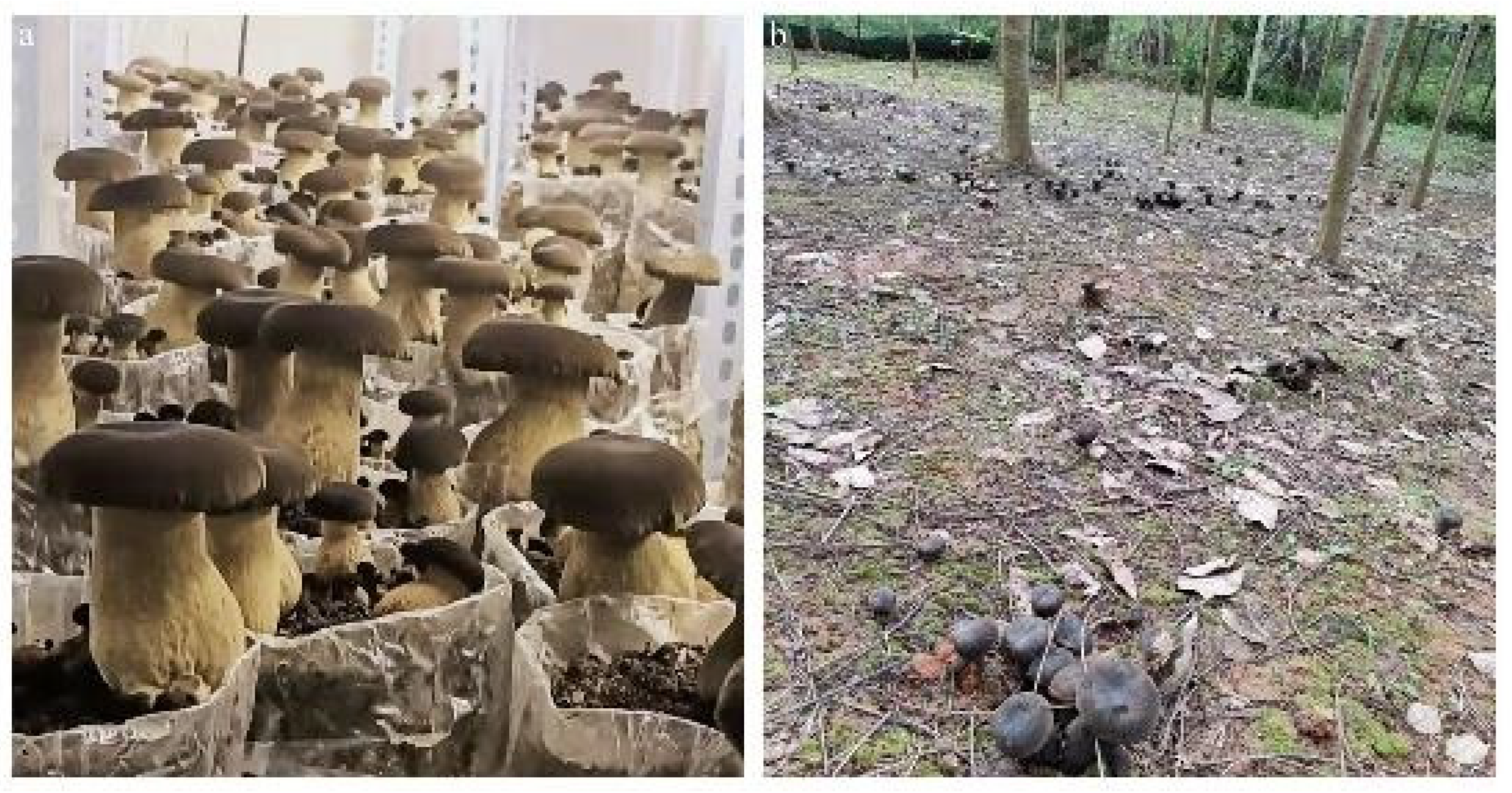
2.2. Field Investigation Sites

Three sites were set up to investigate sclerotium production. Two of them were in the grapefruit orchards the Dongfeng Farm Grapefruit Orchard and Mangajian Village Grapefruit Orchard. They also housed our experimental blocks for the fungal field inoculation tests, and the inoculated strains were PPMU15013. The inoculation trail at the Dongfeng Farm Grapefruit Orchard started in 2015 and produced mushrooms in 2016. The inoculation trail at the Mangajian Village Grapefruit Orchard started in 2017 and produced mushrooms in the same year. The third was the Jinghong Hydropower Station, where wild P. portentosus was growing and fruiting. The three sites were separated, over 30 km away from one another, and all were located in Jinghong City, Xishuangbanna, Yunnan.

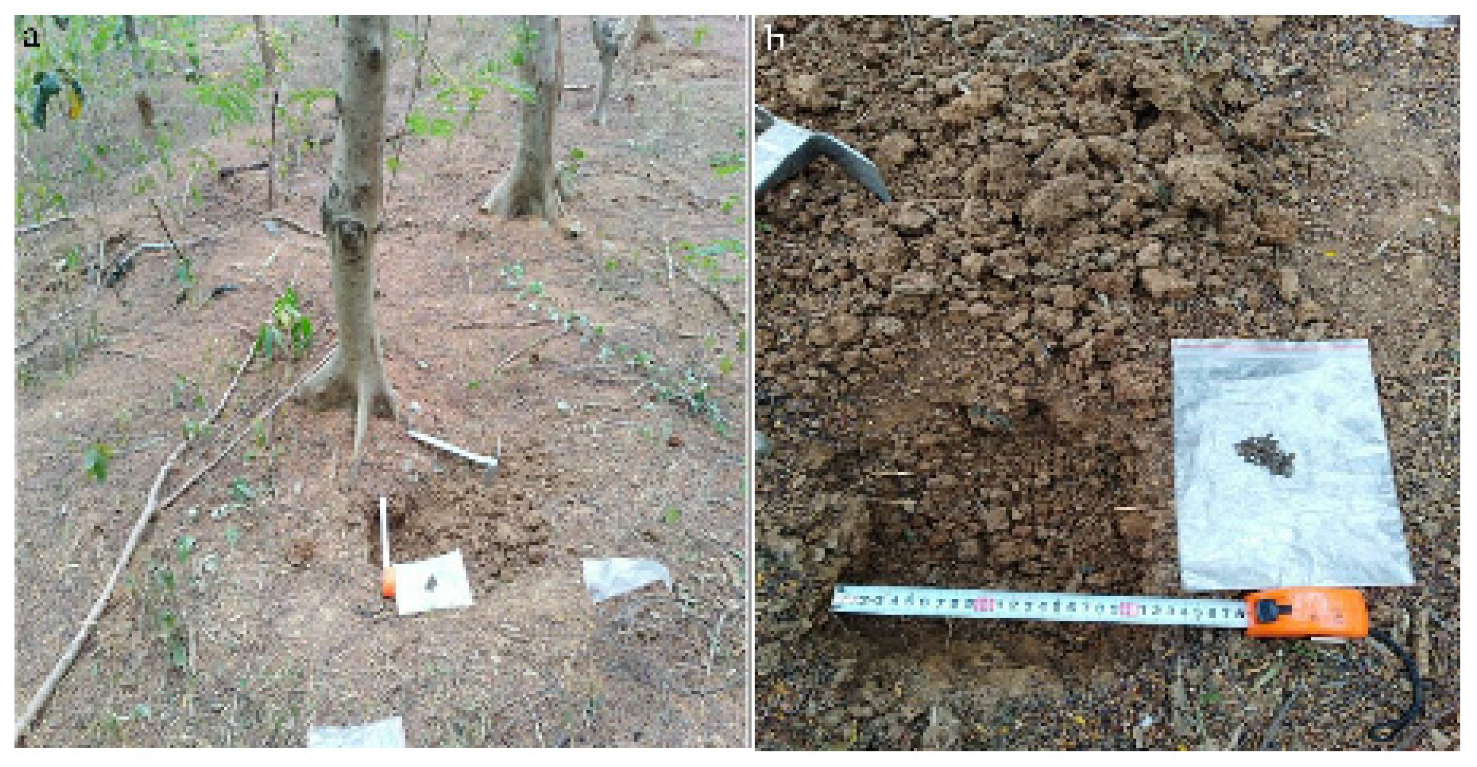
2.3. Field Investigation Methods

The investigation of sclerotium formation was carried out in the second or last ten days of every month from October 2017 to October 2018. At every site, three small pits of 20 × 20 × 20 cm were dug out each time. All the sclerotia and soil from each pit were sampled and taken to the Mycological Lab of the Yunnan Institute for Tropical Crop Research for further examination (Figure 2). The dynamic process of sclerotium development at different collection times was recorded to construct a clear picture of the sclerotium formation under natural conditions. The sclerotia samples were jointly identified by researcher Chunxia Zhang and Dr. Yun Wang.

2.4. Sclerotium Formation Trail in the Lab

M1 agar medium [30] was used for culturing the isolates of PPMU17076, PPMU18004 and PPMU18106, which were incubated at 28–30 °C. Each isolate had 10 plates. The mycelial growth and process of sclerotium formation were examined and recorded.

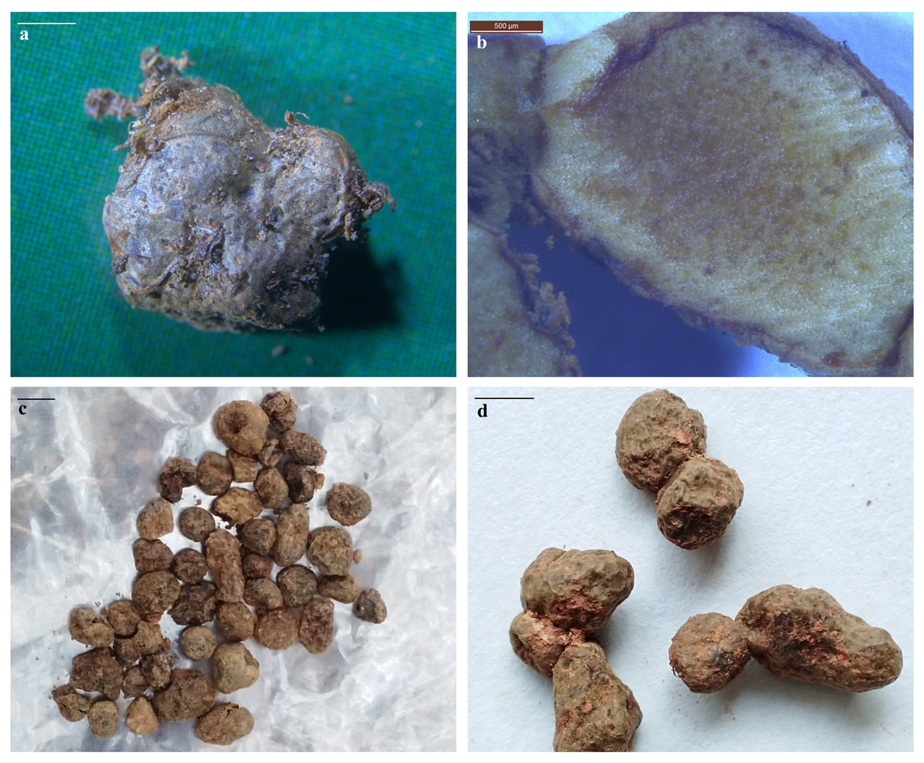
2.5. Morphological Examination of the Sampled Sclerotia in the Lab

Under a stereomicroscope (LEICA M125) and Zeiss upright microscope (ZEISS Axio Scope A1), the morphological characteristics and dynamic process of the sclerotia collected at different times were examined and recorded. The sizes of the sclerotia were measured with an electronic digital display Vernier caliper (GUANGLU 0~200 mm). The fresh weight of sclerotia was determined by electronic balance (Mettler Toledo ME204E/02).

2.6. Sclerotia Germination Experiment

The vegetable garden soil was collected and dried naturally after removing impurities. Sixty percent vegetable garden soil and forty percent grass charcoal were mixed evenly according to volume ratio, then sprayed with water slowly and stirred until the water content reached about 60%. (When the soil was grasped by the hand, the soil could form a mass, and when the hand was loosened, the soil could disperse naturally; at this time, the water content reached about 60%.) A 200 g amount of the above-mentioned soil was put into a glass bottle, and 5 sclerotia were buried in each bottle and cultured at 28~30 °C. The collection of sclerotia from the field and agar medium was repeated 3 times, respectively. The germination of sclerotia and the formation of fruiting bodies were observed.

2.7. Molecular Identification of Sclerotia and DNA Extraction

Genomic DNA was extracted from a small (100 mg) piece of the sclerotia with CWBIO’s fungal genomic DNA isolation kit (Beijing CWBIO Biotech China). Sclerotia were identified based on the internal transcribed spacer (ITS) region of the nuclear ribosomal RNA gene cluster. Two universal primers of ITS1 (5′-CCGTAGGTGAACCTGCGG-3′) and ITS4 (5′-TCCTCCGCTTATTGATATGC-3′) were used for DNA amplification. The amplification conditions were as follows: 94 °C for 5 min, 30 cycles at 94 °C for 30 s, 57 °C for 30 s, and 72 °C for 2 min and then 72 °C for 10 min. The PCR product was gel-purified and sequenced by TSINGKE (Kunming, China).

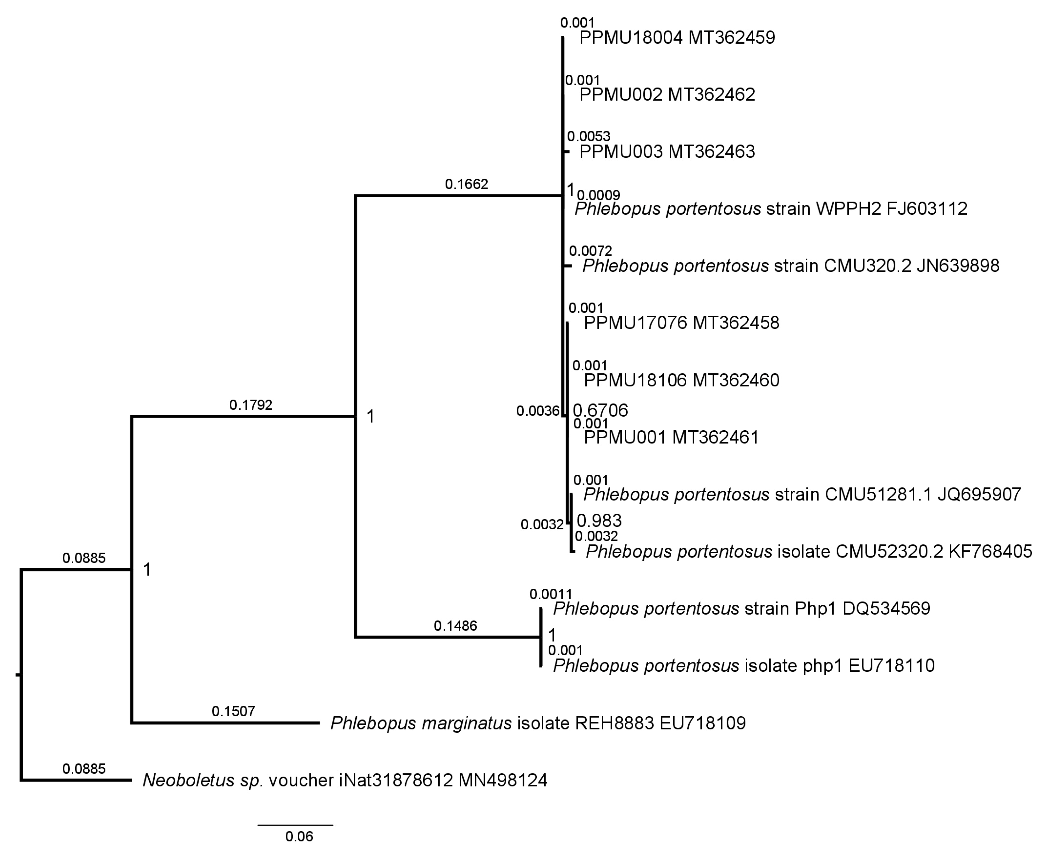
2.8. Phylogenetic Analysis

Comparative analysis was performed on the nucleotide sequences downloaded from the GenBank database (http://www.ncbi.nlm.nih.gov/, accessed on 21 April 2020). Sequence alignments were adjusted using MEGA6 software. Bayesian analysis with MrBayes 3.1, implementing the Markov chain Monte Carlo technique and parameters predetermined with MrModeltest 3.0, was performed [24].

3. Results

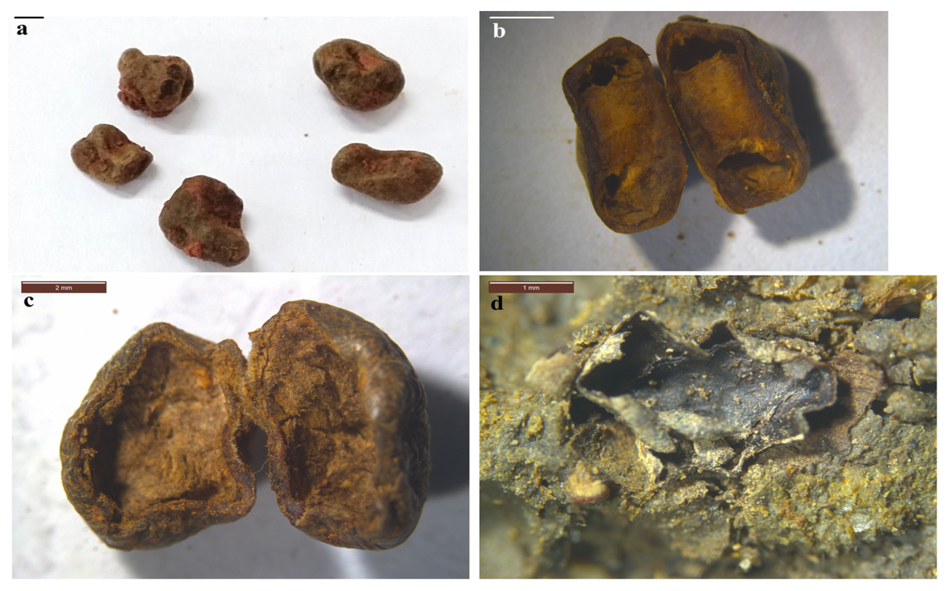
3.1. Formation of Sclerotia in the Field

When the environmental conditions became non-conducive (lower temperature, drought or both) to fungal growth, the fungal mycelia and rhizomorphs in the soil began to converge and tangle (Figure 3a) into a pale-yellow, globose or irregular soft hyphal ball (Figure 3b). As the hyphal ball gradually grew bigger, its tissue became tighter and darker and then dense air hyphae emerged from its surface (Figure 3c). At this stage, the ball case was thickening and covered by rhizomorphs (Figure 3d), and it contained dark-brown, honey-like juice inside (Figure 3e). Finally, the hyphal ball solidified and separated from the surrounding hyphae, becoming a matured sclerotium (Figure 4a).
The matured sclerotium consisted of solid, dark, glabrous, vein-like rhizomorphs scattered on the surface (Figure 4a). The peridium and internal tissues (pith) were differentiated. The peridium was made from dark hyphae, and the internal tissue was made from densely interwoven hyphae, wax-like and grayish to brownish with brown spots (Figure 4b). The sclerotium typically produced singly (Figure 4c), rarely in beads (Figure 4d), and could be globose, subglobose or irregular, 1.42~14.98 mm in diameter. The mature sclerotium consisted of a dark-brown rind layer and a yellowish-brown medulla layer; the latter was composed of a densely interwoven mycelium (Figure 5).

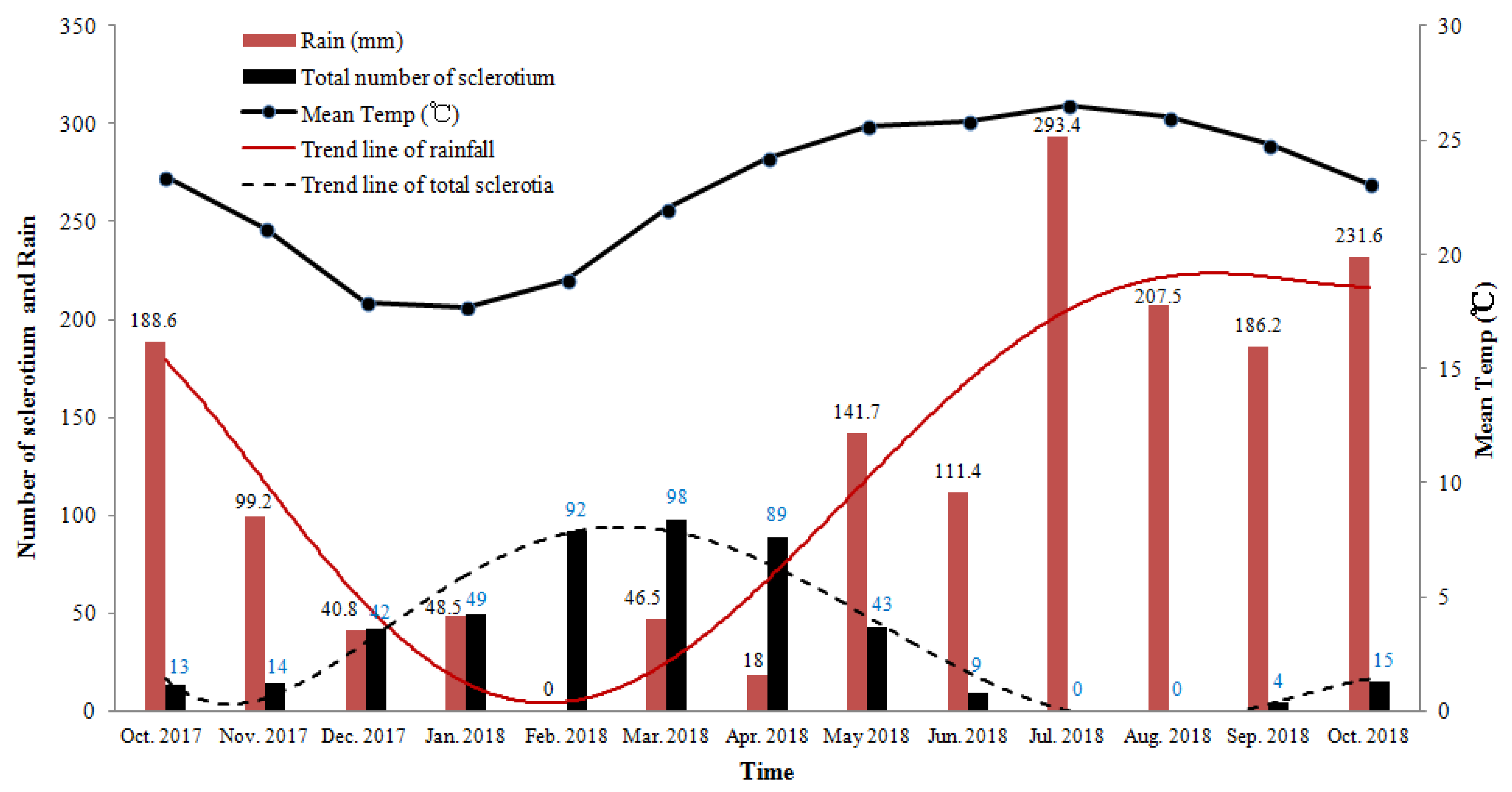
3.2. Seasonal Dynamics of Sclerotia Occurrence in the Field

In the field, the sclerotium developed all year round, except in July and August. From December to April of the following year, the sclerotia produced abundantly in the soil due to the lower temperature and scarce rainfall (Table 1, Figure 6). The number and weight of sclerotia were the largest from February to April. From December to February of the following year, both young and matured sclerotia were discovered simultaneously. From March to June, only matured and aging sclerotia were found. After April, the sclerotia started to germinate as the temperature and rainfall gradually increased; subsequently, the number of sclerotia that were discovered gradually declined. In the meantime, a few mushrooms of P. portentosus emerged. From May to June, a few matured and aging sclerotia were still present in the soil.

3.3. Aging and Dead Young of Sclerotia in the Field

When the weather became extremely dry and the soil was short of water, the sclerotia dried out, becoming shriveled (Figure 7a). Their surfaces wrinkled or became uneven. At this moment, the hyphal growth and accumulation of nutrients in the sclerotia were interrupted due to water loss. As a result, the sclerotia became hollow (Figure 7b), dried (Figure 7c) and finally ruptured to death, with only the dried and fragile peridium being left (Figure 7d). The quantity of dead sclerotia increased in the soil as the dry weather continued, especially in the upper soil layer (0–10 cm). However, some of the matured sclerotia could survive the harsh period even though they almost dried out. When the dried, matured sclerotia were put in a Petri dish lined with moistened double-filter paper for 24 h, they recovered and achieved rebirth.

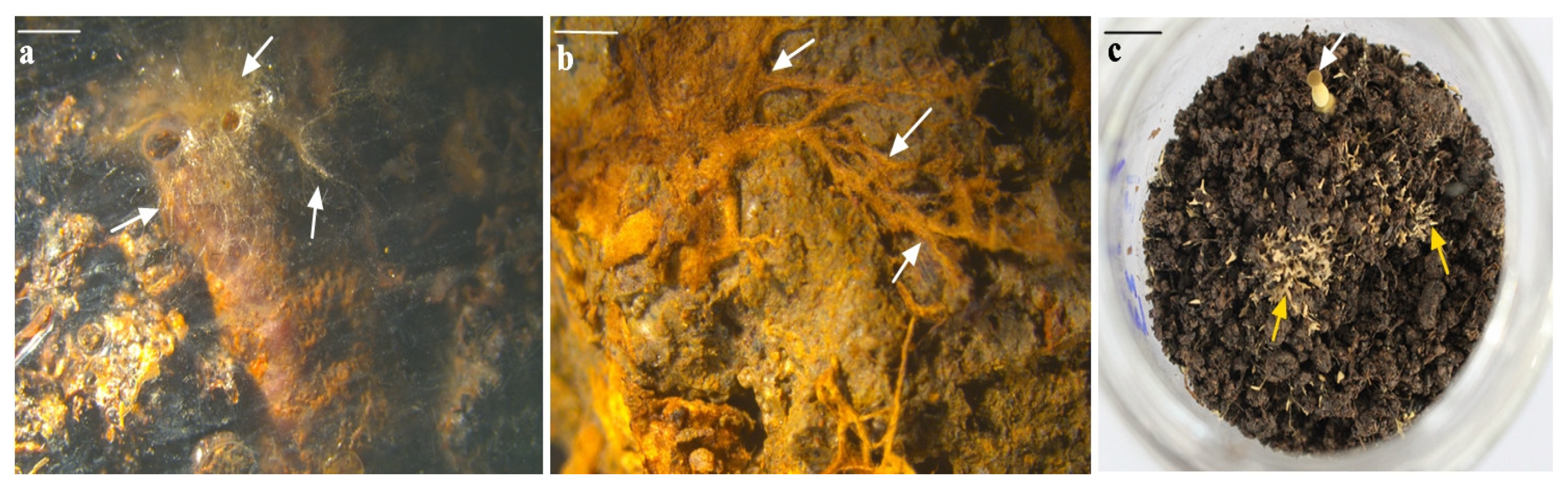
3.4. Germination of Sclerotium in Soil

When the temperature and moisture of the soil reached the optimum conditions, the sclerotia that had survived began to germinate. New mycelia developed from the sclerotium and gradually covered it (Figure 8a). When the nutrients accumulated in the sclerotia ran out, with more mycelia developed, a large number of mycelia and rhizomorphs appeared in the soil, and the sclerotia eventually disappeared (Figure 8b). A new colony of P. portentosus was then created, from which fruiting bodies might develop when the environment is suitable (Figure 8c).

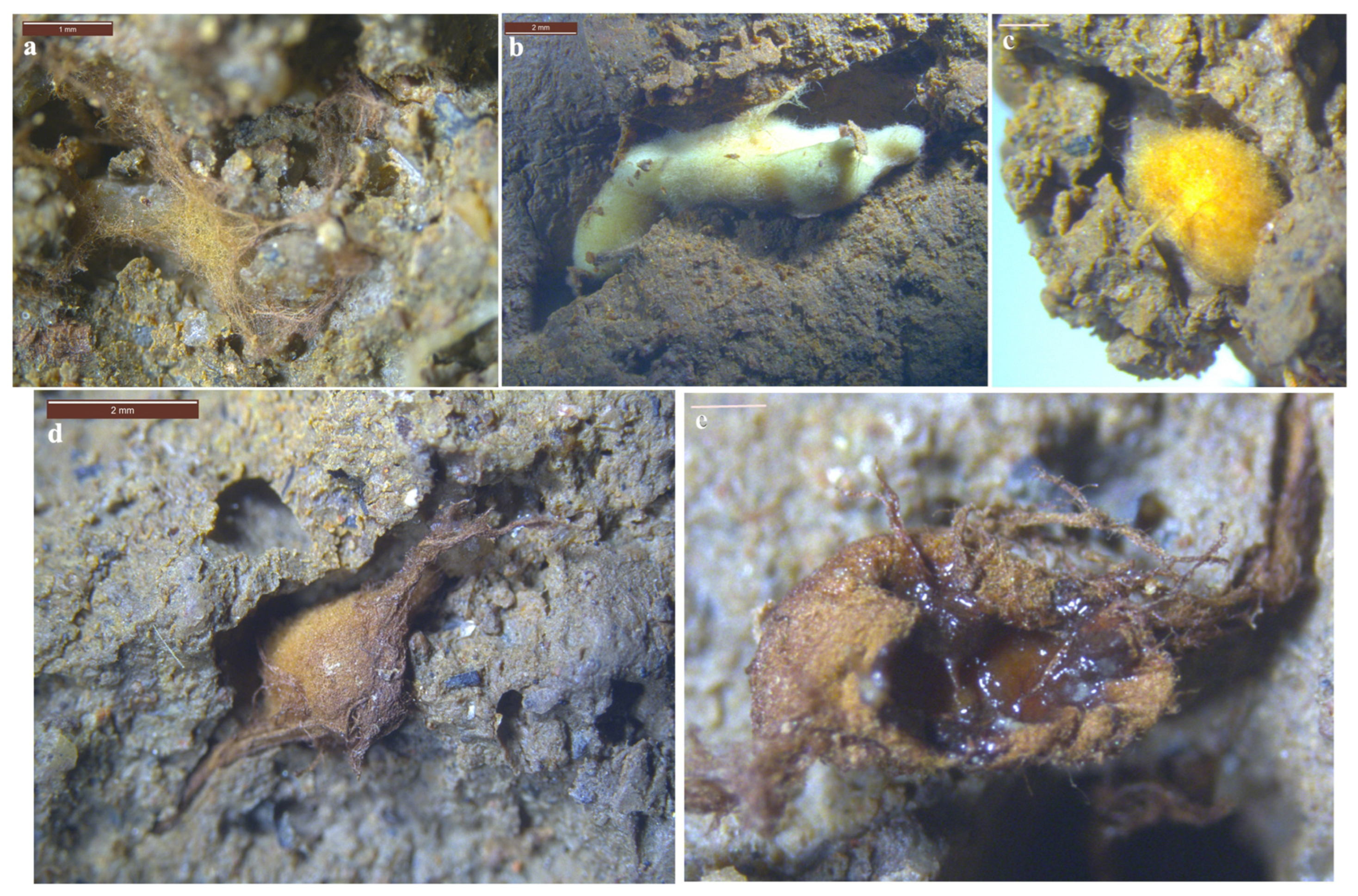
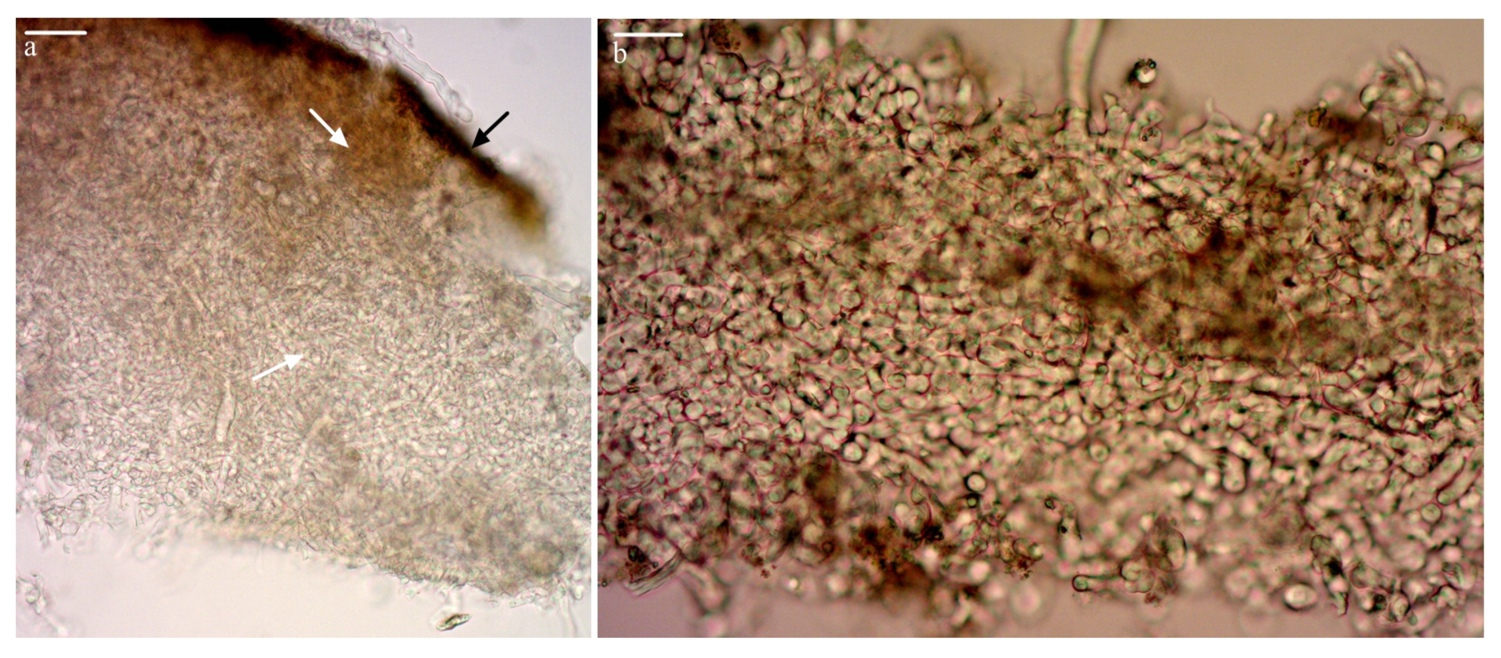
3.5. Culture of Sclerotium in the Lab

The three isolates of P. portentosus grew well on the agar media and produced round colonies. When cotton-wool-like, tangled, interwoven hyphae started to develop on the colony, a few small, transparent liquid droplets appeared on its surface (Figure 9a). More and more liquid droplets developed and became bigger and darker as the fungal colonies grew further. A mass of curly, fluffy, tangled mycelia knots then emerged from the surface of the colony (Figure 9b). They soon developed into dense mycelia balls with a lot of liquid droplets on their surfaces. These were baby sclerotia (Figure 9c). At this stage, the peridium was differentiated from its juicy internal tissue (Figure 9d). As the sclerotium grew up, the interior tissue solidified with nutrient accumulation and intensive hyphal growth (Figure 9e). Then, the sclerotium became harder and harder, and the liquid droplets turned dark red-brown (Figure 9f). At this stage, the fluffy hyphae disappeared from the hardening peridium surface, and a few dark pits appeared (Figure 9g). The interior tissue was composed of dense mycelium, brown to dark brown, with more nutrients accumulated (Figure 9h and Figure 10). Most of the sclerotia were globose to subglobose when young and became irregular cluster shapes as adjacent sclerotia fused together, which could grow up to 20 mm long or longer (Figure 9g).
The time and positions of the sclerotia development on the agar media were different among the three isolates (Figure 11; Table 2). Isolate PPMU17076 started producing liquid droplets as soon as it colonized the agar plate. The sclerotia developed near to the isolated lump and spread out. Isolate PPMU18004 started producing sclerotia on the ninth day after the inoculation, which formed a ring around the isolate lump 1 cm away. However, isolate PPMU18106 would not produce sclerotia until it colonized the whole agar plate, and most of the sclerotia scattered around the edge of the colony. The sclerotia were smaller but abundant. The sclerotia produced by the agar medium could germinate into hyphae after being cultured at 4 °C for 24 h but could not produce fruiting bodies.

3.6. Molecular Identification of the Sclerotium

For phylogenetic analysis, GenBank accession numbers for sequences obtained in the present study are listed in Table 3. Six DNA fragments, 690 bp, were obtained from the six sclerotium samples. A total of 14 ITS sequences were included in the phylogenetic analysis. Neoboletus sp. iNat31878612 (MN498124.1) was used as an outgroup. The results indicate that the nucleotide sequences of the six sclerotium samples were almost the same and grouped with four sequences of P. portentosus. However, they were well separated from Phlebopus marginatus REH8883 (EU718109) (Figure 12).

4. Discussion

The whole process of the sclerotium formation of P. portentosus both in field and lab conditions was reported. In natural conditions, the occurrence of sclerotia was closely related to seasonal weather changes, especially to the rainfall (Figure 6). More sclerotia were discovered from December to April of the following year, the dry season in the Xishuangbanna region. During the dry season, the rainfall was less than 50 mm/month. When the rainfall was more than 100 mm/month, the number of sclerotia discovered was less than 15/month. The more rainfall there was, the fewer sclerotia were discovered. A lower temperature may increase the formation of sclerotia; however, the relationship between sclerotium formation and temperature is not as clear as that between sclerotium formation and rainfall.
It is not easy to distinguish the different developing stages of sclerotia; distinguishing the dead from shriveled but still alive ones is a particular challenge. During the sclerotium germination tests, the dead sclerotia could not germinate, but the shriveled ones could. Fruit bodies of P. tuberregium form from a true sclerotium in the wild [32], and sclerotium development from hyphae to a fruiting body is a necessary stage for Morchella [33]. In this study, the sclerotia of P. portentosus collected in the field could germinate to form mycelium and fruiting bodies under a suitable temperature and humidity, while the sclerotia produced on agar medium could only germinate into mycelia and would not form fruiting bodies; the reason for this needs to be further studied. When the sclerotia of P. portentosus germinates, the whole sclerotia is quickly wrapped by the mycelium, and the nutrients in the sclerotia are gradually consumed, and the sclerotia disappears. Finally, a large number of mycelia and rhizomorphs appear in the soil, which can form fruiting bodies in the field.
On the agar medium, all three of the isolates produced abundant sclerotia under the optimum conditions. The sclerotium formation during artificial cultivation was always accompanied by the appearance of water droplets on the colony (Figure 9). Fungal colonies on agar medium producing liquid droplets is a common phenomenon known as “spitting water”, which indicates a shift from a vegetative to a reproductive phase [5,34]. The results of recent research on Morchella cultivation revealed that the occurrence of fruiting bodies follows the sclerotium formation, which is accompanied by water spitting [35]. Morchella spitting water is due to the changes in the cytoplasmic movement and osmotic pressure caused by the internal physiological balance or mycelial metabolism [5,36]. The occurrence of sclerotia is a prerequisite for the fruitification of culturing Morchella [11,37,38].
In nature, sclerotium formation is an important strategy of the fungus P. portentosus to survive harsh environments, while sclerotia can germinate and form fruiting bodies under suitable environmental conditions. During the cultivation of P. portentosus, the fungus produces abundant sclerotia on its surface when it fully colonizes the substrate. At this moment, the fungus reaches its reproductive stage and is ready to produce mushrooms. The characteristics such as rapid mycelium growth, “spitting water” and sclerotia formation on the cultivation substrate can be used as the main criteria for screening excellent strains of P. portentosus [39]. It shows that sclerotia play an important role in the life cycle of P. portentosus. The process of sclerotia formation in the field and agar medium is the same, but the reasons for formation are different. It can be said that, under artificial conditions (on the agar medium), the sclerotium formation of P. portentosus is caused by its physiological maturation rather than environmental stress. However, in nature, during the mushroom season (June to October), few sclerotia are produced in the soil.

5. Conclusions

In this study, the whole process of birth, growth, death and rebirth of the sclerotium of P. portentosus both in field and lab conditions was reported for the first time. Environmental stresses such as low temperature and drought are the main reasons for the sclerotia formation of P. portentosus in the field, and the sclerotia can germinate rapidly to form a mycelium and fruiting bodies under suitable temperature and water conditions. Under lab conditions, the sclerotium formation of P. portentosus is related to the fungus’ physiological maturation. The sclerotia produced under lab conditions can only germinate into a mycelium but cannot form fruiting bodies. Further research on the relationship between sclerotium formation and the fruitification of P. portentosus is needed in the future.

Author Contributions

Conception and design: T.Y., Y.W. and C.Z.; Investigation: T.Y., J.L., X.X., M.H. and C.Z.; Acquisition of data: X.X., M.H., F.G., Y.F. and W.W.; Analysis and interpretation of data: T.Y., J.L., L.D. and Y.W.; Writing original draft: T.Y., Y.W. and C.Z. All authors have read and agreed to the published version of the manuscript.

Funding

This study was supported by the National Natural Science Foundation of China (no. 32060707), the Funds of Sci-Tech Innovation System Construction for Tropical Crops of Yunnan Province (no. RF2022-15), the Project of Sci-Tech Talents and the Platform of Yunnan Province (No. 2019HB069) and the Youth Growth Project of the Yunnan Institute of Tropical Crops (no. QNCZ 2020-9).

Data Availability Statement

The sequence data generated in this study can be obtained from NCBI GenBank (http://blast.ncbi.nlm.nih.gov/Blast.cgi, accessed on 21 April 2020). The data included in this study are available through contacting the authors.

Acknowledgments

We are very grateful to Qiying Yang and Wangguang Qi for their help in the field investigation and to the Xishuangbanna Meteorological Bureau for providing us with rainfall and temperature data.

Conflicts of Interest

The authors declare that they have no competing interest.

References

  1. Willetts, H.J. Developmental mycology, Sclerotium formation. Filamentous Fungi 1978, 3, 197–213. [Google Scholar]
  2. Smith, M.E.; Henkel, T.W.; Rollins, J.A. How many fungi make sclerotia? Fungal Ecol. 2015, 13, 211–220. [Google Scholar] [CrossRef]
  3. Song, C.; Liu, M.; Xing, Y.; Guo, S. ESTs analysis of putative genes engaged in Polyporus umbellatus sclerotial development. Int. J. Mol. Sci. 2014, 15, 15951–15962. [Google Scholar] [CrossRef] [PubMed] [Green Version]
  4. Volk, T.J.; Leonard, T.J. Physiological and environmental studies of sclerotium formation and maturation in isolates of Morchella crassipes. Appl. Environ. Microb. 1989, 55, 3095–3100. [Google Scholar] [CrossRef] [Green Version]
  5. Liu, S.W.; Liang, Z.Q.; Liu, A.Y. Sclerotia an important part in morchella life cycle. Guizhou Agr. Sci. 1997, 25, 55–59. [Google Scholar]
  6. Liu, W.; Cai, Y.L.; He, P.X.; Chen, L.F.; Bian, Y.B. Comparative transcriptomics reveals potential genes involved in the vegetative growth of Morchella importuna. 3 Biotech 2019, 9, 81. [Google Scholar] [CrossRef]
  7. Lee, S.R.; Lee, S.; Moon, E.; Park, H.J.; Park, H.B.; Kim, K.H. Bioactivity guided isolation of anti-inflammatory triterpenoids from the sclerotia of Poria cocos using lps-stimulated raw264.7 cells. Bioorgan. Chem. 2016, 70, 94–99. [Google Scholar] [CrossRef]
  8. Zhou, J.; Zhang, Z.M.; Wu, S.Y.; Chen, L.; Ding, Z.Y. Study on cultivation of Pleurotus tuberregium sclerotia via plate and the formation mechanism. Food Ferment. Ind. 2022, 48, 84–90. [Google Scholar] [CrossRef]
  9. Liu, Y.Y.; Zhang, X.L.; Duan, J.F.; Qin, Y.T.; Xiao, Z.L. Isolation and Identification of symbiotic armillaria of wild Polyporus umbellatus in Ziwuling Forest Region of Qingyang City. J. Chin. Med. Mate. 2022, 45, 1296–1300. [Google Scholar] [CrossRef]
  10. Ower, R.D.; Mills, G.L.; Malachowski, J.A. Cultivation of Morchella: USA. US4594809, 17 June 1986. [Google Scholar]
  11. Liu, W.; Zhang, Y.; He, P.X. Morel Biology and Cultivation; Jilin Science and Technology Press: Changchun, China, 2017. [Google Scholar]
  12. Liu, Q.Z.; Dong, C.H. Research Progress on Sclerotium F Ormation in Morchella and Reflection on Its Application in Cultivation. J. Edible Fungi 2020, 27, 172–178. [Google Scholar] [CrossRef]
  13. Alvarado-Castillo, G.; Vázquez, A.P.; Martínez-Carrera, D.; Tablada, M.E.N.; Osorio-Acosta, F. Morchella sclerotia production through grain supplementation. Interciencia 2011, 36, 768–773. [Google Scholar] [CrossRef]
  14. Liu, W.Y.; Guo, H.B.; Bi, K.X.; Sibirina, L.A.; Qi, X.J.; Yu, X.D. Determining why continuous cropping reduces the production of the morel Morchella sextelata. Front. Microbiol. 2022, 13, 903983. [Google Scholar] [CrossRef]
  15. Ji, K.P.; Cao, Y.; Zhang, C.X.; He, M.X.; Liu, J.; Wang, W.B.; Wang, Y. Cultivation of Phlebopus portentosus in southern China. Mycol. Prog. 2011, 10, 293–300. [Google Scholar] [CrossRef]
  16. Zhang, C.X.; Ji, K.P.; He, M.X.; Cao, Y.; Liu, J.; Wang, W.B. Analysis on nutrient components of Phlebopus portentosus fruit bodies. J. Yunnan Univ. 2010, 32, 702–704. [Google Scholar]
  17. Sanmee, R.; Lumyong, P.; Dell, B.; Lumyong, S. In vitro cultivation and fruit body formation of the black bolete, Phlebopus portentosus, a popular edible ectomycorrhizal fungus in thailand. Mycoscience 2010, 51, 15–22. [Google Scholar] [CrossRef]
  18. Raghoonundon, B.; Raspé, O.; Thongklang, N.; Hyde, K.D. Phlebopus (boletales, boletinellaceae), a peculiar bolete genus with widely consumed edible species and potential for economic development in tropical countries. Food Biosci. 2021, 41, 100962. [Google Scholar] [CrossRef]
  19. Ji, K.P.; He, M.X.; Zhang, C.X.; Liu, J.; Wang, W.B.; Hou, J.Y. Semi-Artificial Simulate Cultivation of Phlebopus portentosus and the Durability of Hyphae on Host Roots. Microbiol. China 2009, 36, 377–382. [Google Scholar]
  20. Zhang, C.X.; He, M.X.; Liu, J.; Xu, X.J.; Cao, Y.; Gao, F.; Fang, Y.W.; Wang, W.B.; Wang, Y. Brief introduction to a unique edible bolete—Phlebopus portentosus in Southern China. J. Agr. Sci. Technol. B 2017, 7, 386–394. [Google Scholar] [CrossRef] [Green Version]
  21. Ji, K.P.; Zhang, C.X.; Zeng, Y.; Liu, C.F.; He, M.X.; Wang, W.B. Artificial Fungal Colony and Its Fruiting of Phlebopus portentosus (Boletaceae) in Pot. Acta Botanica Yunnanica 2007, 5, 554–558. [Google Scholar] [CrossRef]
  22. Cao, Y.; Ji, G.Y.; Luo, S.Z.; Gao, L.X.; Tao, L.; Wang, Q.L.; Yang, R.H.; Bao, D.P.; Ji, K.P. Domestication and artificially cultivation of Phlebopus portentosus: Retrospect and prospect. Mycosystema 2021, 40, 3064–3080. [Google Scholar] [CrossRef]
  23. Kumla, J.; Bussaban, B.; Suwannarach, N.; Lumyong, S.; Danell, E. Basidiome formation of an edible wild, putatively ectomycorrhizal fungus, Phlebopus portentosus without host plant. Mycologia 2012, 104, 597–603. [Google Scholar] [CrossRef] [PubMed]
  24. Xie, H.J.; Zhang, C.X.; He, M.X.; Liang, Z.Q.; Deng, X.H.; Zeng, N.K. Buchwaldoboletus xylophilus and Phlebopus portentosus, two non-ectomycorrhizal boletes from tropical China. Phytotaxa 2021, 520, 137–154. [Google Scholar] [CrossRef]
  25. Zhang, C.X.; He, M.X.; Cao, Y.; Liu, J.; Gao, F.; Wang, W.B.; Ji, K.P.; Shao, S.C.; Wang, Y. Fungus-insect gall of Phlebopus portentosus. Mycologia 2015, 107, 12–20. [Google Scholar] [CrossRef]
  26. Zhang, C.X.; He, M.X.; Liu, J.; Cao, Y.; Gao, F.; Xu, X.J.; Wang, W.B.; Wang, Y. Preliminary studies on trophic relationships of Phlebopus portentosus, soil mealybug and plant. Edible Med. Mushrooms 2015, 23, 359–363. [Google Scholar]
  27. Fang, Y.W.; Wang, W.B.; He, M.X.; Xu, X.J.; Gao, F.; Liu, J.; Yang, T.W.; Cao, Y.; Yang, T.; Wang, Y.; et al. Relationship between the honeydew of mealy bugs and the growth of Phlebopus portentosus. PLoS ONE 2020, 15, e0233710. [Google Scholar] [CrossRef] [PubMed]
  28. He, M.X.; Yang, T.W.; Gao, F.; Liu, J.; Xu, X.J.; Fang, Y.W.; Wang, W.B.; Zhang, C.X. Seasonal Variation of Fungus-Insect Gall and Its Correlation with Fruiting of Phlebopus portentosus. Acta Edulis Fungi 2019, 26, 73–81. [Google Scholar] [CrossRef]
  29. Zhang, C.X.; He, M.X.; Ji, K.P.; Cao, Y.; Liu, J.; Wang, W.B. Studies on Ecological Characteristics of Phlebopus portentosus. Southwest China J. Agr. Sci. 2012, 25, 614–619. [Google Scholar] [CrossRef]
  30. Zhang, C.X.; He, M.X.; Ji, K.P.; Cao, Y.; Liu, J.; Wang, W.B. Study on Selection of Mother Culture Media of Phlebopus portentosus. Southwest China J. Agric. Sci. 2009, 22, 1699–1701. [Google Scholar] [CrossRef]
  31. Binder, M.; Hibbett, D.S. Molecular systematics and biological diversification of Boletales. Mycologia 2006, 98, 971–981. [Google Scholar] [CrossRef]
  32. Isikhuemhen, O.S.; Stamets, P.E.; Vilgalys, R. Biology, food, medicinal, and biotechnological applications of the tropical mushroom Pleurotus tuberregium (Rumph.:Fr.) Singer. Int. J. Med. Mushrooms 2005, 7, 415–416. [Google Scholar] [CrossRef]
  33. And, H.K.K.; Reddy, M.S. Influence of sclerotia formation on ligninolytic enzyme production in Morchella crassipes. J. Basic Microbiol. 2014, 54, S63–S69. [Google Scholar] [CrossRef]
  34. Amir, R.; Levanon, D.; Hadar, Y.; Chet, I. Morphology and physiology of Morchella esculenta during sclerotial formation. Mycol. Res. 1993, 97, 683–689. [Google Scholar] [CrossRef]
  35. Cheng, L.J. Study on the Cultural and Genetic Characteristics of the Sclerotia Producing by Morchella. Yunnan Univ. 2012, 11, 92p. [Google Scholar]
  36. He, P.X.; Wang, K.; Cai, Y.L.; Hu, X.L.; Zheng, Y.; Zhang, J.J.; Liu, W. Involvement of autophagy and apoptosis and lipid accumulation in sclerotial morphogenesis of Morchella importuna. Micron 2018, 109, 34–40. [Google Scholar] [CrossRef]
  37. Liu, Q.Z.; Zhao, Z.H.; Dong, H.; Dong, C.H. Reactive oxygen species induce sclerotial formation in Morchella importuna. Appl. Microbiol. Biot. 2018, 102, 7997–8009. [Google Scholar] [CrossRef] [PubMed]
  38. Liu, Q.Z.; He, G.Q.; Wei, J.K.; Dong, C.H. Comparative transcriptome analysis of cells from diferent areas reveals ROS responsive mechanism at sclerotial initiation stage in Morchella importuna. Sci. Rep. 2021, 11, 9418. [Google Scholar] [CrossRef] [PubMed]
  39. He, M.X.; Xu, X.J.; Gao, F.; Liu, J.; Cao, Y.; Wang, W.B.; Fang, Y.W.; Yang, T.W.; Wang, Y.; Zhang, C.X. Isolation and screening of high quality Phlebopus portentosus strains. Acta Edulis Fungi 2017, 24, 33–38. [Google Scholar] [CrossRef]
Figure 1. Phlebopus portentosus produced mushrooms in the mushroom house (a) and around inoculated trees in the field (b).
Forests 14 01096 g001
Figure 2. Field investigation of sclerotium (a), and Collection of sclerotium samples from the pit (b).
Forests 14 01096 g002
Figure 3. Early stage of sclerotium formation in the field. (a) Converged and tangled mycelia and rhizomorphs. (b) A pale yellow, irregular soft hyphal ball formed. (c) The hyphal ball gradually grew bigger, the tissue becoming tighter and darker with dense air hyphae on the surface. (d) The ball case thickened and was covered by rhizomorphs. (e) Dark-brown, honey-like juice inside. Scale bars: (c,e) = 2 mm. (The samples were collected from Dongfeng Farm in November 2017).
Forests 14 01096 g003
Figure 4. Matured sclerotium produced in the field. (a) A matured sclerotium. (b) Cross-section of a matured sclerotium showing a differentiated peridium and pith. (c) Sclerotia in different shapes. (d) Bead-like sclerotia. Scale bars: (a) = 2 mm, (c,d) = 5 mm. (The samples were collected from the hydropower station in February 2018).
Forests 14 01096 g004
Figure 5. Microscopic characteristics of sclerotia. (a) The sclerotium has two layers; the outermost layer is a dark-brown rind (black arrow); the inner layer is the medulla, composed of densely interwoven mycelium (white arrow) and (b) a dense mycelium in the medullary layer. Scale bars: (a) = 100 μm, (b) = 50 μm.
Forests 14 01096 g005
Figure 6. The relationship between the production of sclerotia and monthly average temperature and rainfall. (The climate data were provided by Xishuangbanna Meteorological Bureau).
Forests 14 01096 g006
Figure 7. Aging and dead young sclerotia. (a) Shriveled sclerotia. (b) The sclerotium becoming hollow. (c) Cross-section of dead sclerotium. (d) A dead sclerotium with only a dried and fragile peridium left. Scale bars: (a) = 3 mm, (b) = 2 mm. (The samples were collected from the hydropower station in April 2018).
Forests 14 01096 g007
Figure 8. Germination of a sclerotium. (a) Mycelium grows from the surface of sclerotia (white arrow). (b) Mycelium and rhizomorph (white arrow) appeared in the soil after sclerotia germination. (c) Primordia (yellow arrow) and fruiting bodies (white arrow) formed by sclerotia germination. Scale bars: (a) = 2 mm, (b,c) = 3 mm. (The samples were collected from Mangajian in April 2018).
Forests 14 01096 g008
Figure 9. Sclerotium formation on agar medium. (a) Liquid droplets appeared on the colony. (b) Curly, fluffy, tangled mycelia knots emerging. (c) A baby sclerotium. (d) Inside the young sclerotium, juicy and soft. (e) The internal tissue solidified with nutrients and hyphae accumulated. (f) Liquid droplets became dark brown. (g) Brown and fluffy hyphae disappeared, and pits appeared. (h) Cross-sections show the interior hyphae and stored nutrients. Scale bars: (a,ce) = 1 mm; (b) = 5 mm; (fh) = 2 mm. ((ae) form the isolate PPMU18004; (fh) form the isolate PPMU17076).
Forests 14 01096 g009
Figure 10. The microstructure of mature sclerotia. (a) Longitudinal section of sclerotia. (b) The dense mycelium inside the sclerotia. Scale bars: (b) = 100 μm.
Forests 14 01096 g010
Figure 11. Sclerotium formation of three strains PPMU17076 (a), PPMU18004 (b) and PPMU18106 (c) on the agar medium.
Forests 14 01096 g011
Figure 12. Phylogram inferred from ITS region dataset by the Bayesian method.
Forests 14 01096 g012
Table 1. Seasonal dynamics of sclerotia formation from 2017 to 2018.
Investigation TimeDongfeng FarmHydropower StationMangajianTotal Number of Sclerotia
Number of SclerotiaWeight (g) **Number of SclerotiaWeight (g)Number of SclerotiaWeight (g)
October 201780.42250.2710-13
November 201770.35530.15540.19814
December 2017170.860160.78990.47742
January 2018150.702201.032140.72949
February 2018271.241341.554311.60392
March 2018190.897432.103361.67898
April 2018241.015361.494291.39889
May 201870.358251.130110.58043
June 2018- *-40.19150.2469
July 2018------0
August 2018------0
September 201840.219----4
October 201850.26970.36530.14615
Note: * no sclerotia were found in the soil; ** the total weight of sclerotia.
Table 2. Characteristics of sclerotia formation of the three isolates on the agar medium.
IsolatesTime of Appearance Amount *Weight (g) **Size (mm) ***Position on the Medium
PPMU170764d1328.304 1.80~21.77Near the isolate lump
PPMU180049d19410.030 1.31~12.341 cm away from the isolate lump
PPMU1810616d2178.854 1.61~9.76Far away from the isolate lump
Note: * total number of sclerotia from 10 Petri dishes. ** Total weight of sclerotia. *** Size ranges of sclerotia.
Table 3. Sequences used in the analysis.
SpeciesVoucher No.GenBank Accession No. DNA Size (bp)Reference
Phlebopus portentosusPhp1DQ534569702[31]
Phlebopus portentosusPhp1EU718110707unpublished
Phlebopus portentosusWPPH2FJ603112813[17]
Phlebopus portentosusCMU320.2 JN639898797unpublished
Phlebopus portentosusCMU51281.1JQ695907678unpublished
Phlebopus portentosusCMU52320.2KF768405750unpublished
Phlebopus marginatusREH8883EU718109687unpublished
Neoboletus sp.iNat31878612MN498124726unpublished
Phlebopus portentosusPPMU17076MT362458690This paper
Phlebopus portentosusPPMU18004MT362459690This paper
Phlebopus portentosusPPMU18106MT362460690This paper
Phlebopus portentosusPPMU001MT362461690This paper
Phlebopus portentosusPPMU002MT362462690This paper
Phlebopus portentosusPPMU003MT362463690This paper
Note: Sclerotia sample PPMU001 was collected from Dongfeng Farm; PPMU002 was collected from the hydropower station; and PPMU003 was collected from Mangajian.
Disclaimer/Publisher’s Note: The statements, opinions and data contained in all publications are solely those of the individual author(s) and contributor(s) and not of MDPI and/or the editor(s). MDPI and/or the editor(s) disclaim responsibility for any injury to people or property resulting from any ideas, methods, instructions or products referred to in the content.

