Article

Exploring the Potential Applications of the Noninvasive Reporter Gene RUBY in Plant Genetic Transformation

1
State Key Laboratory for Conservation and Utilization of Subtropical Agro-Bioresources, College of Forestry, Guangxi University, Nanning 530000, China
2
Key Laboratory of National Forestry and Grassland Administration on Cultivation of Fast-Growing Timber in Central South China, College of Forestry, Guangxi University, Nanning 530000, China
*
Authors to whom correspondence should be addressed.
Forests 2023, 14(3), 637; https://doi.org/10.3390/f14030637
Submission received: 18 January 2023 / Revised: 6 March 2023 / Accepted: 14 March 2023 / Published: 21 March 2023
(This article belongs to the Section Genetics and Molecular Biology)

Abstract

:
Betalains can be conveniently observed and quantified and, accordingly, have the potential as naked-eye visual screening reporters during plant transformation. RUBY is a new reporter system that uses “2A” peptides to fuse three key genes, CYP76AD1, DODA, and glucosyl transferase, for betalain biosynthesis, and has been successfully used for transformation of rice, Arabidopsis, and cotton, but its potential applications in the genetic transformation of various other plant species remain to be verified. In this study, RUBY was transferred into the hairy roots of Plukenetia volubilis and Nicotiana benthamiana, and was transferred into Arabidopsis by the floral-dip method. The expression levels of CYP76AD1, DODA, and glucosyl transferase were detected by RT−PCR and RT−qPCR, the relationship between the expression level of RUBY and red coloration was analyzed, and the genetic stability of RUBY in transgenic Arabidopsis was studied. The results showed that the expression of RUBY could reconstruct the betalain biosynthesis pathway in the hairy roots of P. volubilis, N. benthamiana, and Arabidopsis plants, indicating that it has the potential for versatile use across species. As a reporter, betalain did not affect callus induction, plant regeneration, development, or fertility. However, when used in plant transformation for observation and visual screening, it needed to accumulate to a certain extent to show red coloration, and it was positively correlated with gene expression. In general, RUBY is a convenient reporter for plant transformation, and has no obvious side effects during plant growth and development. However, the potential application of RUBY for visual screening is highly determined by the expression level, and further improvement is needed.

1. Introduction

Betalains, anthocyanins, and carotenoids are three major natural pigments in plants. Of the three pigments, betalains have the simplest biosynthesis pathway: only CYP76AD, DODA, and glucosyl transferase are needed to synthesize betalain in four steps of catalysis, using tyrosine as the substrate [1,2,3]. Previous studies have shown that betalain could be used as a noninvasive reporter, which means that unlike the reporter gene GUS, it does not require staining and sacrificing of plant tissues to track the process of arbuscular mycorrhizal fungal colonization by Rhizophagus irregularis in Medicago truncatula and N. benthamiana roots [4,5]. The expression of CYP76AD, DODA, and glucosyl transferase driven by tissue-specific promoters led to the accumulation of betalain in specific tissues, such as fruits and endosperm [6,7]. When replaced with inducible promoters, such as AtYUC4 and DR5, betalain was induced and accumulated where auxin was distributed [8]. Compared with those of MYB and the basic HELIX-LOOP-HELIX (bHLH) transcription factors that regulate anthocyanin synthesis, the biosynthesis of betalain is more universal across species, and it has now been reconstructed by genetic engineering in multiple betalain-free species, such as A. thaliana, Nicotiana tabacum, Petunia hybrida, Oryza sativa, Solanum lycopersicum, S. tuberosum, and S. melongena [4,6,9,10,11]. In angiosperms, betalains and anthocyanins might be mutually exclusive [12], but the overexpression of CYP76AD, DODA, and glucosyl transferase in tomatoes resulted in the production of both anthocyanins and betalains in fruits, which presented uniform coloration distinct from WT fruit pigmentation [11,13]; hence, betalain is expected to be a more efficient candidate reporter than anthocyanins [12].
RUBY is a naked-eye visual reporter based on the characteristics of betalain. It uses a 2A peptide to link CYP76AD, DODA, and glucosyl transferase into a fusion gene. With the help of “ribosome jumping” and “self-cleavage technology”, it generates three enzymes that catalyze the biosynthesis of betalain [8]. Arabidopsis and rice transformed with RUBY displayed a prominent red color, thus allowing the differentiation of transgenic callus or plants from wild-type, and enabling the efficient screening of positive transformants in genetic transformation [8,14,15,16,17,18]. However, the generality and stability of RUBY in plants remain to be verified.
P. volubilis is a woody vine oil crop whose seeds contain approximately 45%–50% lipids and have high economic value [19,20,21,22,23,24]. In recent years, using Agrobacterium rhizogenes to induce transgenic hairy roots has been successful in a variety of woody plants [25,26,27,28]. Those studies provide new insight into the genetic transformation of recalcitrant woody plants such as P. volubilis. To assess the potential applications of RUBY and increase the efficiency of the genetic transformation of P. volubilis, the 346 bp Cauliflower mosaic virus (CaMV) 35S promoter was designed to drive RUBY ectopic expression in this study. Transgenic hairy roots of P. volubilis were obtained by A. rhizogenes-mediated transformation, and red transgenic callus was successfully induced. At the same time, RUBY was transferred into the model plants N. benthamiana and Arabidopsis to explore the relationship between RUBY’s red coloration and its expression, and to analyze the expression stability of RUBY in transgenic Arabidopsis. These results provided a reference for the screening of reporter genes for the majority of woody plants that currently lack an established and efficient genetic transformation system.

2. Materials and Methods

2.1. Plant Materials and Growth Conditions

The seeds of P. volubilis and N. benthamiana were surface-sterilized with 0.1% mercuric chloride for 5–10 min and then washed five times with sterile water. Subsequently, the seeds of P. volubilis and N. benthamiana were sown on 1/2 woody plant medium (WPM) and 1/2 Murashige and Skoog (MS) solid medium, respectively, and cultured in a growth chamber at 100–150 μmol/m−2·s−1 under a 16 h light/8 h darkness cycle at 24–26 °C. The stem segments and leaves of sterile P. volubilis and N. benthamiana seedlings were taken as explants for genetic transformation. Sterilized seeds of Col-0 were sown on 1/2 MS solid medium and grown for 14 days in greenhouse, and then seedlings were transplanted to soil to grow until bolting for floral-dip transformation.

2.2. Construction of the 35S346 bp::RUBY Expression Vector

The pRN114 plasmid [29] was used as a template, and the specific primer was designed to amplify the CaMV 35S promoter with a length of 346 bp without an enhancer [30], named 35S346 bp. This fragment was ligated into DR5::RUBY [8], which had been digested by Sma I and Pst I to construct 35S346 bp::RUBY by an In-fusion PCR Cloning Kit (Vazyme, Code No. C112−01, China). The recombinant plasmid 35S346 bp::RUBY was verified by sequencing and transferred into A. tumefaciens GV3101 and A. rhizogenes K599 by the heat shock method for genetic transformation [31].

2.3. Genetic Transformation of RUBY

Single colonies of A. rhizogenes strain K599 and A. tumefaciens strain GV3101 harboring 35S346 bp::RUBY were isolated from plates, inoculated in 10 mL of LB liquid medium with 50 mg·L−1 kanamycin and 20 mg·L−1 rifampicin, with shaking (180 rpm) at 28 °C for 12–14 h, transferred into 200 mL LB liquid medium to incubate until the A600 nm was 0.6, and centrifuged at 4000 rpm for 5 min to collect the bacteria. The cells were resuspended in 200 mL 1/2 MS liquid medium (pH = 5.8) containing 100 μM acetosyringone to an A600 nm of 0.4–0.8, which was prepared to infect explants of P. volubilis and N. benthamiana. When infecting P. volubilis by A. rhizogenes strain K599, the hypocotyls of 10-day-old sterile seedlings or young stem segments located at the top of 2–3 cm on 40-day-old sterile seedlings were selected as explants. The induction of P. volubilis transgenic hairy roots in planta was carried out by a “one-step A. rhizogenes-mediated” (ARM) method with some modifications [32], and three chimeric plants were obtained. When infecting N. benthamiana by A. tumefaciens strain GV3101, the leaves of 30-day-old sterile seedlings were selected as explants, and 29 RUBY transgenic plants were obtained.

2.4. Genetic Transformation of 35S346 bp::RUBY Arabidopsis

Arabidopsis transformation was performed by the floral-dip method [33]. The seeds of transformed Arabidopsis were sterilized by chlorine gas and sown evenly on the screening medium (25 mg·L−1 hygromycin). The genomic DNA from resistant Arabidopsis plants was extracted with the modified SDS-alkali lysis method [34]. A pair of specific primers, RUBY−F and RUBY−R (Table S1), were designed to amplify full-length RUBY to identify transgenic Arabidopsis.

2.5. RNA Extraction, RT−PCR, and RT−qPCR Analysis

The total RNA of transgenic Arabidopsis, transgenic hairy roots of P. volubilis, and N. benthamiana was extracted by TRIzol reagent (Thermo, Code No.15596026, Massachusetts, USA). The synthesis of cDNA from RNA was performed by using a HiScript® III All-In-One RT MasterMix kit (Vazyme, Code No. R333−01, Nanjing, China) and diluted 10 times as a template. Specific primers (Supplementary Table S1) were designed to detect the expression of CYP76AD1, DODA, and glucosyl transferase by RT−PCR and RT−qPCR. Actin homologs in Arabidopsis, P. volubilis, and N. benthamiana were used as reference RNA. The SYBR green fluorescence RT−qPCR analysis was performed with three biological replicates for each N. benthamiana transgenic hairy root and transgenic Arabidopsis sample, whereas samples from P. volubilis transgenic hairy roots were analyzed with three technical replicates. The relative expression of genes was calculated using the 2−∆∆Ct method [35]. Graphs were drawn by using GraphPad Prism 8.0.

2.6. Extraction and Quantitative Determination of Pigment of RUBY Transgenic Plants and Tissues

Two grams of fresh RUBY transgenic callus of P. volubilis induced from red hairy roots and the pulp of red dragon fruit were weighed and stirred in 20 mL of 75% ethanol solution (pH = 5.2) as a solvent for 50 min. After stirring, the samples were centrifuged at 4000× g at 25 °C for 5 min. Two kinds of the obtained extracts were analyzed with a Nanodrop spectrophotometer. The absorption spectra of the two pigment extracts were drawn with GraphPad Prism 8.0 [36]. The 14-day-old Arabidopsis seedlings of lines #3 and #6 were classified into fully colored (cotyledons, hypocotyls, and roots were all of red coloration) and semicolored seedlings (only hypocotyls or roots were of red coloration). The extracts of each kind of seedling were pipetted into the wells of a 96-well microtiter plate, and the absorbance was measured at A538 nm and A400 nm with a microplate reader. The contents of red pigments were calculated and converted into the corresponding betalain mass fraction [37].

3. Results

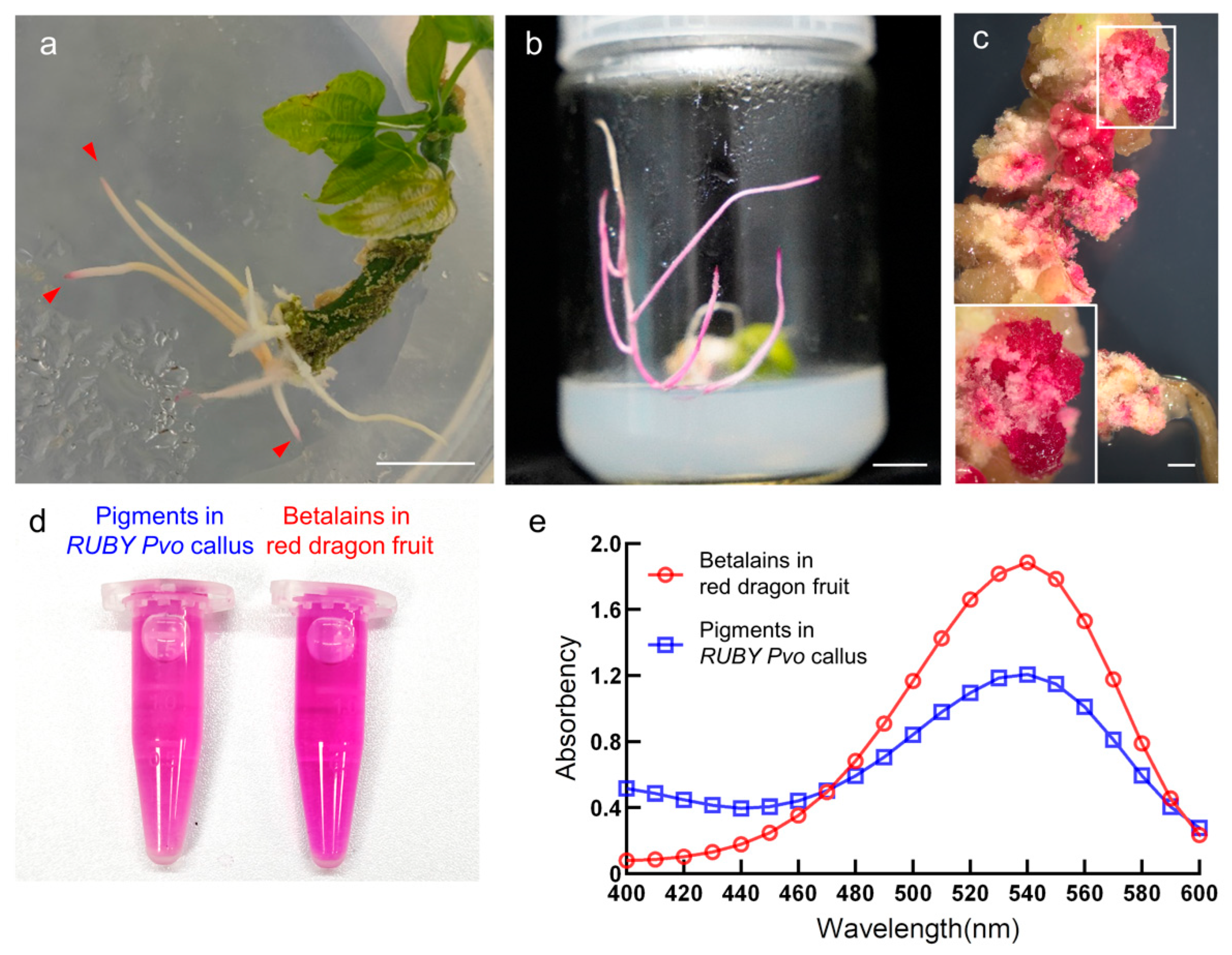
3.1. Ectopic Expression of RUBY Reconstructed the Betalain Biosynthesis Pathway in P. volubilis

To explore the potential applications of RUBY in the genetic transformation of woody plants, we transformed the hypocotyls of P. volubilis with DR5::RUBY by using A. rhizogenes strain K599. We found that only a low rate of RUBY transgenic hairy roots was reddish. DR5::RUBY was transformed into 20 explants, and its transgenic hairy roots were induced after approximately 3 weeks. Among them, a total of 30 hairy roots were induced from 14 explants, only 6 of which were red at the root tips, and the rest were white (Figure 1a). This might be because the expression of the DR5 promoter was affected by auxin concentration at the root tips, as previously reported [8,38,39]. Subsequently, to enhance the expression level of RUBY, the DR5 promoter was replaced by the 346 bp CaMV 35S promoter that did not contain an enhancer [30], which was widely used to drive the expression of selectable markers on the pCAMBIA series of vectors. Next, 35S346 bp::RUBY was transferred into young stems of P. volubilis mediated by A. rhizogenes strain K599. The red transgenic hairy roots were induced (Figure 1b), but the problem of low rates of red coloration remained (Supplementary Figure S1). We extracted pigments from RUBY transgenic callus (Figure 1c) and used red dragon fruit as a control, since red dragon fruit was commonly known as a primary source of betalain [37,40]. Both extracts were bright red (Figure 1d), and their absorption spectra measured with spectrophotometers showed that they had the same maximum absorption wavelength at A538 nm (Figure 1e), which indicates that the pigments from RUBY transgenic callus were most likely the same as those in red dragon fruit [37,40]. At the same time, the absorption peak of the pigment extracted from RUBY callus of P. volubilis was higher than that of red dragon fruit at 400–460 nm, which may contain other pigments. Previous studies have found a similar phenomenon, such as in tomatoes [13]. These results showed that the transformation of RUBY could reconstruct the betalain biosynthesis pathway in the hairy roots of P. volubilis.

3.2. Ectopic Expression of RUBY Has No Adverse Effects on the Growth and Development of Plants

To verify whether continuous expression of RUBY and consumption of tyrosine would affect the normal growth and development of plants, we first used RUBY transgenic red hairy roots of P. volubilis as explants to induce a large number of red calli on WPM medium containing 0.2 mg·L−1 IBA and 2 mg·L−1 6-BA (Figure 2a). Second, three chimeric P. volubilis plants containing red hairy roots were obtained by the ARM (A. rhizogenes-mediated) method [32] (Figure 2b), and the plants grew normally without any abnormalities under the growth conditions tested here. Third, 35S346 bp::RUBY was transferred into Col-0, and 10 resistant seedlings were obtained. PCR detection showed that nine of them were positive (Supplementary Figure S2), and #1–#7 RUBY transgenic Arabidopsis were selected for further analysis. The siliques of those transgenic Arabidopsis plants were dissected, and we did not observe seed abortion (Figure 2c). Finally, the leaves of N. benthamiana were used to perform the genetic transformation with A. tumefaciens strain GV3101, and approximately 29 red RUBY transgenic shoots were obtained (Supplementary Figure S3). These transgenic shoots rooted to form 13 complete plants, which were grown in soil (Figure 3a–c and Figure S4). Compared with the wild-type, RUBY transgenic N. benthamiana plants and their leaves, stems, flowers, fruits, and seeds showed no developmental defects except for the red coloration, due to the accumulation of betalain (Figure 3d–k). These experimental results indicate that the continuous consumption of tyrosine by overexpression of RUBY may not adversely affect plant growth, regeneration, or reproduction.

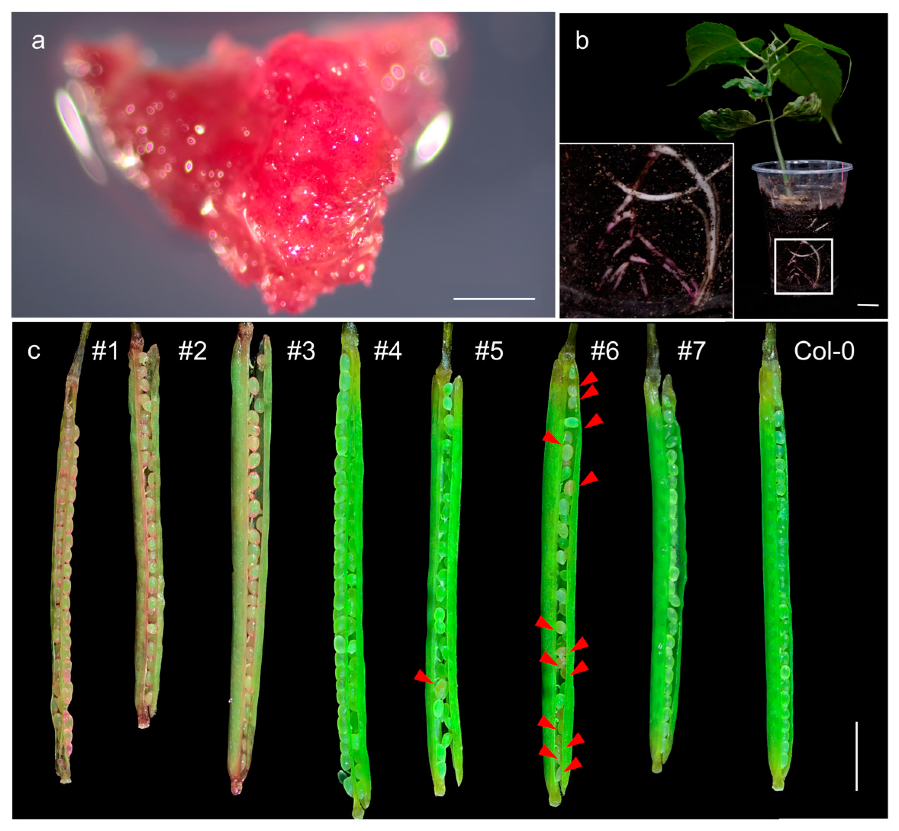
3.3. RUBY Transgenic Hairy Roots and Plants Showed Low Rates of Red Coloration

To explore whether the low rate of red coloration of RUBY was caused by species specificity, we used A. rhizogenes K599 harboring 35S346 bp::RUBY to infect the leaves of N. benthamiana and successfully induced a large number of hairy roots (Figure 4a,b), but the problem of a low rate of red coloration remained. We also found that some white hairy roots of N. benthamiana also grew normally in the selection medium with 15 mg·L−1 hygromycin. Therefore, we randomly selected nine white hairy roots and performed RT-PCR. After 25 cycles of PCR amplification, the transcripts of CYP76AD1, DODA, and glucosyl transferase were not detected in those hairy roots, but when the number of PCR cycles was increased to 30, three gene transcripts were detected except in the roots of WT, #1, and #2 (Figure 4c). It has been reported that when A. rhizogenes transferred the target gene into hairy roots, it simultaneously transferred auxin genes, such as rol A on the Ri plasmid, into the recipient cell genome, resulting in abnormal concentrations of auxin [41,42,43].
To further rule out the possibility that the low rate of red coloration was caused by the characteristics of transgenic hairy roots, we observed nine lines of 35S346 bp::RUBY transgenic Arabidopsis generated with A. tumefaciens and found that only 33.3% were colored. We selected seeds of lines #3 (fully colored), #4 (noncolored), and #6 (semicolored) from transgenic Arabidopsis and sowed them on vertical plates that contained 25 mg·L−1 hygromycin. After 30 days of growth, 15 fully colored plants of RUBY transgenic Arabidopsis line #3 had a total of 216 roots (including taproots and primary lateral roots), of which only 42.1% were red roots, and the remaining 57.9% of roots were white or almost white. In the semicolored RUBY transgenic Arabidopsis line #6, only 5 fully colored plants and the remaining 10 plants were semicolored or noncolored. These plants grew a total of 187 roots, among which only 18.7% of roots were red, and the remaining 81.3% of roots were white. The rate of red coloration of roots was distinctly lower than that in line #3. A total of 240 roots grew from the 15 plants of noncolored line #4, all of which were white. The elongation and number of roots under dark conditions were much lower than those under 16 h light/8 h dark cycles. Nineteen red roots and 28 white roots grew from 15 plants of RUBY transgenic Arabidopsis line #3, while 10 red roots and 39 white roots grew from line #6, and 49 white roots grew from line #4 (Figure 4d,e). The rates of red coloration of RUBY transgenic Arabidopsis were low under both planting conditions, which was consistent with the phenomenon observed in P. volubilis and N. benthamiana. The above experiments showed that RUBY, as a “noninvasive” naked-eye visualization reporter, did have advantages in genetic transformation, but it had the problem of a low rate of red coloration in transgenic hairy roots and plants, which had not been previously reported.

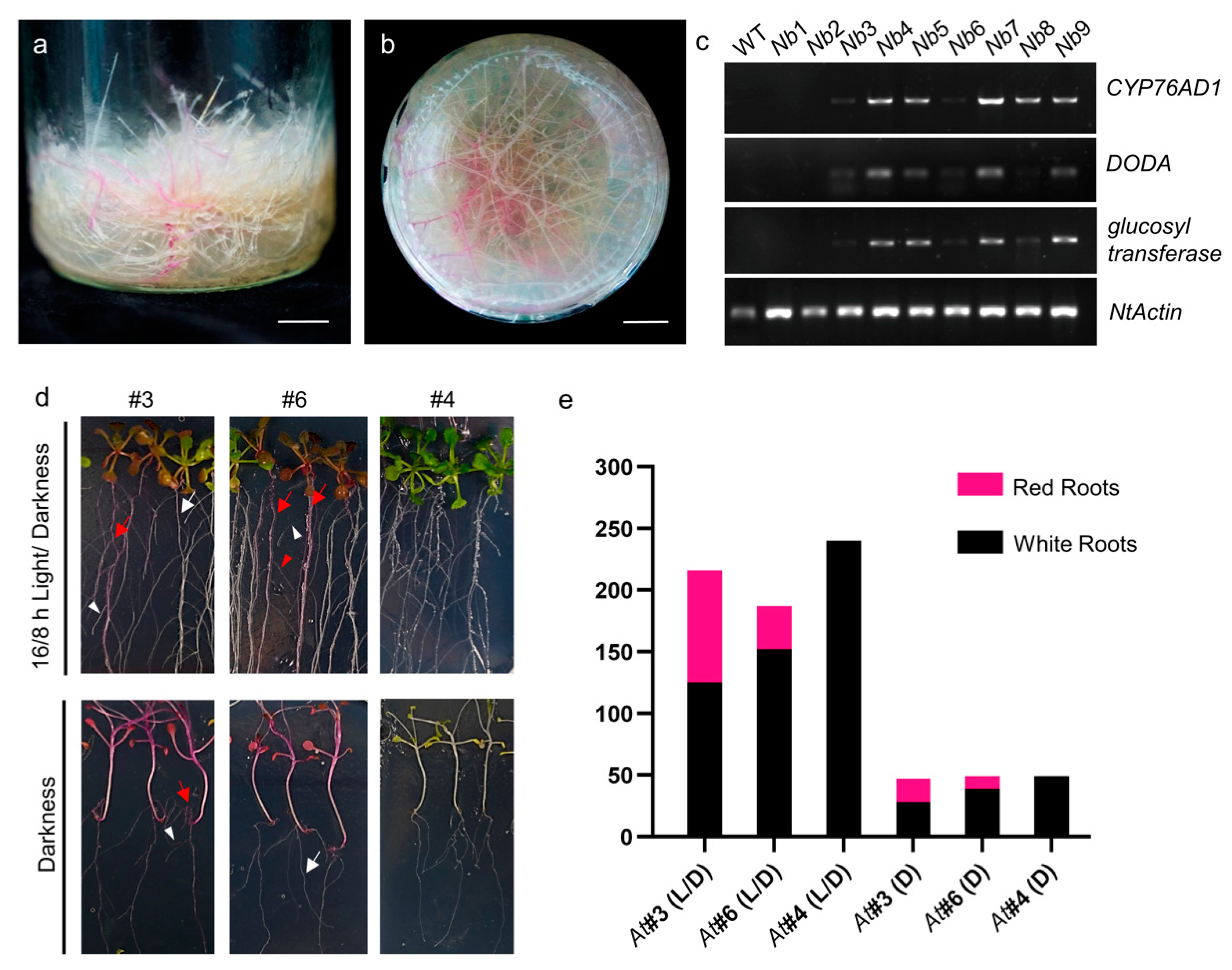
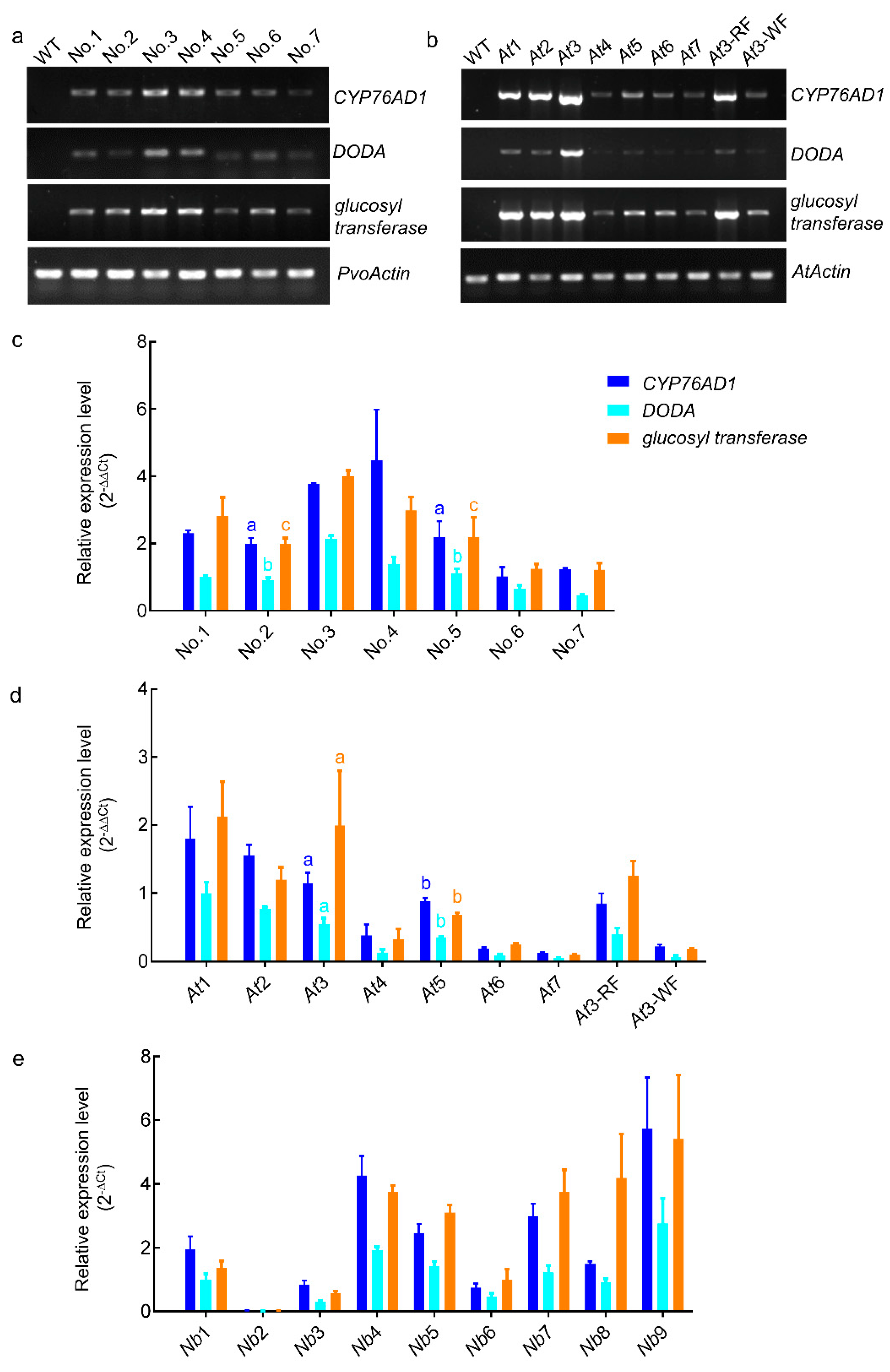
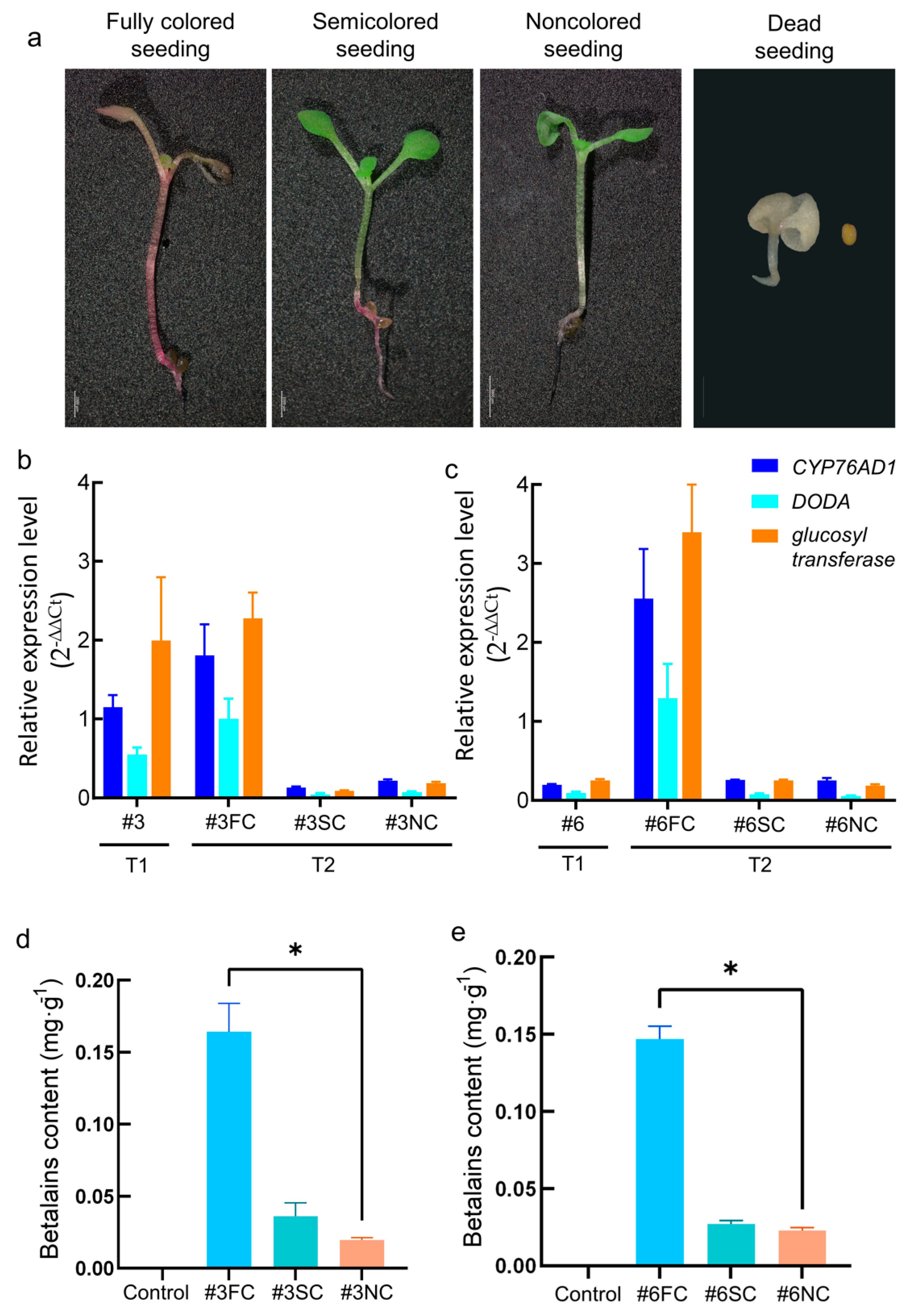
3.4. The Expression Level of RUBY Is Positively Correlated with Red Coloration

We found that one of the RUBY transgenic hairy roots obtained from P. volubilis turned from white to red during subculture: the primary root was white, but the whole or part of the secondary lateral roots were red (Supplementary Figure S5). To explore the reason for the different red coloration in this RUBY transgenic hairy root, we divided it into seven regions for RT−PCR detection. The transcripts of CYP76AD1, DODA, and glucosyl transferase were detected in regions no. 1–4 and region no. 6 after 25 cycles of PCR amplification, but region no. 5 and region no. 7 had lower levels (Figure 5a).
Among the T1 generation of RUBY transgenic Arabidopsis, only lines #1, #2, and #3 had red siliques and seeds. The siliques of line #6 were as green as those of the wild-type, but some seeds were red (Figure 2c). In addition, we also found that the fully colored line #3 had white inflorescence and green siliques that were consistent with those of the wild-type at a later stage of growth (Supplementary Figure S6). We selected RUBY transgenic Arabidopsis lines #1–#6, and inflorescences on line #6 for RT−PCR detection, and the results showed that CYP76AD1, DODA, and glucosyl transferase were expressed in all transgenic lines: lines #1, #2, and #3 had high expression levels, while lines #4, #5, and #6 had lower expression levels; and the same results in inflorescences of line #6 (Figure 5b).
To further quantitatively analyze the relationship between the expression of RUBY genes and red coloration, we first detected the expression levels of CYP76AD1, DODA, and glucosyl transferase in the transgenic hairy roots of P. volubilis and transgenic Arabidopsis inflorescences by RT−qPCR. The expression of the three genes mentioned above could be detected in RUBY transgenic hairy roots of P. volubilis in each region, and the expression levels of those genes in regions no. 1–3 were higher than those in regions no. 5–7 (Figure 5c). However, the expression levels of those genes in region no. 2 had no significant difference from that of region no. 5. In Arabidopsis, the expression levels of the three genes in colored lines #1, #2, and #3 were higher than those in the noncolored lines, while in the white inflorescence of line #3, the expression levels of those genes were as low as those in the noncolored lines (Figure 5d). The expression level in line #3 was the lowest among those of the colored lines, and was significantly different from that of line #5, which had the highest expression among the noncolored lines.
In addition, we used RT−qPCR to detect the expression of RUBY in nine white transgenic hairy roots of N. benthamiana (Figure 5e). The results showed that three genes comprising RUBY were detected in all roots except no. 2, and the expression abundance was less than 0.08 times that of Actin (Figure 5e). The red coloration of nine white transgenic hairy roots could not be observed with the naked eye, probably due to the relatively low levels of expression. Based on the results of the above experiments, we speculated that the high expression of RUBY would lead to the accumulation of betalain and red coloration in transgenic plants.

3.5. Instability of RUBY Expression in Transgenic Arabidopsis

According to a previous study, the fusion gene design of RUBY might cause abnormal transcription events, and there might be unknown risks in inheritance in the use of RUBY as a reporter [3]. We counted and calculated the rates of red coloration of Arabidopsis T2 seedlings in three fully colored lines (#1, #2, and #3) and three noncolored lines (#4, #5, and #6) (Supplementary Figure S7), and the results are shown in Table 1. The results showed that the ratios of surviving seedlings to dead seedlings from #1–#6 lines were all close to 3:1. It suggests that a single RUBY transgene could be stably passed on to the next generation according to Mendelian inheritance. However, the offspring of the fully colored lines #1, #2, and #3 had a considerable amount of semicolored seedlings with only the hypocotyls or roots colored (Figure 6a), which accounted for approximately 6.36% and 11.44% of lines #1 and #2, respectively; line #3 had simultaneous red and white inflorescences in the T1 generation, and the proportion of semicolored seedlings in the T2 generation was slightly higher (approximately 19.1%), but not significantly different from that of lines #1 and #2. In addition, statistics showed that the offspring of noncolored line #6 included fully colored, semicolored, noncolored, and dead seedlings, and the ratio of those four kinds of seedlings was close to 1:2:1:1. Moreover, it was found that two seedlings among the offspring of noncolored line #5 were colored (Figure 2f and Figure S7). We speculated that the expression of RUBY might be unstable in different generations of Arabidopsis.
To further explore the relationship between RUBY expression stability and red coloration in heredity, two representative RUBY transgenic Arabidopsis lines, #3 and #6, were selected for further analysis. First, the RT−qPCR analysis showed that the expression levels of CYP76AD1, DODA, and glucosyl transferase in fully colored seedlings of the T2 generation of line #3 were the same as those of the T1 generation (Figure 6b). The expression levels of three genes in the T1 generation of line #6 were very low, but they were highly expressed in the T2 generation of fully colored seedling (Figure 6c). Moreover, we found that the expression levels of the three genes were very low in the semicolored and noncolored seedlings of the T2 generation of lines #3 and #6, and they showed no significant difference (Figure 6b,c). Second, we extracted betalain from the fully colored, semicolored, and noncolored seedlings in the T2 generation of lines #3 and #6. The results showed that the contents of betalain were consistent with RT−qPCR detection: the betalain contents of the fully colored seedlings were high, while the semicolored and noncolored seedlings had lower contents, and the difference between the latter two was not significant (Figure 6d,e). The above experiments further proved that the inconsistent red coloration in RUBY transgenic Arabidopsis was caused by its unstable expression. It is implied that the RUBY fusion gene might have transcriptional abnormalities in transgenic Arabidopsis, but it could also be due to RNA processing or other post-transcriptional regulation not known for RUBY.

4. Discussion

In this study, the reporter RUBY was used to reconstruct the betalain biosynthesis pathway in transgenic hairy roots of P. volubilis and in the model plants Arabidopsis and N. benthamiana to evaluate the potential applications of RUBY in plant genetic transformation.

4.1. RUBY Has Broad Prospects as a Naked-Eye Visual Screening Reporter

As a noninvasive reporter, RUBY is not only convenient for observation, but also does not affect callus induction, plant regeneration, or fertility. Generally, it has advantages in the genetic transformation of plants, allowing a nondestructive, easily visible evaluation of tissues without the need for fluorescence or light imaging, which can improve efficiency of genetic transformation and reduce the cost. Furthermore, due to the relatively simple synthetic pathway for betalains, RUBY has the potential to be applied on more plant species. Currently, the genetic transformation of woody plants is still relatively difficult [43]. The establishment of root transgenic systems mediated by A. rhizogenes has been widely used in studies of secondary metabolites and gene function. Therefore, the selection of an appropriate reporter is essential for the establishment of transgenic hairy roots [3,26,44,45]. Traditional selection reporters in genetic transformation, that generally use antibiotics and herbicides, have disadvantages for the growth of plant roots [46,47,48], and various fluorescent proteins or GUS require special equipment or expensive chemical substrates, which makes them inconvenient to observe [49,50]. Anthocyanin has been used as a naked-eye visual reporter, but its synthesis pathway involving at least eight genes is complex, and there were great differences between species [48,50,51,52,53]. Compared with the abovementioned reporters, RUBY is a novel, low-cost reporter that does not need instrument assistance and is an excellent alternative reporter in plant genetic transformation.

4.2. The Risk of RUBY in Genetic Transformation

As shown above (Supplementary Figure S1, Figure 2c and Figure 4), a low rate of red coloration was observed in transgenic hairy roots of P. volubilis and N. benthamiana and Arabidopsis plants, in which the CaMV 35S promoter was used to drive the expression of RUBY. It was caused by variable expression of RUBY or instability of betalains. RT−qPCR analysis showed that the red coloration of RUBY was directly related to the level of expression. Although the CaMV 35S promoter has been widely used as a constitutive promoter to study gene function, there are differences in expression abundance in different tissues and abiotic stresses at each developmental stage of plants [54,55,56]. This was a good explanation for each of the regions that had different types of red coloration in the same transgenic hairy root of P. volubilis; white inflorescence appeared in fully colored transgenic Arabidopsis line #3, and small numbers of red seeds appeared in noncolored line #6, which might be caused by differential expression of RUBY driven by the CaMV 35S promoter. At the same time, the expression levels of RUBY in regions “no. 2” and “no. 5” were not significantly different (Figure 5c), but the coloration difference was obvious, probably due to the different accumulation of betalain in different tissues. It was reported that betalain accumulated differently in various tissues, affected by phytohormone and light conditions [57,58,59].
On the other hand, using a specific promoter to drive the expression of CYP76AD1, DODA, and glucosyl transferase in plants could enable the synthesis and accumulation of betalain in specific tissues [4,8]. However, we found that CYP76AD1, DODA, and glucosyl transferase, which constituted RUBY, were expressed at varying degrees in transgenic white hairy roots of P. volubilis and N. benthamiana, as well as the transgenic Arabidopsis lines and the white inflorescence of line #3. Relatively low expression of RUBY leads to insufficient accumulation of betalain and a noncolored plant, indicating that RUBY might have lower sensitivity than GUS and LUC [60,61], presenting the problem of false-negatives as a reporter. These results suggest that RUBY as a reporter requires a strong constitutive promoter to drive in transgenic plants, and a specific promoter or an inducible promoter used to drive RUBY may result in a reduced screening efficiency.

4.3. RUBY as a Reporter Needs Further Optimizations

Betalain has begun, in recent years, to be used as a reporter in studies on Arabidopsis and other species. However, we found that some tissues and plants with high expression of RUBY did not show enough red coloration. This may be a common problem with pigment-based reporters. For example, when MdMYB10 was used as a reporter for promoter studies, differences between the transgenic plants and the wild-type could hardly be distinguished in the case of a specific weak promoter, particularly in tissues showing a strongly colored background, such as green leaves. Even when using the 35S strong promoter to trigger plant coloration, not all the plants showed uniform coloration [62], and comparable results were shown in our study.
On the other hand, the accumulation of betalain depends on expression levels of biosynthetic genes and the stability of betalains. As a natural pigment, betalain is reported to have poor stability and is easily affected by factors such as pH, light, and temperature [63,64,65]. In this study, we compared the red coloration rates of Arabidopsis seedlings cultivated in a light–dark cycle and total darkness, and the red coloration of seedlings under different lighting conditions had no significant difference. However, this does not rule out the possibility that other factors we mentioned above affect betalain stability and accumulation as a reporter during the experiments, and further studies are needed to confirm this hypothesis.
The RNA expression levels of the three genes that make up the RUBY reporter gene system were variable in transgenic P. volubilis and N. benthamiana hairy roots, and transgenic Arabidopsis plants. The statistical data showed that pigment production and RNA expression over two generations of transgenic Arabidopsis were unstable. It is supposed that 2A peptide-mediated “self-cleavage” or ribosomal “skipping” to cleave multiple proteins does not always work efficiently [3]. There is more possibility: some unexpected post-transcriptional processes might happen in RUBY “polycistronic” mRNA for the three genes. Recent emerging evidence has shown that polycistronic mRNAs are also transcribed in eukaryotes. In Arabidopsis, 271 polycistronic loci have been identified and the post-transcriptional regulation for these loci remains to be elucidated [66]. Therefore, it is necessary to further fully understand the RUBY system and optimize it for wider use.

5. Conclusions

RUBY is a naked-eye visual reporter that has obvious advantages, such as a simpler synthesis mechanism, an easier experimental operation, and a lower cost in the genetic transformation of plants, and might be used as a high-quality reporter for the genetic engineering breeding of woody plants. However, the expression of this system was shown to be unstable in different generations of transgenic plants, which might be caused by post-transcriptional regulation. In order to achieve red coloration, the amount of accumulated betaines might need to reach a certain threshold. The sensitivity of RUBY system presents the problem of false-negatives as a reporter, but it still has broad potential application prospects after improvement.

Supplementary Materials

The following supporting information can be downloaded at https://www.mdpi.com/article/10.3390/f14030637/s1: Figure S1: Low red coloration of RUBY transgenic hairy roots of P. volubilis induced by A. rhizogenes K599. Figure S2: PCR identification of RUBY transgenic Arabidopsis lines. Figure S3: RUBY transgenic shoots of N. benthamiana induced by leaf-disk transformation methods. Figure S4: RUBY transgenic N. benthamiana plants grown in the pots. Figure S5: A nonuniform red coloration of RUBY transgenic hairy root of P. volubilis. Figure S6: Characterization of RUBY transgenic Arabidopsis. Figure S7: RUBY transgenic Arabidopsis offspring screening and red coloration.

Author Contributions

Conceptualization, Y.W. and Z.-F.X.; methodology, Y.W.; software, J.Y., S.D. and H.H.; validation, J.Y. and S.D.; formal analysis, J.Y.; investigation, J.Y., S.D., Y.W. and Z.-F.X.; resources, J.Y.; data curation, J.M.; writing—original draft preparation, J.Y. and Y.W.; writing—review and editing, Y.W. and Z.-F.X.; visualization, J.Y.; supervision, H.H. and J.M.; project administration, H.H. and J.M.; funding acquisition, J.Y., S.D. and H.H. All authors have read and agreed to the published version of the manuscript.

Funding

This work was supported by startup research funds from Guangxi University, Project of Guangxi Natural Science Foundation (2022GXNSFAA035491), and Key Research and Development Projects of Nanning (No. 20182005-1).

Institutional Review Board Statement

Not applicable.

Informed Consent Statement

Not applicable.

Data Availability Statement

Not applicable.

Acknowledgments

We would like to thank He Yubing of Nanjing Agricultural University for providing the DR5::RUBY vector, and Meng Dong of Beijing Forestry University for providing the A. rhizogenes K599 strain.

Conflicts of Interest

The authors declare no conflict of interest.

References

  1. Tanaka, Y.; Sasaki, N.; Ohmiya, A. Biosynthesis of plant pigments: Anthocyanins, betalains and carotenoids. Plant J. 2008, 54, 733–749. [Google Scholar] [CrossRef] [PubMed]
  2. Gandía-Herrero, F.; García-Carmona, F. Biosynthesis of betalains: Yellow and violet plant pigments. Trends Plant Sci. 2013, 18, 334–343. [Google Scholar] [CrossRef] [PubMed]
  3. Kiryushkin, A.S.; Ilina, E.L.; Guseva, E.D.; Pawlowski, K.; Demchenko, K.N. Hairy CRISPR: Genome editing in plants using hairy root transformation. Plants 2021, 11, 51. [Google Scholar] [CrossRef]
  4. Timoneda, A.; Yunusov, T.; Quan, C.; Gavrin, A.; Brockington, S.F.; Schornack, S. MycoRed: Betalain pigments enable in vivo real-time visualisation of arbuscular mycorrhizal colonisation. PLoS Biol. 2021, 19, e3001326. [Google Scholar] [CrossRef] [PubMed]
  5. Kumar, A.; Lin, H.; Li, Q.; Ruan, Y.; Cousins, D.; Li, F.; Gao, S.; Jackson, K.; Wen, J.; Murray, J.D.; et al. Anthocyanin pigmentation as a quantitative visual marker for arbuscular mycorrhizal fungal colonization of Medicago truncatula Roots. New Phytol. 2022, 236, 1988–1998. [Google Scholar] [CrossRef]
  6. Polturak, G.; Breitel, D.; Grossman, N.; Sarrion-Perdigones, A.; Weithorn, E.; Pliner, M.; Orzaez, D.; Granell, A.; Rogachev, I.; Aharoni, A. Elucidation of the first committed step in betalain biosynthesis enables the heterologous engineering of betalain pigments in plants. New Phytol. 2015, 210, 269–283. [Google Scholar] [CrossRef] [Green Version]
  7. Zhang, L.; Liu, X.; Li, J.; Meng, Y.; Zhao, G.R. Improvement of betanin biosynthesis in Saccharomyces cerevisiae by metabolic engineering. Synth. Syst. Biotechnol. 2023, 8, 54–60. [Google Scholar] [CrossRef] [PubMed]
  8. He, Y.; Zhang, T.; Sun, H.; Zhan, H.; Zhao, Y. A reporter for noninvasively monitoring gene expression and plant transformation. Hortic. Res. 2020, 7, 152. [Google Scholar] [CrossRef]
  9. Tian, Y.S.; Fu, X.Y.; Yang, Z.Q.; Wang, B.; Gao, J.J.; Wang, M.Q.; Xu, J.; Han, H.J.; Li, Z.J.; Yao, Q.H.; et al. Metabolic engineering of rice endosperm for betanin biosynthesis. New Phytol. 2019, 225, 1915–1922. [Google Scholar] [CrossRef]
  10. Harris, N.N.; Javellana, J.; Davies, K.M.; Lewis, D.H.; Jameson, P.E.; Deroles, S.C.; Calcott, K.E.; Gould, K.S.; Schwinn, K.E. Betalain production is possible in anthocyanin-producing plant species given the presence of DOPA-dioxygenase and L-DOPA. BMC Plant Biol. 2012, 12, 34. [Google Scholar] [CrossRef] [Green Version]
  11. Polturak, G.; Grossman, N.; Vela-Corcia, D.; Dong, Y.; Nudel, A.; Pliner, M.; Levy, M.; Rogachev, I.; Aharoni, A. Engineered gray mold resistance, antioxidant capacity, and pigmentation in betalain-producing crops and ornamentals. Proc. Natl. Acad. Sci. USA 2017, 114, 9062–9067. [Google Scholar] [CrossRef] [PubMed] [Green Version]
  12. Clement, J.S.; Mabry, T.J.B.A. Pigment evolution in the Caryophyllales: A systematic overview. Bot. Acta 1996, 109, 360–367. [Google Scholar] [CrossRef]
  13. Grützner, R.; Schubert, R.; Horn, C.; Yang, C.; Vogt, T.; Marillonnet, S. Engineering betalain biosynthesis in tomato for high level betanin production in fruits. Front. Plant Sci. 2021, 12, 682443. [Google Scholar] [CrossRef]
  14. Alagarsamy, K.; Shamala, L.F.; Wei, S. Protocol: High-efficiency in-planta Agrobacterium-mediated transgenic hairy root induction of Camellia sinensis Var. sinensis. Plant Methods 2018, 14, 17. [Google Scholar] [CrossRef]
  15. Chen, K.; Hu, K.; Xi, F.; Wang, H.; Kohnen, M.V.; Gao, P.; Liao, J.; Wei, W.; Liu, X.; Zhang, H.; et al. High-efficient and transient transformation of Moso Bamboo (Phyllostachys edulis) and Ma Bamboo (Dendrocalamus latiflorus Munro). J. Plant Biol. 2023, 66, 75–86. [Google Scholar] [CrossRef]
  16. Cao, X.; Xie, H.; Song, M.; Lu, J.; Ma, P.; Huang, B.; Wang, M.; Tian, Y.; Chen, F.; Peng, J.; et al. Cut–dip–budding delivery system enables genetic modifications in plants without tissue culture. Innovation 2023, 4, 100345. [Google Scholar] [CrossRef]
  17. Mei, G.; Chen, A.; Wang, Y.; Li, S.; Wu, M.; Liu, X.; Hou, X. A simple and efficient in planta transformation method based on the active regeneration capacity of plants. bioRxiv 2023. [Google Scholar] [CrossRef]
  18. Ge, X.; Wang, P.; Wang, Y.; Wei, X.; Chen, Y.; Li, F. Development of an eco-friendly pink cotton germplasm by engineering betalain biosynthesis pathway. Plant Biotechnol. J. 2023, in press. [Google Scholar] [CrossRef]
  19. Kodahl, N. Sacha inchi (Plukenetia volubilis L.)-from lost crop of the Incas to part of the solution to global challenges? Planta 2020, 251, 80. [Google Scholar] [CrossRef]
  20. Wang, S.; Zhu, F.; Kakuda, Y. Sacha inchi (Plukenetia volubilis L.): Nutritional composition, biological activity, and uses. Food Chem. 2018, 265, 316–328. [Google Scholar] [CrossRef] [PubMed]
  21. Ángel, M.R.C.; German, G.A.; María, F.S.M.; Kodahl, N. Ethnobotanical knowledge in the peruvian amazon of the neglected and underutilized crop Sacha Inchi (Plukenetia volubilis L.). Econ. Bot. 2019, 73, 281–287. [Google Scholar]
  22. Fu, Q.; Niu, L.; Zhang, Q.; Pan, B.Z.; He, H.; Xu, Z.F. Benzyladenine treatment promotes floral feminization and fruiting in a promising oilseed crop Plukenetia volubilis. Ind. Crops Prod. 2014, 59, 295–298. [Google Scholar] [CrossRef] [Green Version]
  23. Fu, Q.; Niu, L.; Chen, M.S.; Tao, Y.B.; Wang, X.; He, H.; Pan, B.Z.; Xu, Z.F. De novo transcriptome assembly and comparative analysis between male and benzyladenine-induced female inflorescence buds of Plukenetia Volubilis. J. Plant Physiol. 2018, 221, 107–118. [Google Scholar] [CrossRef] [PubMed]
  24. Hu, X.D.; Pan, B.Z.; Fu, Q.; Niu, L.; Chen, M.S.; Xu, Z.F. De novo transcriptome assembly of the eight major organs of Sacha Inchi (Plukenetia Volubilis) and the identification of genes involved in α-linolenic acid metabolism. BMC Genom. 2018, 19, 380. [Google Scholar] [CrossRef] [PubMed] [Green Version]
  25. Kereszt, A.; Li, D.; Indrasumunar, A.; Nguyen, C.D.T.; Nontachaiyapoom, S.; Kinkema, M.; Gresshoff, P.M. Agrobacterium rhizogenes-mediated transformation of soybean to study root biology. Nat. Protoc. 2007, 2, 948–952. [Google Scholar] [CrossRef] [PubMed]
  26. Meng, D.; Yang, Q.; Dong, B.; Song, Z.; Niu, L.; Wang, L.; Cao, H.; Li, H.; Fu, Y. Development of an efficient root transgenic system for pigeon pea and its application to other important economically plants. Plant Biotechnol. J. 2019, 17, 1804–1813. [Google Scholar] [CrossRef] [Green Version]
  27. Cui, M.L.; Liu, C.; Piao, C.L.; Liu, C.L. A stable Agrobacterium rhizogenes-mediated transformation of cotton (Gossypium hirsutum L.) and plant regeneration from transformed hairy root via embryogenesis. Front. Plant Sci. 2020, 11, 604255. [Google Scholar] [CrossRef]
  28. Qin, Y.; Wang, D.; Fu, J.; Zhang, Z.; Qin, Y.; Hu, G.; Zhao, J. Agrobacterium rhizogenes-mediated hairy root transformation as an efficient system for gene function analysis in Litchi chinensis. Plant Methods 2021, 17, 103. [Google Scholar] [CrossRef]
  29. Maher, M.; Nasti, R.; Vollbrecht, M.; Starker, C.; Clark, M.; Voytas, D. Plant gene editing through de novo induction of meristems. Nat. Biotechnol. 2020, 38, 84–89. [Google Scholar] [CrossRef]
  30. Odell, J.T.; Nagy, F.; Chua, N.H. Identification of DNA sequences required for activity of the cauliflower mosaic virus 35S promoter. Nature 1985, 313, 810–812. [Google Scholar] [CrossRef]
  31. Holsters, M.; de Waele, D.; Depicker, A.; Messens, E.; van Montagu, M.; Schell, J. Transfection and transformation of Agrobacterium tumefaciens. Mol. Genet. Genom. 1978, 163, 181–187. [Google Scholar] [CrossRef]
  32. Fan, Y.L.; Zhang, X.H.; Zhong, L.J.; Wang, X.Y.; Jin, L.S.; Lyu, S.H. One-step generation of composite soybean plants with transgenic roots by Agrobacterium rhizogenes-mediated transformation. BMC Plant Biol. 2020, 20, 208. [Google Scholar] [CrossRef] [PubMed]
  33. Clough, S.; Bent, A.F. Floral dip: A simplified method for Agrobacterium-mediated transformation of Arabidopsis thaliana. Plant J. 1998, 16, 735–743. [Google Scholar] [CrossRef] [PubMed] [Green Version]
  34. Jia, Z.; Ding, M.; Nakano, M.; Hong, K.; Huang, R.; Becker, D.; Glazebrook, J.; Katagiri, F.; Han, X.; Tsuda, K. Letter to the editor: DNA purification-free PCR from plant tissues. Plant Cell Physiol. 2021, 62, 1503–1505. [Google Scholar] [CrossRef] [PubMed]
  35. Arocho, A.; Chen, B.; Ladanyi, M.; Pan, Q. Validation of the 2-DeltaDeltaCt calculation as an alternate method of data analysis for quantitative PCR of BCR-ABL P210 transcripts. Diagn. Mol. Pathol. 2006, 15, 56–61. [Google Scholar] [CrossRef]
  36. Cai, Y.; Sun, M.; Corke, H. Identification and distribution of simple and acylated betacyanins in the Amaranthaceae. J. Agric. Food Chem. 2001, 49, 1971–1978. [Google Scholar] [CrossRef]
  37. Herbach, K.M.; Maier, C.; Stintzing, F.C.; Carle, R. Effects of processing and storage on juice colour and betacyanin stability of purple pitaya (Hylocereus polyrhizus) juice. Eur. Food Res. Technol. 2007, 224, 649–658. [Google Scholar] [CrossRef]
  38. Cheng, Y.; Dai, X.; Zhao, Y. Auxin biosynthesis by the YUCCA flavin monooxygenases controls the formation of floral organs and vascular tissues in Arabidopsis. Gene Dev. 2006, 20, 1790–1799. [Google Scholar] [CrossRef] [Green Version]
  39. Chen, Y.; Yordanov, Y.S.; Ma, C.; Strauss, S.; Busov, V.B. DR5 as a reporter system to study auxin response in Populus. Plant Cell Rep. 2013, 32, 453–463. [Google Scholar] [CrossRef]
  40. Choo, K.Y.; Ong, Y.Y.; Lim, R.L.H.; Tan, C.P.; Ho, C.W. Study on bioaccessibility of betacyanins from red dragon fruit (Hylocereus Polyrhizus). Food Sci. Biotechnol. 2019, 28, 1163–1169. [Google Scholar] [CrossRef]
  41. Piątczak, E.; Kuźma, Ł.; Skała, E.; Żebrowska, M.; Balcerczak, E.; Wysokińska, H. Iridoid and phenylethanoid glycoside production and phenotypical changes in plants regenerated from hairy roots of Rehmannia glutinosa Libosch. Plant Cell Tissue Organ Cult. 2015, 122, 259–266. [Google Scholar] [CrossRef] [Green Version]
  42. Boobalan, S.; Kamalanathan, D. Tailoring enhanced production of aervine in Aerva lanata (L.) Juss. Ex Schult by Agrobacterium rhizogenes- mediated hairy root cultures. Ind. Crop. Prod. 2020, 155, 112814. [Google Scholar] [CrossRef]
  43. Piątczak, E.; Jeleń, A.; Makowczyńska, J.; Zielińska, S.; Kuźma, Ł.; Balcerczak, E. Establishment of hairy root cultures of Rehmannia elata N.E. Brown ex Prain and production of iridoid and phenylethanoid glycosides. Ind. Crop. Prod. 2019, 137, 308–314. [Google Scholar] [CrossRef]
  44. Osakabe, Y.; Sugano, S.S.; Osakabe, K. Genome engineering of woody plants: Past, present and future. J. Wood Sci. 2016, 62, 217–225. [Google Scholar] [CrossRef] [Green Version]
  45. Habibi, P.; de Sa, M.F.G.; da Silva, A.L.L.; Makhzoum, A.; da Costa, J.L.; Borghetti, I.A.; Soccol, C.R. Efficient genetic transformation and regeneration system from hairy root of Origanum vulgare. Physiol. Mol. Biol. Plants 2016, 22, 271–277. [Google Scholar] [CrossRef] [Green Version]
  46. Gantait, S.; Mukherjee, E. Hairy root culture technology: Applications, constraints and prospect. Appl. Microbiol. Biot. 2021, 105, 35–53. [Google Scholar] [CrossRef] [PubMed]
  47. Velini, E.D.; Trindade, M.; Duke, B.S.O. Growth regulation and other secondary effects of herbicides. Weed Sci. 2010, 58, 351–354. [Google Scholar] [CrossRef]
  48. Wang, X.; Ryu, D.; Houtkooper, R.H.; Auwerx, J. Antibiotic use and abuse: A threat to mitochondria and chloroplasts with impact on research, health, and environment. BioEssays 2015, 37, 1045–1053. [Google Scholar] [CrossRef]
  49. Liang, J.; He, J. Protective role of anthocyanins in plants under low nitrogen stress. Biochem. Biophys. Res. Commun. 2018, 498, 946–953. [Google Scholar] [CrossRef]
  50. Jefferson, R.A.; Kavanagh, T.A.; Bevan, M.W. GUS fusions: Beta-glucuronidase as a sensitive and versatile gene fusion marker in higher plants. EMBO J. 1987, 6, 3901–3907. [Google Scholar] [CrossRef]
  51. Mitiouchkina, T.; Mishin, A.; Somermeyer, L.; Markina, N.; Chepurnyh, T.; Guglya, E.; Karataeva, T.; Palkina, K.; Shakhova, E.; Fakhranurova, L.; et al. Plants with genetically encoded autoluminescence. Nat. Biotechnol. 2020, 38, 1000–1001. [Google Scholar] [CrossRef]
  52. Zhu, Q.; Yu, S.; Zeng, D.; Liu, H.; Wang, H.; Yang, Z.; Xie, X.; Shen, R.; Tan, J.; Li, H.; et al. Development of “Purple Endosperm Rice” by engineering anthocyanin biosynthesis in the endosperm with a high-efficiency transgene stacking system. Mol. Plant 2017, 10, 918–929. [Google Scholar] [CrossRef] [PubMed] [Green Version]
  53. Zheng, J.; Wu, H.; Zhu, H.; Huang, C.; Liu, C.; Chang, Y.; Kong, Z.; Zhou, Z.; Wang, G.; Lin, Y.; et al. Determining factors, regulation system, and domestication of anthocyanin biosynthesis in rice leaves. New Phytol. 2019, 223, 705–721. [Google Scholar] [CrossRef] [PubMed]
  54. He, Y.; Zhu, M.; Wu, J.; Ouyang, L.; Wang, R.; Sun, H.; Yan, L.; Wang, L.; Xu, M.; Zhan, H.; et al. Repurposing of anthocyanin biosynthesis for plant transformation and genome editing. Front. Genome Ed. 2020, 2, 607982. [Google Scholar] [CrossRef] [PubMed]
  55. Sunilkumar, G.; Mohr, L.A.; Lopata-Finch, E.; Emani, C.; Rathore, K.S. Developmental and tissue-specific expression of CaMV35S promoter in cotton as revealed by GFP. Plant Mol. Biol. 2002, 50, 463–474. [Google Scholar] [CrossRef]
  56. Amack, S.C.; Antunes, M.S. CaMV35S promoter—A plant biology and biotechnology workhorse in the era of synthetic biology. Curr. Plant Biol. 2020, 24, 100179. [Google Scholar] [CrossRef]
  57. Zhao, S.Z.; Sun, H.Z.; Chen, M.; Wang, B.S. Light-regulated betacyanin accumulation in Euhalophyte Suaeda Salsa Calli. Plant Cell Tissue Organ Cult. 2010, 102, 99–107. [Google Scholar] [CrossRef]
  58. Shin, K.S.; Murthy, H.N.; Heo, J.W.; Paek, K.Y. Induction of betalain pigmentation in hairy roots of red beet under different radiation sources. Biol. Plant. 2003, 47, 149–152. [Google Scholar] [CrossRef]
  59. Zheng, X.; Liu, S.; Cheng, C.; Guo, R.; Chen, Y.; Xie, L.; Mao, Y.; Lin, Y.; Zhang, Z.; Lai, Z. Cloning and expression analysis of betalain biosynthesis genes in Amaranthus Tricolor. Biotechnol. Lett. 2016, 38, 723–729. [Google Scholar] [CrossRef]
  60. Kiselev, K.; Aleynova, O.; Ogneva, Z.; Suprun, A.; Dubrovina, A. 35S promoter-driven transgenes are variably expressed in different organs of Arabidopsis thaliana and in response to abiotic stress. Mol. Biol. Rep. 2021, 48, 2235–2241. [Google Scholar] [CrossRef]
  61. Jefferson, R.A. The GUS reporter gene system. Nature 1989, 342, 837–838. [Google Scholar] [CrossRef]
  62. Koo, J.; Kim, Y.; Kim, J.; Yeom, M.; Lee, I.C.; Nam, H.G. A GUS/Luciferase fusion reporter for plant gene trapping and for assay of promoter activity with luciferin-dependent control of the reporter protein stability. Plant Cell Physiol. 2007, 48, 1121–1131. [Google Scholar] [CrossRef] [Green Version]
  63. Khidr, Y.A.; Flachowsky, H.; Haselmair-Gosch, C.; Thill, J.; Miosic, S.; Hanke, M.V.; Stich, K.; Halbwirth, H. Evaluation of a MdMYB10/GFP43 fusion gene for its suitability to act as reporter gene in promoter studies in Fragaria vesca L. ‘Rügen’. Plant Cell Tissue Organ Cult. 2017, 130, 345–356. [Google Scholar] [CrossRef] [PubMed] [Green Version]
  64. Pérez-Ramírez, E.; Lima, E.; Guzmán, A. Natural betalains supported on γ-alumina: A wide family of stable pigments. Dye. Pigment. 2015, 120, 161–168. [Google Scholar] [CrossRef]
  65. Khan, M.I. Stabilization of Betalains: A Review. Food Chem. 2016, 197, 1280–1285. [Google Scholar] [CrossRef] [PubMed]
  66. Kurihara, Y.; Makita, Y.; Kawauchi, M.; Kageyama, A.; Kuriyama, T.; Matsui, M. Intergenic splicing-stimulated transcriptional readthrough is suppressed by nonsense-mediated mRNA decay in Arabidopsis. Commun. Biol. 2022, 5, 1390. [Google Scholar] [CrossRef] [PubMed]
Figure 1. Induction of RUBY transgenic hairy roots and callus of P. volubilis and pigment identification. (a) DR5::RUBY transgenic hairy roots, the red triangles indicate the RUBY-colored root tips. (b) 35S346 bp::RUBY transgenic hairy roots. (c) Red callus induced from RUBY transgenic hairy roots of P. volubilis. The inset in the lower left corner is an enlarged image of the white box in the upper right corner, showing red callus induced from RUBY transgenic hairy roots. (d) Extracted pigment from RUBY callus of P. volubilis and red dragon fruit. (e) Absorption spectrum of the pigment from transgenic RUBY callus of P. volubilis and red dragon fruit (scale bar = 1 cm).
Forests 14 00637 g001
Figure 2. Nondestructive analysis of ectopic expression of RUBY in P. volubilis and Arabidopsis. (a) Red transgenic callus induced from RUBY transgenic hairy roots of P. volubilis as explants. (b) P. volubilis chimeric plants. (c) Dissected silique of RUBY transgenic Arabidopsis lines #1–#7, the red triangles indicate red coloration (scale bars in (a,b) = 1 cm, scale bar in (c) = 200 μm).
Forests 14 00637 g002
Figure 3. The phenotype analysis of RUBY transgenic N. benthamiana plants. (a,b) RUBY transgenic adventitious bud regenerated from N. benthamiana leaf. (c) RUBY transgenic N. benthamiana plant induced roots. (d) The whole plant morphology of RUBY transgenic N. benthamiana (left) and WT (right), the same below. The leaf (e), stem (f), flower (g,h), and fruit (i) morphology of RUBY transgenic N. benthamiana and WT. Peeled semimature fruit (j) and seeds (k). (Scale bars in (ae,g,h) = 1 cm, scale bar in (f,ik) = 1 mm).
Forests 14 00637 g003
Figure 4. Analysis of the red coloration rate of overexpressed RUBY in N. benthamiana hairy roots and Arabidopsis. (a,b) RUBY transgenic hairy roots induced from N. benthamiana leaves, (a) side view of the bottle, (b) bottom view of the bottle. (c) RT−PCR detecting the expression levels of CYP76AD1, DODA, and glucosyl transferase in nine noncolored hairy roots of N. benthamiana. The roots of wild-type were used as controls. “Nb” indicates templates from N. benthamiana. (d) Root growth and red coloration rate of T2 seedlings of RUBY transgenic Arabidopsis lines under 16/8 h (light/darkness) and darkness (red arrows or triangles indicate colored roots, the white triangles indicate noncolored roots). (e) The statistics of root red coloration rate of RUBY transgenic Arabidopsis lines #3 (fully colored), #4 (noncolored), and #6 (semicolored). “At” indicates samples of A. thaliana.
Forests 14 00637 g004
Figure 5. The expression analysis of CYP76AD1, DODA, and glucosyl transferase in transgenic hairy roots and plants. (a) RT−PCR detection of the expression of three genes in different regions of RUBY transgenic hairy root of P. volubilis. No. 1−7 are different regions of RUBY transgenic of P. volubilis hairy roots, and WT is the wild-type root. (b) RT−PCR detection of the expression of three genes in RUBY transgenic Arabidopsis. At1−At3 are fully colored lines, and At4−At7 are noncolored lines. At3−RF and At3−WF were red inflorescence and white inflorescence of line #3, respectively. (ce) RT−PCR detection of three gene expression levels in RUBY transgenic hairy roots of P. volubilis (c) and N. benthamiana (e), and RUBY transgenic Arabidopsis lines #1–#7 and red and white inflorescence in line #3 (d). Vertical bars indicate standard errors of means (n = 3), different letters indicate significant differences at p < 0.05 (Fisher’s LSD test) between the corresponding values of “No.2” and “No.5”and “At3” and “At5”, respectively.
Forests 14 00637 g005
Figure 6. The unstable expression analysis of RUBY in transgenic Arabidopsis. (a) Different phenotypes of 35S346 bp::RUBY transgenic Arabidopsis T2 generation (scale bars = 2000 μm). (b,c) RT−qPCR detection of RUBY gene expression of lines #3 and #6 T1 generation seedlings and fully colored, semicolored, and noncolored T2 generation seedlings. (d,e) Betalain content detection of lines #3 and #6 fully colored, semicolored, and noncolored T2 generation seedlings. WT seedlings were used as controls. * Indicates a significant difference in expression (p < 0.05).
Forests 14 00637 g006
Table 1. Segregation of phenotypes in different lines of the 35S346 bp::RUBY transgenic Arabidopsis T2 generation.
35S::RUBY Col-0 T2Total Number of SeedlingsSurviving Seedlings (%)Dead Seedlings (%)The Ratio of Surviving and Dead SeedlingsChi-Test (p-Value)
Fully Colored Seedlings (%)Semicolored
Seedlings (%)
Noncolored Seedlings (%)
#142063.45 ± 2.9911.44 ± 0.170.22 ± 0.2224.89 ± 1.563.04 ± 0.43:10.97
#267673.31 ± 3.906.36 ± 1.010.00 ± 0.0020.34 ± 2.814.09 ± 1.08:10.30
#348159.10 ± 1.8819.10 ± 1.840.19 ± 0.1921.64 ± 0.183.62 ± 0.07:10.44
#44450.00 ± 0.000.00 ± 0.0075.13 ± 2.0224.51 ± 2.363.08 ± 0.6:10.99
#55210.97 ± 0.370.00 ± 0.0078.25 ± 1.1520.77 ± 0.103.84 ± 0.39:10.80
#667617.51 ± 4.1733.40 ± 5.1526.78 ± 2.0422.31 ± 1.603.53 ± 0.56:10.48
Disclaimer/Publisher’s Note: The statements, opinions and data contained in all publications are solely those of the individual author(s) and contributor(s) and not of MDPI and/or the editor(s). MDPI and/or the editor(s) disclaim responsibility for any injury to people or property resulting from any ideas, methods, instructions or products referred to in the content.

