Communication

Sooty Bark Disease in Sycamore: Seasonal and Vertical Variation in Spore Release of Cryptostroma corticale

1
Department of Forest Protection, Bavarian State Institute of Forestry, Hans-Carl-von-Carlowitz-Platz 1, 85354 Freising, Germany
2
Department of Forestry, Weihenstephan-Triesdorf University of Applied Sciences, Hans-Carl-von-Carlowitz-Platz 3, 85354 Freising, Germany
*
Author to whom correspondence should be addressed.
Forests 2022, 13(11), 1956; https://doi.org/10.3390/f13111956
Submission received: 15 October 2022 / Revised: 14 November 2022 / Accepted: 15 November 2022 / Published: 19 November 2022
(This article belongs to the Special Issue Forest Pathogens and Diseases under Changing Climate)

Abstract

:
Fungal pathogens that benefit from climate change have become more prevalent as a consequence of extended drought periods and higher temperatures. Since the dry and hot years of 2018/2019, the causative agent of sooty bark disease Cryptostroma corticale led to an increasing die back in sycamore forest stands in Germany. Thus, in 2019, spore traps were set up in one forest stand to investigate the dispersal of the pathogen under temporal and spatial aspects. The mortality rate in the study site due to sooty bark disease was quite high: 31% in spring, increasing to 49% in autumn 2019. Quantifying the airborne spores of C. corticale in consecutive periods of the year, a correlation with seasonal aspects was detected. In winter and spring, spore release was relatively low compared with summer months. In summer, high abundances of conidia of C. corticale were released. In July, 399 spores were detected per cm2 per day. From June to August, spore release was 12.6 times higher than in springtime. In autumn 2019, a decrease was visible, although the number of conidia was still higher than in spring. The vertical gradient of spore abundances changed over time: the lowest spore release was found along a gradient at 14 m of height in spring, which shifted down to 2 m in autumn. According to our results of spore release, sooty bark disease is strongly driven by the impact of seasonal factors.

1. Introduction

The fungal pathogen Cryptostroma corticale (Ellis & Everh.) P.H. Greg. & S. Waller [1] has been introduced to Europe from North America, where it is known as a saprobe on sugar maples (Acer saccharum Marshall) [2]. The first European detection occurred in England in 1945 [3,4], where in the 1970s as a consequence of hot and dry summers, high mortality rates due to sooty bark disease (SBD) caused by C. corticale in sycamore maples (Acer pseudoplatanus L.) were reported [5,6,7]. Next in Europe, the disease was detected in parks in Paris in 1951 [8]. In Germany, the pathogen was identified in 1964 in Berlin on stored firewood [9]. However, the ascomycete C. corticale became more abundant after 2005 [10,11,12]. In other Mid-European countries, the disease was detected after 2003 [13,14,15,16,17,18,19]. In addition to the primary infected sycamore maple in Europe, C. corticale has been found in Norway maple (A. platanoides L.), field maple (A. campestre L.), box elder (A. negundo L.) and silver maple (A. saccharinum L.) [5,16]. The fungus seems to be mainly restricted to the genus of Acer spp. but SBD has actually also been reported in Seattle, USA, on horse chestnut (Aesculus hippocastanum L.) [20], which is closely related to the genus of maple trees. The pathogen C. corticale is probably widespread as a latent endophyte in sycamore maple [21]. The actual outbreaks of SBD are likely to be triggered by disposing climatical factors. The formation of the extensive spore layers of C. corticale has been observed in Bavaria [22] in areas with an average of 9.4 °C annual temperature in the long-term mean between 1991 and 2020, but not in regions with a mean annual temperature below 8.1 °C (Figure 1a). Regarding the annual precipitation in the long-term mean, SBD has been detected in areas with 686 mm in average, but not in regions above 1240 mm rain per year (Figure 1b). In the south-east of Germany, spores of the causative pathogen of SBD were observed in areas between 175 and 650 m above sea level.
Infection with C. corticale comes from airborne conidia, which colonize the plant via dead branches or wounds [1]. If the infection is successful, the pathogen spreads horizontally in the heartwood. The first external symptoms of SBD in maple trees are wilting and chlorosis of leaves. Further on, crown deadwood and a formation of water sprouts on the trunk can be observed. Under favorable climatic conditions, the fungus colonizes the heartwood and grows outwards radially, which becomes visible by a greenish brown discoloration of the wood [1,11,21]. As soon as the hyphae invade the cambial area, the stroma of C. corticale with the dark brown conidia is formed under the bark (Figure 2a). This leads to a breaking of bark in pieces or stripes in the whole stem area up to thicker branches. According to the observation of 250 sycamore trees in four study sites in Bavaria, diseased trees died back without exception [23]. In some incidents, the formation of spore layers of C. corticale was also observed on stored firewood, subsequent to the felling of trees [23]. This has been described by Gregory and Waller [1], and was observed at the first detection of the pathogen in Germany [9].
The stroma of the fungus is formed with 30 to 170 million asexually conidia per square centimeter [1]. It develops a powder-like layer several millimeters thick under the surface periderm of the bark tissue. When felling and removing trees with SBD, the spores can pose a health risk if inhaled intensively [24,25].
Knowledge about the temporal and spatial variation in the abundance of conidia is essential for understanding the epidemiology of C. corticale. Spore dispersal of the causative agent of SBD in sycamore has not been investigated to date. This is highly relevant for the execution of management strategies in infected forest stands, due to containment aspects and employment protection. Therefore, spore abundance according to seasonal and vertical aspects was focused within the following research.

2. Materials and Methods
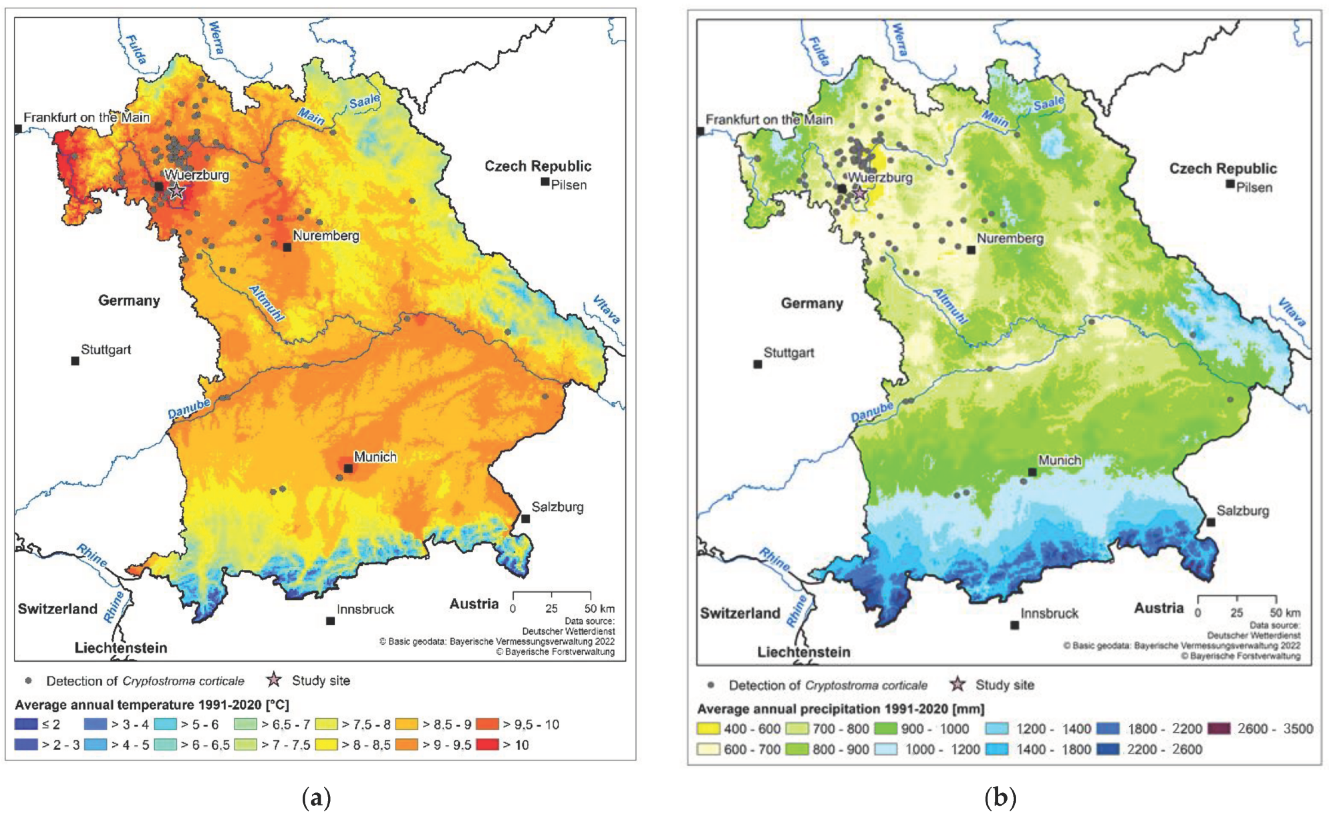
2.1. Study Site

The study of the pathogen’s spread of conidia was conducted in a heavily damaged forest stand close to Wuerzburg in Germany (49°46′24.4″ N, 10°05′23.7″ E). In the year of investigation in 2019, the mean annual temperature in Wuerzburg was 11.7 °C and total annual precipitation amounted to 469 mm. In this year, the weather station of the German Meteorological Service close to the study site recorded mean monthly temperatures between 20.4 and 20.9 °C in June, July and August [26]. In the previous year, with the first detected outbreaks of SBD in the area, mean temperatures in summer months ranged between 19.6 and 22.2 °C.
The square study site with a size of 0.25 ha was dominated by sycamore maple. Those represented 95% of the overstory with a mean diameter at breast height of 17.8 cm. In spring 2019, spore layers of C. corticale were observed at 40% of the 67 sycamore trees within the study site [23]. In October 2019, spores were visible at 54% of the trees. The mortality rate of sycamores due to SBD was very high with 31% in spring 2019, increasing to 49% in autumn of the same year. In summer 2020 and 2021, a decrease in the infection rates was observed, with a relatively stable share of 61% and 62% of sycamores showing spore layers of the pathogen, respectively.

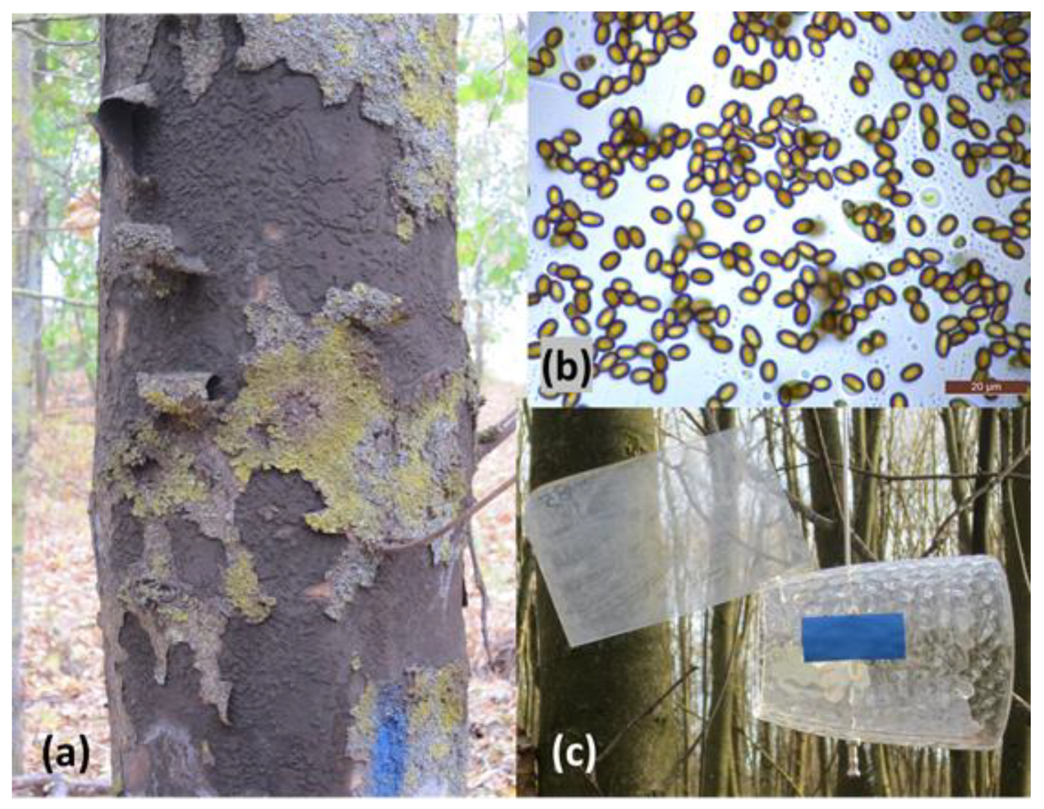
2.2. Detection of C. corticale

The species determination of the fungus has been performed morphologically by microscopy of the species-specific spores (Figure 2b) according to Ellis and Ellis [27]. To confirm the morphological determination, a molecular approach was followed. Samples of spores for DNA extraction were taken from five diseased trees. To detect the causative pathogen of SBD in the wood of symptomatic trees, six sycamore maple trees close to the study site were felled. Wood disks were carried into the laboratory and broken up in tangential direction with a log splitter to guarantee a clean surface to take the wood samples for DNA extraction. Shaving samples were taken from the discolored wood with a sterilized drill (Makita DDF451, Makita, Aichi, Japan), lyophilized, and finely ground with a vibratory bead mill (Retsch MM 400, Retsch GmbH, Haan, Germany). The extraction of DNA was conducted with a DNeasy Plant Mini Kit (Qiagen, Hilden, Germany) according to the manufacturers’ protocol. The PCR with species-specific primers was carried out according to Kelnarová et al. [21].

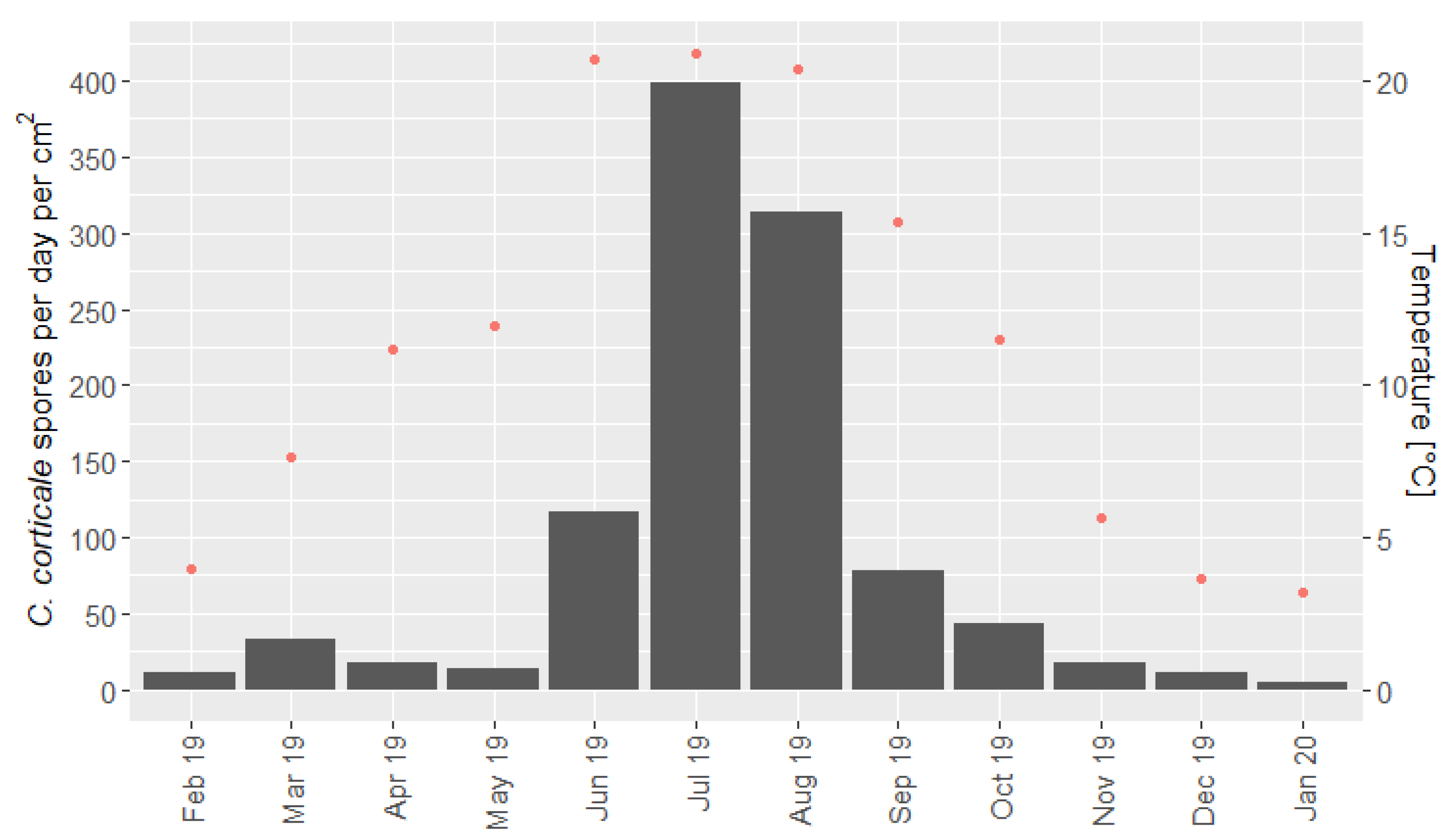
2.3. Spore Release According to Season

Since commercial spore traps are sparsely available, self-constructed traps consisting of plastic pipes with fixed microscope slides were used (Figure 2c). The pipes with a length of 19.5 cm were open on both sides. The microscope slides were vertically installed, right in the center. The spore traps were moved by wind by a fixed sail at the top of the pipe to face the wind steadily and filter spores out of a high volume of air. Rain protection was carried out through the pipes themselves. The microscope object slides with a surface of 19.76 cm2 were coated with a thin layer of Vaseline on the exposed side, to which fungal spores to which fungal spores stuck. The spore traps were fixed on poles at a height of 2 m. The sampling period was about three weeks on average, ranging between 7 and 34 days. Two to three traps were used simultaneously, which were allocated randomly with a minimum distance of 10 m among them and with a minimum distance of 2 m to the next sycamore stem.

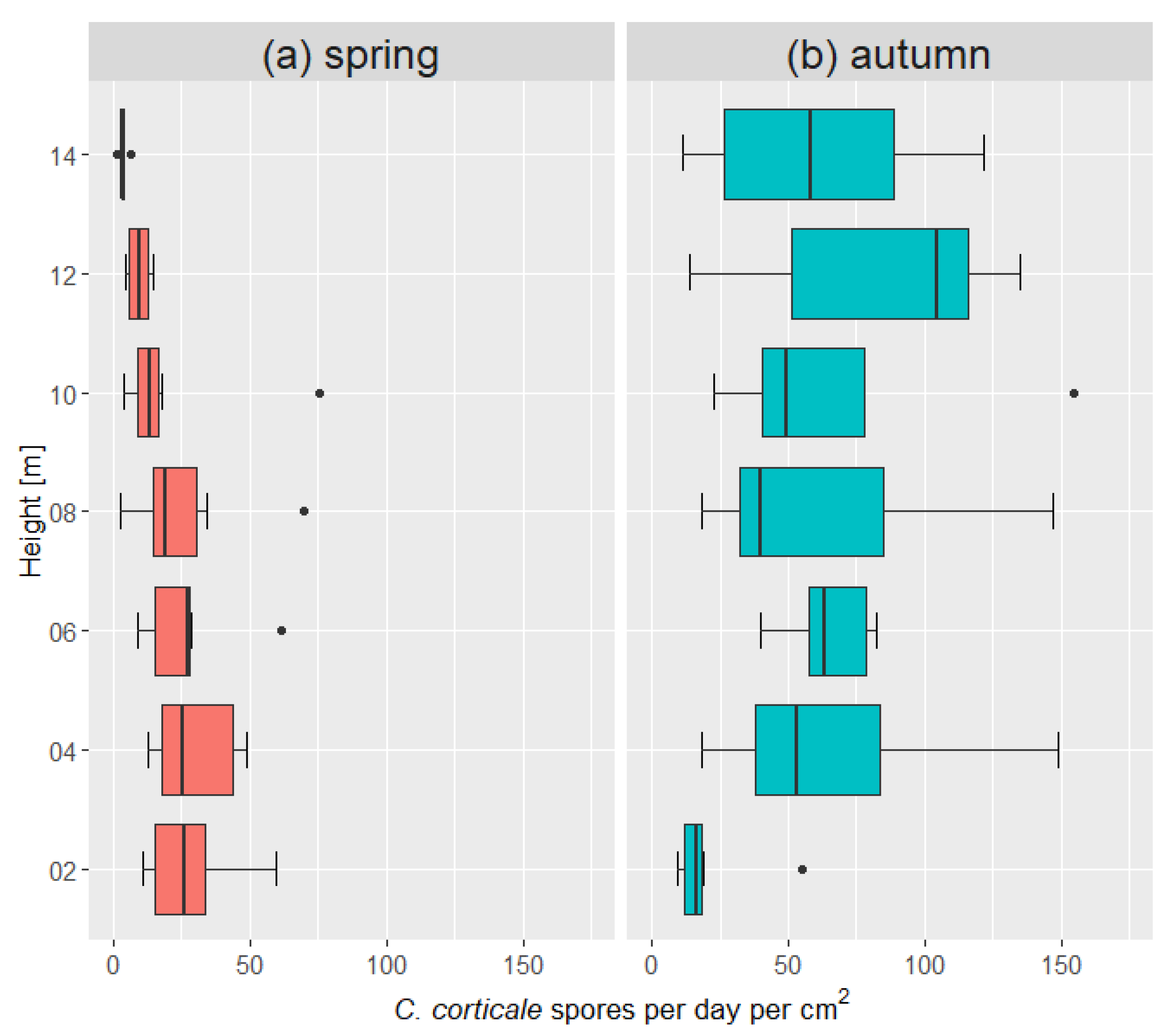
2.4. Spore Release along Vertical Gradient

In spring and autumn 2019, a vertical gradient with seven spore traps at a fixed rope from 2 to 14 m was examined. Two ropes were used simultaneously in three consecutive time series in spring (7 March–18 March, 18 March–8 April, 8 April–2 May 2019), and also in three consecutive time series in autumn (17 October–7 November, 7 November–26 November, 26 November–17 December 2019). The ropes have been shot on higher side branches of maple trees in the study site. The vertical traps were also flexible in orientation by small flags on the top of the pipe. Altogether, 84 microscope slides were counted. In October 2019, two additional spore traps were exposed on the top of a wind turbine at the height of 120 m in distance of 1.2 km to the sycamore maple forest stand for one month.
The statistical analysis for the variable season of spore abundances in the vertical gradient was conducted in R 4.0.5 (R Core Team, Vienna, Austria) using a generalized additive mixed model with the package ‘gamm4’ version 0.2-6.

2.5. Counting Spores by Microscopy

After replacement in the forest, the microscope slides were stored at 5 °C until examination of microcopy in the laboratory. The conidia on the object slides were visually counted by brightfield light microscopy (Leica DM 1000, Leica Camera AG, Wetzlar, Germany) according to the description of the oval, melanized spores of C. corticale [1,27]. When the hydrophobic spores appeared in aggregated clusters, their number was estimated by microscopy across the spore layers. From June to October, when spore abundances increased, the object slides were divided into four parallel sections and the second from the left was counted. The abundance of spores per day per cm2 was calculated by the exposure time of each microscope slide. The area where the pipe covered the microscope slide was subtracted out of the area of counted spores.

3. Results

3.1. Detection of C. corticale

The identification of C. corticale by microscopy of the spores of diseased trees was obvious according to the description of the dark brown, melanized, elliptical spores with a size of 4–6 × 3.5–4 µm described by Gregory and Waller and Ellis and Ellis [1,27]. The detection of C. corticale by extraction of DNA and PCR with species-specific primers according to Kelnarová et al. [21] by spores of five trees and in the discolored wood of six felled diseased maples was positive for all samples.

3.2. Spore Release According to Season

Conidia of C. corticale have been found on each object slide exposed in the damaged sycamore forest stand. At the end of winter and in spring 2019, the abundance of spores was relatively low (Figure 3). In the colder season from February to May 2019, on average, 19 spores were detected per day per cm2. In June, the number of spores on the object slides increased up to 117 spores per day per cm2. In July, a further increase up to 399 spores per day per cm2 was detected. In September, a decrease in the release was determined with a mean value of 78 spores per day per cm2.
The abundance of conidia in summer 2019 was higher compared with the other seasons. In summer, in average 277 spores were caught per day per cm2. From June to August the counts were increased up to the 12.6-fold compared with previous spring and up to 6-fold compared with following autumn. Taking the average of the entire year, 88 spores were caught per day.

3.3. Spore Release along Vertical Gradient

Spore dispersal of C. corticale in the vertical gradient was investigated in spring and autumn 2019. In spring, the highest abundance of spores was found at the height of 4 m with 30 spores per day per cm2 and the lowest abundance was found at 14 m (Figure 4a). At the height over 8 m, a slight decrease in spore numbers was detected. In autumn, more than 115 spores per day per cm2 were counted at 4, 8 and 10 m, lowest abundance was found at 2 m above ground (Figure 4b). In autumn, the deviation of the spore abundances at the different heights was higher than in springtime. Additionally, in autumn compared with spring, the abundance of spores was significantly increased (p ≤ 0.01). This result is consistent with the findings of the seasonal variation. Before summertime, spore release was lowest, in summer highest and after summer, the spore abundance was higher compared with winter and spring, where spores of the preceding time had been drifted away.
In the two spore traps on a wind turbine 180 and 30 spores were counted on the object slides with an exposure time of 30 days in total.

4. Discussion and Conclusions

Growth and formation of fruiting bodies of most fungi is usually highest under humid conditions. Therefore, it is remarkable that the fungal pathogen C. corticale forms its hydrophobic spores particularly under drought conditions. It is well known that the causative pathogen of SBD profits by warm temperatures and periods without precipitation. C. corticale is described to have an optimal growth at 25 °C [7,19,28] and seems to grow better in its woody host under warm conditions with drought stress [7,19].
Since the second finding of C. corticale in 2005 [10], the occurrence of SBD has increased in Germany, especially in urban areas [11,12]. In Bavaria, the pathogen was first recorded in 2018 as a consequence of heat and drought [29], when mainly small-scaled forests and urban areas were damaged. Since then, SBD has been increasing in many areas of Germany as a result of the extreme heat and drought periods in 2018 and 2019 [23,30,31]. The disease was especially observed in sycamore maple (A. pseudoplatanus) since this species is most widespread and considered to be more sensitive towards a deficit of precipitation than the also common but fewer affected Norway and field maple (A. platanoides, A. campestre).
In the year of the experiments, SBD was strongly driven by the impact of high temperatures and precipitation deficits in the generally warm and dry area close to Wuerzburg. The finding of an increased spore dispersal of C. corticale in 2019 especially in summer months coincides with the occurrence of SBD after heat and drought periods [7,19]. Mean monthly temperatures were above the long-time average, but not as high as 23 °C mean temperature, such as Young [5] describes as a favorable trigger for outbreaks of SBD in England. Nevertheless, the weather station of the German Meteorological Service [26] in Wuerzburg recorded 28 heat days of more than 30 °C in the year of investigation in 2019, and even 37 heat days in the previous year 2018. In contrast, 11 heat days per year build the average of the period from 1951 to 2020 at the weather station close to Wuerzburg. Thus, the number of heat days above 30 °C was 2.5 times above the long-term average. Therefore, consecutive high temperature peaks or the increasing frequency of heat days with temperatures above 30 °C might be more favorable for the epidemiology of C. corticale than mean temperatures. The high abundance of heat days in the years 2018/2019 could have been the main trigger of the massive outbreaks of SBD with high spore releases in the relatively warm and dry region around Wuerzburg.
Investigations about the spread of spores have been carried out for some forest pathogens. In studies about the causative agent of pine canker, Fusarium circinatum, spore dispersal up to 13 spores per cm2 was found in California using passive spore traps [32]. In the investigation of dispersal of Lecanosticta acicola, high abundances of spores were found in autumn, with a maximum amount of 145 spores per cm2 per day [33]. Our finding of an average of 277 spores of C. corticale per day per cm2 in summertime from June to August 2019 seems to be very high and may be an indicator for the intense inoculum pressure of this pathogen. Abundances of spores are obviously dependent on the development of recent spore layers in the forest stand. Thus, the maximum of spore release of SBD is unknown. Our measurements of spore release have been carried out in summer 2019 in a period of severe outbreaks of SBD, when in spring 40% and in autumn 54% of the trees in the study site showed spore layers of the pathogen [23]. The disease dynamic was very high in 2018 and 2019; thus, we assume that the average number of spores in summer represents a heavy outbreak situation of this disease.
In investigations using rotating arm spore traps on spore dispersal of the fungus causing ash decline, Hymenoscyphus fraxineus, peaks of spore dispersal were detected between the end of June and the beginning of September [34] and in August [35]. The dispersal of this pathogen is dependent on the formation of rather transient fruiting bodies; therefore, the time interval is much smaller than in SBD. C. corticale forms the large-scaled stroma with heavy loads of spores, which seem to be released into the air for a longer time period. In studies of spore dispersal of L. acicola, performed by Mesanza et al. [33], high abundances of spores were found from September to November. In this study in Spain, the daily maximum temperature and daily cumulative precipitation were associated with higher spore numbers in Pinus radiata stands. In studies by Wyka et al. [36] in the USA, highest abundances of spores of L. acicola were found in Pinus strobus stands from May to August with the highest impact of relative humidity and rainfall. Therefore, the abundance of spores of L. acicola seems to be strongly dependent on precipitation and/or humidity. In contrast, in our study, C. corticale was released in a hot year with a lack of precipitation, what indicates the potential of this pathogen for a further spread under changing climatic conditions with increasing periods of heat and drought.
In the vertical dispersal, spore abundance in spring was significantly lower compared with autumn; it was lowest at the height of 14 m. In contrast, in autumn, spores were mainly caught in higher altitudes and lowest spore dispersal was found at the minimum height. This might be a consequence of the height of spore layers on the stem in the time of trapping spores. This finding could be a consequence of the building of the fungal stroma. In contrast to previous findings, where starting of SBD waslocalized in the crown [21], based on our observations, one could conclude that the building of fungal stroma starts primarily at the bottom of the trunk and precedes up the stem. Future research could clarify this inconsistency. On the other hand, a volumetric reference of filtered air of the spore traps is missing and turbulent wind conditions in autumn could affect the spore dispersal of C. corticale in this time of the year.
Focusing on the pattern of spore dispersal, management operations in forests with SBD should be carried out in an early stage of the disease, before spore layers are abundant. As C. corticale seems to be widespread, in these regions, such containment measures seem to be obsolete. However, the variation in spore release within a year could be an important criterion for the timing of forest work: the load of spores is reduced between autumn and early spring. In addition, spore abundance on a vertical gradient in autumn was lowest at 2 m above ground, which enables management operations with a lower contamination compared with other seasons and heights.
The detection of C. corticale at a level of 120 m above ground suggests that the pathogen can spread in higher altitudes and hence over wide areas by thermal movements. The conidia of C. corticale are very small and melanized, which makes them more resilient against radiation. Therefore, a further spread of the causative pathogen of SBD resulting in the colonization of new habitats in previously uninfected areas can be assumed.
C. corticale is locally widespread asymptomatically as an endophyte in sycamore trees [21]. Under actual climatical changes, favorable disposing conditions for a further spread of the pathogen will progress in future. If a high inoculum pressure increases the success of infection with SBD, the massive aerial dispersal of conidia could increase infections of maple trees in colder forests that are not yet colonized. Therefore, maple trees in previously colder areas with increasing temperatures or in higher altitudes in mountain areas could be threatened in future.

Author Contributions

Conceptualization, N.B. and L.H.; methodology, N.B. and L.H.; software, N.B.; investigation, N.B. and L.H.; data curation, N.B.; writing—original draft preparation, N.B.; writing—review and editing, N.B. and W.A.H.; visualization, N.B.; supervision, W.A.H.; project administration, N.B. and W.A.H.; funding acquisition, N.B. and W.A.H. All authors have read and agreed to the published version of the manuscript.

Funding

We are grateful for financial support by the Bavarian State Ministry of Food, Agriculture and Forestry (Bayerisches Staatsministerium für Ernährung, Landwirtschaft und Forsten), support code ST 344, project title “Sooty bark disease of sycamore maple in Bavaria-Heat hotspots and possible risk areas for maple cultivation”.

Institutional Review Board Statement

Not applicable.

Informed Consent Statement

Not applicable.

Data Availability Statement

Data is available upon request to the first author.

Acknowledgments

We thank Ulrich Skatulla for conception and performance of the spore traps and Emily Lampertsdörfer and Britta Schnappauf for patient counting of C. corticale spores at the microscope. We are grateful to Britta Hausknecht for the preparation of climate data of the German Meteorological Service and the execution of the climatic maps for Bavaria and Ludwig Straßer for the idea and initiation of the research project. Finally, we thank the two reviewers for their valuable comments on the manuscript.

Conflicts of Interest

The authors declare no conflict of interest.

References

  1. Gregory, P.H.; Waller, S. Cryptostroma corticale and sooty bark disease of sycamore (Acer pseudoplatanus). Mycol. Res. 1951, 34, 579–597. [Google Scholar] [CrossRef]
  2. Ellis, J.B.; Everhart, B.M. New species of hyphomycetes fungi. J. Mycol. 1889, 5, 68–72. [Google Scholar] [CrossRef]
  3. Gregory, P.H.; Peace, T.R.; Waller, S. Death of sycamore trees associated with an unidentified fungus. Nature 1949, 164, 275. [Google Scholar] [CrossRef] [Green Version]
  4. Peace, T.R. Sooty bark disease of sycamore. A disease in eclipse. Q. J. For. 1955, 49, 197–204. [Google Scholar]
  5. Young, C.W.T. Sooty Bark Disease of Sycamore. Arboric. Leafl. 1978, 3, 8. [Google Scholar]
  6. Gibbs, J.N. Fifty years of sooty bark disease of sycamore. Q. J. For. 1997, 91, 215–221. [Google Scholar]
  7. Dickenson, S.; Wheeler, B.E.J. Effects of temperature, and water stress in sycamore, on growth of Cryptostroma corticale. Trans. Br. Mycol. Soc. 1981, 76, 181–185. [Google Scholar] [CrossRef]
  8. Moreau, C.; Moreau, M. La suie des Sycamores a Paris. Bull. La Société Mycol. Fr. 1951, 67, 404–418. [Google Scholar]
  9. Plate, H.-P.; Schneider, R. Ein Fall von asthmaartiger Allergie, verursacht von Cryptostroma corticale. Nachr. Des Dtsch. Pflanzenschutzd. 1965, 17, 100–101. [Google Scholar]
  10. Metzler, B. Cryptostroma corticale an Berg-Ahorn nach dem Trockenjahr 2003. Mitt. Biol. Bundesanst. Land Forstwirtsch. 2006, 400, 161–162. [Google Scholar]
  11. Kehr, R. Neue Krankheiten an Platane, Linde und Ahorn. Jahrb. Baumpflege 2007, 2007, 144–156. [Google Scholar]
  12. Robeck, P.; Heinrich, R.; Schumacher, J.; Feindt, R.; Kehr, R. Status der Rußrindenkrankheit des Ahorns in Deutschland. Jahrb. Baumpflege 2008, 2008, 238–245. [Google Scholar]
  13. Cech, T.L. Bemerkenswerte Krankheiten in 2004. Forstsch. Aktuell 2004, 32, 31–34. [Google Scholar]
  14. Cochard, B.; Crovadore, J.; Bovigny, P.Y.; Chablais, R.; Lefort, F. First reports of Cryptostroma corticale causing sooty bark disease in Acer sp. in Canton Geneva, Switzerland. New Dis. Rep. 2015, 31, 8. [Google Scholar] [CrossRef] [Green Version]
  15. Verkooijen, R.; Willems, J. Roetschorsziekte nu ook in Nederland. Tijdschr Bedr. Verzek. 2016, 24, 166–167. [Google Scholar] [CrossRef]
  16. Bencheva, S. First report of Cryptostroma corticale (Ellis & Everh.) P.H. Greg. & S. Waller on Acer platanoides L. in Bulgaria. Silva Balc. 2014, 15, 101–104. [Google Scholar]
  17. Koukol, O.; Kelnarová, I.; Černý, K. Recent observations of sooty bark disease of sycamore maple in Prague (Czech Republic) and the phylogenetic placement of Cryptostroma corticale. For. Pathol. 2015, 45, 21–27. [Google Scholar] [CrossRef]
  18. Longa, C.M.O.; Vai, N.; Maresi, G. Cryptostroma corticale in the northern Apennines (Italy). Phytopathol. Mediterr. 2016, 55, 136–138. [Google Scholar]
  19. Ogris, N.; Brglez, A.; Piškur, B. Drought Stress Can Induce the Pathogenicity of Cryptostroma corticale, the Causal Agent of Sooty Bark Disease of Sycamore Maple. Forests 2021, 12, 377. [Google Scholar] [CrossRef]
  20. Forest Health Watch (USA). Available online: https://foresthealth.org/sooty-bark-disease-on-maples-in-seattle/ (accessed on 14 October 2022).
  21. Kelnarová, I.; Černý, K.; Zahradník, D.; Koukol, O. Widespread latent infection of Cryptostroma corticale in asymptomatic Acer pseudoplatanus as a risk for urban plantations. For. Pathol. 2017, 47, e12344. [Google Scholar] [CrossRef]
  22. Burgdorf, N.; Härtl, L.; Straßer, L. Ahorn-Rußrindenkrankheit: Untersuchungen zur Verbreitung von Cryptostroma corticale. Jahrb. Baumpflege 2020, 2020, 277–284. [Google Scholar]
  23. Burgdorf, N.; Straßer, L. Schadverlauf und Mortalität durch die Rußrindenkrankheit in Bergahornbeständen Bayerns. Forstsch. Aktuell 2021, 66, 30–35. [Google Scholar]
  24. Braun, M.; Klingelhöfer, D.; Groneberg, D.A. Sooty bark disease of maples: The risk for hypersensitivity pneumonitis by fungal spores not only for woodman. J. Occup. Med. Toxicol. 2021, 16, 2. [Google Scholar] [CrossRef] [PubMed]
  25. Towey, J.W.; Sweany, H.C.; Huron, W.H. Severe bronchial asthma apparently due to fungus spores found in maple bark. J. Am. Med. Assoc. 1932, 99, 453–459. [Google Scholar] [CrossRef]
  26. German Meteorological Service (Deutscher Wetterdienst). Available online: https://www.dwd.de/DE/leistungen/klimadatendeutschland/klarchivtagmonat.html?nn=16102 (accessed on 30 August 2022).
  27. Ellis, M.B.; Ellis, J.P. Microfungi on Land Plants: An Identification Handbook; Croom Helm: London, UK, 1985. [Google Scholar]
  28. Härtl, L. Untersuchungen zur Biologie und Verbreitung von Cryptostroma corticale, Erreger der Ahorn-Rußrindenkrankheit. Bachelor’s Thesis, Department of Forestry, University of Applied Sciences Weihenstephan-Triesdorf, Freising, Germany, 2020. [Google Scholar]
  29. Bork, K. Rußrindenkrankheit an Ahorn—Erstfund in Bayern. AFZ Wald 2018, 20, 34–35. [Google Scholar]
  30. Grüner, J.; Berens, A.; Delb, H. Die Ahorn-Rußrindenkrankheit in Südwestdeutschland: Gefahren, Prognose und Empfehlungen. Waldschutz Info 2020, 2, 1–8. [Google Scholar]
  31. Schlösser, R.; Langer, G. Verbreitung der Rußrindenkrankheit des Ahorns in Deutschland. AFZ Wald 2021, 24, 28–32. [Google Scholar]
  32. Garbelotto, M.; Smith, T.; Schweigkofler, W. Variation in rates of spore deposition of Fusarium circinatum, the causal agent of pine pitch canker, over a 12-month-period at two locations in Northern California. Phytopathology 2008, 98, 137–143. [Google Scholar] [CrossRef] [Green Version]
  33. Mesanza, N.; García-García, D.; Raposo, E.R.; Raposo, R.; Iturbide, M.; Pascual, M.T.; Barrena, I.; Urkola, A.; Berano, N.; Sáez de Zerain, A.; et al. Weather Variables Associated with Spore Dispersal of Lecanosticta acicola Causing Pine Needle Blight in Northern Spain. Plants 2021, 10, 2788. [Google Scholar] [CrossRef]
  34. Chandelier, A.; Helson, M.; Dvorak, M.; Gischer, F. Detection and quantification of airborne inoculum of Hymenoscyphus pseudoalbidus using real-time PCR assays. Plant Pathol. 2014, 63, 1296–1305. [Google Scholar] [CrossRef]
  35. Dvorak, M.; Rotkova, G.; Botella, L. Detection of airborne inoculum of Hymenoscyphus fraxineus and H. albidus during seasonal fluctuations associated with absence of apothecia. Forests 2015, 7, 1. [Google Scholar] [CrossRef] [Green Version]
  36. Wyka, S.A.; McIntire, C.D.; Smith, C.; Munck, I.A.; Rock, B.N.; Asbjornsen, H.; Broders, K.D. Effect of climatic variables on abundance and dispersal of Lecanosticta acicola spores and their impact on defoliation on eastern white pine. Phytopathology 2018, 108, 374–383. [Google Scholar] [CrossRef] [PubMed]
Figure 1. Distribution of sooty bark disease caused by Cryptostroma corticale in the south-east of Germany (Bavaria) from the first detection in summer 2018 to summer 2022, with a hotspot in Lower Franconia around Wuerzburg; grey dots symbolize the points of detection of C. corticale, and the grey star south-east of Wuerzburg symbolizes the study site: (a) average annual temperature 1991–2020 °C; (b) average annual precipitation 1991–2020 mm.
Forests 13 01956 g001
Figure 2. (a) Sycamore maple with sooty bark disease: extensive spore layers of Cryptostroma corticale; (b) conidia of C. corticale viewed under a light microscope; (c) spore trap with an applied microscope slide in vertical orientation behind the blue stripe.
Forests 13 01956 g002
Figure 3. Seasonal abundance of spores of C. corticale per day per cm2 (grey columns) from February 2019 to January 2020 in spore traps in the forest site close to Wuerzburg, Germany; n = 2–3; right y-axis: mean monthly temperatures 2019 in the weather station Wuerzburg (red dots) of the German Meteorological Service.
Forests 13 01956 g003
Figure 4. Abundances of spores of C. corticale per day per cm2 along the vertical gradient at the height from 2 to 14 m in the study site close to Wuerzburg in (a) spring (7 March–2 May 2019), and (b) autumn (17 October–17 December 2019); box plots: central line: median, boxes: second and third quartile, whiskers: interquartile range, dots: outliers; n = 6.
Forests 13 01956 g004
Publisher’s Note: MDPI stays neutral with regard to jurisdictional claims in published maps and institutional affiliations.

