Article

Formation of PLGA–PEDOT: PSS Conductive Scaffolds by Supercritical Foaming

1
Department of Chemical Engineering and Food Technology, Faculty of Sciences, University of Cadiz, International Excellence Agrifood Campus (CeiA3), 11510 Puerto Real, Cadiz, Spain
2
Department Condensed Matter Physics and Institute of Electron Microscopy and Materials, Faculty of Sciences, University of Cadiz, International Excellence Agrifood Campus (CeiA3), 11510 Puerto Real, Cadiz, Spain
*
Author to whom correspondence should be addressed.
Materials 2023, 16(6), 2441; https://doi.org/10.3390/ma16062441
Submission received: 19 December 2022 / Revised: 13 March 2023 / Accepted: 16 March 2023 / Published: 18 March 2023

Abstract

:
The usage of conjugated materials for the fabrication of foams intended to be used as therapeutic scaffolds is gaining relevance these days, as they hold certain properties that are not exhibited by other polymer types that have been regularly used until the present. Hence, this work aims to design a specific supercritical CO2 foaming process that would allow the production of porous polymeric devices with improved conductive properties, which would better simulate matrix extracellular conditions when used as therapeutic scaffolds (PLGA–PEDOT:PSS) systems. The effects of pressure, temperature, and contact time on the expansion factor, porosity, mechanical properties, and conductivity of the foam have been evaluated. The foams have been characterized by scanning electron and atomic force microscopies, liquid displacement, PBS degradation test, compression, and resistance to conductivity techniques. Values close to 40% porosity were obtained, with a uniform distribution of polymers on the surface and in the interior, expansion factors of up to 10 orders, and a wide range of conductivity values (2.2 × 10⁻⁷ to 1.0 × 10⁻⁵ S/cm) and mechanical properties (0.8 to 13.6 MPa Young’s modulus in compression test). The conductive and porous scaffolds that have been produced by supercritical CO2 in this study show an interesting potential for tissue engineering and for neural or cardiac tissue regeneration purposes due to the fact that electrical conductivity is a crucial factor for proper cell function and tissue development.

1. Introduction

Nowadays, composite 3D structures are increasingly gaining interest, particularly because of their proven ability to support cell attachment in in vitro cultures [1]. Flexibility and porosity are two of the most vital properties that these structures must exhibit if they are to be used for tissue engineering purposes. Therefore, polymers or polymer mixtures have been commonly used based on their advantageous mechanical properties [2]. To effectively mimic the behavior of extracellular matrices, these therapeutical devices must be made of materials with an internal structure formed by large voids or interconnected pores that facilitate the diffusion of oxygen and nutrients. These devices are commonly referred to as scaffolds [3].
Recent research studies have highlighted that biodegradable and conductive copolymers promote both cell proliferation and tissue repair [4,5,6]. Conductivity also plays a significant role with regard to supporting certain biological processes, such as the transmission of signals through the nervous system or the healing of damaged tissue. The potential benefits of using electrical stimulation to promote and enhance these processes is also a matter of increasing interest [7].
Conductive polymers have been demonstrated to have, either with or without electrical stimulation, a beneficial impact on cell proliferation and adhesion as well as high levels of biocompatibility [8,9]. Therapeutic scaffolds have been recently designed and made of conductive copolymers for their use in tissue engineering or as drug delivery systems that are stimulated by electrical signals. The most frequently used are polyaniline [10], polythiophenes [11], polypyrrole [12,13], or poly(2,3-dihydrothieno-1,4-dioxin) (PEDOT) [14]. In our work, we have investigated PEDOT:PSS (Poly(2,3-dihydrothieno-1,4-dioxin)–poly(styrenesulfonate)) scaffolds. To embed PEDOT:PSS into porous materials and to improve their mechanical properties through the addition of different aggregates [15], these types of scaffolds have been prepared in the literature using different techniques as follows: electrochemical polymerization surrounding a pre-synthesized scaffold [16]; vapor-phase polymerization for neural stem cell proliferation and differentiation [17]; or freeze-drying. The capacity of the body cells to attach to porous scaffolds plays a relevant role with regard to the impedance of polymeric networks and can, therefore, be used as a means to control cell growth. Compared to other polythiophene derivatives, PEDOT:PSS presents high chemical and thermal stability as well as a high and adjustable electrical conductivity [18]. Other advantages of PEDOT:PSS include its low oxidative strength, good visible light transparency, and high biocompatibility that makes it suitable for its usage with biodegradable polymeric scaffolds. PEDOT:PSS is commonly available in the form of an aqueous dispersion, which makes it appropriate for an array of coating methods [19].
Conductive polymers generally fail to possess some of the essential characteristics required for tissue engineering with regard to strength, flexibility, solubility, or durability. Furthermore, in some cases, when conductive polymers are in a liquid phase at room temperature, they require rather complex processing techniques [20]. In order to overcome these limitations, these polymers are often combined with or incorporated into other non-conductive polymeric structures. These structures provide the mechanical properties that are especially required, particularly for tissue repair. Poly(lactic-co-glycolic acid) (PLGA), polyvinylpyrrolidone (PVP), polylactide (PLA), polycaprolactone (PCL), polyvinyl acetate (PVAc), or polyvinyl alcohol (PVA) are some of the compounds often used for this purpose [21]. PLGA, in particular, is often preferred for this type of biomedical objective for a number of reasons, namely, due to its mechanical properties, its composition-dependent biodegradability, and its easy production by different methods [22,23,24]. PLGA also has exceptional processing capabilities that allow to produce different pore-size scaffolds that can be used in combination with other materials for the regeneration of a wide assortment of tissues. Its mechanical properties also make it a suitable choice for tissue regeneration treatments in terms of biocompatibility and degradability. Thus, PLGA’s chemical properties enable its hydrolytic degradation by deesterification, and its monomeric components are easily removed by natural means following its degradation. Nevertheless, and although PLGA’s degradation process may sometimes lead to a local acidic microenvironment when compared against other commonly used polymers, PLGA allows superior control over its degradation properties by modifying its monomers ratio (lactic:glycolic). In addition, the broad range of degradation rates exhibited by PLGA can be controlled by adjusting the composition of its chains, its crystallinity, and its hydrophobic/hydrophilic balance. It has been recently combined with other materials, such as ceramics, to enhance some of its properties and improve its performance in tissue repair treatments [25,26].
Currently, there are numerous techniques that have been commonly applied to generate porous scaffolds over the last decades, such as gas foaming [27], freeze drying [28], solvent-casting [29], 3D printing [30], electrospinning [31] or phase separation [32]. Nevertheless, on the one hand, the usage of high temperatures needs to be avoided in order to reduce the degradation that biodegradable polyesters may suffer over their processing. On the other hand, many of the above-mentioned processes are not capable of producing morphologically similar polymer matrices with a uniform pore size and a high level of interconnectivity between them [33].
Supercritical carbon dioxide (scCO2) foaming has emerged as a promising method to produce functional porous scaffolds that avoid some of the drawbacks associated with other methods, such as high temperatures, the usage of to organic solvents, or the poor homogeneity of the final structures [34]. CO2 is not only highly soluble in polymers, but it is also a non-toxic, inexpensive, and reusable compound that makes this a greener technique [35]. In its supercritical state, CO2 exhibits properties, such as density, diffusivity, and low viscosity, that enable it to permeate the polymeric matrix, resulting in the plasticization of the matrix by decreasing its glass transition temperature. After a controlled contact time, depressurization of the system leads to supersaturation, which induces cell nucleation during phase separation, ultimately resulting in the formation of the porous scaffold structure [36,37,38]. This approach requires that the polymer has a certain affinity with the CO2 phase. Therefore, this scaffold-producing technique is particularly suitable for its use with amorphous materials.
Several studies can be found in the literature that investigate the development of polymeric scaffolds produced by supercritical CO2 foaming. Thus, a variety of polymeric scaffolds were obtained via different approaches. Poly(vinyl alcohol)/poly(ethylene glycol) produced bimodal open-celled structures with interconnected networks [39]. Poly(ɛ-caprolactone) resulted in structures with porosities between 60–80%, which represents a good potential for bone tissue engineering [40]. PLGA was combined with bioactive lipids for bone regenerative purposes [41], or PCL/PLGA blends [42], among others.
As far as the authors know, no previously published study has investigated scaffolds with this mixture of polymers, PLGA–PEDOT:PSS, generated by the supercritical foaming process presented herein. The objective of this study is to produce porous systems using a combination of non-conductive and conductive polymers for their potential application in tissue-repairing treatments. For this purpose, the authors evaluated the effects of certain key variables of the process, namely, temperature, pressure, and contact time, on the expansion factor, conductivity, mechanical strength, porosity, and the degradability of the final scaffolds.

2. Materials and Methods

2.1. Materials

PLGA (lactide:glycolide 75:25) (poly(lactic-co-glycolic acid)) with Mw 76,000–115,000 and PEDOT:PSS (Poly(2,3-dihydrothieno-1,4-dioxin)-poly(styrenesulfonate)) 3.0–4.0% in H2O and conductivity > 200 S/cm were purchased from Sigma–Aldrich (Spain). CO2 (99.8% purity) was supplied by Linde (Spain). Disodium-hydrogen phosphate (Na2HPO4) with 99.9% purity, potassium chloride (KCl) with 99.0% purity, potassium dihydrogen phosphate (KH2PO4) with 99.9% purity, and sodium chloride (NaCl) with 99.9% purity were purchased from Panreac Applychem (Barcelona, Spain).

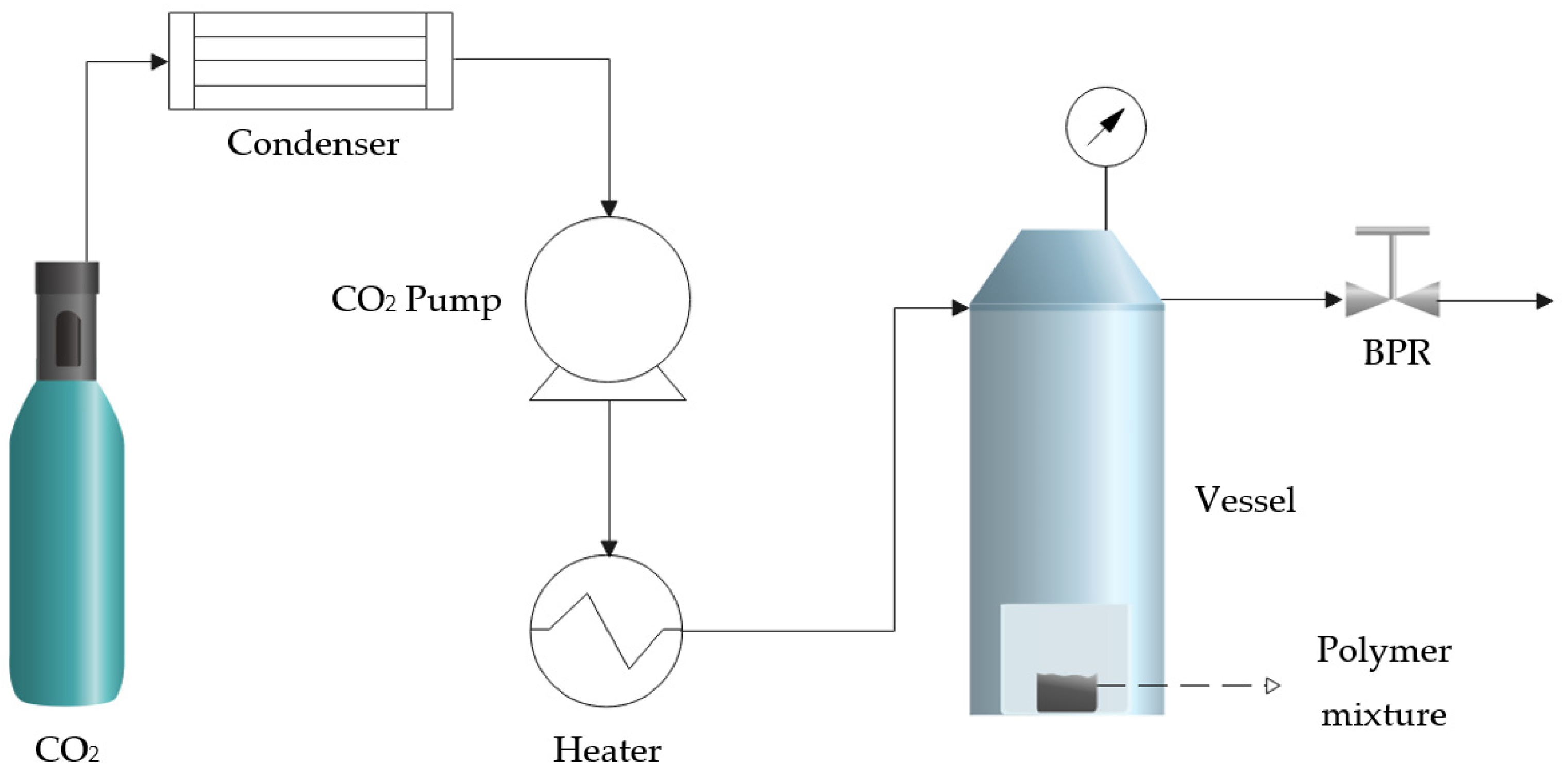
2.2. Supercritical Foaming Procedure

The scaffold foaming experiments were conducted at a pilot plant (TharProcess, Pittsburgh, PA, USA). A simplified diagram can be seen in Figure 1. The plant was equipped with a CO2 storage tank and a condenser to transfer the CO2 to the liquid state and to be able to pump it into the vessel (250 mL) with a high-pressure pump. Afterward, both the inlet CO2 and the interior of the vessel are heated by a heat exchanger and a heating jacket. Finally, the system was fitted with a micrometric valve (Back-Pressure Regulator—BPR) to regulate the depressurization rate and achieve the full venting of the CO2.
A total of 25 mg of PLGA 75:25 and 200 µL of PEDOT:PSS were mixed for each experiment and then shaped into a round tablet (30 mm3) by means of a tablet press machine. The tablets were placed in an aluminum foil holder and inserted into a high-pressure chamber. The system was then adjusted to the desired pressure and temperature, and CO2 was pumped in until supercritical conditions were reached. The polymeric mixture pellet was kept in contact with the supercritical phase for a specified amount of time to allow the CO2 to penetrate and cause polymer plasticization. After each specific saturation time, the system was depressurized at a controlled rate by means of the BPR. The foaming effect, i.e., the sample size and porosity increments, was achieved thanks to the rapid depressurization, and the PEDOT:PSS molecules were found to be evenly distributed throughout the entire tablet. The effect of temperature (30–60 °C), pressure (60–300 bar), and contact time (0.5–24 h) on the final porous structure was studied. Figure 2 shows one of the tablets used for the experiments before and after being subjected to the foaming process.

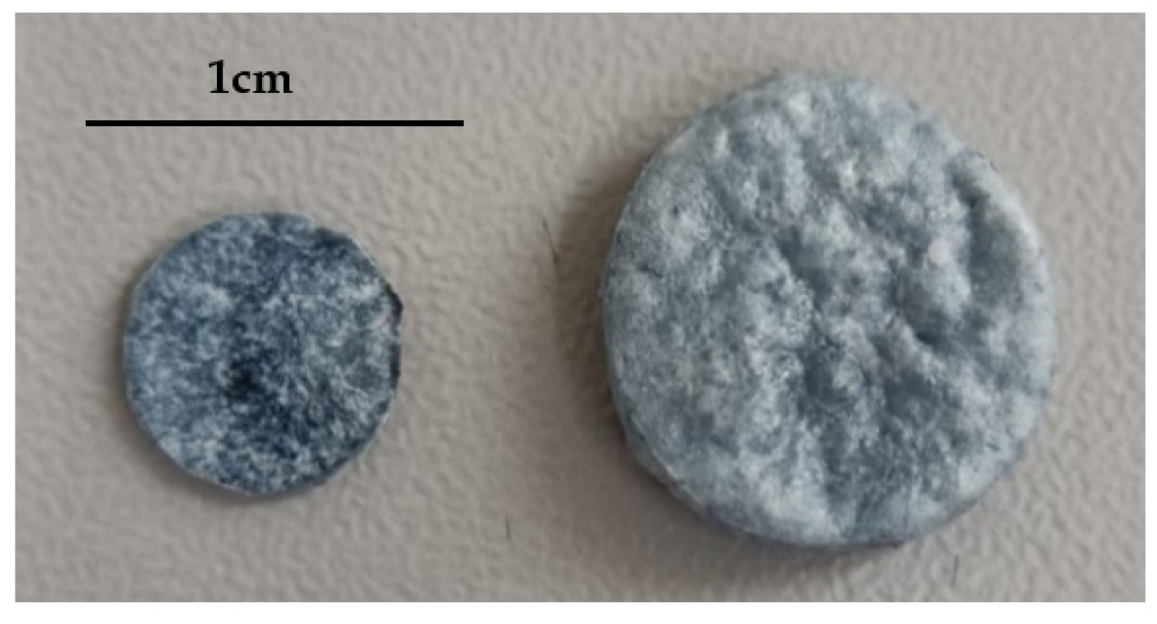
2.3. Scanning Electron Microscopy (SEM)

The morphology and pore diameter of the foamed scaffolds were examined by Scanning Electron Microscope Nova NanoSEM 450TM (Elecmi, Zaragoza, Spain) with 30 kV accelerating voltage. To improve the conductivity of samples in SEM and provide better image quality, a cross-section of each sample was coated with a 10 nm film of gold prior to analysis. The SEM images were used to calculate the pore diameter by Scion image software, measuring a minimum number of 300 pores for each experiment.

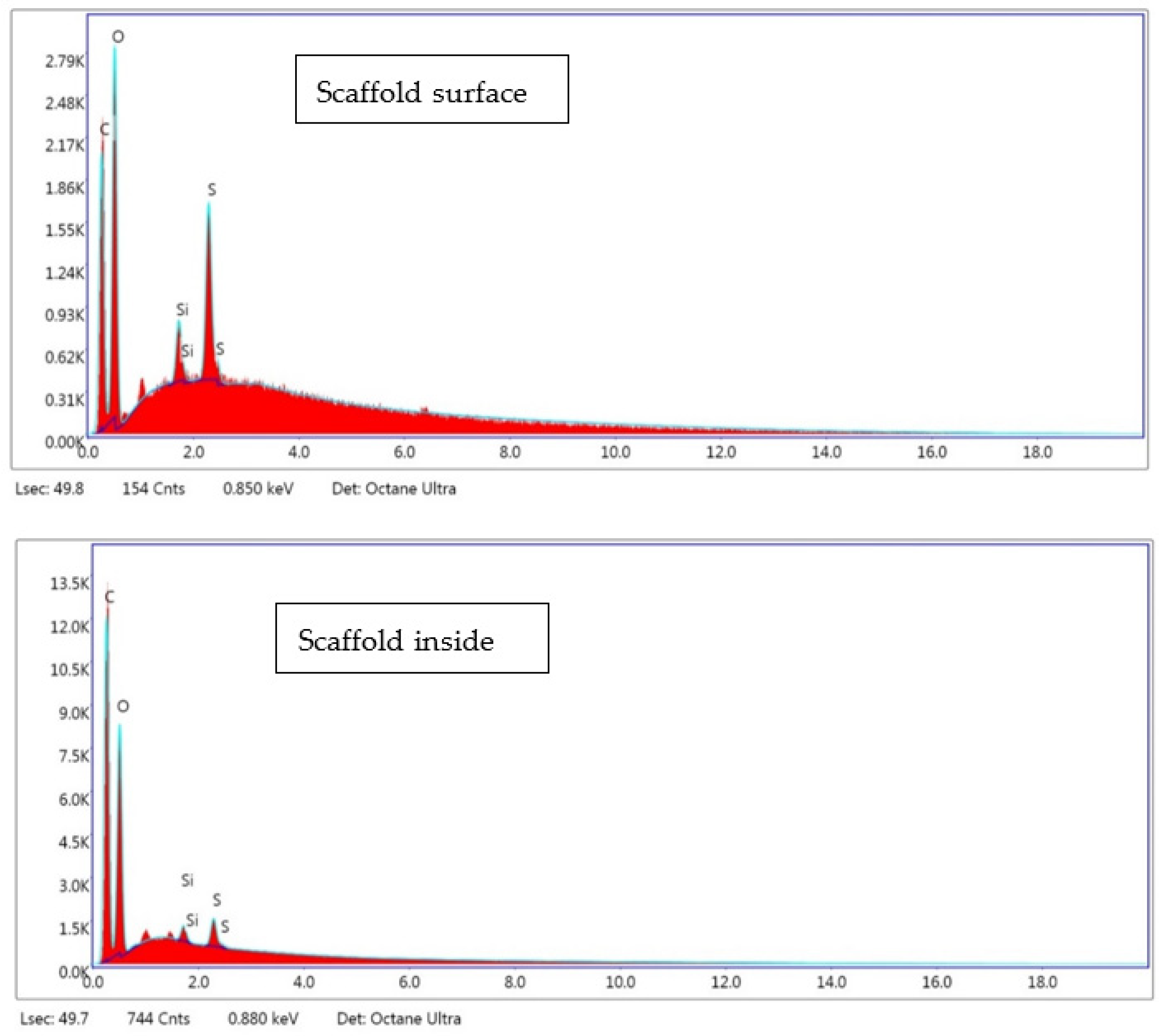
2.4. Polymer Distribution

Energy-dispersive X-ray spectroscopy (EDX) was used to analyze the chemical composition of different sections of the samples. The samples were directly deposited on carbon tape sections fixed on aluminum supports, and no gold coating was applied in order to avoid any interferences between sulfur and gold.
The Atomic Force Microscopy (AFM) images were obtained using a Bruker Dimension Icon microscope. A thin slice of the sample was mounted onto a metallic disc glued with double-sided tape, and, for the electrical measurements, a thin layer of silver paint was used to connect the sample surface to the metallic disc and the microscope stage. Given the fact that Young’s moduli of PEDOT:PSS and PLGA are of the order of a few GPa [43,44], for the nanomechanical measurements performed using the Quantitative Nanomechanics kit from Bruker, a Bruker RTESPA probe (nominal spring constant, k = 40 N/m, tip radius 8 nm) was selected since it is the one recommended for Young’s modulus ranging from 0.2 to 8.1 GPa. On the other hand, to obtain the surface potential maps by means of Kelvin Probe Force Microscopy (KPFM), a Bruker PFQNE-AL probe was used (k = 0.8 N/m, tip radius 5 nm). In this case, the work function of the tip was measured by using a gold/aluminum calibration sample (Bruker PFKPFM-SMPL) since the work function of gold can be averaged to 5.38 eV [45].

2.5. Expansion Factor of Samples

Volume variation of the different samples after processing with supercritical CO2 was quantified by the determination of the final expansion factor. The initial volume of each pellet could be directly measured thanks to its uniform surface and form, while its final volume was determined by submerging them into ethanol and following the liquid displacement method. The expansion factor was calculated according to the following expression:
E F = F i n a l   v o l u m e I n i t i a l   v o l u m e

2.6. Estimated Porosity

The porosity of the formed scaffolds was determined by liquid displacement. Ethanol (96%) was chosen as the working fluid because it is capable of penetrating through the polymeric structure without any disruption. Porosity was determined by the rate between the void volume (the pores) and the total volume of the scaffolds. This estimation is based on Archimedes’ principle [46,47]. Thus, an estimation of porosity was calculated according to the following equation:
P o r o s i t y % = v o i d   v o l u m e S c a f   f o l d   v o l u m e = V 1 V 3 V 2 V 3 · 100
where V1 is the initial volume of ethanol, V2 is the volume with the sample immersed till saturation, and V3 is the ethanol residual volume after sample removal.

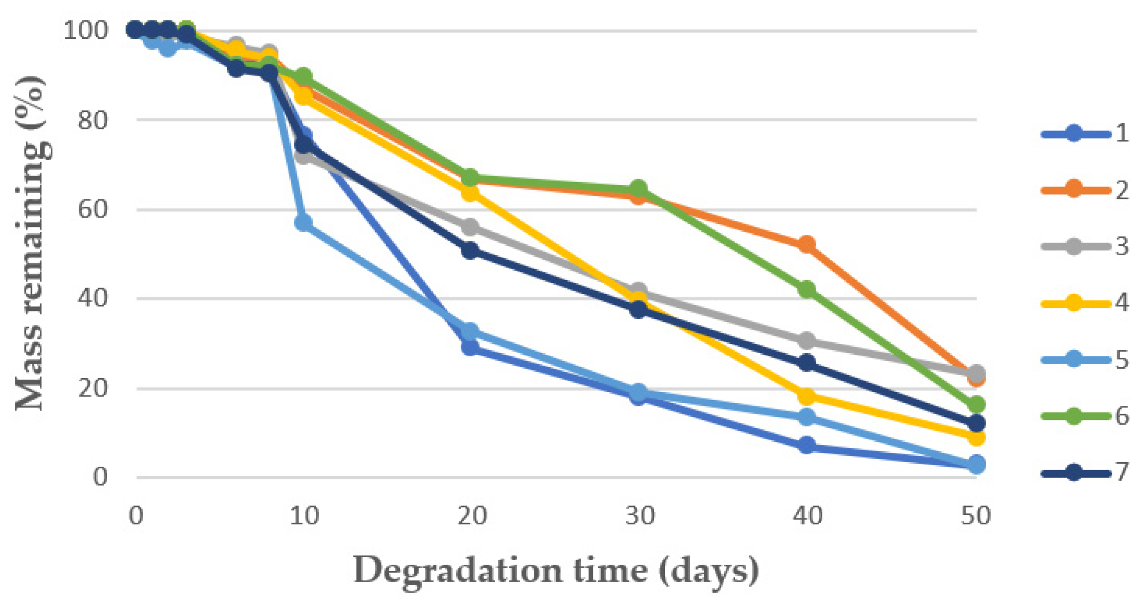
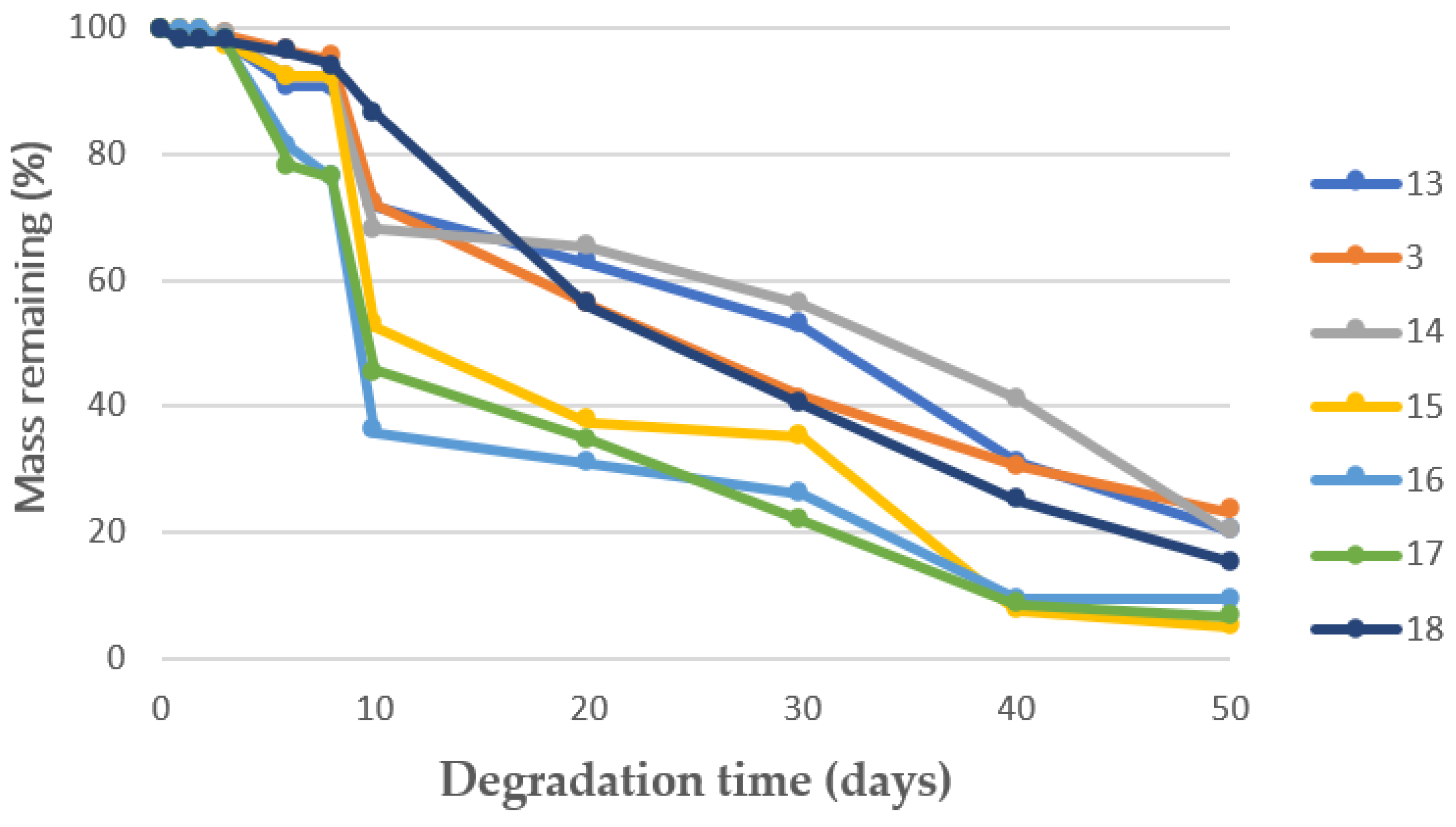
2.7. Degradability Test in PBS

The degradability of the scaffolds was evaluated by their degradation in phosphate buffer saline (PBS) 0.01 M solution at 37 °C. The weight of the samples as a function of degradation time was calculated gravimetrically [47]. Excess PBS was removed by filter paper at each measurement. The assessments were taken every 2 days during the first 10 days of analysis and every 10 days during the following 40 days (50 days in total). The scaffolds’ mass remaining after 50 days were determined according to the following equation:
M a s s   r e m a i n i n g % = W 2 W 1 · 100
where W1 is the initial weight before the test and W2 is the final weight after 50 days in PBS.

2.8. Mechanical Properties

In order to study the mechanical endurance of the scaffolds, Young’s modulus (E) was determined by a compression test and was calculated as the slope of the elastic region in the stress–strain curve of the compression test [48]. The compression tests were carried out by means of an MTS Criterion C45 tester, where scaffolds were compressed to a total strain of 70% using a compression rate of 0.02 mm/s and a maximum load of 10 kN. The sizes of the samples were adjusted to 15 mm3 prior to analysis.

2.9. Conductive Properties

The electrical conductivity properties of the polymer scaffolds were measured by the two-probe method [49], using a resistance meter (TRMS Fluke 87-V). The samples were cut into 13 mm2-sized pieces. Each of the surfaces was put in contact with a copper sheet, which had a thickness of 250 µm. Electrical resistivity was measured using the 0–10 V voltage range. This analysis was carried out in duplicate.

3. Results and Discussion

In preliminary experiments with PLGA and PEDOT:PSS, a large amount of PEDOT:PSS outside the scaffold was detected in excess. Accordingly, the PLGA–PEDOT:PSS ratio was optimized to avoid this large excess of the PEDOT:PSS. Different amounts of conductive polymer were tested with respect to PLGA, which were 200, 400, and 600 µL. This represented a mass ratio of 1:8, 1:16, and 1:24, respectively. These previous experiments were carried out at average operating conditions, i.e., 200 bar pressure, 40 °C temperature, and 2 h contact time, where the foaming of PLGA is favored. Moreover, at 1:16 and 1:24 mass ratios, a greater amount of PEDOT:PSS was found outside the scaffold. The part of it that was found on its surface was easily detached. An amount of 200 µL of the conductive polymer was therefore used for the subsequent experiments, as this amount did not result in any excess polymer, and the scaffolds were reasonably homogeneous across their surface.
Under these conditions, the rest of the experiments were successfully carried out, and it was observed that the PLGA was foamed to a greater or lesser extent as a scaffold. On the other hand, the PEDOT:PSS was evenly distributed throughout their entire structure. In order to evaluate the reproducibility of the process, a replication (experiment 7) was performed. The similarity of the results from experiments 6 and 7 confirmed the reproducibility of the process. The effects of temperature, pressure, and contact time on the morphology, pore diameter, porosity, expansion factor, conductivity, and degradability were studied in order to establish a connection between the operating conditions and the scaffold properties and potential applications.

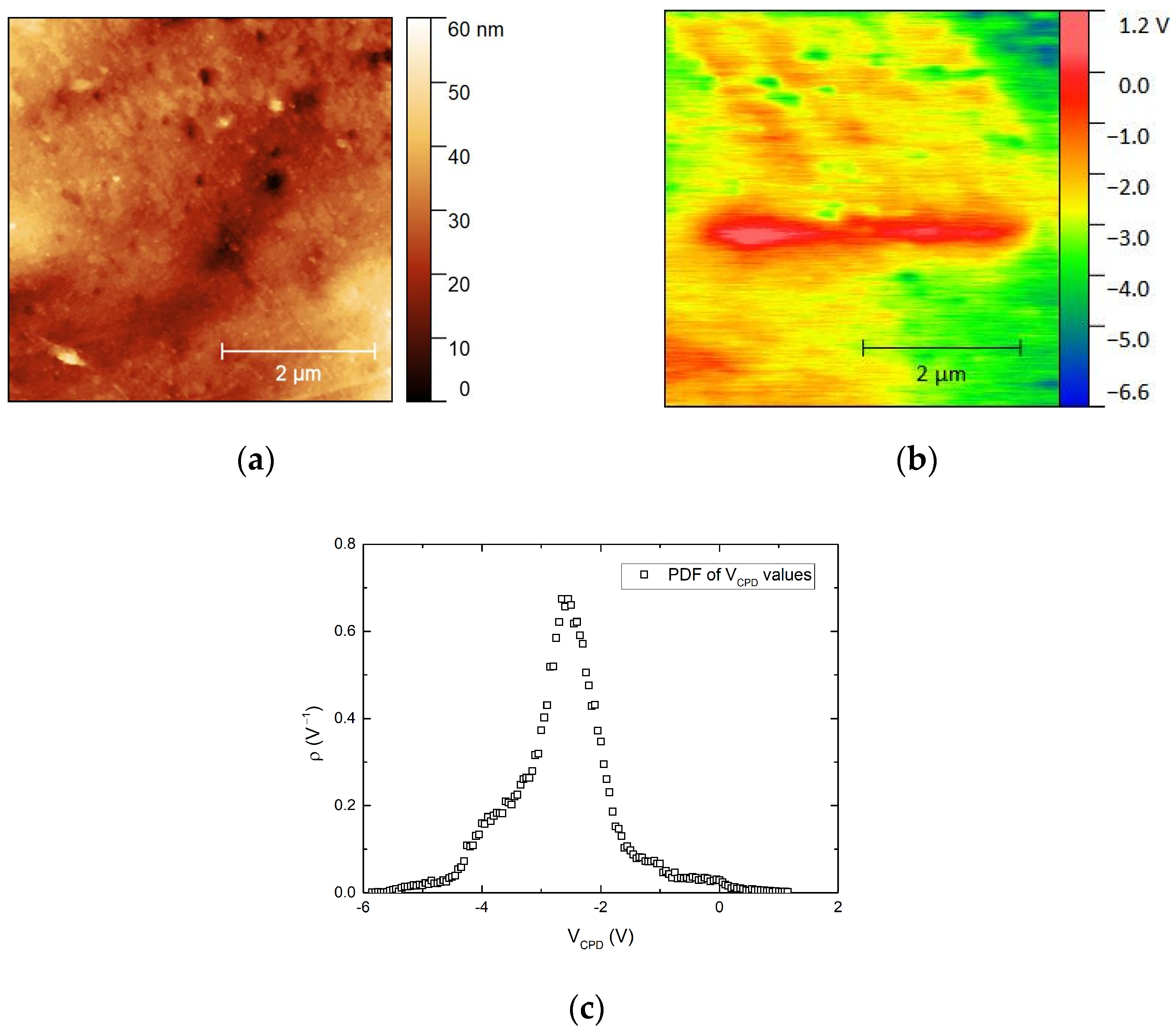
3.1. Polymer Distribution

A number of EDX analyses were carried out on different areas of the scaffolds (experiment 3) in order to identify any present PEDOT:PSS. As this polymer, unlike PLGA, contains sulfur, this compound was selected as the indicative of the PEDOT: PSS presence. An example of EDX spectra can be seen in Figure 3, where sulfur is clearly identifiable. Sulfur contents ranging from 0.8 up to 4.7 wt% on different surface areas and from 1.3 up to 2.6 wt% inside the scaffold were registered. It appears that there was a higher proportion of sulfur on the surface and, therefore, a higher amount of PEDOT: PSS. However, it should also be emphasized that in several areas within the scaffold, the amount of sulfur was noticeable. In line with EDX analyses, it was determined that PEDOT:PSS is located on the top and inside the scaffold. In order to discern if PEDOT was covering the scaffold surface as a film, AFM analyses were carried out for this scaffold.
Since the surface of the sample generally showed high roughness, a flat region of the sample surface was selected for nanomechanical and electrical measurements to reduce the interference of the purely topographic features on these parameters. Figure 4a shows the optical image of this flat area selected for the AFM scans, together with the topography of a 5 × 5 µm scanned area (b) and its corresponding Young’s modulus map, which looks quite homogeneous. Thus, the probability density function for Young’s modulus is shown in (d). These values were fitted to a gaussian function, and an average value of 9.5 GPa was obtained, which was close to the values previously reported for PEDOT:PSS and PLGA thin films [43,44].
Since PLGA shows similar Young’s modulus values to those of PEDOT:PSS [50], a different method is needed to demonstrate the presence of both polymers on the sample surface. PEDOT:PSS is a conducting polymer, and a large difference in work function is expected with respect to that of PLGA. The relationship between the work function of the AFM tip (χtip), the work function of the sample surface (χsample), and the contact potential difference (VCPD) between the tip and the sample is the following:
V C P D = χ t i p χ s a m p l e e
where  e is the elementary charge and VCPD is the parameter measured as surface potential in KPFM experiments. The KPFM probe was calibrated using a gold standard sample, and an average VCPD of 0.50 V was obtained for gold, whose work function value is 5.10 eV. Then, the work function of the tip was calculated as follows:
χ t i p = χ A u + e · V C P D = 5.10   eV + 0.50 eV = 5.60 eV  
Considering this calibrated value, the work function of the samples measured with this tip can be deduced from the following equation:
χ s a m p l e = 5.60   eV e · V C P D
Thus, Figure 5a shows the topography of another 5 × 5 µm area of the same flat region shown in Figure 4, together with the corresponding surface potential map (i.e., VCPD map) (Figure 5b), and the probability density function of VCPD values all over the scanned surface (Figure 5c). It may be readily deduced that the work function is not homogenous along the scanned area.
From these VCPD values, work function values ranging from 5.5 to 11.2 eV are deduced from Figure 5b. The reported value for the work function of PEDOT:PSS in thin films is 5.1 eV [51]. No reported values of the work function of PLGA have been found, but the most probable explanation for this result is that the surface sample is composed of a mixture of PLGA and PEDOT:PSS arranged in such a way that it is not possible to distinguish which regions are occupied by both polymers, given the lateral resolution of the KPFM technique (50–100 nm).
To confirm this, additional KPFM measurements were made in a pure PEDOT:PSS film and a pure PLGA pressed pellet, as shown in the Supplementary Information. The PEDOT:PSS film was deposited by spin coating using a 3% PEDOT:PSS aqueous solution onto a HOPG (highly oriented pyrolytic graphite) substrate. The work function deduced for this PEDOT:PSS film was 5.16 eV, which is very close to the previously reported value. However, for the pressed pellet of PLGA, the KPFM images showed that its VCPD values were so close to the lower limit of detection of the instrument (−10 V) that only in a few small areas of the sample VCPD values are within the available range, and its variation can be registered in the experiments. Its average value must be then beyond this −10 V limit. Even considering an average value of VCPD in PLGA of −10 V, its work function would be, according to this experiment, of the order of 15 eV at least. Then, the range of VCPD values found for the sample would necessarily imply that the surface of the sample is made of a combination of PEDOT:PSS and PLGA.

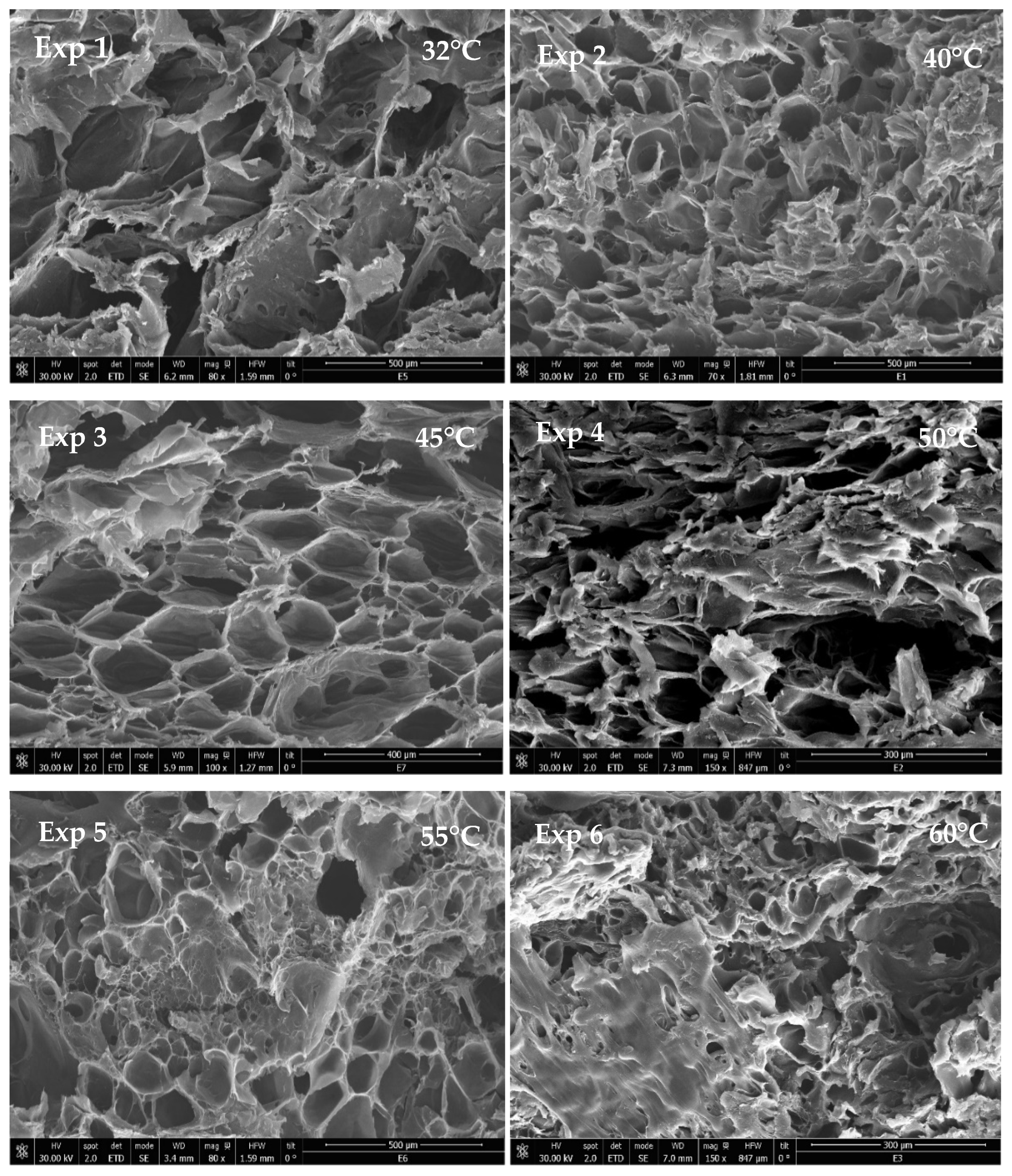
3.2. Effect of Temperature

The temperature was varied from 32 to 60 °C, keeping constant the rest of the parameters, as can be observed in Table 1. The morphology of the samples can be observed in Figure 6, where SEM images are shown. The formed scaffolds are based on PLGA, but PEDOT:PSS did not appear as particles. If both the AFM analyses and [18,24] are taken into account, it follows that PEDOT:PSS is well integrated, and it is the covering part (i.e., film) of the PLGA surface.
It can be observed in Table 1 that pore diameter increased significantly at the lowest temperature tested. Then, as the temperature was increased, the pore diameter fluctuated until the temperature reached 60 °C, after which smaller pore diameters were obtained. The same trend was observed with regard to standard deviation, so lower-standard deviations were registered at higher temperatures. In any case, and according to the SEM images, the highest geometry of the pores was achieved at 45 °C. With regard to porosity rate, it seemed to decrease at higher temperatures since their smaller pore diameters resulted in lower porosity rates, while the scaffolds with larger pores also had greater porosity rates. Therefore, the total porosity of the scaffolds seems to be more dependent on pore size rather than on pore number, even if the number of small pores was greater in those scaffolds that had been processed under higher temperatures. In this way, when the temperature is increased while pressure remains constant, the density of CO2 decreases, and so does its solvent power. On the other hand, at temperatures above the glass transition of PLGA (45–47 °C) [52], the polymer reaches its rubber state, and CO2 diffuses easily into the polymer matrix.
Different research studies in the literature deal with the formation of PLGA porous scaffolds using other techniques, such as 3D printing, electrospinning, or electrospun. This last one produced larger size pores. On the other hand, supercritical foaming generated macropores between 150–500 µm [53,54,55,56] with a larger pore size distribution and also allowed better control of this parameter just by implementing minor changes in its key variables.
Thus, the expansion factor proved to be strongly influenced by temperature, so greater expansion factors were achieved at lower temperatures. At constant pressure, an increase in temperature reduces the density of CO2 and its ability to solubilize and penetrate into the polymer matrix. This poorer soaking of CO2 into the polymer results in a lower number of bubbles and, therefore, a lower volume growth. The same behavior had been observed in a previous work, where PLGA was foamed while simultaneously impregnated with rutin [36].
With regard to the conductivity levels measured (Table 1), it can be observed that the higher the expansion factor, the lower the conductivity. The lower conductivity of the scaffolds [33] can be explained by the higher porosity resulting in a higher amount of trapped air and by a higher disorder between the PEDOT:PSS molecules. The conductive scaffolds prepared in this work have useful electrical characteristics, achieving a maximum conductivity of 1.6 × 10⁻⁵ S/cm, and therefore, they may be a suitable candidate for nerve, bone, vascular, or cardiac muscle tissue replacements that generally need to conduct electrical signals [57,58].
The degradability of the scaffolds, as a function of their weight loss, was measured for fifty days, as can be seen in Figure 7. The mass remaining at the end of the assay can be seen in Table 1. This factor is crucial to find out the suitability of the scaffolds for tissue engineering purposes. According to the data in Table 1, the scaffolds that were generated in experiments 2 (40 °C) and 3 (45 °C) presented the lowest degradation, with a final mass remaining or around 22–23 wt.%. Contrarily, the scaffolds that were most degraded had been produced at 32 and 50 °C. Nevertheless, no clear correlation could be established between temperature and degradability. During the first stages of the degrading process, a small number of glycolic and lactic units are hydrolyzed and released. The length of this first stage of degradation depends on each polymer’s lactic content, so that the greater the content, the longer this first stage is. In all the cases, a second degrading phase started after approximately ten days. This can be explained by the fact that during the period of 50 days, the medium becomes very acidic due to the autocatalyzed PLGA hydrolysis reaction, which increases the degradation rate [59,60].

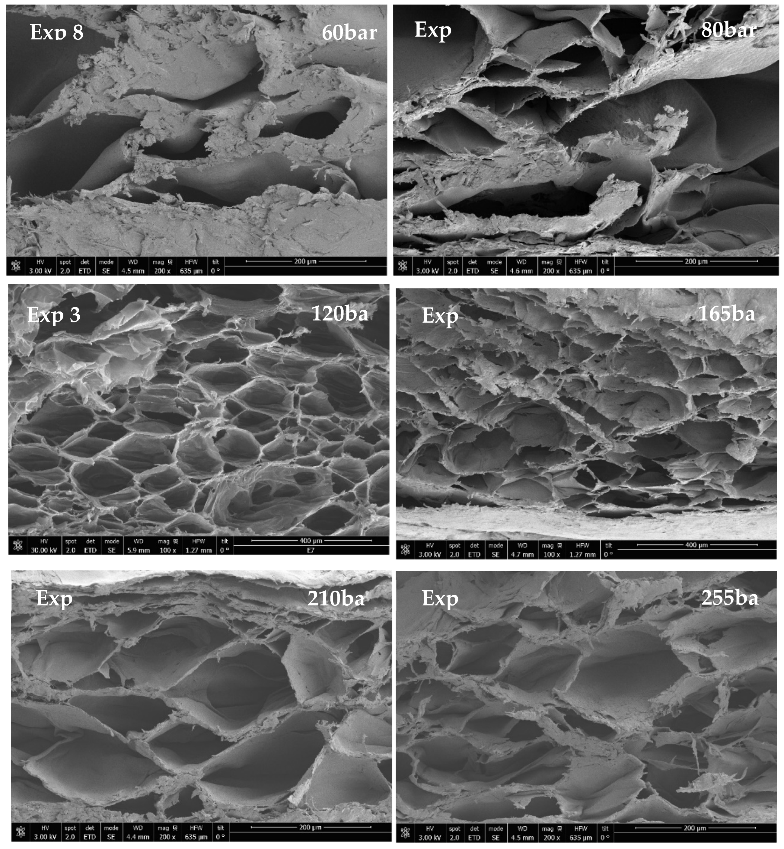
3.3. Effect of Pressure

In order to evaluate the effect of pressure on the scaffolds’ properties, the temperature (45 °C) and the contact time (0.5 h) were held constant, and different pressure levels were tested. Figure 8 displays the SEM images of the scaffolds. Foaming under subcritical conditions (experiment 8) was also evaluated, and the greatest average pore diameter, as well as the lowest porosity rate, were recorded. The size of the polymers’ pores is correlated with the number of CO2 bubbles that are produced according to the different experimental conditions. Thus, it can be observed that pore diameters are much greater when subcritical pressure is used for the foaming process, which is possibly due to the particular expansion profile that foam exhibits under these conditions [61].
In this sense, when pressure rises over CO2 critical pressure, the average diameter of the pores obtained is smaller. This is due to the higher solubility of the CO2 in the polymeric matrix, which decreases the nucleation energy barrier [46]. Similar behaviors have been observed when foaming other polymers [62,63,64]. Not only did the average pore diameter decrease with higher pressure levels, but pore uniformity was also enhanced. This can be observed in Table 2, where experiment 12 (255 bar) produced the scaffold with the smallest pore average diameter and the most regular pores.
Throughout all the experiments, pore diameters generally between 30 and 200 nm and porosity rates up to 50% were obtained. According to the literature, this would allow the use of the scaffolding for a diversity of applications depending on their actual porosity. Their applications could include osteogenesis (150–200 nm pore diameter, 35% porosity) [65], skin regeneration (20–125 nm pore diameter) [66], or smooth muscle cell differentiation (20–250 nm pore diameter) [67], among others.
The porosity of the scaffolds increased as the pressure was escalated up to 165 bar. Thus, as pressure was increased, more CO2 was absorbed by the polymer matrix, resulting in more CO2 bubbles and greater porosity. However, at pressures above 165 bar and up to 255 bar, the porosity rate dropped. This occurs because higher pressure increases the diffusivity of the CO2 into the polymer up to a point where pores cannot withstand the excessive pressure, and they collapse [64,65,66]. The expansion factor proved to be highly dependent on porosity rate so that it followed the same trend, i.e., it increased with pressure until 165 bar and then went down in the range from 165 to 255 bar.
Conductivity seemed to be inversely correlated with the expansion factor, i.e., as the pressure and expansion factor were increased, the conductivity was reduced. Hence, the scaffolds were more conductive when produced at lower pressure levels (Table 2). As above said, the larger population of voids in the polymer foams affected the electrical conductivity of the scaffolds and resulted in disorder between PEDOT:PSS molecules. For this reason, the scaffolds that exhibited the highest conductivity were those that had been produced under the pressure range between 165 and 255 bar, which resulted in a lower expansion factor. Overall, the conductivity was higher when the growth of the polymer was lower, thus avoiding disruption between PEDOT:PSS molecules along the polymer structure.
With regard to degradability, it can be observed from Table 2, and Figure 9 that experiments 8 and 11 had the highest degradability rates, and experiment 3 had the lowest one, but no correlation with pressure could be identified. It seems that the degradation around the tenth day resulting from the hydrolysis and acidification of the media does not take place with the same intensity in every scaffold. This could be explained by the different distribution of the PEDOT:PSS molecules inside the PLGA structure.

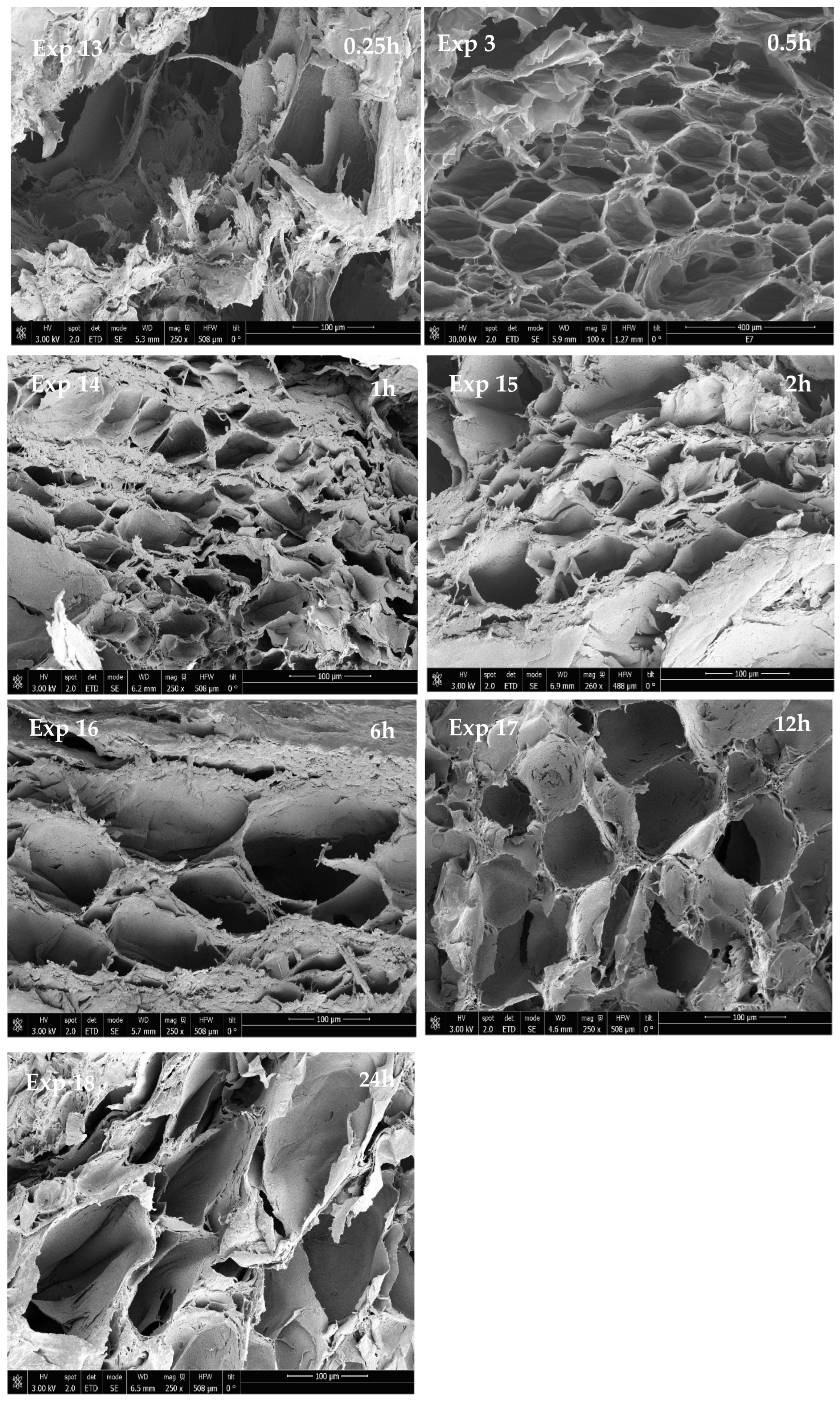
3.4. Effect of Contact Time

In order to determine if there was any correlation between the contact time of the scCO2 with the polymer and the obtained scaffold properties, a series of experiments were performed (see data in Table 3). The morphology of the scaffolds resulting from this set of experiments can be observed in Figure 10.
According to the images, 0.25 h contact time does not seem to be long enough to produce a scaffold structure. The foams obtained under these conditions presented poor porosity, with a large pore average diameter and widely variable shape. However, despite this lack of uniformity, these structures exhibited high conductivity values. This was probably explained by their low expansion factor, which contributed to avoiding the disruption of the PEDOT:PSS molecules and, in turn, favored electrical conductivity.
Contact time does not seem to have a clear effect on average pore diameter. Nevertheless, the maximum porosity rate was registered after 0.5 h contact time. The same trend was observed with respect to the expansion factor, which reached its maximum value also after 0.5h contact time. This was in consonance with CO2 sorption, which did not seem to increase with contact times longer than 0.5 h. Conductivity, on the other hand, remained unaltered regardless of the contact time used. Finally, it seems that the scaffolds exhibited higher resistance to degradation in PBS when they were produced using shorter contact times, as can be seen in Table 3 and Figure 11. However, the scaffolds that had been produced after 24 h contact time (experiment 18) also registered higher mass remaining than some of the scaffolds obtained after shorter contact times. Therefore, no definite trends could be established.

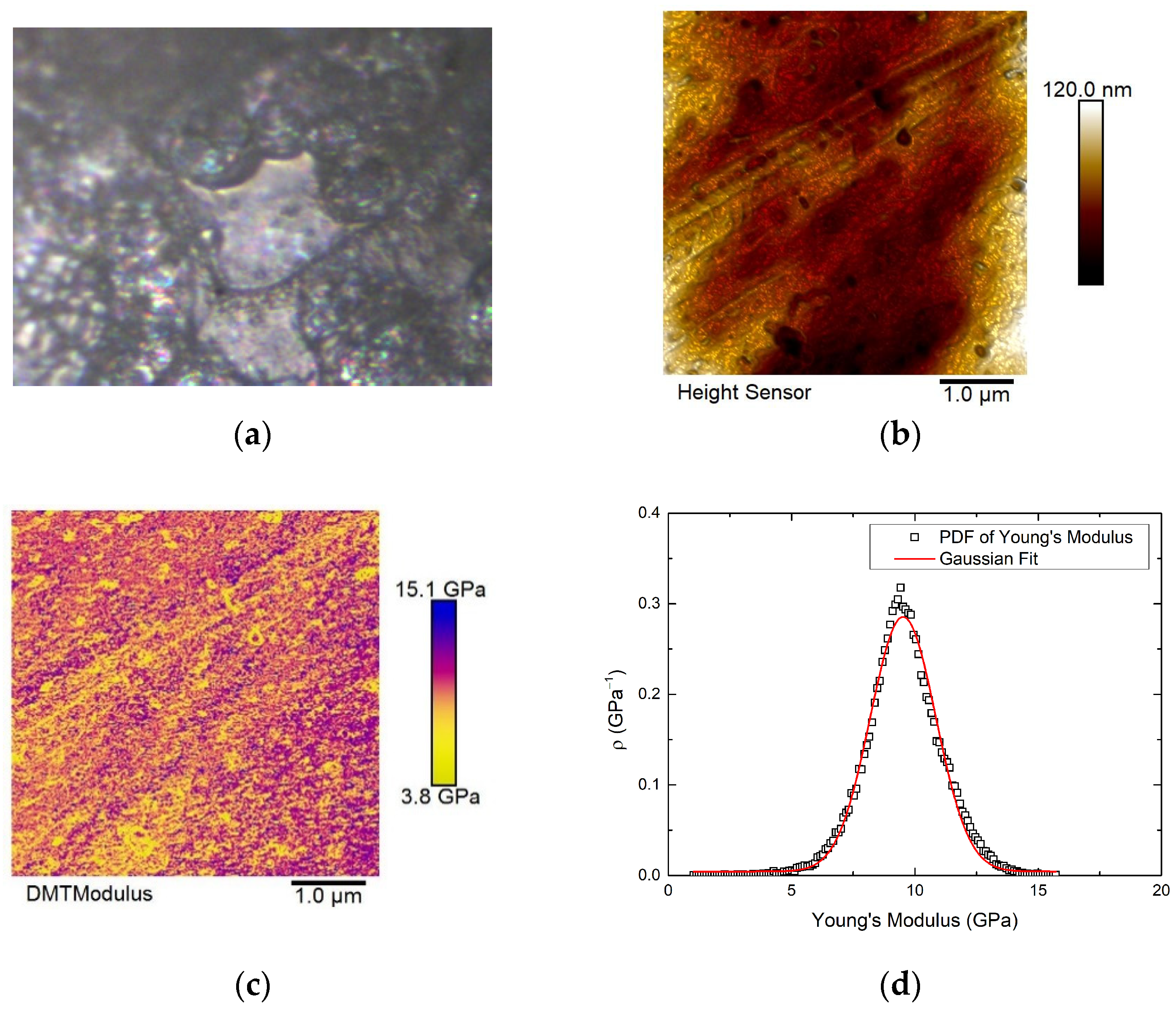
3.5. Mechanical Properties

The mechanical properties of several of the scaffolds that had been produced were evaluated to determine the influence of temperature, pressure, and contact time on the toughness of the scaffolds. This is an essential factor to take into account if the scaffolds are intended to be used as cell-supporting matrices for tissue regeneration.
Table 4 summarizes the various measured properties and Young’s modulus. The modulus has been determined by the slope of the linear region of the stress–strain curves, which indicates the material’s elastic behavior. The resistance of body tissues ranges between 2 and 6000 MPa depending on the type of tissue so that these values would correspond to a mature bone or to fibrous tissue, respectively [67,68,69,70].
The scaffolds obtained by experiment 11 exhibited the highest Young’s modulus and, therefore, the best mechanical properties. The scaffolds from experiments 10 and 14, which had been produced at 45 °C, also registered high modulus. A priori, the expansion factor should be inversely related to the toughness of the material. In this sense, the scaffold from experiment 11, with the lowest expansion factor, met this trend. Likewise, the scaffold obtained from experiment 1 and presenting the highest expansion factor, had a low Young’s modulus. However, the scaffold from experiment 4, with the same expansion factor as the latter, exhibited the poorest mechanical resistance. In the rest of the cases, no relationship between the expansion factor and mechanical resistance could be established. This was probably due to the specific distribution of the PEDOT:PSS molecules within the polymer structure, which might confer certain fragility to the scaffolds.
Most of the tests that have been performed revealed an improvement in the mechanical strength of the PLGA foamed. This is in line with the results reported in the literature when this material was foamed using other techniques, such as 3D printing, and where Young’s modulus compression tests registered values between 1.5 and 3.7 MPa [53].

4. Conclusions

PLGA–PEDOT:PSS scaffolds have been produced by means of a reproducible super-critical foaming process. According to EDX analyses, PEDOT:PSS was distributed on the surfaces and the inside of the PLGA scaffolds. It has also been confirmed, via the AFM analyses, that the surface of the scaffolds obtained was formed by a combination of PEDOT:PSS and PLGA. The effects of temperature, pressure, and contact time on the morphology, average pore diameter, porosity rate, expansion factor, conductivity, and degradability have been evaluated. This evaluation has allowed us to determine the trends that should be taken into account for the final scaffolds to exhibit the desirable properties for their intended applications. Thus, the expansion factor of the foams was strongly influenced by temperature, so that the lower the temperature, the higher the expansion factor. The foams’ conductivity, on the contrary, became lower as their expansion factor increased. Temperature presented an inverse relation with porosity so that the largest pore diameters were obtained at the lowest temperatures tested. Although the porosity of the scaffolds went slightly down as the temperature increased, their maximum porosity was reached at 165 bar regardless of the temperature. At pressure levels above 165 bar, porosity decreased, possibly due to the collapsing of the pores because of the excessive pressure. Moreover, smaller and more uniform pore diameters were obtained under the highest pressure levels tested.
Contact times shorter than thirty minutes did not seem to be adequate to form a proper scaffold structure, even if the foams obtained after thirty minutes exhibited a higher conductivity. The maximum porosity and expansion factors were obtained after a thirty-minute contact time, while longer times did not improve the sorption of the CO2, neither did the porosity rate nor the expansion of the foams. In any case, no clear contact time trends could be established, even if the scaffolds that had been produced with shorter times presented a greater resistance to degradation and a greater mass remaining (22–23 wt%). In this sense, the scaffolds produced at 210 bar, 45 °C, and using a thirty-minute contact time had the greatest mechanical resistance but the lowest expansion factor. It should be added that under subcritical conditions, the foams presented the largest average pore diameter and the lowest porosity rate.
Using supercritical CO2 to produce therapeutic scaffolds has allowed us to obtain foam structures with a wide range of average pore diameters (30–200 nm), porosity rates (up to 50%), and conductivity (up to 1.0 × 10⁻⁵ S/cm). This versatility of options corroborates the suitability of the final scaffolds for diverse medical treatments, such as the regeneration of nerve, bone, vascular or cardiac muscle tissues.

Supplementary Materials

The following supporting information can be downloaded at https://www.mdpi.com/article/10.3390/ma16062441/s1, Figure S1. Pristine PEDOT:PSS thin film deposited by spin coating on HOPG. Figure S2. AFM.1. Pressed pellet of PLGA.

Author Contributions

Conceptualization, A.M. and D.V.; methodology, A.M.; software, D.V.; validation, A.M., C.P. and E.M.d.l.O.; formal analysis, A.M., Y.P., D.V. and M.D.; investigation, Y.P. and D.V; resources, Y.P.; data curation, D.V.; writing—original draft preparation, D.V.; writing—review and editing, A.M.; visualization, E.M.d.l.O.; supervision, E.M.d.l.O.; project administration, C.P.; funding acquisition, C.P. All authors have read and agreed to the published version of the manuscript.

Funding

This research was funded by the Spanish Ministry of Science and Innovation: PID2020-116229RB-I00.

Institutional Review Board Statement

Not applicable.

Informed Consent Statement

Not applicable.

Data Availability Statement

Not applicable.

Acknowledgments

We gratefully acknowledge the Spanish Ministry of Science and Innovation (Project PID2020-116229RB-I00) for financial support and the central services of the University of Cádiz. M. Domínguez acknowledges financial support from the Spanish Ministry of Science and Innovation under project reference EQC 2018–004704-P FEDER 2014–2020.

Conflicts of Interest

The authors declare no conflict of interest. The funders had no role in the design of this study, in the collection, analyses, or interpretation of data, in the writing of the manuscript, or in the decision to publish the results.

References

  1. Drury, J.L.; Mooney, D.J. Hydrogels for tissue engineering: Scaffold design variables and applications. Biomaterials 2003, 24, 4337–4351. [Google Scholar] [CrossRef] [PubMed]
  2. Chan, B.P.; Leong, K.W. Scaffolding in tissue engineering: General approaches and tissue-specific considerations. Eur. Spine J. 2008, 17, 467–479. [Google Scholar] [CrossRef] [PubMed] [Green Version]
  3. Lawrence, B.J.; Madihally, S.V. Cell colonization in degradable 3D porous matrices. Cell Adh. Migr. 2008, 2, 9–16. [Google Scholar] [CrossRef]
  4. Hsiao, C.W.; Bai, M.Y.; Chang, Y.; Chung, M.F.; Lee, T.Y.; Wu, C.T.; Maiti, B.; Liao, Z.X.; Li, R.K.; Sung, H.W. Electrical coupling of isolated cardiomyocyte clusters grown on aligned conductive nanofibrous meshes for their synchronized beating. Biomaterials 2013, 34, 1063–1072. [Google Scholar] [CrossRef] [PubMed] [Green Version]
  5. Deng, Z.; Guo, Y.; Zhao, X.; Li, L.; Dong, R.; Guo, B.; Ma, P.X. Stretchable degradable and electroactive shape memory copolymers with tunable recovery temperature enhance myogenic differentiation. Acta Biomater. 2016, 46, 234–244. [Google Scholar] [CrossRef]
  6. Wu, Y.; Wang, L.; Guo, B.; Shao, Y.; Ma, P.X. Electroactive biodegradable polyurethane significantly enhanced Schwann cells myelin gene expression and neurotrophin secretion for peripheral nerve tissue engineering. Biomaterials 2016, 87, 18–31. [Google Scholar] [CrossRef] [PubMed]
  7. Korupalli, C.; Li, H.; Nguyen, N.; Mi, F.L.; Chang, Y.; Lin, Y.J.; Sung, H.W. Conductive Materials for Healing Wounds: Their Incorporation in Electroactive Wound Dressings, Characterization, and Perspectives. Adv. Healthc. Mater. 2021, 10, 2001384. [Google Scholar] [CrossRef]
  8. Yu, R.; Zhang, H.; Guo, B. Conductive Biomaterials as Bioactive Wound Dressing for Wound Healing and Skin Tissue Engineering; Springer: Singapore, 2022; Volume 14. [Google Scholar]
  9. Zhang, Q.; Esrafilzadeh, D.; Crook, J.M.; Kapsa, R.; Stewart, E.M.; Tomaskovic-Crook, E.; Wallace, G.G.; Huang, X.F. Electrical stimulation using conductive polymer polypyrrole counters reduced neurite outgrowth of primary prefrontal cortical neurons from NRG1-KO and DISC1-LI mice. Sci. Rep. 2017, 7, 42525. [Google Scholar] [CrossRef] [Green Version]
  10. Sarvari, R.; Akbari-Alanjaraghi, M.; Massoumi, B.; Beygi-Khosrowshahi, Y.; Agbolaghi, S. Conductive and biodegradable scaffolds based on a five-arm and functionalized star-like polyaniline-polycaprolactone copolymer with a d-glucose core. New J. Chem. 2017, 41, 6371–6384. [Google Scholar] [CrossRef]
  11. Chan, E.W.C.; Bennet, D.; Baek, P.; Barker, D.; Kim, S.; Travas-Sejdic, J. Electrospun Polythiophene Phenylenes for Tissue Engineering. Biomacromolecules 2018, 19, 1456–1468. [Google Scholar] [CrossRef]
  12. Valor, D.; Montes, A.; Cózar, A.; Pereyra, C.; de la Ossa, E.M. Development of Porous Polyvinyl Acetate/Polypyrrole/Gallic Acid Scaffolds Using Supercritical CO2 as Tissue Regenerative Agents. Polymers 2022, 14, 672. [Google Scholar] [CrossRef]
  13. Liang, Y.; Goh, J.C.H. Polypyrrole-Incorporated Conducting Constructs for Tissue Engineering Applications: A Review. Bioelectricity 2020, 2, 101–119. [Google Scholar] [CrossRef] [PubMed]
  14. Lari, A.; Sun, T.; Sultana, N. PEDOT:PSS-Containing Nanohydroxyapatite/Chitosan Conductive Bionanocomposite Scaffold: Fabrication and Evaluation. J. Nanomater. 2016, 2016, 9421203. [Google Scholar] [CrossRef] [Green Version]
  15. Inal, S.; Hama, A.; Ferro, M.; Pitsalidis, C.; Oziat, J.; Iandolo, D.; Pappa, A.M.; Hadida, M.; Huerta, M.; Marchat, D.; et al. Conducting Polymer Scaffolds for Hosting and Monitoring 3D Cell Culture. Adv. Biosyst. 2017, 1, 1700052. [Google Scholar] [CrossRef] [Green Version]
  16. Richardson-Burns, S.M.; Hendricks, J.L.; Martin, D.C. Electrochemical polymerization of conducting polymers in living neural tissue. J. Neural Eng. 2007, 4, L6. [Google Scholar] [CrossRef] [PubMed]
  17. Wang, S.; Guan, S.; Xu, J.; Li, W.; Ge, D.; Sun, C.; Liu, T.; Ma, X. Neural stem cell proliferation and differentiation in the conductive PEDOT-HA/Cs/Gel scaffold for neural tissue engineering. Biomater. Sci. 2017, 5, 2024–2034. [Google Scholar] [CrossRef]
  18. Wichiansee, W.; Sirivat, A. Electrorheological properties of poly(dimethylsiloxane) and poly(3,4-ethylenedioxy thiophene)/poly(stylene sulfonic acid)/ethylene glycol blends. Mater. Sci. Eng. C 2009, 29, 78–84. [Google Scholar] [CrossRef]
  19. Zhang, X.; Yang, W.; Zhang, H.; Xie, M.; Duan, X. PEDOT:PSS: From conductive polymers to sensors. Nanotechnol. Precis. Eng. 2021, 4, 045004. [Google Scholar] [CrossRef]
  20. Nezakati, T.; Seifalian, A.; Tan, A.; Seifalian, A.M. Conductive Polymers: Opportunities and Challenges in Biomedical Applications. Chem. Rev. 2018, 118, 6766–6843. [Google Scholar] [CrossRef]
  21. Marsudi, M.A.; Ariski, R.T.; Wibowo, A.; Cooper, G.; Barlian, A.; Rachmantyo, R.; Bartolo, P.J.D.S. Conductive polymeric-based electroactive scaffolds for tissue engineering applications: Current progress and challenges from biomaterials and manufacturing perspectives. Int. J. Mol. Sci. 2021, 22, 11543. [Google Scholar] [CrossRef]
  22. Pan, Z.; Ding, J. Poly(lactide-co-glycolide) porous scaffolds for tissue engineering and regenerative medicine. Interface Focus. 2012, 2, 366–377. [Google Scholar] [CrossRef] [PubMed] [Green Version]
  23. Ishaug, S.L.; Crane, G.M.; Miller, M.J.; Yasko, A.W.; Yaszemski, M.J.; Mikos, A.G. Bone formation by three-dimensional stromal osteoblast culture in biodegradable polymer scaffolds. J. Biomed. Mater. Res. 1997, 36, 17–28. [Google Scholar] [CrossRef]
  24. Shen, H.; Hu, X.; Bei, J.; Wang, S. The immobilization of basic fibroblast growth factor on plasma-treated poly(lactide-co-glycolide). Biomaterials 2008, 29, 2388–2399. [Google Scholar] [CrossRef]
  25. Gentile, P.; Chiono, V.; Carmagnola, I.; Hatton, P.V. An overview of poly(lactic-co-glycolic) Acid (PLGA)-based biomaterials for bone tissue engineering. Int. J. Mol. Sci. 2014, 15, 3640–3659. [Google Scholar] [CrossRef] [PubMed]
  26. Zhao, D.; Zhu, T.; Li, J.; Cui, L.; Zhang, Z.; Zhuang, X.; Ding, J. Poly(lactic-co-glycolic acid)-based composite bone-substitute materials. Bioact. Mater. 2021, 6, 346–360. [Google Scholar] [CrossRef] [PubMed]
  27. Salerno, A.; Oliviero, M.; Di Maio, E.; Iannace, S.; Netti, P.A. Design of porous polymeric scaffolds by gas foaming of heterogeneous blends. J. Mater. Sci. Mater. Med. 2009, 20, 2043–2051. [Google Scholar] [CrossRef] [PubMed]
  28. Grenier, J.; Duval, H.; Barou, F.; Lv, P.; David, B.; Letourneur, D. Mechanisms of pore formation in hydrogel scaffolds textured by freeze-drying. Acta Biomater. 2019, 94, 195–203. [Google Scholar] [CrossRef]
  29. Sola, A.; Bertacchini, J.; D’Avella, D.; Anselmi, L.; Maraldi, T.; Marmiroli, S.; Messori, M. Development of solvent-casting particulate leaching (SCPL) polymer scaffolds as improved three-dimensional supports to mimic the bone marrow niche. Mater. Sci. Eng. C 2019, 96, 153–165. [Google Scholar] [CrossRef] [Green Version]
  30. Do, A.V.; Khorsand, B.; Geary, S.M.; Salem, A.K. 3D Printing of Scaffolds for Tissue Regeneration Applications. Adv. Healthc. Mater. 2015, 4, 1742–1762. [Google Scholar] [CrossRef] [Green Version]
  31. Arida, I.A.; Ali, I.H.; Nasr, M.; El-Sherbiny, I.M. Electrospun polymer-based nanofiber scaffolds for skin regeneration. J. Drug Deliv. Sci. Technol. 2021, 64, 102623. [Google Scholar] [CrossRef]
  32. Kasoju, N.; Kubies, D.; Sedlačík, T.; Janoušková, O.; Koubková, J.; Kumorek, M.M.; Rypáček, F. Polymer scaffolds with no skin-effect for tissue engineering applications fabricated by thermally induced phase separation. Biomed. Mater. 2016, 11, 015002. [Google Scholar] [CrossRef]
  33. Alegret, N.; Dominguez-Alfaro, A.; Mecerreyes, D. 3D Scaffolds Based on Conductive Polymers for Biomedical Applications. Biomacromolecules 2019, 20, 73–89. [Google Scholar] [CrossRef]
  34. Song, C.; Luo, Y.; Liu, Y.; Li, S.; Xi, Z.; Zhao, L.; Cen, L.; Lu, E. Fabrication of PCL scaffolds by supercritical CO2 foaming based on the combined effects of rheological and crystallization properties. Polymers 2020, 12, 780. [Google Scholar] [CrossRef] [Green Version]
  35. Jacobs, L.J.M.; Kemmere, M.F.; Keurentjes, J.T.F. Sustainable polymer foaming using high pressure carbon dioxide: A review on fundamentals, processes and applications. Green Chem. 2008, 10, 731–773. [Google Scholar] [CrossRef]
  36. Valor, D.; Montes, A.; Monteiro, M.; García-Casas, I.; Pereyra, C.; de la Ossa, E.M. Determining the optimal conditions for the production by supercritical CO2 of biodegradable plga foams for the controlled release of rutin as a medical treatment. Polymers 2021, 13, 1645. [Google Scholar] [CrossRef]
  37. Kosowska, K.; Krzysztoforski, J.; Henczka, M. Foaming of PCL-Based Composites Using scCO2—Biocompatibility and Evaluation for Biomedical Applications. Materials 2022, 15, 3858. [Google Scholar] [CrossRef]
  38. Kosowska, K.; Krzysztoforski, J.; Henczka, M. Foaming of PCL-Based Composites Using scCO2: Structure and Physical Properties. Materials 2022, 15, 1169. [Google Scholar] [CrossRef]
  39. Liu, P.; Chen, W.; Liu, C.; Tian, M.; Liu, P. A novel poly (vinyl alcohol)/poly (ethylene glycol) scaffold for tissue engineering with a unique bimodal open-celled structure fabricated using supercritical fluid foaming. Sci. Rep. 2019, 9, 9534. [Google Scholar] [CrossRef] [Green Version]
  40. Chen, C.X.; Liu, Q.Q.; Xin, X.; Guan, Y.X.; Yao, S.J. Pore formation of poly(ε-caprolactone) scaffolds with melting point reduction in supercritical CO2 foaming. J. Supercrit. Fluids 2016, 117, 279–288. [Google Scholar] [CrossRef]
  41. Li, S.; Song, C.; Yang, S.; Yu, W.; Zhang, W.; Zhang, G.; Xi, Z.; Lu, E. Supercritical CO2 foamed composite scaffolds incorporating bioactive lipids promote vascularized bone regeneration via Hif-1α upregulation and enhanced type H vessel formation. Acta Biomater. 2019, 94, 253–267. [Google Scholar] [CrossRef]
  42. Guo, H.; Jiang, J.; Li, Z.; Jin, Z.; Hou, J.; Wang, X.; Li, Q. Solid-State Supercritical CO2 Foaming of PCL/PLGA Blends: Cell Opening and Compression Behavior. J. Polym. Environ. 2020, 28, 1880–1892. [Google Scholar] [CrossRef]
  43. Benaglia, S.; Drakopoulou, S.; Biscarini, F.; Garcia, R. In operando nanomechanical mapping of PEDOT:PSS thin films in electrolyte solutions with bimodal AFM. Nanoscale 2022, 14, 14146–14154. [Google Scholar] [CrossRef]
  44. Pannuzzo, M.; Horta, B.A.C.; La Rosa, C.; Decuzzi, P. Predicting the Miscibility and Rigidity of Poly(lactic- co-glycolic acid)/Polyethylene Glycol Blends via Molecular Dynamics Simulations. Macromolecules 2020, 53, 3643–3654. [Google Scholar] [CrossRef]
  45. Haynes, W.M. CRC Handbook of Chemistry and Physics; CRC Press: Boca Raton, FL, USA, 2012; pp. 12–124. [Google Scholar]
  46. Mou, Z.L.; Zhao, L.J.; Zhang, Q.A.; Zhang, J.; Zhang, Z.Q. Preparation of porous PLGA/HA/collagen scaffolds with supercritical CO2 and application in osteoblast cell culture. J. Supercrit. Fluids 2011, 58, 398–406. [Google Scholar] [CrossRef]
  47. Moghadam, M.Z.; Hassanajili, S.; Esmaeilzadeh, F.; Ayatollahi, M.; Ahmadi, M. Formation of porous HPCL/LPCL/HA scaffolds with supercritical CO2 gas foaming method. J. Mech. Behav. Biomed. Mater. 2017, 69, 115–127. [Google Scholar] [CrossRef]
  48. White, L.J.; Hutter, V.; Tai, H.; Howdle, S.M.; Shakesheff, K.M. The effect of processing variables on morphological and mechanical properties of supercritical CO2 foamed scaffolds for tissue engineering. Acta Biomater. 2012, 8, 61–71. [Google Scholar] [CrossRef] [Green Version]
  49. Pooshidani, Y.; Zoghi, N.; Rajabi, M.; Haghbin Nazarpak, M.; Hassannejad, Z. Fabrication and evaluation of porous and conductive nanofibrous scaffolds for nerve tissue engineering. J. Mater. Sci. Mater. Med. 2021, 32, 46. [Google Scholar] [CrossRef]
  50. Shirazi, R.N.; Aldabbagh, F.; Erxleben, A.; Rochev, Y.; McHugh, P. Nanomechanical properties of poly(lactic-co-glycolic) acid film during degradation. Acta Biomater. 2014, 10, 4695–4703. [Google Scholar] [CrossRef]
  51. Nardes, A.M.; Kemerink, M.; de Kok, M.M.; Vinken, E.; Maturova, K.; Janssen, R.A.J. Conductivity, work function, and environmental stability of PEDOT:PSS thin films treated with sorbitol. Org. Electron. 2008, 9, 727–734. [Google Scholar] [CrossRef]
  52. Carmagnola, I.; Nardo, T.; Gentile, P.; Tonda-Turo, C.; Mattu, C.; Cabodi, S.; Defilippi, P.; Chiono, V. Poly(lactic acid)-based blends with tailored physicochemical properties for tissue engineering applications: A case study. Int. J. Polym. Mater. Polym. Biomater. 2015, 64, 90–98. [Google Scholar] [CrossRef]
  53. Lai, Y.; Li, Y.; Cao, H.; Long, J.; Wang, X.; Li, L.; Li, C.; Jia, Q.; Teng, B.; Tang, T.; et al. Osteogenic magnesium incorporated into PLGA/TCP porous scaffold by 3D printing for repairing challenging bone defect. Biomaterials 2019, 197, 207–219. [Google Scholar] [CrossRef]
  54. Yang, W.; Yang, F.; Wang, Y.; Both, S.K.; Jansen, J.A. In vivo bone generation via the endochondral pathway on three-dimensional electrospun fibers. Acta Biomater. 2013, 9, 4505–4512. [Google Scholar] [CrossRef]
  55. Zabot, G.L.; Meireles, M.A.A. On-line process for pressurized ethanol extraction of onion peels extract and particle formation using supercritical antisolvent. J. Supercrit. Fluids. 2016, 110, 230–239. [Google Scholar] [CrossRef]
  56. Zhu, X.; Cui, W.; Li, X.; Jin, Y. Electrospun fibrous mats with high porosity as potential scaffolds for skin tissue engineering. Biomacromolecules 2008, 9, 1795–1801. [Google Scholar] [CrossRef]
  57. Zarei, M.; Samimi, A.; Khorram, M.; Abdi, M.M.; Golestaneh, S.I. Fabrication and characterization of conductive polypyrrole/chitosan/collagen electrospun nanofiber scaffold for tissue engineering application. Int. J. Biol. Macromol. 2021, 168, 175–186. [Google Scholar] [CrossRef]
  58. Wibowo, A.; Vyas, C.; Cooper, G.; Qulub, F.; Suratman, R.; Mahyuddin, A.I.; Dirgantara, T.; Bartolo, P. 3D printing of polycaprolactone-polyaniline electroactive Scaffolds for bone tissue engineering. Materials 2020, 13, 512. [Google Scholar] [CrossRef] [Green Version]
  59. Vey, E.; Rodger, C.; Booth, J.; Claybourn, M.; Miller, A.F.; Saiani, A. Degradation kinetics of poly(lactic-co-glycolic) acid block copolymer cast films in phosphate buffer solution as revealed by infrared and Raman spectroscopies. Polym. Degrad. Stab. 2011, 96, 1882–1889. [Google Scholar] [CrossRef]
  60. Fouad, H.; Elsarnagawy, T.; Almajhdi, F.N.; Khalil, K.A. Preparation and in vitro thermo-mechanical characterization of electrospun PLGA nanofibers for soft and hard tissue replacement. Int. J. Electrochem. Sci. 2013, 8, 2293–2304. [Google Scholar]
  61. Duarte, R.M.; Correia-Pinto, J.; Reis, R.L.; Duarte, A.R.C. Subcritical carbon dioxide foaming of polycaprolactone for bone tissue regeneration. J. Supercrit. Fluids 2018, 140, 1–10. [Google Scholar] [CrossRef] [Green Version]
  62. Goel, S.K.; Beckman, E.J. Generation fo microcellular polymeric foams using supercritical carbon dioxide. Polym. Mater. Sci. Eng. Proc. ACS Div. Polym. Mater. Sci. Eng. 1992, 67, 506–507. [Google Scholar]
  63. Reverchon, E.; Cardea, S. Production of controlled polymeric foams by supercritical CO2. J. Supercrit. Fluids. 2007, 40, 144–152. [Google Scholar] [CrossRef]
  64. Arora, K.A.; Lesser, A.J.; McCarthy, T.J. Preparation and characterization of microcellular polystyrene foams processed in supercritical carbon dioxide. Macromolecules 1998, 31, 4614–4620. [Google Scholar] [CrossRef]
  65. Aghaloo, T.L.; Moy, P.K.; Freymiller, E.G. Bone graft in the shape of human mandibular condyle reconstruction via seeding marrow-derived osteoblasts into porous coral in a nude mice model. J. Oral Maxillofac. Surg. 2002, 60, 1155–1159. [Google Scholar] [CrossRef]
  66. Yannas, I.V.; Lee, E.; Orgill, D.P.; Skrabut, E.M.; Murphy, G.F. Synthesis and characterization of a model extracellular matrix that induces partial regeneration of adult mammalian skin. Proc. Natl. Acad. Sci. USA 1989, 86, 933–937. [Google Scholar] [CrossRef] [Green Version]
  67. Cho, S.W.; Kim, I.K.; Lim, S.H.; Kim, D.I.; Kang, S.W.; Kim, S.H.; Kim, Y.H.; Lee, E.Y.; Choi, C.Y.; Kim, B.S. Smooth muscle-like tissues engineered with bone marrow stromal cells. Biomaterials 2004, 25, 2979–2986. [Google Scholar] [CrossRef]
  68. Milovanovic, S.; Markovic, D.; Mrakovic, A.; Kuska, R.; Zizovic, I.; Frerich, S.; Ivanovic, J. Supercritical CO2—Assisted production of PLA and PLGA foams for controlled thymol release. Mater. Sci. Eng. C 2019, 99, 394–404. [Google Scholar] [CrossRef]
  69. Teng, X.; Ren, J. GS Preparation and characterization of porous PDLLA/HA composite foams by supercritical carbon dioxide technology. J. Biomed. Mater. Res. B Appl. Biomater. 2007, 81, 185–193. [Google Scholar] [CrossRef]
  70. Byrne, D.P.; Lacroix, D.; Planell, J.A.; Kelly, D.J.; Prendergast, P.J. Simulation of tissue differentiation in a scaffold as a function of porosity, Young’s modulus and dissolution rate: Application of mechanobiological models in tissue engineering. Biomaterials 2007, 28, 5544–5554. [Google Scholar] [CrossRef]
Figure 1. Flow diagram of the supercritical foaming equipment.
Materials 16 02441 g001
Figure 2. Photograph of a sample before (left) and after being subjected to the foaming process (right).
Materials 16 02441 g002
Figure 3. EDX spectra corresponding to composition on the surface and inside the scaffold (experiment 3).
Materials 16 02441 g003
Figure 4. (a) Optical photograph of the flat area scanned for nanomechanical and electrical measurements. (b) Topography of a region of the sample (5 × 5 um scan). The minimum height data value has been shifted to zero. (c) Young’s modulus map of the area scanned in (b). (d) Probability density function of Young’s modulus values in (c).
Materials 16 02441 g004
Figure 5. (a) Topography (5 × 5 µm scan) and (b) KPFM images. (c) Probability density function of the contact potential difference (VCPD) values mapped in (b).
Materials 16 02441 g005
Figure 6. SEM images of formed scaffolds at 120 bar of pressure and 30 min of contact time at different operating temperatures.
Materials 16 02441 g006
Figure 7. Mass remaining vs. degradation time in PBS solution of the scaffolds produced under different temperature levels.
Materials 16 02441 g007
Figure 8. SEM images of formed scaffolds at 45 °C of temperature and 30 min of contact time at different operating pressures.
Materials 16 02441 g008
Figure 9. Mass remaining vs. degradation time in PBS solution of the scaffolds produced under different pressure levels.
Materials 16 02441 g009
Figure 10. SEM images of formed scaffolds at 120 bar of pressure and 45 °C of temperature at different contact times.
Materials 16 02441 g010
Figure 11. Mass remaining vs. degradation time in PBS solution of the scaffolds obtained after varying contact times.
Materials 16 02441 g011
Table 1. Supercritical foaming experiments to evaluate the influence of temperature on the process.
Experiments t
(t)
P
(bar)
T
(°c)
Pore Diameter
(nm)
Porosity (%)Expansion FactorConductivity (S/cm)Mass Remaining (%)
10.512032246.4 ± 77.751.5 ± 1.410.76.3 × 10−72.7 ± 1.2
240107.5 ± 41.442.9 ± 0.15.53 × 10−622.2 ± 3.5
345127.8 ± 52.047.7 ± 2.36.21.2 × 10−623.2 ± 9.7
45086.5 ± 76.040.8 ± 0.85.11.9 × 10−69.1 ± 3.5
555113.5 ± 46.242.3 ± 0.64.94.5 × 10−62.7 ± 2.5
66032.4 ± 16.029.2 ± 4.11.41.6 × 10−515.8 ± 10.1
76031.8 ± 8.629.1 ± 4.01.43.1 × 10−512.0 ± 1.4
Table 2. Supercritical foaming experiments to evaluate the influence of pressure on the process.
Experiments t
(h)
T
(°C)
P
(bar)
Pore Diameter
(nm)
Porosity (%)Expansion FactorConductivity (S/cm)Mass Remaining (%)
80.54560331.3 ± 345.829.2 ± 4.24.92.6 × 10−63.3 ± 1.4
980114.5 ± 100.940.8 ± 0.85.91.1 × 10−612.5 ± 1.1
3120127.8 ± 52.047.7 ± 2.36.21.2 × 10−623.2 ± 6.9
10165116.9 ± 71.150.0 ± 0.17.42.2 × 10−712.8 ± 4.6
11210117.4 ± 49.742.2 ± 2.25.11.1 × 10−64.7 ± 1.1
1225596.2 ± 43.433.3 ± 0.13.43.8 × 10−613.0 ± 8.7
Table 3. Supercritical foaming experiments to evaluate the influence of contact time on the process.
Experiments P
(bar)
T
(°C)
t
(h)
Pore Diameter
(nm)
Porosity (%)Expansion FactorConductivity (S/cm)Mass Remaining (%)
13120450.25213.8 ± 151.930.9 ± 2.42.41.0 × 10−520.2 ± 2.4
30.5127.8 ± 52.047.7 ± 2.36.21.2 × 10−623.2 ± 2.4
14144.5 ± 21.450.0 ± 0.16.05.2 × 10−720.0 ± 5.9
15261.1 ± 30.038.1 ± 4.85.41.6 × 10−65.1 ± 4.3
16894.5 ± 34.453.6 ± 3.66.27.6 × 10−79.5 ± 4.1
171597.4 ± 22.050.1 ± 0.17.73.5 × 10−76.5 ± 1.4
18 24103.7 ± 37.536.7 ± 3.34.95.5 × 10−615.4 ± 5.7
Table 4. Summary of the data, including Young’s modulus, that corresponds to the scaffolds obtained from specific experiments.
ExperimentP
(bar)
T
(°C)
t
(h)
PoreD
(nm)
Porosity
(%)
EF *MR * (%)C *
(S/cm)
E (MPa)
1120320.5246.4 ± 77.751.5 ± 1.410.72.7 ± 1.26.3 × 10−71.3
3120450.5127.8 ± 52.047.7 ± 2.36.223.2 ± 9.71.2 × 10−61.2
4120500.586.5 ± 76.040.8 ± 0.85.19.1 ± 3.51.9 × 10−60.8
980450.5114.5 ± 100.940.8 ± 0.85.912.5 ± 1.11.1 × 10−62.2
10165450.5116.9 ± 71.150.0 ± 0.17.412.8 ± 4.62.2 × 10−711.3
11210450.5117.4 ± 49.742.2 ± 2.25.14.7 ± 1.11.1 × 10−613.6
1412045144.5 ± 21.450.0 ± 0.16.020.0 ± 5.95.2 × 10−78.9
1612045894.5 ± 34.453.6 ± 3.66.29.5 ± 4.17.6 × 10−73.3
17120451597.4 ± 22.050.1 ± 0.17.76.5 ± 1.43.5 × 10−71.3
* EF = Expansion Factor. * MR = Mass Remaining. * C = Conductivity.
Disclaimer/Publisher’s Note: The statements, opinions and data contained in all publications are solely those of the individual author(s) and contributor(s) and not of MDPI and/or the editor(s). MDPI and/or the editor(s) disclaim responsibility for any injury to people or property resulting from any ideas, methods, instructions or products referred to in the content.

