
Non-Ionic Surfactants for Stabilization of Polymeric Nanoparticles for Biomedical Uses

 , , , and
Abstract
:1. Introduction
2. Use of Surfactants for Nanoparticle Stabilization
2.1. Background
2.2. Stabilization Mechanisms
2.3. Ionic and Non-Ionic Surfactants
2.4. New Surfactants
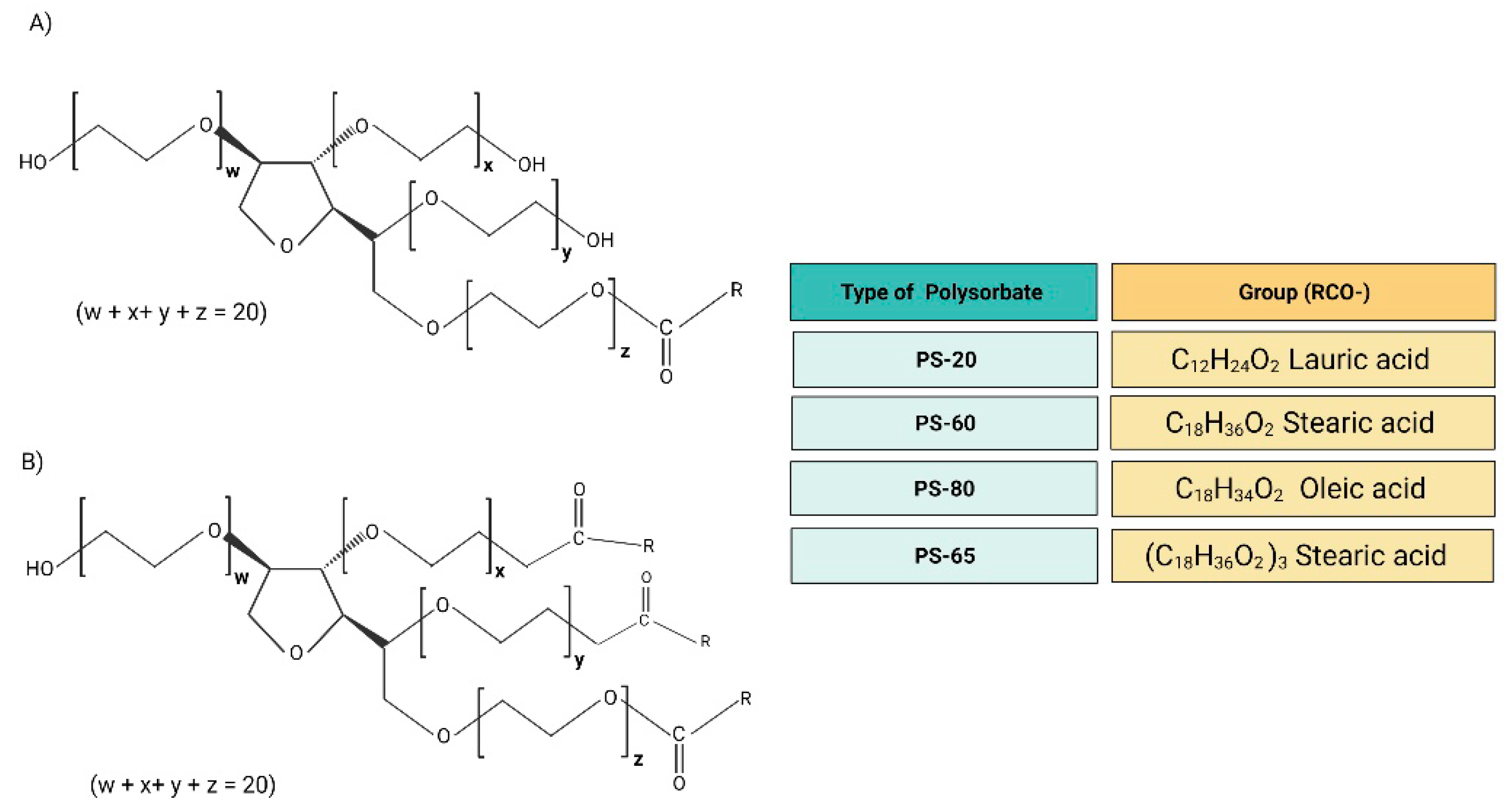
3. Polysorbates
3.1. Physicochemical Properties
3.2. Types
3.3. Uses
3.4. Derivatives
3.5. Examples of NP Applications
3.6. Toxicity in NP
3.7. Advantages and Disadvantages
4. Polyvinyl Alcohol
4.1. Physicochemical Properties
4.2. Types
4.3. Uses
4.4. Derivatives
4.5. Examples of NP Applications
4.6. Toxicity in NP
4.7. Advantages and Disadvantages
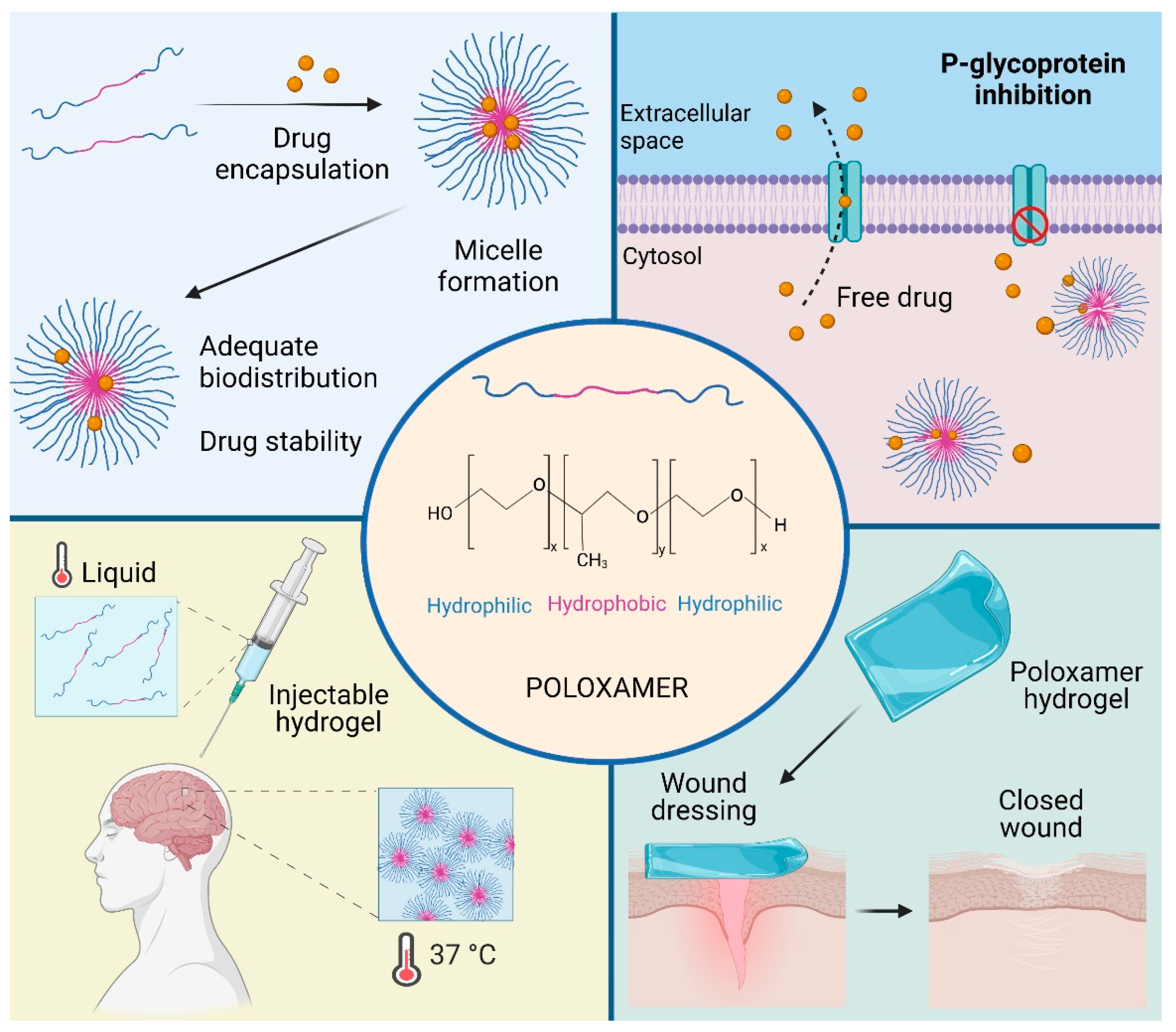
5. Poloxamers
5.1. Physicochemical Properties
5.2. Types
5.3. Uses
5.4. Derivatives
5.5. Examples of NP Applications
5.6. Toxicity in NP
5.7. Advantages and Disadvantages
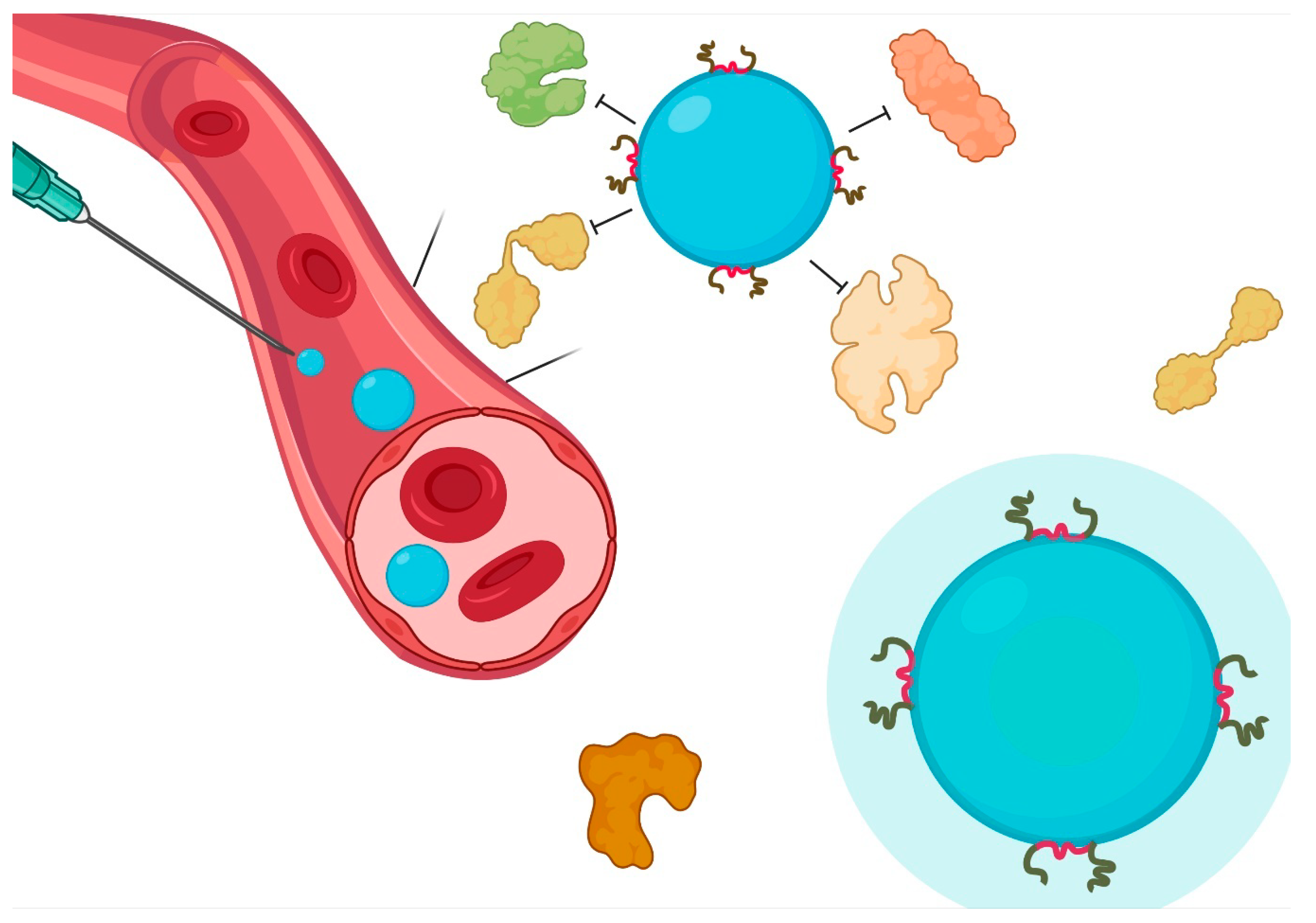
6. Influence of Non-Ionic Surfactants on the Interaction with Biological Barriers
6.1. Blood-Brain Barrier
6.2. Intestinal
6.3. Intranasal
6.4. Pulmonary
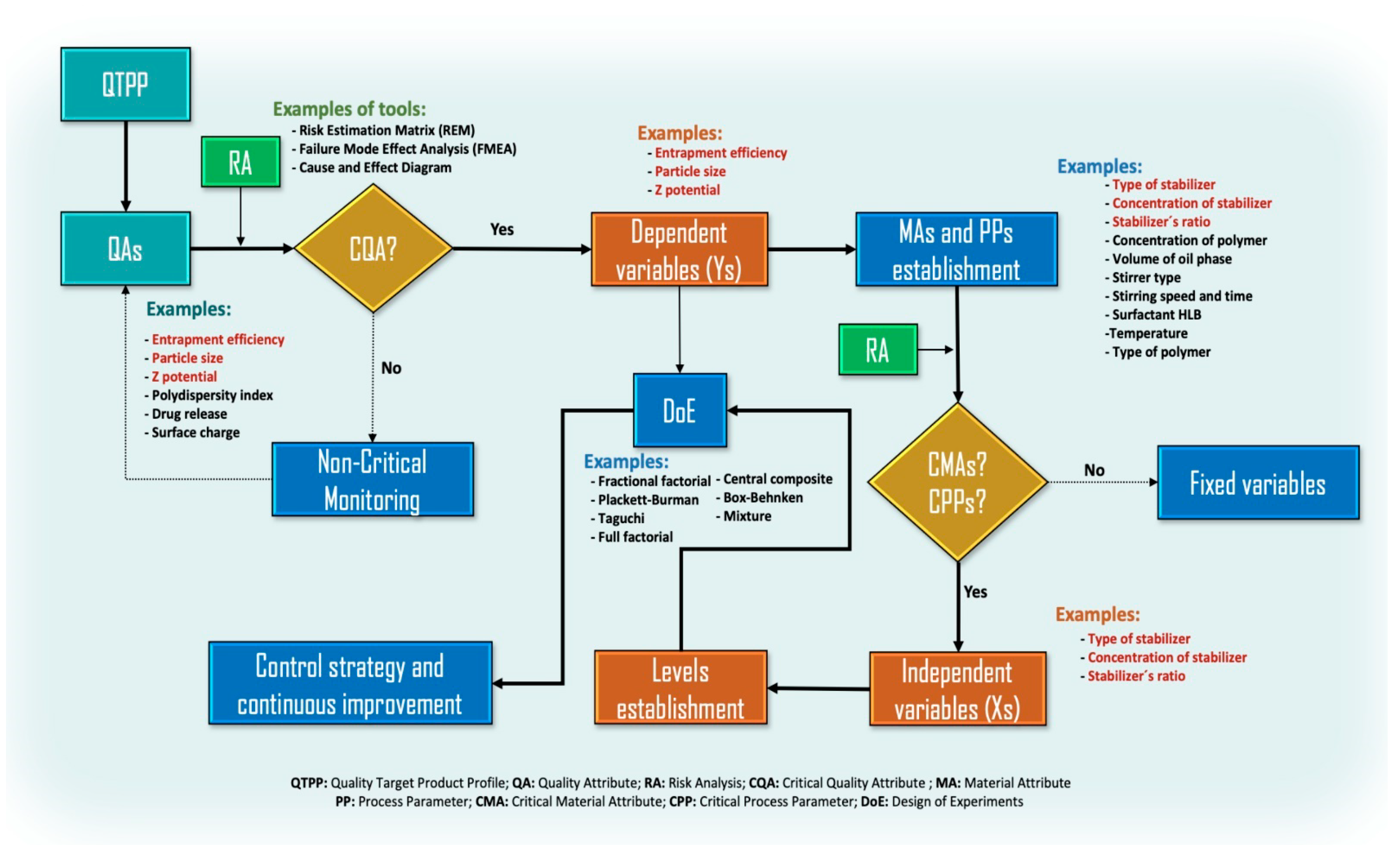
7. Quality by Design in the Choice of a Surfactant: The Royal Road
8. Drawbacks and Future
9. Conclusions
Author Contributions
Funding
Institutional Review Board Statement
Informed Consent Statement
Data Availability Statement
Acknowledgments
Conflicts of Interest
References
- Suhail, M.; Janakiraman, A.K.; Khan, A.; Naeem, A.; Badshah, S.F. Surfactants and their role in pharmaceutical product de-velopment: An overview. J. Pharm. Pharm. 2019, 6, 72–82. [Google Scholar]
- Strickley, R.G. Solubilizing excipients used in commercially available oral and injectable formulations. Pharm. Res. 2004, 21, 201–230. [Google Scholar] [CrossRef] [PubMed]
- Joshi, T. A short history and preamble of surfactants. Int. J. Appl. Chem. 2017, 13, 283–292. [Google Scholar]
- Phan, H.T.; Haes, A.J. What does nanoparticle stability mean? J. Phys. Chem. C 2019, 123, 16495–16507. [Google Scholar] [CrossRef]
- Cheraghian, G. Evaluation of clay and fumed silica nanoparticles on adsorption of surfactant polymer during enhanced oil recovery. J. Japan Pet. Inst. 2017, 60, 85–94. [Google Scholar] [CrossRef] [Green Version]
- Tapia-Guerrero, Y.S.; Del Prado-Audelo, M.L.; Borbolla-Jiménez, F.V.; Giraldo Gomez, D.M.; García-Aguirre, I.; Colín-Castro, C.A.; Morales-González, J.A.; Leyva-Gómez, G.; Magaña, J.J. Effect of UV and gamma irradiation sterilization processes in the properties of different polymeric nanoparticles for biomedical applications. Materials 2020, 13, 1090. [Google Scholar] [CrossRef] [Green Version]
- Kulkarni, S.A.; Feng, S.S. Effects of particle size and surface modification on cellular uptake and biodistribution of polymeric nanoparticles for drug delivery. Pharm. Res. 2013, 30, 2512–2522. [Google Scholar] [CrossRef]
- Salatin, S.; Maleki Dizaj, S.; Yari Khosroushahi, A. Effect of the surface modification, size, and shape on cellular uptake of nanoparticles. Cell Biol. Int. 2015, 39, 881–890. [Google Scholar] [CrossRef]
- Khandelwal, P.; Das, A.; Sen, C.K.; Srinivas, S.P.; Roy, S.; Khanna, S. A surfactant polymer wound dressing protects human keratinocytes from inducible necroptosis. Sci. Rep. 2021, 11, 1–15. [Google Scholar] [CrossRef]
- Piva, S.; DiBlasi, R.M.; Slee, A.E.; Jobe, A.H.; Roccaro, A.M.; Filippini, M.; Latronico, N.; Bertoni, M.; Marshall, J.C.; Portman, M.A. Surfactant therapy for COVID-19 related ARDS: A retrospective case–control pilot study. Respir. Res. 2021, 22, 1–8. [Google Scholar] [CrossRef]
- De Luca, D.; Cogo, P.; Kneyber, M.C.; Biban, P.; Semple, M.G.; Perez-Gil, J.; Conti, G.; Tissieres, P.; Rimensberger, P.C. Surfactant therapies for pediatric and neonatal ARDS: ESPNIC expert consensus opinion for future research steps. Crit. Care 2021, 25, 1–12. [Google Scholar] [CrossRef] [PubMed]
- Wilson, B.; Selvam, J.; Mukundan, G.K.; Premakumari, K.B.; Jenita, J.L. Albumin nanoparticles coated with polysorbate 80 for the targeted delivery of antiepileptic drug levetiracetam into the brain. Drug Deliv. Transl. Res. 2020, 10, 1853–1861. [Google Scholar] [CrossRef] [PubMed]
- Chintamaneni, P.K.; Krishnamurthy, P.T.; Pindiprolu, S.K.S.S. Polysorbate-80 surface modified nano-stearylamine BQCA conjugate for the management of Alzheimer’s disease. RSC Adv. 2021, 11, 5325–5334. [Google Scholar] [CrossRef]
- Yusuf, M.; Khan, M.; Alrobaian, M.M.; Alghamdi, S.A.; Warsi, M.H.; Sultana, S.; Khan, R.A. Brain targeted Polysorbate-80 coated PLGA thymoquinone nanoparticles for the treatment of Alzheimer’s disease, with biomechanistic insights. J. Drug Deliv. Sci. Technol. 2021, 61, 102214. [Google Scholar] [CrossRef]
- Shaban, S.M.; Kang, J.; Kim, D.H. Surfactants: Recent advances and their applications. Compos. Commun. 2020, 22, 100537. [Google Scholar] [CrossRef]
- Hotze, E.M.; Phenrat, T.; Lowry, G.V. Nanoparticle aggregation: Challenges to understanding transport and reactivity in the environment. J. Environ. Qual. 2010, 39, 1909–1924. [Google Scholar] [CrossRef] [PubMed] [Green Version]
- Heinz, H.; Pramanik, C.; Heinz, O.; Ding, Y.; Mishra, R.K.; Marchon, D.; Flatt, R.J.; Estrela-Lopis, I.; Llop, J.; Moya, S.; et al. Nanoparticle decoration with surfactants: Molecular interactions, assembly, and applications. Surf. Sci. Rep. 2017, 72, 1–58. [Google Scholar] [CrossRef]
- Voigt, N.; Henrich-Noack, P.; Kockentiedt, S.; Hintz, W.; Tomas, J.; Sabel, B.A. Surfactants, not size or zeta-potential influence blood-brain barrier passage of polymeric nanoparticles. Eur. J. Pharm. Biopharm. 2014, 87, 19–29. [Google Scholar] [CrossRef]
- Kogawa, A.C.; Cernic, B.G.; do Couto, L.G.D.; Salgado, H.R.N. Synthetic detergents: 100 years of history. Saudi Pharm. J. 2017, 25, 934–938. [Google Scholar] [CrossRef]
- Myers, E.G. Soap and Detergents. In Inedible Meat By-Products; Springer: Berlin/Heidelberg, Germany, 1992; pp. 149–176. [Google Scholar]
- Lagaly, G.; Weiss, A. Anordnung und Orientierung kationischer Tenside auf Silicatoberflächen—II. Paraffinähnliche Strukturen bei den n-Alkylammonium-Schichtsilicaten mit hoher Schichtladung (Glimmer). Kolloid-Zeitschrift Zeitschrift für Polymere 1970, 237, 364–368. [Google Scholar] [CrossRef]
- Meyer, R.A.; Green, J.J. Shaping the future of nanomedicine: Anisotropy in polymeric nanoparticle design. Wiley Interdiscip. Rev. Nanomed. Nanobiotechnology 2016, 8, 191–207. [Google Scholar] [CrossRef] [Green Version]
- Wu, L.; Zhang, J.; Watanabe, W. Physical and chemical stability of drug nanoparticles. Adv. Drug Deliv. Rev. 2011, 63, 456–469. [Google Scholar] [CrossRef]
- Trefalt, G.; Montes Ruiz-Cabello, F.J.; Borkovec, M. Interaction forces, heteroaggregation, and deposition involving charged colloidal particles. J. Phys. Chem. B 2014, 118, 6346–6355. [Google Scholar] [CrossRef]
- Pazmino, E.; Trauscht, J.; Dame, B.; Johnson, W.P. Power law size-distributed heterogeneity explains colloid retention on soda lime glass in the presence of energy barriers. Langmuir 2014, 30, 5412–5421. [Google Scholar] [CrossRef]
- Sun, H.; Jiao, R.; An, G.; Xu, H.; Wang, D.; Lee, D.J. Influence of particle size on the aggregation behavior of nanoparticles: Role of structural hydration layer. J. Environ. Sci. 2021, 103, 33–42. [Google Scholar] [CrossRef] [PubMed]
- Arab, D.; Kantzas, A.; Bryant, S.L. Nanoparticle stabilized oil in water emulsions: A critical review. J. Pet. Sci. Eng. 2018, 163, 217–242. [Google Scholar] [CrossRef]
- Leyva-Gómez, G.; Cortés, H.; Magaña, J.J.; Leyva-García, N.; Quintanar-Guerrero, D.; Florán, B. Nanoparticle technology for treatment of Parkinson’s disease: The role of surface phenomena in reaching the brain. Drug Discov. Today 2015, 20, 824–837. [Google Scholar] [CrossRef]
- Bhakay, A.; Rahman, M.; Dave, R.N.; Bilgili, E. Bioavailability enhancement of poorly water-soluble drugs via nanocomposites: Formulation–Processing aspects and challenges. Pharmaceutics 2018, 10, 86. [Google Scholar] [CrossRef] [PubMed] [Green Version]
- Bilgili, E.; Li, M.; Afolabi, A. Is the combination of cellulosic polymers and anionic surfactants a good strategy for ensuring physical stability of BCS Class II drug nanosuspensions? Pharm. Dev. Technol. 2016, 21, 499–510. [Google Scholar] [CrossRef]
- Anestopoulos, I.; Kiousi, D.E.; Klavaris, A.; Galanis, A.; Salek, K.; Euston, S.R.; Pappa, A.; Panayiotidis, M.I. Surface active agents and their health-promoting properties: Molecules of multifunctional significance. Pharmaceutics 2020, 12, 688. [Google Scholar] [CrossRef]
- Rapp, B.E. Surface Tension. In Microfluidics: Modelling, Mechanics and Mathematics; Elsevier: Amsterdam, The Netherlands, 2017; pp. 421–444. [Google Scholar]
- Clendennen, S.K.; Boaz, N.W. Betaine Amphoteric Surfactants—Synthesis, Properties, and Applications. In Biobased Surfactants; AOCS Press: Urbana, IL, USA, 2019; pp. 447–469. [Google Scholar]
- Afra, S.; Samouei, H.; Truong, P.; Nasr-El-Din, H. Micellar growth and network formation in acidic solutions of a sulfobetaine zwitterionic surfactant triggered by an inorganic salt. Soft Matter 2020, 16, 4494–4501. [Google Scholar] [CrossRef] [PubMed]
- Lin, W.; Kampf, N.; Klein, J. Designer Nanoparticles as Robust Superlubrication Vectors. ACS Nano 2020, 14, 7008–7017. [Google Scholar] [CrossRef] [PubMed]
- Sekhon, B.S. Surfactants: Pharmaceutical and Medicinal Aspects. J. Pharm. Technol. Res. Manag. 2013, 1, 43–68. [Google Scholar] [CrossRef] [Green Version]
- Zakharova, L.Y.; Pashirova, T.N.; Doktorovova, S.; Fernandes, A.R.; Sanchez-Lopez, E.; Silva, A.M.; Souto, S.B.; Souto, E.B. Cationic surfactants: Self-assembly, structure-activity correlation and their biological applications. Int. J. Mol. Sci. 2019, 20, 5534. [Google Scholar] [CrossRef] [PubMed] [Green Version]
- Benhur, A.M.; Diaz, J.; Amin, S. Impact of polyelectrolyte-surfactant interactions on the rheology and wet lubrication performance of conditioning shampoo. Int. J. Cosmet. Sci. 2021, 43, 246–253. [Google Scholar] [CrossRef]
- Smułek, W.; Burlaga, N.; Hricovíni, M.; Medveďová, A.; Kaczorek, E.; Hricovíniová, Z. Evaluation of surface active and antimicrobial properties of alkyl D-lyxosides and alkyl L-rhamnosides as green surfactants. Chemosphere 2021, 271, 129818. [Google Scholar] [CrossRef]
- Keck, C.M.; Kovačević, A.; Müller, R.H.; Savić, S.; Vuleta, G.; Milić, J. Formulation of solid lipid nanoparticles (SLN): The value of different alkyl polyglucoside surfactants. Int. J. Pharm. 2014, 474, 33–41. [Google Scholar] [CrossRef]
- Souza, F.D.; Souza, B.S.; Tondo, D.W.; Leopoldino, E.C.; Fiedler, H.D.; Nome, F. Imidazolium-based zwitterionic surfactants: Characterization of normal and reverse micelles and stabilization of nanoparticles. Langmuir 2015, 31, 3587–3595. [Google Scholar] [CrossRef]
- Kovačević, A.B.; Müller, R.H.; Savić, S.D.; Vuleta, G.M.; Keck, C.M. Solid lipid nanoparticles (SLN) stabilized with polyhydroxy surfactants: Preparation, characterization and physical stability investigation. Colloids Surf. A Physicochem. Eng. Asp. 2014, 444, 15–25. [Google Scholar] [CrossRef]
- Voulgaridou, G.P.; Mantso, T.; Anestopoulos, I.; Klavaris, A.; Katzastra, C.; Kiousi, D.E.; Mantela, M.; Galanis, A.; Gardikis, K.; Banat, I.M.; et al. Toxicity profiling of biosurfactants produced by novel marine bacterial strains. Int. J. Mol. Sci. 2021, 22, 2383. [Google Scholar] [CrossRef]
- Sanjivkumar, M.; Deivakumari, M.; Immanuel, G. Investigation on spectral and biomedical characterization of rhamnolipid from a marine associated bacterium Pseudomonas aeruginosa (DKB1). Arch. Microbiol. 2021. [Google Scholar] [CrossRef]
- Razafindralambo, H. Carbohydrate-Based Surfactants: Structure-Activity Relationships; Blecker, C., Ed.; IntechOpen: Rijeka, Croatia, 2012; pp. 215–228. [Google Scholar]
- Charoensit, P.; Pompimon, W.; Khorana, N.; Sungthongjeen, S. Effect of amide linkage of PEG-lipid conjugates on the stability and cytotoxic activity of goniodiol loaded in PEGylated liposomes. J. Drug Deliv. Sci. Technol. 2019, 50, 1–8. [Google Scholar] [CrossRef]
- Su, Y.; Tang, W.; Song, Y.; Wang, C.; Tian, Q.; Wang, X.; Quan, J.; Li, B.; Wang, S.; Deng, Y. Mixed PEGylated surfactant modifying system decrease the accelerated blood clearance phenomenon of nanoemulsions in rats. Asian J. Pharm. Sci. 2017, 12, 28–36. [Google Scholar] [CrossRef] [PubMed] [Green Version]
- Summary of the Public Assessment Report for Pfizer/BioNTech COVID-19 Vaccine. Available online: https://www.gov.uk/government/publications/regulatory-approval-of-pfizer-biontech-vaccine-for-covid-19/summary-public-assessment-report-for-pfizerbiontech-covid-19-vaccine (accessed on 23 May 2021).
- Polack, F.P.; Thomas, S.J.; Kitchin, N.; Absalon, J.; Gurtman, A.; Lockhart, S.; Perez, J.L.; Pérez Marc, G.; Moreira, E.D.; Zerbini, C.; et al. Safety and Efficacy of the BNT162b2 mRNA Covid-19 Vaccine. N. Engl. J. Med. 2020, 383, 2603–2615. [Google Scholar] [CrossRef] [PubMed]
- Hofman, K.; Shenoy, G.N.; Chak, V.; Balu-Iyer, S.V. Pharmaceutical Aspects and Clinical Evaluation of COVID-19 Vaccines. Immunol. Investig. 2021, 0, 1–37. [Google Scholar] [CrossRef]
- Devalapally, H.; Duan, Z.; Seiden, M.V.; Amiji, M.M. Paclitaxel and ceramide co-administration in biodegradable polymeric nanoparticulate delivery system to overcome drug resistance in ovarian cancer. Int. J. Cancer 2007, 121, 1830–1838. [Google Scholar] [CrossRef]
- Chevalier, Y.; Bolzinger, M.A. Emulsions stabilized with solid nanoparticles: Pickering emulsions. Colloids Surf. A Physicochem. Eng. Asp. 2013, 439, 23–34. [Google Scholar] [CrossRef]
- Pickering, S.U. CXCVI.—Emulsions. J. Chem. Soc. Trans. 1907, 91, 2001–2021. [Google Scholar] [CrossRef] [Green Version]
- Robin, B.; Albert, C.; Beladjine, M.; Legrand, F.-X.; Geiger, S.; Moine, L.; Nicolas, V.; Canette, A.; Trichet, M.; Tsapis, N.; et al. Tuning morphology of Pickering emulsions stabilised by biodegradable PLGA nanoparticles: How PLGA characteristics influence emulsion properties. J. Colloid Interface Sci. 2021, 595. [Google Scholar] [CrossRef]
- Food Safety Commission. Evaluation Report of Food Additives Polysorbates (Polysorbates 20, 60, 65 and 80); Food Safety Commission: Tokyo, Japan, 2007. [Google Scholar]
- Martos, A.; Koch, W.; Jiskoot, W.; Wuchner, K.; Winter, G.; Friess, W.; Hawe, A. Trends on analytical characterization of polysorbates and their degradation products in biopharmaceutical formulations. J. Pharm. Sci. 2017, 106, 1722–1735. [Google Scholar] [CrossRef]
- Kishore, R.S.K.; Kiese, S.; Fischer, S.; Pappenberger, A.; Grauschopf, U.; Mahler, H.C. The degradation of polysorbates 20 and 80 and its potential impact on the stability of biotherapeutics. Pharm. Res. 2011, 28, 1194–1210. [Google Scholar] [CrossRef] [PubMed]
- Kerwin, B.A. Polysorbates 20 and 80 used in the formulation of protein biotherapeutics: Structure and degradation pathways. J. Pharm. Sci. 2008, 97, 2924–2935. [Google Scholar] [CrossRef]
- Moore, J. Final Report on the Safety Assessment of Polysorbates 20, 21, 40, 60, 61, 65, 80, 81, and 85. J. Am. Coll. Toxicol. 1984, 3, 1–82. [Google Scholar]
- Hoda, M.; Pajaniradje, S.; Shakya, G.; Mohankumar, K.; Rajagopalan, R. Anti-proliferative and apoptosis-triggering potential of disulfiram and disulfiram-loaded polysorbate 80-stabilized PLGA nanoparticles on hepatocellular carcinoma Hep3B cell line. Nanomed. Nanotechnol. Biol. Med. 2016, 12, 1641–1650. [Google Scholar] [CrossRef]
- Sinha, N.; Joshi, A.S.; Thakur, A.K. Analytical validation of an ATR-FTIR based method for quantifying the amount of polysorbate 80 adsorbed on PLGA nanoparticles. Anal. Methods 2020, 12, 5360–5366. [Google Scholar] [CrossRef]
- Katiyar, S.S.; Muntimadugu, E.; Rafeeqi, T.A.; Domb, A.J.; Khan, W. Co-delivery of rapamycin- and piperine-loaded polymeric nanoparticles for breast cancer treatment. Drug Deliv. 2016, 23, 2608–2616. [Google Scholar] [CrossRef]
- Onyema, H.N.; Berger, M.; Musyanovych, A.; Bantz, C.; Maskos, M.; Freese, C. Uptake of polymeric nanoparticles in a human induced pluripotent stem cell-based blood–brain barrier model: Impact of size, material, and protein corona. Biointerphases 2021, 16, 021004. [Google Scholar] [CrossRef]
- Nguyen, H.T.; Tran, T.H.; Kim, J.O.; Yong, C.S.; Nguyen, C.N. Enhancing the in vitro anti-cancer efficacy of artesunate by loading into poly-d,l-lactide-co-glycolide (PLGA) nanoparticles. Arch. Pharm. Res. 2015, 38, 716–724. [Google Scholar] [CrossRef] [PubMed]
- Jose, S.; Sowmya, S.; Cinu, T.A.; Aleykutty, N.A.; Thomas, S.; Souto, E.B. Surface modified PLGA nanoparticles for brain targeting of Bacoside-A. Eur. J. Pharm. Sci. 2014, 63, 29–35. [Google Scholar] [CrossRef] [PubMed]
- Tahara, K.; Yamamoto, H.; Kawashima, Y. Cellular uptake mechanisms and intracellular distributions of polysorbate 80-modified poly (d,l-lactide-co-glycolide) nanospheres for gene delivery. Eur. J. Pharm. Biopharm. 2010, 75, 218–224. [Google Scholar] [CrossRef] [PubMed]
- Yuan, X.; Ji, W.; Chen, S.; Bao, Y.; Tan, S.; Lu, S.; Wu, K.; Chu, Q. Anovel paclitaxel-loaded poly(d,l-lactide-co-glycolide)-Tween 80 copolymer nanoparticle overcoming multidrug resistance for lung cancer treatment. Int. J. Nanomed. 2016, 11, 2119–2131. [Google Scholar]
- Xu, R.; Wang, J.; Xu, J.; Song, X.; Huang, H.; Feng, Y.; Fu, C. Rhynchophylline loaded-mPEG-PLGA nanoparticles coated with tween-80 for preliminary study in Alzheimer’s disease. Int. J. Nanomed. 2020, 15, 1149–1160. [Google Scholar] [CrossRef] [PubMed] [Green Version]
- Das, D.; Lin, S. Double-coated poly (butylcynanoacrylate) nanoparticulate delivery systems for brain targeting of dalargin via oral administration. J. Pharm. Sci. 2005, 94, 1343–1353. [Google Scholar] [CrossRef] [PubMed]
- Al-Kassas, R.; Madni, A.; Buchanan, C.; Shelling, A.N. pH-Sensitive Nanoparticles Developed and Optimized Using Factorial Design for Oral Delivery of Gliclazide. J. Pharm. Innov. 2021. [Google Scholar] [CrossRef]
- Neibloom, D.; Bevan, M.A.; Frechette, J. Surfactant-Stabilized Spontaneous 3-(Trimethoxysilyl) Propyl Methacrylate Nanoemulsions. Langmuir 2020, 36, 284–292. [Google Scholar] [CrossRef] [PubMed]
- Mahmoudi, M.; Saeidian, H.; Mirjafary, Z.; Mokhtari, J. Preparation and characterization of memantine loaded polycaprolactone nanocapsules for Alzheimer’s disease. J. Porous Mater. 2021, 28, 205–212. [Google Scholar] [CrossRef]
- Ahsaei, S.M.; Talebi-Jahromi, K.; Amoabediny, G. Insecticidal activity of polycaprolactone nanoparticles decorated with chitosan containing two essential oils against Tribolium confusum. Int. J. Pest Manag. 2020, 1, 1–9. [Google Scholar] [CrossRef]
- Badri, W.; Miladi, K.; Nazari, Q.A.; Fessi, H.; Elaissari, A. Effect of process and formulation parameters on polycaprolactone nanoparticles prepared by solvent displacement. Colloids Surf. A Physicochem. Eng. Asp. 2017, 516, 238–244. [Google Scholar] [CrossRef]
- Elmowafy, M.; Alruwaili, N.K.; Shalaby, K.; Alharbi, K.S.; Altowayan, W.M.; Ahmed, N.; Zafar, A.; Elkomy, M. Long-acting paliperidone parenteral formulations based on polycaprolactone nanoparticles; the influence of stabilizer and chitosan on in vitro release, protein adsorption, and cytotoxicity. Pharmaceutics 2020, 12, 160. [Google Scholar] [CrossRef] [Green Version]
- Amrite, A.C.; Edelhauser, H.F.; Singh, S.R.; Kompella, U.B. Effect of circulation on the disposition and ocular tissue distribution of 20 nm nanoparticles after periocular administration. Mol. Vis. 2008, 14, 150–160. [Google Scholar]
- Fu, J.; Wu, C. Laser light scattering study of the degradation of poly(sebacic anhydride) nanoparticles. J. Polym. Sci. Part B Polym. Phys. 2001, 39, 703–708. [Google Scholar] [CrossRef] [Green Version]
- Shakeri, F.; Shakeri, S.; Hojjatoleslami, M. Preparation and characterization of carvacrol loaded polyhydroxybutyrate nanoparticles by nanoprecipitation and dialysis methods. J. Food Sci. 2014, 79, N697–N705. [Google Scholar] [CrossRef]
- Sukmawati, A.; Utami, W.; Yuliani, R.; Da’I, M.; Nafarin, A. Effect of tween 80 on nanoparticle preparation of modified chitosan for targeted delivery of combination doxorubicin and curcumin analogue. IOP Conf. Ser. Mater. Sci. Eng. 2018, 311, 012024. [Google Scholar] [CrossRef]
- Talib, S.; Ahmed, N.; Khan, D.; Khan, G.M.; Rehman, A. Ur Chitosan-chondroitin based artemether loaded nanoparticles for transdermal drug delivery system. J. Drug Deliv. Sci. Technol. 2021, 61, 102281. [Google Scholar] [CrossRef]
- Bhattacharya, S. Fabrication and characterization of chitosan-based polymeric nanoparticles of Imatinib for colorectal cancer targeting application. Int. J. Biol. Macromol. 2020, 151, 104–115. [Google Scholar] [CrossRef]
- Govindaraju, R.; Karki, R.; Chandrashekarappa, J.; Santhanam, M.; Shankar, A.K.K.; Joshi, H.K.; Divakar, G. Enhanced Water Dispersibility of Curcumin Encapsulated in Alginate-Polysorbate 80 Nano Particles and Bioavailability in Healthy Human Volunteers. Pharm. Nanotechnol. 2019, 7, 39–56. [Google Scholar] [CrossRef]
- Masotti, A.; Vicennati, P.; Alisi, A.; Marianecci, C.; Rinaldi, F.; Carafa, M.; Ortaggi, G. Novel Tween® 20 derivatives enable the formation of efficient pH-sensitive drug delivery vehicles for human hepatoblastoma. Bioorganic Med. Chem. Lett. 2010, 20, 3021–3025. [Google Scholar] [CrossRef] [PubMed]
- Hartl, N.; Adams, F.; Merkel, O.M. From adsorption to covalent bonding: Apolipoprotein E functionalization of polymeric nanoparticles for drug delivery across the blood–brain barrier. Adv. Ther. 2021, 4, 1–21. [Google Scholar] [CrossRef]
- Kreuter, J.; Ramge, P.; Petrov, V.; Hamm, S.; Gelperina, S.E.; Engelhardt, B.; Alyautdin, R.; Von, H.; Begley, D.J. Direct evidence that poly (butylcyanoacrylate) nanoparticles deliver drugs to the CNS via specific mechanisms requiring prior binding of drug to the nanoparticles. Pharm. Res. 2003, 20, 409–416. [Google Scholar] [CrossRef] [PubMed]
- Sun, W.; Xie, C.; Wang, H.; Hu, Y. Specific role of polysorbate 80 coating on the targeting of nanoparticles to the brain. Biomaterials 2004, 25, 3065–3071. [Google Scholar] [CrossRef]
- Ravichandran, V.; Nguyen Cao, T.G.; Choi, D.G.; Kang, H.C.; Shim, M.S. Non-ionic polysorbate-based nanoparticles for efficient combination chemo/photothermal/photodynamic therapy. J. Ind. Eng. Chem. 2020, 88, 260–267. [Google Scholar] [CrossRef]
- National Center for Biotechnology Information. PubChem Compound Summary for CID 443314, Polysorbate 20. 2021. Available online: https://pubchem.ncbi.nlm.nih.gov/compound/Polysorbate-20 (accessed on 27 April 2021).
- Chang, J.; Jallouli, Y.; Kroubi, M.; Yuan, X.; Feng, W.; Kang, C.; Pu, P.; Betbeder, D. Characterization of endocytosis of transferrin-coated PLGA nanoparticles by the blood–brain barrier. Int. J. Pharm. 2009, 379, 285–292. [Google Scholar] [CrossRef] [PubMed]
- Coors, E.A.; Seybold, H.; Merk, H.F.; Mahler, V. Polysorbate 80 in medical products and nonimmunologic anaphylactoid reactions. Ann. Allergy Asthma Immunol. 2005, 95, 593–599. [Google Scholar] [CrossRef]
- Schwartzberg, L.S.; Navari, R.M. Safety of Polysorbate 80 in the Oncology Setting. Adv. Ther. 2018, 35, 754–767. [Google Scholar] [CrossRef] [PubMed] [Green Version]
- Helgason, T.; Awad, T.S.; Kristbergsson, K.; McClements, D.J.; Weiss, J. Effect of surfactant surface coverage on formation of solid lipid nanoparticles (SLN). J. Colloid Interface Sci. 2009, 334, 75–81. [Google Scholar] [CrossRef]
- Grabarek, A.D.; Bozic, U.; Rousel, J.; Menzen, T.; Kranz, W.; Wuchner, K.; Jiskoot, W.; Hawe, A. What Makes Polysorbate Functional? Impact of Polysorbate 80 Grade and Quality on IgG Stability During Mechanical Stress. J. Pharm. Sci. 2020, 109, 871–880. [Google Scholar] [CrossRef] [PubMed] [Green Version]
- Alade, O.S.; Al Shehri, D.; Mahmoud, M.; Mokheimer, E.M.A.; Al Hamad, J.; Kamal, M.S.; Al-Nakhli, A.; Sasaki, K. A novel technique for heavy oil recovery using poly vinyl alcohol (PVA) and PVA-NaOH with ethanol additive. Fuel 2021, 285, 119128. [Google Scholar] [CrossRef]
- Park, M.J.; Gonzales, R.R.; Abdel-Wahab, A.; Phuntsho, S.; Shon, H.K. Hydrophilic polyvinyl alcohol coating on hydrophobic electrospun nanofiber membrane for high performance thin film composite forward osmosis membrane. Desalination 2018, 426, 50–59. [Google Scholar] [CrossRef]
- Hago, E.E.; Li, X. Interpenetrating polymer network hydrogels based on gelatin and PVA by biocompatible approaches: Synthesis and characterization. Adv. Mater. Sci. Eng. 2013, 2013, 328763. [Google Scholar] [CrossRef] [Green Version]
- Zarekhalili, Z.; Bahrami, S.H.; Ranjbar-Mohammadi, M.; Milan, P.B. Fabrication and characterization of PVA/Gum tragacanth/PCL hybrid nanofibrous scaffolds for skin substitutes. Int. J. Biol. Macromol. 2017, 94, 679–690. [Google Scholar] [CrossRef]
- Scholes, P.D.; Coombes, A.G.A.; Illum, L.; Daviz, S.S.; Vert, M.; Davies, M.C. The preparation of sub-200 nm poly(lactide-co-glycolide) microspheres for site-specific drug delivery. J. Control. Release 1993, 25, 145–153. [Google Scholar] [CrossRef]
- Zambaux, M.F.; Bonneaux, F.; Gref, R.; Maincent, P.; Dellacherie, E.; Alonso, M.J.; Labrude, P.; Vigneron, C. Influence of experimental parameters on the characteristics of poly(lactic acid) nanoparticles prepared by a double emulsion method. J. Control. Release 1998, 50, 31–40. [Google Scholar] [CrossRef]
- Carrio, A.; Schwach, G.; Coudane, J.; Vert, M. Preparation and degradation of surfactant-free PLAGA microspheres. J. Control. Release 1995, 37, 113–121. [Google Scholar] [CrossRef]
- Hallensleben, M.L. Polyvinyl Compounds, Others. In Ullmann’s Encyclopedia of Industrial Chemistry; Wiley-VCH: Weinheim, Germany, 2000; pp. 1–23. [Google Scholar]
- Kawai, F.; Hu, X. Biochemistry of microbial polyvinyl alcohol degradation. Appl. Microbiol. Biotechnol. 2009, 84, 227–237. [Google Scholar] [CrossRef]
- C Haweel, S.A. Preparation of polyvinyl alcohol from local raw material. Iraqi J. Chem. Pet. Eng. 2008, 9, 15–21. [Google Scholar]
- Shahbazi, M.; Rajabzadeh, G.; Rafe, A.; Ettelaie, R.; Ahmadi, S.J. The physico-mechanical and structural characteristics of blend film of poly (vinyl alcohol) with biodegradable polymers as affected by disorder-to-order conformational transition. Food Hydrocoll. 2016, 60, 393–404. [Google Scholar] [CrossRef] [Green Version]
- Chiellini, E.; Corti, A.; D’Antone, S.; Solaro, R. Biodegradation of poly (vinyl alcohol) based materials. Prog. Polym. Sci. 2003, 28, 963–1014. [Google Scholar] [CrossRef]
- Baker, M.I.; Walsh, S.P.; Schwartz, Z.; Boyan, B.D. A review of polyvinyl alcohol and its uses in cartilage and orthopedic applications. J. Biomed. Mater. Res. 2012, 100, 1451–1457. [Google Scholar] [CrossRef] [PubMed]
- Tacx, J.C.J.F.; Schoffeleers, H.M.; Brands, A.G.M.; Teuwen, L. Dissolution behavior and solution properties of polyvinylalcohol as determined by viscometry and light scattering in DMSO, ethyleneglycol and water. Polymer 2000, 41, 947–957. [Google Scholar] [CrossRef]
- Tang, X.; Alavi, S. Recent advances in starch, polyvinyl alcohol based polymer blends, nanocomposites and their biodegradability. Carbohydr. Polym. 2011, 85, 7–16. [Google Scholar] [CrossRef]
- Wang, Y.; Zhang, W.; Li, J.; Fu, J. A novel LEuH/PVA luminescent hydrogel with ammonia response and self-recovery luminescence behavior. New J. Chem. 2019, 43, 5133–5138. [Google Scholar] [CrossRef]
- Bhatt, R.; Bajpai, A.K. Solubility behavior of poly (diaminonaphthalene) doped poly (vinyl alcohol) films in different solvents and structural and electrical characterization of corresponding films. Mater. Res. Express 2019, 6, 105330. [Google Scholar] [CrossRef]
- Jelinska, N.; Kalnins, M.; Tupureina, V.; Dzene, A. Poly (vinyl alcohol)/poly (vinyl acetate) blend films. Sci. J. Riga Tech. Univ. 2010, 21, 55–61. [Google Scholar]
- Gholap, S.G.; Jog, J.P.; Badiger, M.V. Synthesis and characterization of hydrophobically modified poly(vinyl alcohol) hydrogel membrane. Polymer 2004, 45, 5863–5873. [Google Scholar] [CrossRef]
- Nguyen, D.; Balsamo, V. Emulsification of heavy oil in aqueous solutions of poly(vinyl alcohol): A method for reducing apparent viscosity of production fluids. Energy and Fuels 2013, 27, 1736–1747. [Google Scholar] [CrossRef]
- Raffa, P.; Broekhuis, A.A.; Picchioni, F. Polymeric surfactants for enhanced oil recovery: A review. J. Pet. Sci. Eng. 2016, 145, 723–733. [Google Scholar] [CrossRef] [Green Version]
- Demerlis, C.C.; Schoneker, D.R. Review of the oral toxicity of polyvinyl alcohol (PVA). Food Chem. Toxicol. 2003, 41, 319–326. [Google Scholar] [CrossRef]
- Alves, M.H.; Jensen, B.E.B.; Smith, A.A.A.; Zelikin, A.N. Poly(vinyl alcohol) physical hydrogels: New vista on a long serving biomaterial. Macromol. Biosci. 2011, 11, 1293–1313. [Google Scholar] [CrossRef]
- Turk, C.T.S.; Oz, U.C.; Serim, T.M.; Hascicek, C. Formulation and optimization of nonionic surfactants emulsified nimesulide-loaded PLGA-based nanoparticles by design of experiments. AAPS PharmSciTech 2014, 15, 161–176. [Google Scholar] [CrossRef] [Green Version]
- Rowe, R.C.; Sheskey, P.J.; Quinn, M.E. Handbook of Pharmaceutical Excipients; Libros Digitales-Pharmaceutical Press: London, UK, 2009; ISBN 9782705690229. [Google Scholar]
- Gul, M.O.; Jones, S.A.; Dailey, L.A.; Nacer, H.; Ma, Y.; Sadouki, F.; Hider, R.; Araman, A.; Forbes, B. A poly(vinyl alcohol) nanoparticle platform for kinetic studies of inhaled particles. Inhal. Toxicol. 2009, 21, 631–640. [Google Scholar] [CrossRef]
- Marin, E.; Rojas, J.; Ciro, Y. A review of polyvinyl alcohol derivatives: Promising materials for pharmaceutical and biomedical applications. Afr. J. Pharm. Pharmacol. 2014, 8, 674–684. [Google Scholar]
- Rodríguez-Cruz, I.M.; Domínguez-Delgado, C.L.; Escobar-Chávez, J.J.; Leyva-Gómez, G.; Ganem-Quintanar, A.; Quintanar-Guerrero, D. Nanoparticle infiltration to prepare solvent-free controlled drug delivery systems. Int. J. Pharm. 2009, 371, 177–181. [Google Scholar] [CrossRef]
- Gebreel, R.M.; Edris, N.A.; Elmofty, H.M.; Tadros, M.I.; El-Nabarawi, M.A.; Hassan, D.H. Development and characterization of PLGA nanoparticle-laden hydrogels for sustained ocular delivery of norfloxacin in the treatment of pseudomonas keratitis: An experimental study. Drug Des. Devel. Ther. 2021, 15, 399–418. [Google Scholar] [CrossRef]
- Qiu, S.; Ge, N.J.; Sun, D.K.; Zhao, S.; Sun, J.F.; Guo, Z.B.; Hu, K.; Gu, N. Synthesis and characterization of magnetic Polyvinyl Alcohol (PVA) hydrogel microspheres for the embolization of blood vessel. IEEE Trans. Biomed. Eng. 2016, 63, 730–736. [Google Scholar]
- Covey, A.M.; Tuorto, S.; Brody, L.A.; Sofocleous, C.T.; Schubert, J.; von Tengg-Kobligk, H.; Getrajdman, G.I.; Schwartz, L.H.; Fong, Y.; Brown, K.T. Safety and efficacy of preoperative portal vein embolization with polyvinyl alcohol in 58 patients with liver metastases. Am. J. Roentgenol. 2005, 185, 1620–1626. [Google Scholar] [CrossRef]
- Lang, R.A.; Grüntzig, P.M.; Weisgerber, C.; Weis, C.; Odermatt, E.K.; Kirschner, M.H. Polyvinyl alcohol gel prevents abdominal adhesion formation in a rabbit model. Fertil. Steril. 2007, 88, 1180–1186. [Google Scholar] [CrossRef]
- Weis, C.; Odermatt, E.K.; Kressler, J.; Funke, Z.; Wehner, T.; Freytag, D. Poly(vinyl alcohol) membranes for adhesion prevention. J. Biomed. Mater. Res. 2004, 70, 191–202. [Google Scholar] [CrossRef]
- Zhao, X.; Xiong, D.; Liu, Y. Improving surface wettability and lubrication of polyetheretherketone (PEEK) by combining with polyvinyl alcohol (PVA) hydrogel. J. Mech. Behav. Biomed. Mater. 2018, 82, 27–34. [Google Scholar] [CrossRef]
- Zhang, J.; Lei, W.; Chen, J.; Liu, D.; Tang, B.; Li, J.; Wang, X. Enhancing the thermal and mechanical properties of polyvinyl alcohol (PVA) with boron nitride nanosheets and cellulose nanocrystals. Polymer 2018, 148, 101–108. [Google Scholar] [CrossRef]
- Habiba, U.; Lee, J.J.L.; Joo, T.C.; Ang, B.C.; Afifi, A.M. Degradation of methyl orange and congo red by using chitosan/polyvinyl alcohol/TiO2 electrospun nanofibrous membrane. Int. J. Biol. Macromol. 2019, 131, 821–827. [Google Scholar] [CrossRef]
- Mahmoodi, N.M.; Mokhtari-Shourijeh, Z.; Abdi, J. Preparation of mesoporous polyvinyl alcohol/chitosan/silica composite nanofiber and dye removal from wastewater. Environ. Prog. Sustain. Energy 2019, 38, S100–S109. [Google Scholar] [CrossRef]
- Karim, M.R.; Aijaz, M.O.; Alharth, N.H.; Alharbi, H.F.; Al-Mubaddel, F.S.; Awual, M.R. Composite nanofibers membranes of poly(vinyl alcohol)/chitosan for selective lead(II) and cadmium(II) ions removal from wastewater. Ecotoxicol. Environ. Saf. 2019, 169, 479–486. [Google Scholar] [CrossRef]
- Do Nascimento, F.C.; de Aguiar, L.C.V.; Costa, L.A.T.; Fernandes, M.T.; Marassi, R.J.; de S. Gomes, A.; de Castro, J.A. Formulation and characterization of crosslinked polyvinyl alcohol (PVA) membranes: Effects of the crosslinking agents. Polym. Bull. 2021, 78, 917–929. [Google Scholar] [CrossRef]
- Reddy, N.; Chen, L.; Zhang, Y.; Yang, Y. Reducing environmental pollution of the textile industry using keratin as alternative sizing agent to poly(vinyl alcohol). J. Clean. Prod. 2014, 65, 561–567. [Google Scholar] [CrossRef]
- Kim, J.W.; Park, H.; Lee, G.; Jeong, Y.R.; Hong, S.Y.; Keum, K.; Yoon, J.; Kim, M.S.; Ha, J.S. Paper-like, thin, foldable, and self-healable electronics based on PVA/CNC nanocomposite film. Adv. Funct. Mater. 2019, 29, 1–14. [Google Scholar] [CrossRef]
- Sajjan, A.M.; Naik, M.L.; Kulkarni, A.S.; Fazal-E-Habiba Rudgi, U.; M, A.; Shirnalli, G.G.; A, S.; Kalahal, P.B. Preparation and characterization of PVA-Ge/PEG-400 biodegradable plastic blend films for packaging applications. Chem. Data Collect. 2020, 26, 100338. [Google Scholar] [CrossRef]
- Kaboorani, A.; Riedl, B. Effects of adding nano-clay on performance of polyvinyl acetate (PVA) as a wood adhesive. Compos. Part A Appl. Sci. Manuf. 2011, 42, 1031–1039. [Google Scholar] [CrossRef]
- Abdulsalam, S. Production of emulsion house paint using polyvinyl acetate and gum arabic as binder. Int. J. Mater. Sci. Appl. 2015, 4, 350. [Google Scholar] [CrossRef] [Green Version]
- Abdullah, Z.W.; Dong, Y.; Davies, I.J.; Barbhuiya, A. PVA, PVA blends and their nanocomposites for biodegradable packaging application. Polym. Plast. Technol. Eng. 2017, 56, 1307–1344. [Google Scholar] [CrossRef] [Green Version]
- Hassan, C.M.; Peppas, N.A. Structure and applications of poly(vinyl alcohol) hydrogels produced by conventional crosslinking or by freezing/thawing methods. Adv. Polym. Sci. 2000, 153, 37–65. [Google Scholar]
- Schubert, J.; Chanana, M. Coating matters: Review on colloidal stability of nanoparticles with biocompatible coatings in biological media, living cells and organisms. Curr. Med. Chem. 2018, 25, 4553–4586. [Google Scholar] [CrossRef] [PubMed]
- Bolto, B.; Tran, T.; Hoang, M.; Xie, Z. Crosslinked poly(vinyl alcohol) membranes. Prog. Polym. Sci. 2009, 34, 969–981. [Google Scholar] [CrossRef]
- Chen, K.; Chen, G.; Wei, S.; Yang, X.; Zhang, D.; Xu, L. Preparation and property of high strength and low friction PVA-HA/PAA composite hydrogel using annealing treatment. Mater. Sci. Eng. C 2018, 91, 579–588. [Google Scholar] [CrossRef]
- Cieśla, K.; Abramowska, A. Effect of absorbed dose on starch:PVA films irradiated with gamma rays. Radiat. Phys. Chem. 2021, 180, 109290. [Google Scholar] [CrossRef]
- Yu, C.; Li, B. Preparation and Characterization of carboxymethyl polyvinyl alcohol-graphite nanosheet composites. Polym. Compos. 2008, 29, 998–1005. [Google Scholar] [CrossRef]
- Zhao, X.; Zhang, Q.; Chen, D.; Lu, P. Enhanced mechanical properties of graphene-based polyvinyl alcohol composites. Macromolecules 2010, 43, 2357–2363. [Google Scholar] [CrossRef]
- Gohil, J.M.; Bhattacharya, A.; Ray, P. Studies on the cross-linking of poly (vinyl alcohol). J. Polym. Res. 2006, 13, 161–169. [Google Scholar] [CrossRef]
- Patel, A.R.; Vavia, P.R. Evaluation of synthesized cross linked polyvinyl alcohol as potential disintegrant. J. Pharm. Pharm. Sci. 2010, 13, 114–127. [Google Scholar] [CrossRef] [PubMed] [Green Version]
- Mansur, H.S.; Sadahira, C.M.; Souza, A.N.; Mansur, A.A.P. FTIR spectroscopy characterization of poly (vinyl alcohol) hydrogel with different hydrolysis degree and chemically crosslinked with glutaraldehyde. Mater. Sci. Eng. C 2008, 28, 539–548. [Google Scholar] [CrossRef]
- Yu, Q.; Song, Y.; Shi, X.; Xu, C.; Bin, Y. Preparation and properties of chitosan derivative/poly(vinyl alcohol) blend film crosslinked with glutaraldehyde. Carbohydr. Polym. 2011, 84, 465–470. [Google Scholar] [CrossRef]
- Han, B.; Li, J.; Chen, C.; Xu, C.; Wickramasinghe, S.R. Effects of degree of formaldehyde acetal treatment and maleic acid crosslinking on solubility and diffusivity of water in PVA membranes. Chem. Eng. Res. Des. 2003, 81, 1385–1392. [Google Scholar] [CrossRef]
- Riyajan, S.A.; Chaiponban, S.; Tanbumrung, K. Investigation of the preparation and physical properties of a novel semi-interpenetrating polymer network based on epoxised NR and PVA using maleic acid as the crosslinking agent. Chem. Eng. J. 2009, 153, 199–205. [Google Scholar] [CrossRef]
- Tsai, C.E.; Lin, C.W.; Hwang, B.J. A novel crosslinking strategy for preparing poly(vinyl alcohol)-based proton-conducting membranes with high sulfonation. J. Power Sources 2010, 195, 2166–2173. [Google Scholar] [CrossRef]
- Loughlin, R.G.; Tunney, M.M.; Donnelly, R.F.; Murphy, D.J.; Jenkins, M.; McCarron, P.A. Modulation of gel formation and drug-release characteristics of lidocaine-loaded poly(vinyl alcohol)-tetraborate hydrogel systems using scavenger polyol sugars. Eur. J. Pharm. Biopharm. 2008, 69, 1135–1146. [Google Scholar] [CrossRef] [PubMed]
- Gulrez, S.K.H.; Al-Assaf, S.; Phillips, G.O. Hydrogels: Methods of preparation, characterization and applications. J. Vasc. Surg. 2010, 51, 1248–1259. [Google Scholar]
- Alupei, I.C.; Popa, M.; Hamcerencu, M.; Abadie, M.J.M. Superabsorbant hydrogels based on xanthan and poly(vinyl alcohol): 1. The study of the swelling properties. Eur. Polym. J. 2002, 38, 2313–2320. [Google Scholar] [CrossRef]
- Liong, M.; Shao, H.; Haun, J.B.; Lee, H.; Weissleder, R. Carboxymethylated polyvinyl alcohol stabilizes doped ferrofluids for biological applications. Adv. Mater. 2010, 22, 5168–5172. [Google Scholar] [CrossRef] [Green Version]
- Drury, J.L.; Mooney, D.J. Hydrogels for tissue engineering: Scaffold design variables and applications. Biomaterials 2003, 24, 4337–4351. [Google Scholar] [CrossRef]
- Sonker, A.K.; Rathore, K.; Nagarale, R.K.; Verma, V. Crosslinking of Polyvinyl Alcohol (PVA) and Effect of Crosslinker Shape (Aliphatic and Aromatic) Thereof. J. Polym. Environ. 2018, 26, 1782–1794. [Google Scholar] [CrossRef]
- González-Guisasola, C.; Ribes-Greus, A. Dielectric relaxations and conductivity of cross-linked PVA/SSA/GO composite membranes for fuel cells. Polym. Test. 2018, 67, 55–67. [Google Scholar] [CrossRef]
- El-Aziz, A.M.A.; El-Maghraby, A.; Taha, N.A. Comparison between polyvinyl alcohol (PVA) nanofiber and polyvinyl alcohol (PVA) nanofiber/hydroxyapatite (HA) for removal of Zn2+ ions from wastewater. Arab. J. Chem. 2017, 10, 1052–1060. [Google Scholar] [CrossRef] [Green Version]
- Lu, Y.; Liu, X.; Luo, G. Synthesis of polystyrene latex via emulsion polymerization with poly(vinyl alcohol) as sole stabilizer. J. Appl. Polym. Sci. 2017, 134, 1–9. [Google Scholar] [CrossRef]
- Siddharth, S.; Nayak, A.; Nayak, D.; Bindhani, B.K.; Kundu, C.N. Chitosan-Dextran sulfate coated doxorubicin loaded PLGA-PVA-nanoparticles caused apoptosis in doxorubicin resistance breast cancer cells through induction of DNA damage. Sci. Rep. 2017, 7, 1–10. [Google Scholar] [CrossRef]
- Liu, Z.; Jiang, M.; Kang, T.; Miao, D.; Gu, G.; Song, Q.; Yao, L.; Hu, Q.; Tu, Y.; Pang, Z.; et al. Lactoferrin-modified PEG-co-PCL nanoparticles for enhanced brain delivery of NAP peptide following intranasal administration. Biomaterials 2013, 34, 3870–3881. [Google Scholar] [CrossRef]
- Mohammadi, G.; Mirzaeei, S.; Taghe, S.; Mohammadi, P. Preparation and Evaluation of Eudragit® L100 Nanoparticles Loaded Impregnated with KT Tromethamine Loaded PVA -HEC Insertions for Ophthalmic Drug Delivery. Adv. Pharm. Bull. 2019, 9, 593–600. [Google Scholar] [CrossRef]
- Suzuki, T.; Sasai, A.; Tsujimoto, H.; Yasunaga, T.; Ogawa, N.; Yamamoto, H. Promoting effect of type 17 collagen production by chlorogenic acid using PLGA nanoparticles in the human epidermal keratinocyte cell. J. Drug Deliv. Sci. Technol. 2020, 58, 101624. [Google Scholar] [CrossRef]
- Fodor-Kardos, A.; Kiss, Á.; Monostroy, K.; Feczhó, T. Sustained in vitro interferon-beta release and in vivo toxicity of PLGA and PEG-PLGA nanoparticles. RSC Adv. 2020, 10, 15893–15900. [Google Scholar] [CrossRef] [Green Version]
- Shahgordi, S.; Sankian, M.; Yazdani, Y.; Mashayekhi, K.; Hasan Ayati, S.; Sadeghi, M.; Saeidi, M.; Hashemi, M. Immune responses modulation by curcumin and allergen encapsulated into PLGA nanoparticles in mice model of rhinitis allergic through sublingual immunotherapy. Int. Immunopharmacol. 2020, 84, 106525. [Google Scholar] [CrossRef] [PubMed]
- Sadozai, S.K.; Khan, S.A.; Karim, N.; Becker, D.; Steinbrück, N.; Gier, S.; Baseer, A.; Breinig, F.; Kickelbick, G.; Schneider, M. Ketoconazole-loaded PLGA nanoparticles and their synergism against Candida albicans when combined with silver nanoparticles. J. Drug Deliv. Sci. Technol. 2020, 56, 101574. [Google Scholar] [CrossRef]
- Sousa, F.; Cruz, A.; Fonte, P.; Pinto, I.M.; Neves-Petersen, M.T.; Sarmento, B. A new paradigm for antiangiogenic therapy through controlled release of bevacizumab from PLGA nanoparticles. Sci. Rep. 2017, 7, 1–13. [Google Scholar] [CrossRef]
- Yenice Gürsu, B. Potential antibiofilm activity of farnesol-loaded poly(DL-lactide-co-glycolide) (PLGA) nanoparticles against Candida albicans. J. Anal. Sci. Technol. 2020, 11, 1–10. [Google Scholar] [CrossRef]
- Ray, S.; Ghosh (Ray), S.; Mandal, S. Development of bicalutamide-loaded PLGA nanoparticles: Preparation, characterization and in-vitro evaluation for the treatment of prostate cancer. Artif. Cells Nanomed. Biotechnol. 2017, 45, 944–954. [Google Scholar] [CrossRef] [Green Version]
- Behnke, M.; Vollrath, A.; Klepsch, L.; Beringer-siemers, B.; Stumpf, S. Optimized Encapsulation of the FLAP / PGES-1 Inhibitor BRP-187 in PVA-Stabilized PLGA Nnanoparticles using microfuidics. Polymers 2020, 12, 2751. [Google Scholar] [CrossRef]
- Shin, N.; Shin, H.J.; Yi, Y.; Beom, J.; Lee, W.; Lee, C.H.; Kim, D.W. p66shc siRNA-encapsulated PLGA nanoparticles ameliorate neuropathic pain following spinal nerve ligation. Polymers 2020, 12, 1014. [Google Scholar] [CrossRef]
- Takeuchi, I.; Kobayashi, S.; Hida, Y.; Makino, K. Estradiol-loaded PLGA nanoparticles for improving low bone mineral density of cancellous bone caused by osteoporosis: Application of enhanced charged nanoparticles with iontophoresis. Colloids Surf. B Biointerfaces 2017, 155, 35–40. [Google Scholar] [CrossRef]
- Vibe, C.B.; Fenaroli, F.; Pires, D.; Wilson, S.R.; Bogoeva, V.; Kalluru, R.; Speth, M.; Anes, E.; Griffiths, G.; Hildahl, J. Thioridazine in PLGA nanoparticles reduces toxicity and improves rifampicin therapy against mycobacterial infection in zebrafish. Nanotoxicology 2016, 10, 680–688. [Google Scholar] [CrossRef] [PubMed]
- Zaid, A.N.; Hassan, M.; Jaradat, N.; Assali, M.; Al-Abbassi, R.; Alkilany, A.; Abulateefeh, S.R. Formulation and characterization of combretastatin A4 loaded PLGA nanoparticles. IOP Sci. 2019, 6, 1250d7. [Google Scholar] [CrossRef]
- Arzani, H.; Adabi, M.; Mosafer, J.; Dorkoosh, F.; Khosravani, M.; Maleki, H.; Nekounam, H.; Kamali, M. Preparation of curcumin-loaded PLGA nanoparticles and investigation of its cytotoxicity effects on human glioblastoma U87MG cells. Biointerface Res. Appl. Chem. 2019, 9, 4225–4231. [Google Scholar]
- Ni, J.; Liu, Y.; Hussain, T.; Li, M.; Liang, Z.; Liu, T.; Zhou, X. Recombinant ArgF PLGA nanoparticles enhances BCG induced immune responses against Mycobacterium bovis infection. Biomed. Pharmacother. 2021, 137, 111341. [Google Scholar] [CrossRef] [PubMed]
- Nassir, A.M.; Shahzad, N.; Ibrahim, I.A.A.; Ahmad, I.; Md, S.; Ain, M.R. Resveratrol-loaded PLGA nanoparticles mediated programmed cell death in prostate cancer cells. Saudi Pharm. J. 2018, 26, 876–885. [Google Scholar] [CrossRef] [PubMed]
- Prabhuraj, R.; Bomb, K.; Srivastava, R.; Bandyopadhyaya, R. Dual drug delivery of curcumin and niclosamide using PLGA nanoparticles for improved therapeutic effect on breast cancer cells. J. Polym. Res. 2020, 27, 1–13. [Google Scholar]
- Anwer, M.K.; Mohammad, M.; Iqbal, M.; Ansari, M.N.; Ezzeldin, E.; Fatima, F.; Alshahrani, S.M.; Aldawsari, M.F.; Alalaiwe, A.; Alzahrani, A.A.; et al. Sustained release and enhanced oral bioavailability of rivaroxaban by PLGA nanoparticles with no food effect. J. Thromb. Thrombolysis 2020, 49, 404–412. [Google Scholar] [CrossRef]
- Maksimenko, O.; Malinovskaya, J.; Shipulo, E.; Osipova, N.; Razzhivina, V.; Arantseva, D.; Yarovaya, O.; Mostovaya, U.; Khalansky, A.; Fedoseeva, V.; et al. Doxorubicin-loaded PLGA nanoparticles for the chemotherapy of glioblastoma: Towards the pharmaceutical development. Int. J. Pharm. 2019, 572, 118733. [Google Scholar] [CrossRef]
- Si, S.; Li, H.; Han, X. Sustained release olmesartan medoxomil loaded PLGA nanoparticles with improved oral bioavailability to treat hypertension. J. Drug Deliv. Sci. Technol. 2020, 55, 101422. [Google Scholar] [CrossRef]
- Madani, F.; Esnaashari, S.S.; Bergonzi, M.C.; Webster, T.J.; Younes, H.M.; Khosravani, M.; Adabi, M. Paclitaxel/methotrexate co-loaded PLGA nanoparticles in glioblastoma treatment: Formulation development and in vitro antitumor activity evaluation. Life Sci. 2020, 256, 117943. [Google Scholar] [CrossRef]
- Tosyali, O.A.; Allahverdiyev, A.; Bagirova, M.; Abamor, E.S.; Aydogdu, M.; Dinparvar, S.; Acar, T.; Mustafaeva, Z.; Derman, S. Nano-co-delivery of lipophosphoglycan with soluble and autoclaved leishmania antigens into PLGA nanoparticles: Evaluation of in vitro and in vivo immunostimulatory effects against visceral leishmaniasis. Mater. Sci. Eng. C 2021, 120, 111684. [Google Scholar] [CrossRef] [PubMed]
- Öztürk, A.A.; Namlı, İ.; Güleç, K.; Kıyan, H.T. Diclofenac sodium loaded PLGA nanoparticles for inflammatory diseases with high anti-inflammatory properties at low dose: Formulation, characterization and in vivo HET-CAM analysis. Microvasc. Res. 2020, 130, 103991. [Google Scholar] [CrossRef] [PubMed]
- Kizilbey, K. Optimization of rutin-loaded PLGA nanoparticles synthesized by single-emulsion solvent evaporation method. ACS Omega 2019, 4, 555–562. [Google Scholar] [CrossRef]
- Chung, K.; Ullah, I.; Kim, N.; Lim, J.; Shin, J.; Lee, S.C.; Jeon, S.; Kim, S.H.; Kumar, P.; Lee, S.K. Intranasal delivery of cancer-targeting doxorubicin-loaded PLGA nanoparticles arrests glioblastoma growth. J. Drug Target. 2020, 28, 617–626. [Google Scholar] [CrossRef]
- Rençber, S.; Aydın Köse, F.; Karavana, S.Y. Dexamethasone loaded PLGA nanoparticles for potential local treatment of oral precancerous lesions. Pharm. Dev. Technol. 2020, 25, 149–158. [Google Scholar] [CrossRef] [PubMed]
- Ladnut, A.; Mamoon, K.; Thammasit, P.; Pawichai, S.; Tima, S.; Preechasuth, K.; Kaewkod, T.; Tragoolpua, Y.; Tragoolpua, K. In vitro antifungal and antivirulence activities of biologically synthesized ethanolic extract of propolis-loaded PLGA Nanoparticles against Candida albicans. Evid.-Based Complement. Altern. Med. 2019, 2019, 3715481. [Google Scholar]
- Ahmad, N.; Ahmad, R.; Al Qatifi, S.; Alessa, M.; Al Hajji, H.; Sarafroz, M. A bioanalytical UHPLC based method used for the quantification of thymoquinone-loaded-PLGA-nanoparticles in the treatment of epilepsy. BMC Chem. 2020, 14, 10. [Google Scholar] [CrossRef]
- Ali, H.; Weigmann, B.; Collnot, E.M.; Khan, S.A.; Windbergs, M.; Lehr, C.M. Budesonide loaded PLGA nanoparticles for targeting the inflamed intestinal mucosa—Pharmaceutical characterization and fluorescence imaging. Pharm. Res. 2016, 33, 1085–1092. [Google Scholar] [CrossRef] [PubMed]
- Almeida, K.B.; Ramos, A.S.; Nunes, J.B.B.; Silva, B.O.; Ferraz, E.R.A.; Fernandes, A.S.; Felzenszwalb, I.; Amaral, A.C.F.; Roullin, V.G.; Falcão, D.Q. PLGA nanoparticles optimized by Box-Behnken for efficient encapsulation of therapeutic Cymbopogon citratus essential oil. Colloids Surf. B Biointerfaces 2019, 181, 935–942. [Google Scholar] [CrossRef]
- Anwer, M.K.; Al-Mansoor, M.A.; Jamil, S.; Al-Shdefat, R.; Ansari, M.N.; Shakeel, F. Development and evaluation of PLGA polymer based nanoparticles of quercetin. Int. J. Biol. Macromol. 2016, 92, 213–219. [Google Scholar] [CrossRef]
- Attias Cohen, S.; Kingma, P.S.; Whitsett, J.A.; Goldbart, R.; Traitel, T.; Kost, J. SP-D loaded PLGA nanoparticles as drug delivery system for prevention and treatment of premature infant’s lung diseases. Int. J. Pharm. 2020, 585, 119387. [Google Scholar] [CrossRef]
- Baishya, R.; Nayak, D.K.; Kumar, D.; Sinha, S.; Gupta, A.; Ganguly, S.; Debnath, M.C. Ursolic acid loaded PLGA nanoparticles: In vitro and in vivo evaluation to explore tumor targeting ability on B16F10 melanoma cell lines. Pharm. Res. 2016, 33, 2691–2703. [Google Scholar] [CrossRef]
- Haggag, Y.A.; Abosalha, A.K.; Tambuwala, M.M.; Osman, E.Y.; El-Gizawy, S.A.; Essa, E.A.; Donia, A.A. Polymeric nanoencapsulation of zaleplon into PLGA nanoparticles for enhanced pharmacokinetics and pharmacological activity. Biopharm. Drug Dispos. 2021, 42, 12–23. [Google Scholar] [CrossRef]
- Arasoğlu, T.; Derman, S.; Mansuroğlu, B.; Uzunoğlu, D.; Koçyiğit, B.; Gümüş, B.; Acar, T.; Tuncer, B. Preparation, characterization, and enhanced antimicrobial activity: Quercetin-loaded PLGA nanoparticles against foodborne pathogens. Turk. J. Biol. 2017, 41, 127–140. [Google Scholar] [CrossRef]
- Chaves, L.L.; Costa Lima, S.A.; Vieira, A.C.C.; Barreiros, L.; Segundo, M.A.; Ferreira, D.; Sarmento, B.; Reis, S. Development of PLGA nanoparticles loaded with clofazimine for oral delivery: Assessment of formulation variables and intestinal permeability. Eur. J. Pharm. Sci. 2018, 112, 28–37. [Google Scholar] [CrossRef]
- Chen, X.T.; Wang, T. Preparation and characterization of atrazine-loaded biodegradable PLGA nanospheres. J. Integr. Agric. 2019, 18, 1035–1041. [Google Scholar] [CrossRef]
- Chourasiya, V.; Bohrey, S.; Pandey, A. Formulation, optimization, characterization and in-vitro drug release kinetics of atenolol loaded PLGA nanoparticles using 33 factorial design for oral delivery. Mater. Discov. 2016, 5, 1–13. [Google Scholar] [CrossRef]
- Fonte, P.; Lino, P.R.; Seabra, V.; Almeida, A.J.; Reis, S.; Sarmento, B. Annealing as a tool for the optimization of lyophilization and ensuring of the stability of protein-loaded PLGA nanoparticles. Int. J. Pharm. 2016, 503, 163–173. [Google Scholar] [CrossRef]
- Jonderian, A.; Maalouf, R. Formulation and in vitro interaction of rhodamine-B loaded PLGA nanoparticles with cardiac myocytes. Front. Pharmacol. 2016, 7, 1–7. [Google Scholar] [CrossRef] [PubMed] [Green Version]
- Colzani, B.; Pandolfi, L.; Hoti, A.; Iovene, P.A.; Natalello, A.; Avvakumova, S.; Colombo, M.; Prosperi, D. Investigation of antitumor activities of trastuzumab delivered by PLGA nanoparticles. Int. J. Nanomed. 2018, 13, 957–973. [Google Scholar] [CrossRef] [Green Version]
- Anwer, M.K.; Mohammad, M.; Ezzeldin, E.; Fatima, F.; Alalaiwe, A.; Iqbal, M. Preparation of sustained release apremilast-loaded PLGAlga nanoparticles: In vitro characterization and in vivo pharmacokinetic study in rats. Int. J. Nanomed. 2019, 14, 1587–1595. [Google Scholar] [CrossRef] [Green Version]
- Liang, Q.; Xiang, H.; Li, X.; Luo, C.; Ma, X.; Zhao, W.; Chen, J.; Tian, Z.; Li, X.; Song, X. Development of rifapentine-loaded PLGA-based nanoparticles: In vitro characterisation and in vivo study in mice. Int. J. Nanomed. 2020, 15, 7491–7507. [Google Scholar] [CrossRef]
- Lin, W.; Li, C.; Xu, N.; Watanabe, M.; Xue, R.; Xu, A.; Araki, M.; Sun, R.; Liu, C.; Nasu, Y.; et al. Dual-functional PLGA nanoparticles co-loaded with indocyanine green and resiquimod for prostate cancer treatment. Int. J. Nanomed. 2021, 16, 2775–2787. [Google Scholar] [CrossRef]
- Jeon, S.G.; Cha, M.Y.; Il Kim, J.; Hwang, T.W.; Kim, K.A.; Kim, T.H.; Song, K.C.; Kim, J.J.; Moon, M. Vitamin D-binding protein-loaded PLGA nanoparticles suppress Alzheimer’s disease-related pathology in 5XFAD mice. Nanomed. Nanotechnol. Biol. Med. 2019, 17, 297–307. [Google Scholar] [CrossRef]
- Malinovskaya, Y.; Melnikov, P.; Baklaushev, V.; Gabashvili, A.; Osipova, N.; Mantrov, S.; Ermolenko, Y.; Maksimenko, O.; Gorshkova, M.; Balabanyan, V.; et al. Delivery of doxorubicin-loaded PLGA nanoparticles into U87 human glioblastoma cells. Int. J. Pharm. 2017, 524, 77–90. [Google Scholar] [CrossRef] [PubMed]
- Ribeiro, S.B.; de Araújo, A.A.; Oliveira, M.M.B.; dos Silva, A.M.; da Silva-Júnior, A.A.; Guerra, G.C.B.; de Brito, G.A.; de Leitão, R.F.; de Júnior, R.F.; Garcia, V.B.; et al. Effect of dexamethasone-loaded PLGA nanoparticles on oral mucositis induced by 5-fluorouracil. Pharmaceutics 2021, 13, 53. [Google Scholar] [CrossRef]
- Sah, A.K.; Suresh, P.K.; Verma, V.K. PLGA nanoparticles for ocular delivery of loteprednol etabonate: A corneal penetration study. Artif. Cells Nanomed. Biotechnol. 2017, 45, 1156–1164. [Google Scholar] [CrossRef] [PubMed] [Green Version]
- Babos, G.; Biró, E.; Meiczinger, M.; Feczkó, T. Dual drug delivery of sorafenib and doxorubicin from PLGA and PEG-PLGA polymeric nanoparticles. Polymers 2018, 10, 895. [Google Scholar] [CrossRef] [PubMed] [Green Version]
- Silveira, N.; Longuinho, M.M.; Leitão, S.G.; Silva, R.S.F.; Lourenço, M.C.; Silva, P.E.A.; Pinto, M.D.C.F.R.; Abraçado, L.G.; Finotelli, P.V. Synthesis and characterization of the antitubercular phenazine lapazine and development of PLGA and PCL nanoparticles for its entrapment. Mater. Sci. Eng. C 2016, 58, 458–466. [Google Scholar] [CrossRef] [PubMed]
- Abamor, E.S.; Tosyali, O.A.; Bagirova, M.; Allahverdiyev, A. Nigella sativa oil entrapped polycaprolactone nanoparticles for leishmaniasis treatment. IET Nanobiotechnology 2018, 12, 1018–1026. [Google Scholar] [CrossRef] [PubMed]
- Alex, A.T.; Joseph, A.; Shavi, G.; Rao, J.V.; Udupa, N. Development and evaluation of carboplatin-loaded PCL nanoparticles for intranasal delivery. Drug Deliv. 2016, 23, 2144–2153. [Google Scholar] [CrossRef] [PubMed]
- Öztürk, K.; Mashal, A.R.; Yegin, B.A.; Çalış, S. Preparation and in vitro evaluation of 5-fluorouracil-loaded PCL nanoparticles for colon cancer treatment. Pharm. Dev. Technol. 2017, 22, 635–641. [Google Scholar] [CrossRef] [PubMed]
- Grabowski, N.; Hillaireau, H.; Vergnaud, J.; Tsapis, N.; Pallardy, M.; Kerdine-Römer, S.; Fattal, E. Surface coating mediates the toxicity of polymeric nanoparticles towards human-like macrophages. Int. J. Pharm. 2015, 482, 75–83. [Google Scholar] [CrossRef] [PubMed]
- Schlachet, I.; Halamish, H.M.; Sosnik, A. Mixed amphiphilic polymeric nanoparticles of chitosan, poly(vinyl alcohol) and poly(methyl methacrylate) for intranasal drug delivery: A preliminary in vivo study. Molecules 2020, 25, 4496. [Google Scholar] [CrossRef]
- Jain, A.K.; Swarnakar, N.K.; Godugu, C.; Singh, R.P.; Jain, S. The effect of the oral administration of polymeric nanoparticles on the efficacy and toxicity of tamoxifen. Biomaterials 2011, 32, 503–515. [Google Scholar] [CrossRef]
- Westedt, U.; Kalinowski, M.; Wittmar, M.; Merdan, T.; Unger, F.; Fuchs, J.; Schäller, S.; Bakowsky, U.; Kissel, T. Poly(vinyl alcohol)-graft-poly(lactide-co-glycolide) nanoparticles for local delivery of paclitaxel for restenosis treatment. J. Control. Release 2007, 119, 41–51. [Google Scholar] [CrossRef]
- Nuttelman, C.R.; Mortisen, D.J.; Henry, S.M.; Anseth, K.S. Attachment of fibronectin to poly(vinyl alcohol) hydrogels promotes NIH3T3 cell adhesion, proliferation, and migration. J. Biomed. Mater. Res. 2001, 57, 217–223. [Google Scholar] [CrossRef]
- Sahoo, S.K.; Panyam, J.; Prabha, S.; Labhasetwar, V. Residual polyvinyl alcohol associated with poly (d,l-lactide-co-glycolide) nanoparticles affects their physical properties and cellular uptake. J. Control. Release 2002, 82, 105–114. [Google Scholar] [CrossRef]
- Zarrintaj, P.; Ramsey, J.D.; Samadi, A.; Atoufi, Z.; Yazdi, M.K.; Ganjali, M.R.; Amirabad, L.M.; Zangene, E.; Farokhi, M.; Formela, K.; et al. Poloxamer: A versatile tri-block copolymer for biomedical applications. Acta Biomater. 2020, 110, 37–67. [Google Scholar] [CrossRef] [PubMed]
- Pitto-Barry, A.; Barry, N.P.E. Pluronic® block-copolymers in medicine: From chemical and biological versatility to rationalisation and clinical advances. Polym. Chem. 2014, 5, 3291–3297. [Google Scholar] [CrossRef] [Green Version]
- Batrakova, E.V.; Kabanov, A.V. Pluronic block copolymers: Evolution of drug delivery concept from inert nanocarriers to biological response modifiers. J. Control. Release 2008, 130, 98–106. [Google Scholar] [CrossRef] [Green Version]
- Werle, M. Natural and synthetic polymers as inhibitors of drug efflux pumps. Pharm. Res. 2008, 25, 500–511. [Google Scholar] [CrossRef] [Green Version]
- Kabanov, A.V.; Batrakova, E.V.; Miller, D.W. Pluronic block copolymers as modulators of drug efflux transporter activity in the blood-brain barrier. Adv. Drug Deliv. Rev. 2003, 55, 151–164. [Google Scholar] [CrossRef]
- Fischer, S.M.; Brandl, M.; Fricker, G. Effect of the non-ionic surfactant Poloxamer 188 on passive permeability of poorly soluble drugs across Caco-2 cell monolayers. Eur. J. Pharm. Biopharm. 2011, 79, 416–422. [Google Scholar] [CrossRef] [Green Version]
- Zhang, Y.; Tang, L.; Sun, L.; Bao, J.; Song, C.; Huang, L.; Liu, K.; Tian, Y.; Tian, G.; Li, Z.; et al. A novel paclitaxel-loaded poly(epsilon-caprolactone)/Poloxamer 188 blend nanoparticle overcoming multidrug resistance for cancer treatment. Acta Biomater. 2010, 6, 2045–2052. [Google Scholar] [CrossRef] [PubMed]
- Saxena, V.; Hussain, M.D. Poloxamer 407/TPGS mixed micelles for delivery of gambogic acid to breast and multidrug-resistant cancer. Int. J. Nanomed. 2012, 7, 713–721. [Google Scholar]
- Peng, H.; Ali, A.; Lanan, M.; Hughes, E.; Wiltberger, K.; Guan, B.; Prajapati, S.; Hu, W. Mechanism investigation for poloxamer 188 raw material variation in cell culture. Biotechnol. Prog. 2016, 32, 767–775. [Google Scholar] [CrossRef] [PubMed]
- Zarrintaj, P.; Ahmadi, Z.; Reza Saeb, M.; Mozafari, M. Poloxamer-based stimuli-responsive biomaterials. Mater. Today Proc. 2018, 5, 15516–15523. [Google Scholar] [CrossRef]
- Wang, R.; Hughes, T.; Beck, S.; Vakil, S.; Li, S.; Pantano, P.; Draper, R.K. Generation of toxic degradation products by sonication of Pluronic® dispersants: Implications for nanotoxicity testing. Nanotoxicology 2013, 7, 1272–1281. [Google Scholar] [CrossRef]
- Petri, B.; Bootz, A.; Khalansky, A.; Hekmatara, T.; Müller, R.; Uhl, R.; Kreuter, J.; Gelperina, S. Chemotherapy of brain tumour using doxorubicin bound to surfactant-coated poly(butyl cyanoacrylate) nanoparticles: Revisiting the role of surfactants. J. Control. Release 2007, 117, 51–58. [Google Scholar] [CrossRef] [PubMed]
- Kabanov, A.V.; Batrakova, E.V.; Alakhov, V.Y. Pluronic® block copolymers for overcoming drug resistance in cancer. Adv. Drug Deliv. Rev. 2002, 54, 759–779. [Google Scholar] [CrossRef]
- Gelperina, S.; Maksimenko, O.; Khalansky, A.; Vanchugova, L.; Shipulo, E.; Abbasova, K.; Berdiev, R.; Wohlfart, S.; Chepurnova, N.; Kreuter, J. Drug delivery to the brain using surfactant-coated poly(lactide-co-glycolide) nanoparticles: Influence of the formulation parameters. Eur. J. Pharm. Biopharm. 2010, 74, 157–163. [Google Scholar] [CrossRef] [PubMed]
- Hosseinzadeh, H.; Atyabi, F.; Dinarvand, R.; Ostad, S.N. Chitosan-Pluronic nanoparticles as oral delivery of anticancer gemcitabine: Preparation and in vitro study. Int. J. Nanomed. 2012, 7, 1851–1863. [Google Scholar]
- Pan, J.F.; Liu, N.H.; Sun, H.; Xu, F. Preparation and characterization of electrospun PLCL/ poloxamer nanofibers and Dextran/Gelatin Hydrogels for skin tissue engineering. PLoS ONE 2014, 9, e112885. [Google Scholar] [CrossRef] [PubMed] [Green Version]
- Leyva-Gómez, G.; Santillan-Reyes, E.; Lima, E.; Madrid-Martínez, A.; Krötzsch, E.; Quintanar-Guerrero, D.; Garciadiego-Cázares, D.; Martínez-Jiménez, A.; Hernández Morales, M.; Ortega-Peña, S.; et al. A novel hydrogel of poloxamer 407 and chitosan obtained by gamma irradiation exhibits physicochemical properties for wound management. Mater. Sci. Eng. C 2017, 74, 36–46. [Google Scholar] [CrossRef]
- Park, K.M.; Lee, S.Y.; Joung, Y.K.; Na, J.S.; Lee, M.C.; Park, K.D. Thermosensitive chitosan-Pluronic hydrogel as an injectable cell delivery carrier for cartilage regeneration. Acta Biomater. 2009, 5, 1956–1965. [Google Scholar] [CrossRef]
- Qiu, Y.; Hamilton, S.K.; Temenoff, J. Improving mechanical properties of injectable polymers and composites. In Injectable Biomaterials; Brent, V., Ed.; Woodhead Publishing Series in Biomaterials; Woodhead Publishing: Sawston, Cambridge, UK, 2011; pp. 61–91. [Google Scholar]
- Gómez-Ballesteros, M.; Andrés-Guerrero, V.; Parra, F.J.; Marinich, J.; De-las-Heras, B.; Molina-Martínez, I.T.; Vázquez-Lasa, B.; Román, J.S.; Herrero-Vanrell, R. Amphiphilic acrylic nanoparticles containing the poloxamer star Bayfit® 10WF15 as ophthalmic drug carriers. Inject. Biomater. 2019, 11, 1213. [Google Scholar] [CrossRef] [Green Version]
- Popescu, I.; Turtoi, M.; Suflet, D.M.; Dinu, M.V.; Darie-Nita, R.N.; Anghelache, M.; Calin, M.; Constantin, M. Alginate/poloxamer hydrogel obtained by thiol-acrylate photopolymerization for the alleviation of the inflammatory response of human keratinocytes. Int. J. Biol. Macromol. 2021, 180, 418–431. [Google Scholar] [CrossRef]
- Del Prado-Audelo, M.L.; Magaña, J.J.; Mejía-Contreras, B.A.; Borbolla-Jiménez, F.V.; Giraldo-Gomez, D.M.; Piña-Barba, M.C.; Quintanar-Guerrero, D.; Leyva-Gómez, G. In vitro cell uptake evaluation of curcumin-loaded PCL/F68 nanoparticles for potential application in neuronal diseases. J. Drug Deliv. Sci. Technol. 2019, 52, 905–914. [Google Scholar] [CrossRef]
- Del Prado Audelo, M.L.; Rodríguez Martínez, G.; Martínez López, V.; Carmina, O.S.; Velasquillo Martínez, C.; Magaña, J.; González Torres, M.; Quintanar Guerrero, D.; Sánchez Sánchez, R.; Leyva Gómez, G. Curcumin-loaded poly-ε-caprolactone nanoparticles show antioxidant and cytoprotective effects in the presence of reactive oxygen species. J. Bioact. Compat. Polym. 2020, 35, 270–285. [Google Scholar]
- Machado, M.E.; Furtado, P.D.S.; De Moraes, M.C.; Cláudio, L.; Pereira, R. Novel rivaroxaban — loaded poly (lactic-co-glycolic acid)/poloxamer nanoparticles: Preparation, physicochemical characterization, in vitro evaluation of time-dependent anticoagulant activity and toxicological profile. Nanotechnology 2021, 32, 135101. [Google Scholar] [CrossRef]
- Saqib, M.; Shabbir Ali Bhatti, A.; Ahmad, N.M.; Ahmed, N.; Shahnaz, G.; Lebaz, N.; Elaissari, A. Amphotericin b loaded polymeric nanoparticles for treatment of leishmania infections. Nanomaterials 2020, 10, 1152. [Google Scholar] [CrossRef]
- Moura, S.; Noro, J.; Cerqueira, P.; Silva, C.; Cavaco-Paulo, A.; Loureiro, A. Poloxamer 407 based-nanoparticles for controlled release of methotrexate. Int. J. Pharm. 2020, 575, 118924. [Google Scholar] [CrossRef] [PubMed] [Green Version]
- Loureiro, A.; Noro, J.; Abreu, A.S.; Nogueira, E.; Soares Da Costa, D.; Silva, C.; Cavaco-Paulo, A. Absence of Albumin Improves in Vitro Cellular Uptake and Disruption of Poloxamer 407-Based Nanoparticles inside Cancer Cells. Mol. Pharm. 2018, 15, 527–535. [Google Scholar] [CrossRef] [PubMed]
- Le, T.M.P.; Pham, V.P.; Dang, T.M.L.; La, T.H.; Le, T.H.; Le, Q.H. Preparation of curcumin-loaded pluronic F127/chitosan nanoparticles for cancer therapy. Adv. Nat. Sci. Nanosci. Nanotechnol. 2013, 4, 025001. [Google Scholar]
- Allotey-Babington, G.L.; Nettey, H.; D’Sa, S.; Braz Gomes, K.; D’Souza, M.J. Cancer chemotherapy: Effect of poloxamer modified nanoparticles on cellular function. J. Drug Deliv. Sci. Technol. 2018, 47, 181–192. [Google Scholar] [CrossRef]
- Yan, F.; Zhang, C.; Zheng, Y.; Mei, L.; Tang, L.; Song, C.; Sun, H.; Huang, L. The effect of poloxamer 188 on nanoparticle morphology, size, cancer cell uptake, and cytotoxicity. Nanomed. Nanotechnol. Biol. Med. 2010, 6, 170–178. [Google Scholar] [CrossRef]
- Jain, D.; Athawale, R.; Bajaj, A.; Shrikhande, S.; Goel, P.N.; Gude, R.P. Studies on stabilization mechanism and stealth effect of poloxamer 188 onto PLGA nanoparticles. Colloids Surf. B Biointerfaces 2013, 109, 59–67. [Google Scholar] [CrossRef]
- Mei, L.; Zhang, Y.; Zheng, Y.; Tian, G.; Song, C.; Yang, D.; Chen, H.; Sun, H.; Tian, Y.; Liu, K.; et al. A novel docetaxel-loaded poly (ε-caprolactone)/Pluronic F68 nanoparticle overcoming multidrug resistance for breast cancer treatment. Nanoscale Res. Lett. 2009, 4, 1530–1539. [Google Scholar] [CrossRef] [Green Version]
- Beck-broichsitter, M.; Bohr, A.; Ruge, C.A. Poloxamer-decorated polymer nanoparticles for lung surfactant compatibility Poloxamer-decorated polymer nanoparticles for lung surfactant compatibility. Mol. Pharm. 2017, 14, 3464–3472. [Google Scholar] [CrossRef]
- Scheeren, L.E.; Nogueira, D.R.; Macedo, L.B.; Vinardell, M.P.; Infante, M.R.; Rolim, C.M.B. PEGylated and poloxamer-modified chitosan nanoparticles incorporating a lysine-based surfactant for pH-triggered doxorubicin release. Colloids Surf. B Biointerfaces 2016, 138, 117–127. [Google Scholar] [CrossRef] [Green Version]
- Mandal, B.B.; Kundu, S.C. Self-assembled silk sericin/poloxamer nanoparticles as nanocarriers of hydrophobic and hydrophilic drugs for targeted delivery. Nanotechnology 2009, 20, 355101. [Google Scholar] [CrossRef] [PubMed]
- Gupta, P.N.; Jain, S.; Nehate, C.; Alam, N.; Khare, V.; Dhar, R.; Saneja, A.; Kour, S.; Singh, S.K. Development and evaluation of paclitaxel loaded PLGA:poloxamer blend nanoparticles for cancer chemotherapy. Int. J. Biol. Macromol. 2014, 69, 393–399. [Google Scholar] [CrossRef] [PubMed]
- Mayol, L.; Serri, C.; Menale, C.; Crispi, S.; Teresa, M.; Mita, L.; Giarra, S.; Forte, M.; Saija, A.; Biondi, M.; et al. Curcumin loaded PLGA – poloxamer blend nanoparticles induce cell cycle arrest in mesothelioma cells. Eur. J. Pharm. Biopharm. 2015, 93, 37–45. [Google Scholar] [CrossRef] [PubMed]
- Cosco, D.; Cilurzo, F.; Maiuolo, J.; Federico, C.; Di Martino, M.T.; Cristiano, M.C.; Tassone, P.; Fresta, M.; Paolino, D. Delivery of miR-34a by chitosan/PLGA nanoplexes for the anticancer treatment of multiple myeloma. Sci. Rep. 2015, 5, 17579. [Google Scholar] [CrossRef] [PubMed] [Green Version]
- Lombardo, G.E.; Maggisano, V.; Celano, M.; Cosco, D.; Mignogna, C.; Baldan, F.; Lepore, S.M.; Allegri, L.; Moretti, S.; Durante, C.; et al. Anti-htert sirna-loaded nanoparticles block the growth of anaplastic thyroid cancer xenograft. Mol. Cancer Ther. 2018, 17, 1187–1195. [Google Scholar] [CrossRef] [PubMed] [Green Version]
- Aljaeid, B.M.; El-Say, K.M.; Hosny, K.M. Chitosan-TPP nanoparticles stabilized by poloxamer for controlling the release and enhancing the bioavailability of doxazosin mesylate: In vitro, and in vivo evaluation. Drug Dev. Ind. Pharm. 2019, 45, 1130–1139. [Google Scholar] [CrossRef] [PubMed]
- Pereverzeva, E.; Treschalin, I.; Treschalin, M.; Arantseva, D.; Ermolenko, Y.; Kumskova, N.; Maksimenko, O.; Balabanyan, V.; Kreuter, J.; Gelperina, S. Toxicological study of doxorubicin-loaded PLGA nanoparticles for the treatment of glioblastoma. Int. J. Pharm. 2019, 554, 161–178. [Google Scholar] [CrossRef]
- Zhang, X.; Sun, M.; Zheng, A.; Cao, D.; Bi, Y.; Sun, J. Preparation and characterization of insulin-loaded bioadhesive PLGA nanoparticles for oral administration. Eur. J. Pharm. Sci. 2012, 45, 632–638. [Google Scholar] [CrossRef]
- Zhu, B.; Zhang, H.; Yu, L. Novel transferrin modified and doxorubicin loaded Pluronic 85/lipid-polymeric nanoparticles for the treatment of leukemia: In vitro and in vivo therapeutic effect evaluation. Biomed. Pharmacother. 2017, 86, 547–554. [Google Scholar] [CrossRef]
- Tang, X.; Liang, Y.; Feng, X.; Zhang, R.; Jin, X.; Sun, L. Co-delivery of docetaxel and Poloxamer 235 by PLGA-TPGS nanoparticles for breast cancer treatment. Mater. Sci. Eng. C 2015, 49, 348–355. [Google Scholar] [CrossRef] [PubMed]
- Lin, Y.H.; Lin, J.H.; Hong, Y.S. Development of chitosan/poly-γ-glutamic acid/pluronic/curcumin nanoparticles in chitosan dressings for wound regeneration. J. Biomed. Mater. Res. 2017, 105, 81–90. [Google Scholar] [CrossRef]
- Kalita, S.; Devi, B.; Kandimalla, R.; Sharma, K.K.; Sharma, A.; Kalita, K.; Kataki, A.C.; Kotoky, J. Chloramphenicol encapsulated in poly-ε-caprolactone-pluronic composite: Nanoparticles for treatment of MRSA-infected burn wounds. Int. J. Nanomed. 2015, 10, 2971–2984. [Google Scholar]
- Luo, Y.Y.; Xiong, X.Y.; Cheng, F.; Gong, Y.C.; Li, Z.L.; Li, Y.P. The targeting properties of folate-conjugated Pluronic F127/poly (lactic-co-glycolic) nanoparticles. Int. J. Biol. Macromol. 2017, 105, 711–719. [Google Scholar] [CrossRef]
- Niu, J.; Yuan, M.; Chen, C.; Wang, L.; Tang, Z.; Fan, Y.; Liu, X.; Ma, Y.J.; Gan, Y. Berberine-loaded thiolated pluronic f127 polymeric micelles for improving skin permeation and retention. Int. J. Nanomed. 2020, 15, 9987–10005. [Google Scholar] [CrossRef]
- Gyulai, G.; Magyar, A.; Rohonczy, J.; Orosz, J.; Yamasaki, M.; Bősze, S. Kiss Preparation and characterization of cationic pluronic for surface modification and functionalization of polymeric drug delivery nanoparticles. Express Polym. Lett. 2016, 10, 216–226. [Google Scholar] [CrossRef] [Green Version]
- Wang, Y.; Wang, Y.; Wang, J.; Lei, W.; Li, K.; Wu, D.; Wang, X. Pharmacokinetics, biodistribution, and bioavailability of gossypol-loaded Pluronic® F127 nanoparticles. J. Drug Deliv. Sci. Technol. 2018, 45, 388–396. [Google Scholar] [CrossRef]
- Li, S.; Xiong, Y.; Zhang, X. Poloxamer surface modified trimethyl chitosan nanoparticles for the effective delivery of methotrexate in osteosarcoma. Biomed. Pharmacother. 2017, 90, 872–879. [Google Scholar] [CrossRef] [PubMed]
- Redhead, H.M.; Davis, S.S.; Illum, L. Drug delivery in poly (lactide-co-glycolide) nanoparticles surface modified with poloxamer 407 and poloxamine 908: In vitro characterisation and in vivo evaluation. J. Control. Release 2001, 70, 353–363. [Google Scholar] [CrossRef]
- Angelo, I.; Garcia-fuentes, M.; Parajo, Y.; Welle, A. Nanoparticles Based on PLGA: Poloxamer Blends for the Delivery of Proangiogenic Growth Factors. Mol. Pharm. 2010, 260, 25–33. [Google Scholar]
- Leyva-Gómez, G.; González-Trujano, M.E.; López-Ruiz, E.; Couraud, P.-O.; Wekslerg, B.; Romero, I.; Miller, F.; Delie, F.; Allémann, E.; Quintanar-Guerrero, D. Nanoparticle formulation improves the anticonvulsant effect of clonazepam on the pentylenetetrazole-induced seizures: Behavior and electroencephalogram. J. Pharm. Sci. 2014, 103, 2509–2519. [Google Scholar] [CrossRef]
- Li, Y.; Sun, L.; Yao, L.; Xiong, X.; Li, Z. Acute and subacute toxicity studies of Pluronic P85 / poly ( lactic acid ) nanoparticles in mice. Micro Nano Lett. 2013, 8, 796–800. [Google Scholar] [CrossRef]
- Fullagar, B.; Rao, W.; Gilor, C.; Xu, F.; He, X.; Adin, C.A. Nano-Encapsulation of Bilirubin in Pluronic F127—Chitosan Improves Uptake in b Cells and Increases Islet Viability and Function after Hypoxic Stress. Cell Transplant. 2017, 26, 1703–1715. [Google Scholar] [CrossRef] [Green Version]
- Tao, Y.; Han, J.; Wang, X.; Dou, H. Biointerfaces Nano-formulation of paclitaxel by vitamin E succinate functionalized pluronic micelles for enhanced encapsulation, stability and cytotoxicity. Colloids Surf. B Biointerfaces 2013, 102, 604–610. [Google Scholar] [CrossRef]
- Bruschi, M.L.; Borghi-pangoni, F.B.; Junqueira, M.V.; Ferreira, S.B.D.S. Nanostructured therapeutic systems with bioadhesive and thermoresponsive properties. In Nanostructures for Novel Therapy; Fica, D., Grumezescu, A., Eds.; Elsevier Inc.: Amsterdam, The Netherlands, 2017; pp. 313–342. [Google Scholar]
- Russo, E.; Villa, C. Poloxamer Hydrogels for Biomedical Applications. Pharmaceutics 2019, 11, 671. [Google Scholar] [CrossRef] [Green Version]
- Grindel, J.M.; Jaworski, T.; Piraner, O.; Emanuele, R.M.; Balasubramanian, M. Distribution, metabolism, and excretion of a novel surface-active agent, purified poloxamer 188, in rats, dogs, and humans. J. Pharm. Sci. 2002, 91, 1936–1947. [Google Scholar] [CrossRef]
- d’Avanzo, N.; Celia, C.; Barone, A.; Carafa, M.; Di Marzio, L.; Santos, H.A.; Fresta, M. Immunogenicity of Polyethylene Glycol Based Nanomedicines: Mechanisms, Clinical Implications and Systematic Approach. Adv. Ther. 2020, 3, 1900170. [Google Scholar] [CrossRef]
- Jarak, I.; Varela, C.L.; Tavares da Silva, E.; Roleira, F.F.M.; Veiga, F.; Figueiras, A. Pluronic-based nanovehicles: Recent advances in anticancer therapeutic applications. Eur. J. Med. Chem. 2020, 206, 112526. [Google Scholar] [CrossRef] [PubMed]
- Chanan-Khan, A.; Szebeni, J.; Savay, S.; Liebes, L.; Rafique, N.M.; Alving, C.R.; Muggia, F.M. Complement activation following first exposure to pegylated liposomal doxorubicin (Doxil): Possible role in hypersensitivity reactions. Ann. Oncol. Off. J. Eur. Soc. Med. Oncol. 2003, 14, 1430–1437. [Google Scholar] [CrossRef] [PubMed]
- Escalona-Rayo, O.; Fuentes-Vázquez, P.; Leyva-Gómez, G.; Cisneros, B.; Villalobos, R.; Magaña, J.J.; Quintanar-Guerrero, D. Nanoparticulate strategies for the treatment of polyglutamine diseases by halting the protein aggregation process. Drug Dev. Ind. Pharm. 2017, 43, 871–888. [Google Scholar] [CrossRef] [PubMed]
- Wang, Y.; Pi, C.; Feng, X.; Hou, Y.; Zhao, L.; Wei, Y. The influence of nanoparticle properties on oral bioavailability of drugs. Int. J. Nanomed. 2020, 15, 6295–6310. [Google Scholar] [CrossRef]
- Li, Y.; Li, J.; Zhang, X.; Ding, J.; Mao, S. Non-ionic surfactants as novel intranasal absorption enhancers: In vitro and in vivo characterization. Drug Deliv. 2016, 23, 2272–2279. [Google Scholar] [CrossRef]
- Morales, J.O.; Peters, J.I.; Williams, R.O. Surfactants: Their critical role in enhancing drug delivery to the lungs. Ther. Deliv. 2011, 2, 623–641. [Google Scholar] [CrossRef]
- ICH Expert Working Group. Pharmaceutical Development Q8(R2); ICH: Geneva, Switzerland, 2009; pp. 1–24. [Google Scholar]
- Beg, S.; Rahman, M.; Kohli, K. Quality-by-design approach as a systematic tool for the development of nanopharmaceutical products. Drug Discov. Today 2019, 24, 717–725. [Google Scholar] [CrossRef]
- Zhang, L.; Mao, S. Application of quality by design in the current drug development. Asian J. Pharm. Sci. 2017, 12, 1–8. [Google Scholar] [CrossRef] [Green Version]
- Li, J.; Qiao, Y.; Wu, Z. Nanosystem trends in drug delivery using quality-by-design concept. J. Control. Release 2017, 256, 9–18. [Google Scholar] [CrossRef]
- Yerlikaya, F.; Ozgen, A.; Vural, I.; Guven, O.; Karaagaoglu, E.; Khan, M.A.; Capan, Y. Development and Evaluation of Paclitaxel Nanoparticles Using a Quality-by-Design Approach. J. Pharm. Sci. 2013, 102, 3748–3761. [Google Scholar] [CrossRef] [PubMed]
- Soni, G.; Kale, K.; Shetty, S.; Gupta, M.K.; Yadav, K.S. Quality by design (QbD) approach in processing polymeric nanoparticles loading anticancer drugs by high pressure homogenizer. Heliyon 2020, 6, e03846. [Google Scholar] [CrossRef] [PubMed]
- Saha, M.; Saha, D.R.; Ulhosna, T.; Sharker, S.M.; Shohag, M.H.; Islam, M.S.; Ray, S.K.; Rahman, G.S.; Reza, H.M. QbD based development of resveratrol-loaded mucoadhesive lecithin/chitosan nanoparticles for prolonged ocular drug delivery. J. Drug Deliv. Sci. Technol. 2021, 63, 102480. [Google Scholar] [CrossRef]
- Patel, D.; Patel, M.; Soni, T.; Suhagia, B. Topical arginine solid lipid nanoparticles: Development and characterization by QbD approach. J. Drug Deliv. Sci. Technol. 2021, 61, 102329. [Google Scholar] [CrossRef]
- Cunha, S.; Costa, C.P.; Moreira, J.N.; Sousa Lobo, J.M.; Silva, A.C. Using the quality by design (QbD) approach to optimize formulations of lipid nanoparticles and nanoemulsions: A review. Nanomed. Nanotechnol. Biol. Med. 2020, 28, 102206. [Google Scholar] [CrossRef]
- Rapalli, V.K.; Banerjee, S.; Khan, S.; Jha, P.N.; Gupta, G.; Dua, K.; Hasnain, M.S.; Nayak, A.K.; Dubey, S.K.; Singhvi, G. QbD-driven formulation development and evaluation of topical hydrogel containing ketoconazole loaded cubosomes. Mater. Sci. Eng. C 2021, 119, 111548. [Google Scholar] [CrossRef]
- Shekhawat, P.; Pokharkar, V. Risk assessment and QbD based optimization of an Eprosartan mesylate nanosuspension: In-vitro characterization, PAMPA and in-vivo assessment. Int. J. Pharm. 2019, 567, 118415. [Google Scholar] [CrossRef]
- Beg, S.; Saini, S.; Bandopadhyay, S.; Katare, O.P.; Singh, B. QbD-driven development and evaluation of nanostructured lipid carriers (NLCs) of Olmesartan medoxomil employing multivariate statistical techniques. Drug Dev. Ind. Pharm. 2018, 44, 407–420. [Google Scholar] [CrossRef] [PubMed]
- Mahmood, A.; Rapalli, V.K.; Waghule, T.; Gorantla, S.; Singhvi, G. Luliconazole loaded lyotropic liquid crystalline nanoparticles for topical delivery: QbD driven optimization, in-vitro characterization and dermatokinetic assessment. Chem. Phys. Lipids 2021, 234, 105028. [Google Scholar] [CrossRef]


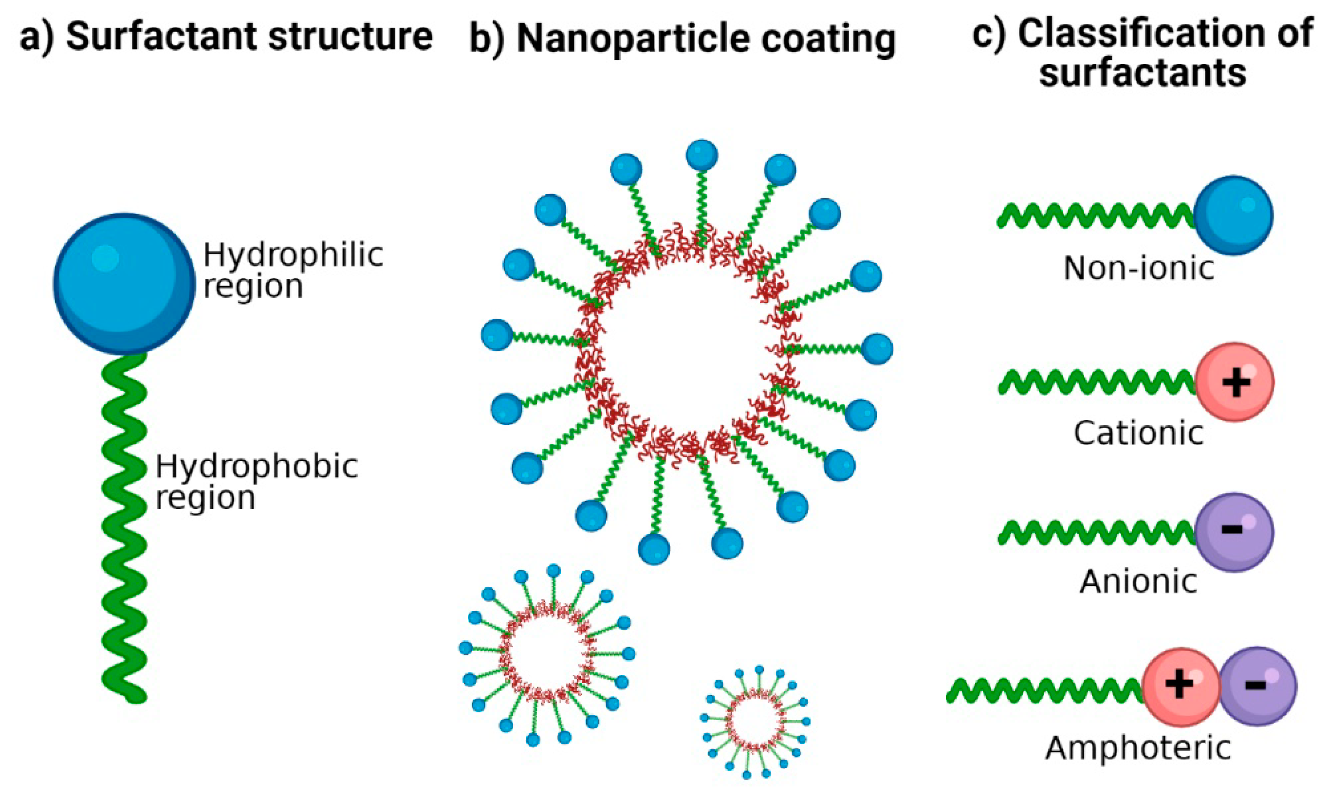
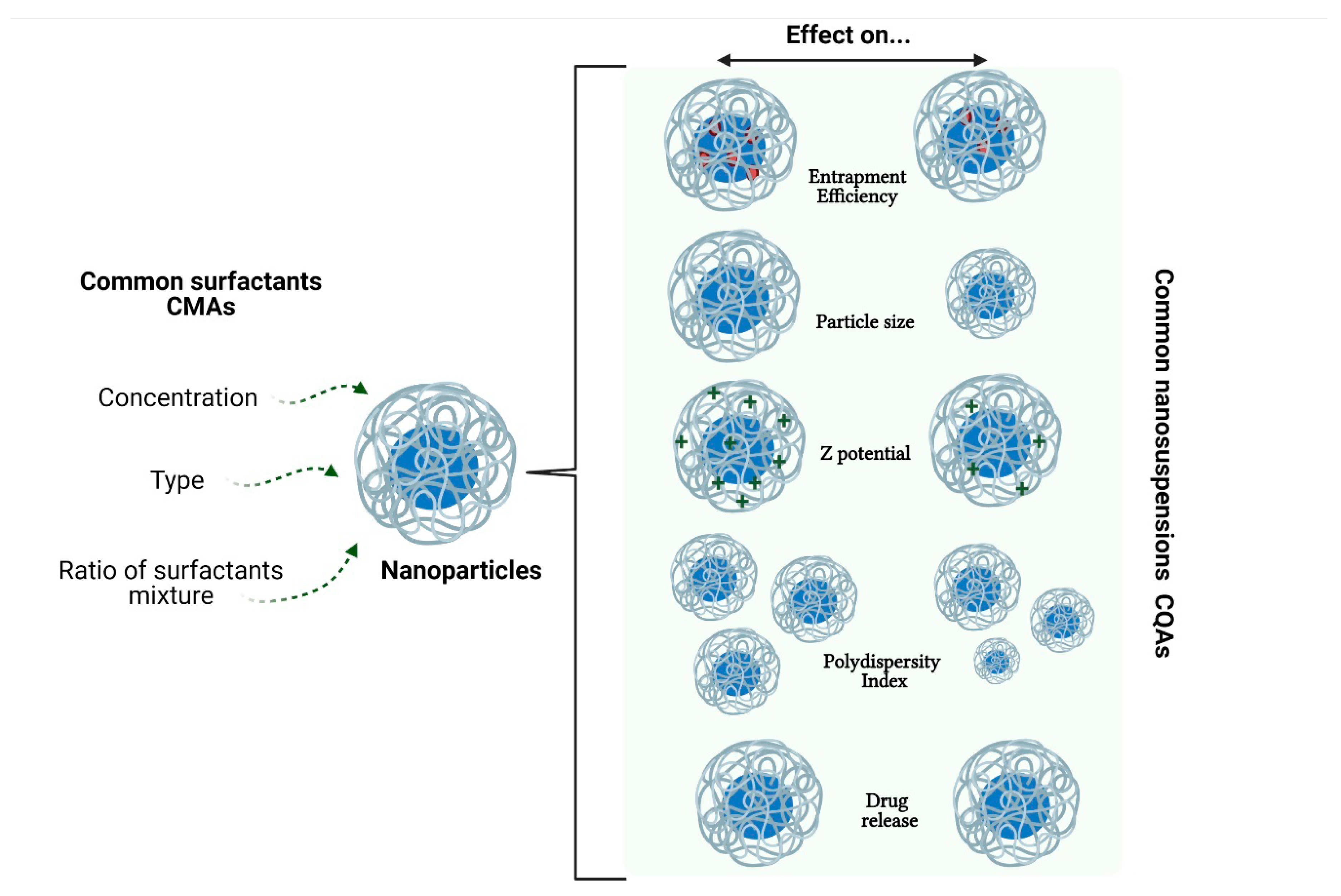
| Type | Surfactants | References |
|---|---|---|
| Anionic | Carboxylates (alkyl carboxylates-fatty acid salts). Sulfates (sodium lauryl sulfate, alkyl ether sulfates). Sulfonates (dioctyl sodium sulfosuccinate, alkyl benzene-sulfonates). Phosphate esters (alkyl aryl ether phosphates, alkyl ether phosphates). | [1,36] |
| Cationic | Quaternary ammonium (cetrimonium bromide, cetylpyridinium chloride, dimethyldioctadecylammonium chloride). Amine-Based (triethylamine hydrochloride, octenidine dihydrochloride). Pyridinium surfactants (benzethonium chloride) | [32,37] |
| Non-ionic | Polyol esters (fatty acid esters of sorbitan). Polyoxyethylene esters (polysorbates). Poloxamers (poloxamer 188). | [1,36] |
| Amphoteric | Phospholipids (phosphatidylcholine or lecithin). Carboxylic Acid/Quaternary Ammonium (cocamidopropyl betaine or amidosulfobetaine-16). Phosphoric Acid/Quaternary Ammonium (hexadecyl phosphocholine). Betaines (alkylamidopropyl betaine). | [32,36,38] |
| Polymer | Surfactant | NP Size (nm) | PDI | Z-Potential (mV) | Drug | EE or DL | Application | Reference |
|---|---|---|---|---|---|---|---|---|
| PLGA | PS-80 1.0% | ~120 | N.A. | N.A. | Disulfiram | EE = 24% | Anticancer potential in Hep3B cell lines, in vitro model of hepatocellular carcinoma | [60] |
| PLGA | PS-80 0.1–7.5 mg/mL 10–30 mg/mL | 160.5 ± 1.37 | 0.043 ± 0.0023 | −21.53 ± 1.47 | N.A. | N.A. | ATR-FTIR method for quantifying the PS-80 adsorbed on the PLGA NP | [61] |
| PLGA | PS-80 1.0% | ~226 | 0.143 | −45.6 | Thymoquinone | EE = 69.5 ± 2.97% | Alzheimer’s disease model in albino mice | [14] |
| PLGA | PS-20 2.0% | 155.6 ± 21.8 | 0.112 | N.A. | Rapamycin Piperine | EE ≈ 70% | Polymeric NP for breast cancer treatment | [62] |
| PLGA PLLA | PS-80 1.0% | 50–100 | N.A. | −26/−32 | N.A. | N.A. | NP crossing BBB model | [63] |
| PLGA | PS-80 1.5% | ~160 | 0.183 | N.A. | Artesunate | DL = 23.67 ± 0.61% | Anticancer activity of artesunate | [64] |
| PLGA | PS-80 1.0% | 77 ± 1 | N.A. | −19 ± 0.89 | Bacoside-A | EE = 57.11 ± 7.11% | NP for brain targeting of bacoside-A | [65] |
| PLGA-PS80 16 | PS-80 1.0% | ~248.1 | 0.084 | −30.9 | plasmid DNA | DL = 9.3% | Nanopartcles for gene delivery | [66] |
| PLGA-Tween 80 | PS-80 0.6 mmol | 156.5 ± 8.6 | 0.14 | −15.4 ± 1.1 | Paclitaxel | DL = 5% | Multidrug resistance lung cancer model | [67] |
| mPEG-PLGA | PS-80 1% | ~145.2 | 0.133 | N.A. | Rhynchophylline | EE = 60% | Neuroprotective effects in an Alzheimer disease model | [68] |
| PBCA | PS-80 0.0–2.0% | ~100 | 0.018 | −2.44 | Dalargin | N.A. | Brain targeting of dalargin via oral administration | [69] |
| Poly (methyl methacrylate-co-methacrylic acid) | PS-60 0.1% | 364.03 ± 5.7 | N.A. | −29.1 ± 1.9 | Gliclazide | EE = 57.46 ± 5.6% | Oral delivery of gliclazide, a hypoglycemic agent | [70] |
| 3-(trimethoxysilyl) propyl methacrylate (TPM) | PS-20 0.01–0.05 mM | ~ 15–500 | N.A. | −49.0 ± 0.5 | N.A. | N.A. | Surfactants influence on spontaneous monodisperse nanoemulsions of TPM | [71] |
| Polycaprolactone | PS-80 | 193 | 0.15 | −26.5 | Memantine | EE = 80 ± 3% | Alzheimer’s disease approach | [72] |
| Polycaprolactone | PS-80 200 and 25 (mg/mL) | 181–407 | 0.3–0.5 | +11.60 to +29.20 | Rosmarinus officinalis and Zataria multiflora essential oils | EE = 75.8–84.4% | Entrapment of two essential oils against Tribolium confusum | [73] |
| Polycaprolactone | PS-80 2.65 mg/mL 5.3 mg/mL 10.6 mg/mL 21.2 mg/mL | ~200 | N.A. | −6.73 | N.A. | N.A. | Optimization of nanoprecipitation method | [74] |
| Polycaprolactone | PS-80 0.5% | ~170 (uncoated) ~260–360 (chitosan coated) | 0.181 (uncoated) 0.345 (chitosan coated) | −12.91 (uncoated) +31.73 (chitosan coated) | Paliperidone | EE ≈ 60% | Influence of PCL/drug ratio, stabilizer type, and high molecular weight of chitosan coating | [75] |
| Polystyrene | PS-20 0.1% | 20–200 | N.A. | N.A. | N.A. | N.A. | NP distribution after periocular administration | [76] |
| Poly (sebacicanhydride) (PSA) | PS-60 PS-20 | 200–160 | N.A. | N.A. | N.A. | N.A. | Degradation of PSA NP | [77] |
| Polyhydroxybutyrate (PHB) | PS-80 1% v/v | 146 ± 30 | N.A. | −26 | Carvacrol | DL = 12.5% | Preparation and characterization of PHB NP by nanoprecipitation and dialysis methods | [78] |
| Chitosan-folate conjugated | PS-80 0.5% v/v | 111.8 ± 4.11 | 0.50 ± 0.21 | N.A. | Doxorubicin and curcumin analog | N.A. | Concentration of PS-80 decreased the size of NP | [79] |
| Chitosan and chondroitin sulfate | PS-80 15 mg | ~234 | 0.2 | +30.0 | Artemether | EE = 83 ± 0.28% | Transdermal antimalarial drug delivery system | [80] |
| Chitosan | PS-80 1.25% | 208 ± 0.01 | N.A. | −32.56 ± 0.03 | Imatinib | EE = 68.52 ± 0.01% | Colorectal cancer targeting application | [81] |
| Sodium Alginate | PS-80 | ~383 | 0.2 | 200 | Curcumin | EE = 95% | Bioavailability in healthy human volunteers | [82] |
| Polymer | Surfactant | NP Size (nm) | PDI | Z-Potential (mV) | Drug | EE or DL | Application | Reference |
|---|---|---|---|---|---|---|---|---|
| PLGA | 2% | 215.3 ± 23 | 0.071 | −10.3 ± 2.3 | Chlorogenic acid | DL = 2.25 ± 0.21% | As a promotor of type 17 collagen production | [165] |
| PLGA | 1–2% w/v and 1.5% w/v | 150 ± 10.4 | 0.081 ± 0.030 | 17.7 | IFN-beta-1a | EE = 96.2% | To diminish symptoms of relapsing-remitting multiple sclerosis | [166] |
| PLGA | 0.1% | 213.8 ± 34.99 | 0.232 ± 0.021 | −52.6 ± 9.483 | Curcumin and ovalbumin | EE = 30% Cur; 16% Ova | Use as sublingual immunotherapy (SLIT) in a mouse model of rhinitis allergic | [167] |
| PLGA | 2% | 172.6 ± 6.20 to 271.9 ± 18.2 | 0.070 ± 0.02 to 0.301 ± 0.03 | N.A. | Ketoconazole | EE = 94.99% ± 3.45 to 97.53% ± 2.33 | Treatment against Candida albicans | [168] |
| PLGA | 2% | 198.6 ± 5.4 (before freeze-drying) 299.8 ± 2.2 (after freeze-drying) | 0.160 ± 0.033 (before) 0.412 ± 0.028 (after) | −20.8 ± 1.4 (before) −16.6 ± 1.1 (after) | Bevacizumab | DL = 1.62 ± 0.01% (before freeze-drying) | Antiangiogenic therapy | [169] |
| PLGA | 0.3% (w/v) | 140 | 0.463 | N.A. | Farnesol | N.A. | Antibiofilm activity, against Candida albicans | [170] |
| PLGA | 0.5% to 5% | 127 ± 0.90 to 289 ± 1.56 | 0.191 ± 2.66 to 0.259 ± 2.67 | −30.43 to −30.89 | Bicalutamide | N.A. | For the treatment of prostate cancer | [171] |
| PLGA | 0.3% (w/w), 1.0% (w/w), and 3.0% (w/w) | 121 to 259 | 0.05 to 0.20 | −27 to −34.4 | FLAP/PGES-1 Inhibitor BRP-187 | DL = 0.5 to 7.29% | A promising drug candidate due to its improved anti-inflammatory efficacy with potentially reduced side-effects in comparison with NSAIDs | [172] |
| PLGA | 1% | 183.7 ± 72.21 | N.A. | −41.1 ± 4.81 mV | p66shc siRNA | EE = 32.3% | To ameliorate neuropathic pain following spinal nerve ligation | [173] |
| PLGA | 1.0% (w/v) | 110.0 ± 41.0 | N.A. | N.A. | 17 beta-estradiol | N.A. | To improve low bone mineral density of cancellous bone caused by osteoporosis | [174] |
| PLGA | 4% | 211 ± 74 | N.A. | −14.2 ± 0.8 | Thioridazine | DL = 26.5% | To reduce toxicity against mycobacterial infection in zebrafish | [175] |
| PLGA | 1% | 198 ± 0 | 0.16 ± 0.01 | −78 ± 1 | Combretastatin A4 | EE = 32 ± 3% DL = 0.41 ± 0.02% | To improve physicochemical characteristics of combretastatin A4, a natural potent tubulin polymerization inhibitor | [176] |
| PLGA | 1% | 119 ± 9 to 206 ± 27 | 0.220 to 0.401 | −4.38 to −5.24 | Curcumin | EE = 77.81 to 92.64% DL = 7.86 to 10.53% | Toxicity on human glioblastoma U87MG cells | [177] |
| PLGA | 0.5% w/v | 186.6 | 0.108 | −28.8 | Recombinant ArgF | EE = 76% DL = 2.6% | For potential use for the prevention of Mycobacterium bovis infection | [178] |
| PLGA | 5.21 mg/mL | 202.8 ± 2.64 | 0.17 ± 0.016 | N.A. | Resveratrol | EE = 89.32 ± 3.51% | For prostate cancer cells | [179] |
| PLGA | 1% | 225.9 | 0.257 | −10.9 | Curcumin and Niclosamide | EE = 58.31% Cur and 84.8% Nic DL = 2.92% Cur and 4.24% Nic | To improve therapeutic effect on breast cancer cells | [180] |
| PLGA | 0.5% w/w | 496 ± 8.5 | 0.607 | −18.41 ± 3.14 | Rivaroxaban | EE = 87.9 ± 8.6% DL = 9.5 ± 1.6% | Anticoagulant medication to prevent blood clots | [181] |
| PLGA | 1% w/v | 110 ± 1 | 0.117 ± 0.003 | −1.29 ± 0.35 | Doxorubicin | EE = 80% | For chemotherapy of glioblastoma | [182] |
| PLGA | 1% w/v | 527 ± 50.21 | 0.26 | N.A. | Olmesartan medoxomil | EE = 78.65 ± 4.31% | To increase the bioavailability of the drug to treat hypertension | [183] |
| PLGA | 1% w/v | 180 ± 8 | 0.04 | −8.59 ± 0.20 | Paclitaxel and methotrexate | EE = 70% MTX and 88% PTX DL = 4% MTX and 5% PTX | Treatment against glioblastoma | [184] |
| PLGA | 3% w/v | 152.8 ± 2.65 | 0.187 ± 0.024 | −30.9 ± 1.67 | Lipophosphoglycan molecule (LPG) and leishmania antigen (ALA) | EE = 14% ALA, 28% LPG DL = 28% ALA, 12% LPG | For a potential nanovaccine to prevent leishmaniasis | [185] |
| PLGA | 0.5% w/v | 114.7 to 124.8 | 0.113 to 0.147 | N.A. | Diclofenac sodium | EE = 41.4% to 77.8% | For inflammatory diseases | [186] |
| PLGA | 3% w/v | 252.6 ± 2.854 | 0.209 ± 0.008 | −23.7 ± 1.36 | Rutin | EE = 81 ± 5% | As a candidate for further multidisciplinary studies (support blood circulation, allergies, viruses, etc.) | [187] |
| PLGA | 4% w/v | 182.2 ± 11.40 | 0.147 ± 0.01 | N.A. | Doxorubicin | EE = 75.3% DL = 4.9% | To arrest glioblastoma growth via intranasal delivery | [188] |
| PLGA | 3% | 191.92 ± 2.3 to 273.70 ± 1.9 | 0.070 ± 0.014 to 0.237 ± 0.030 | −6.87 ± 0.1 to −11.5 ± 0.4 | Dexamethasone | EE = 94.39 ± 2.70% to 95.02 ± 2.98% DL = 3.27 ± 0.58% to 5.43 ± 0.63% | Potential treatment of oral precancerous lesions | [189] |
| PLGA | 2% w/v | 229.5 ± 38.4 to 379.2 ± 21.6 | 0.36 ± 0.02 to 0.73 ± 0.13 | −1.2 ± 1.1 to −3.9 ± 0.5 | Ethanolic Extract of Propolis | EE = 89.90 ± 0.8% to 92.1 ± 0.5% DL = 28.6 ± 1.1% to 56.7 ± 3.4% | As a treatment against Candida albicans | [190] |
| PLGA | 1% | 97.36 ± 2.01 | 0.263 ± 0.004 | −17.98 ± 1.09 | Thymoquinone | EE = 82.49 ± 2.38% Dl = 5.09 ± 0.13% | For the treatment of epilepsy | [191] |
| PLGA | 2% w/v | 200 ± 05 | 0.05 ± 0.02 | N.A. | Budesonide | EE = 85 ± 3.5% | To target the inflammation of mucosa | [192] |
| PLGA | 1.0% w/v | 277 | 0.18 | −16 | Cymbopog citratus essential oil | EE = 73% | As a vehicle for this essential oil with anti-inflammatory, antifungal, sedative, antibacterial, antiviral and anticarcinogenic properties | [193] |
| PLGA | 1% w/v | 118 to 279 | 0.103–0.581 | N.A. | Quercetin | EE = 73.55 ± 2.11% to 86.48 ± 1.67% | Potential vehicle for the antioxidant quercetin | [194] |
| PLGA | 5% w/v | 105 ± 3 | N.A. | −36 ± 5 | Surfactant Protein D (SP-D) | EE = 59 ± 4% | As a potential treatment for respiratory distress syndrome in preterm infants | [195] |
| PLGA | 2% w/v | 154 ± 4.56 | 0.29 | −18.4 | Ursolic Acid | EE = 40 ± 3.24% DL = 4 ± 1.12% | Potential vehicle to deliver the drug against different bearing cell lines | [196] |
| PLGA | 0.5–1.5% w/v | 200 | N.A. | −17.5 | Zaleplon | DL = 5% | For treatment of insomnia | [197] |
| PLGA | 1% w/w | 244.3 ± 4 to 262.8 ± 7 | N.A. | −8.8 ± 0.8 to −17.4 ± 1.0 | Quercetin | EE = 96.2 to 97.8% | To treat foodborne pathogens | [198] |
| PLGA | 1% (w/v) | 211 ± 3 | 0.211 ± 0.009 | N.A. | Clofazimine | DL = 12 ± 1% | To decrease toxicity of the antimicrobial drug | [199] |
| PLGA | 1.5% (w/v) | 268 ± 2.7 | 0.110 ± 0.026 | N.A. | Atrazine | EE = 31.6 to 50.5% | Potential herbicide release system for agriculture | [200] |
| PLGA | 1% | 192.6 ± 3.5 | 0.234 ± 0.008 | −32.4 | Atenolol | EE = 71.65 ± 1.8% | Drug carrier of a β-blocker for cardiovascular disorders | [201] |
| PLGA | 2% (w/v) | 294 ± 15 | 0.26 ± 0.02 | −20.4 ± 2.5 | Insulin | DL = 12.1 ± 0.6% | To optimize the PLGA formulation and lyophilization | [202] |
| PLGA | 2.5% | 184 ± 7 | 0.19 ± 0.01 | −21.7 | Rhodamine-B | EE = 40 ± 2.94% | Potential probes for the drug delivery in cardiac myocytes | [203] |
| PLGA | 2% w/v | 133.3 ± 10.4 | 0.087 ± 0.009 | −16.1 ± 4.5 | Trastuzumab | EE = 42.8 ± 1.6% | Potential vehicle for therapeutic antibodies to avoid current limitations | [204] |
| PLGA | 0.5% w/v | 281.9 to 307.3 | 0.317 to 0.451 | −32.8 ± 1.6 to −43.4 ± 2.6 | Apremilast | EE = 39.5 ±1.1% to 61.1 ± 1.9% DL = 1.3 ± 0.1% to 1.9 ± 0.1% | For the treatment of psoriasis or psoriatic arthritis | [205] |
| PLGA | 3% w/v | 150 ± 7 | 0.16 ± 0.05 | −23.8 ± 0.8 | Rifapentine | EE = 85 ± 8% | For a treatment against tuberculosis | [206] |
| PLGA | 2.5% (w/v) and 0.25% (w/v) | 157.7 | 0.071 | −35.1 | Indocyanine Green and Resiquimod | EE = 65.61 ± 2.09% ICG and 8.363 ± 0.325% R848 | For prostate cancer treatment | [207] |
| PLGA | 1% | 226.6 ± 44.4 | 0.039 ± 0.013 | −0.144 | Ketotifen Fumarate | EE = 89.3 ± 3.3% | Vitamin D binding protein | [208] |
| PLGA | 1% (w/v) | 120 ± 1 | 0.104 ± 0.011 | −11.6 ± 0.8 | Doxorubicin | EE = ~80% | For the delivery of the drug into U87 human glioblastoma cells | [209] |
| PLGA | 0.5% w/v | 210.0 ± 4.8 to 317.5 ± 4.7 | 0.190 ± 0.39 to 0.394 ± 0.53 | −8.3 ± 2.1 to −19.3 ± 0.2 | Dexamethasone | EE = 10.4 ± 2.6% to 64.9 ± 0.6% DL = 0.67 ± 0.2% to 7.17 ± 3.2% | For treatment of oral mucositis | [210] |
| PLGA | 1% w/v | 167.6 ± 0.37 | 0.118 | −16.17 ± 0.53 | Loteprednol etabonate | EE = 96.31 ± 1.68% | For the delivery of the drug into the cornea | [211] |
| PLGA | 1% (w/v) | 164.6 | 0.203 | −17.6 | Doxorubicin and Sorafenib | EE = 74% Dox, 67% Sor | For a cancer treatment using nanotherapeutics | [212] |
| PCL | 3.0% | 188.5 ± 1.7 | 0.160 ± 0.022 | −15.03 ± 2.83 | Lapazine | EE = 35.82 ± 1.47% DL = 54.71% | For antitubercular treatment | [213] |
| PCL | N.A. | 202 ± 24 to 389 ± 37 | 0.08 to 0.164 | −4.92 ± 0.88 to −8.29 ± 1.04 | Nigella sativa oil | EE = 71.6 to 98.6% | For leishmaniasis treatment | [214] |
| PCL | 2.0% w/v | 311.6 ± 4.7 | 0.21 ± 0.03 | −16.3 ± 3.7 | Carboplatin | EE = 27.95 ± 4.21% | Intended to use for intranasal administration to improve brain delivery | [215] |
| PCL | 2% to 3% | 275.23 ± 4.56 to 452.30 ± 9.02 | 0.09 to 0.35 | −4.41 ± 1.21 to −14.77 ± 4.42 | 5-fluorouracil | EE = 94.53 ± 0.23% to 96.82 ± 0.46% | For colon cancer treatment | [216] |
| Polymer | Surfactant | NP Size (nm) | PDI | Z-Potential (mV) | Drug | EE or DL | Application | Reference |
|---|---|---|---|---|---|---|---|---|
| PCL | F-127, 2% | 167 | 0.188 | ~0 | Amphotericin | EE = 85% | Increase the solubility of the drug as treatment for Leishmania infections | [247] |
| Chitosan | F-127, 15% | 146 | N.A | 5.09 | Curcumin | EE = 61.7% | Development of effective delivery system with few side-effects | [250] |
| PCL | F-108, 50% F-127, 50% F-68, 50% | 182 184.7 698.4 | 0.2 0.28 0.88 | –11.7 –1.6 –6.03 | N.A | N.A | Evaluation of the effect of different surfactants | [251] |
| PLGA | F-68, 0.88% | 217.6 ± 8.6 | 0.171 | –23.35 ± 1.17 | Docetaxel | EE = 88% | Development of a delivery system for breast cancer chemotherapy | [252] |
| PLGA | F-68, 0.5% | 160–170 | 0.051 ± 0.012 | −20.5 ± 0.069 | N.A | N.A | Evaluation of the effect of poloxamer as surfactant | [253] |
| PCL/F-68 | PVA, 0.05% | 201.7 ± 10.1 | 0.096 | –12.50 ± 0.86 | Docetaxel | EE = 69.1% DL = 10% | Evaluation of the increased level of uptake NP due to F-68 presence | [254] |
| PCL | F-68, 2% | 149.9 ± 2.2 | 0.087 ± 0.05 | N.A | Curcumin | EE = 96 ± 0.95% DL = 4.9 ± 0.7% | Development of a potential alternative treatment for neuronal diseases based on curcumin | [244] |
| PS | L61, F-68, F-108, L121, F-127 | 97 ± 1, 105 ± 1, 110, 100 ± 2, 108 ± 1 | 0.01, 0.03, 0.02, 0.02, 0.02 | –42 ± 1, –26 ± 2, –14 ± 2, –32 ± 1, –18 ± 2, | N.A | N.A | Analysis of polymer NP modified with different types of poloxamers | [255] |
| Chitosan | F-68 0.5% | 252.80 ± 7.46 | 0.40 ± 0.03 | 17.50 ± 0.93 | Doxorubicin | EE = 61.3 ± 2.28% | Fabrication of DOX-loaded pH-responsive NP for chemotherapy | [256] |
| Silk sericin | F-127 (1:5) F-87 (1:5) | 61.9 ± 5.36 103 ± 1.0 nm | 0.21 0.18 | N.A | Inulin Paclitaxel | EE = 65 ± 10% | Development of silk sericin NP in the presence of poloxamer for successful delivery of both hydrophobic and hydrophilic drugs | [257] |
| PLGA/F-68 | PVA 1% | 179.4 ± 11.2 | 0.309± 0.08 | −22.7 ± 5.7 | Paclitaxel | EE = 65 ± 8.3% | Development of novel PLGA:poloxamer blend NP for intravenous administration of paclitaxel | [258] |
| PLGA/F-68/F-127 | PVA, 1% | 160 ± 31 | 0.671 ± 0.03 | 18.7 ± 1.3 | Curcumin | EE = 90 ± 2.1% | Obtention of PLGA/poloxamer blend NP and evaluation of their interaction with serum proteins and its internalization ability | [259] |
| PLGA-Chitosan | F-68, 1% | 150.7 ± 1.8 | 0.16 ± 0.03 | 25.1 ± 1.6 | miR-34a | EE = 49 ± 2.1% | Anticancer treatment of multiple myeloma | [260] |
| PLGA-Chitosan | F-68, 1% | ∼130 | N.A | 30 | Anti-hTERT siRNA | N.A | Block the growth of anaplastic thyroid cancer xenograft | [261] |
| Chitosan | F-68, 10–50% | ~122 | N.A | 23.63 | Doxazosin mesylate | EE = 99.9% DL = 8.5% | Control release and enhancing the bioavailability of doxazosin mesylate | [262] |
| PLGA | F-68, 1% | ~94 | 0.091 ± 0.010 | –0.3 | Doxorubicin | EE = 92% | Treatment of glioblastoma | [263] |
| PLGA/Chitosan | F-68, 5% | ~134.4 | N.A | 43.1 | Insulin | EE = 52.8% DL = 1.3% | Characterization of bioadhesive NP for oral administration | [264] |
| PLGA | P-85 | 156.7 ± 3.9 | 0.21 ± 0.04 | –45.7 ± 2.9 | Doxorubicin | EE = 85.2 ± 4.1% DL = 7.3 ± 1.2% | Treatment of leukemia | [265] |
| PLGA | P-85/PVA | 180.26 ± 5.60 | 0.184 | −17.47 ± 2.67 | Docetaxel | EE = 82.7% DL = 10% | Breast cancer treatment | [266] |
| Chitosan-γPGA | F-127, 0.25–1% | 193.1 ± 8.9 | 0.29 ± 0.02 | 20.6 ± 2.4 | Curcumin | EE = 52.8 ± 4.7% | Wound regeneration | [267] |
| PCL | F-127, 0.06% | ∼123.5 | N.A | –29.6 | Chloramphenicol | EE = 98.3% | For treatment of MRSA-infected burn wounds | [268] |
| Folated F127/PLGA | F-127 | 107.6 ± 4.25 | 0.308 ± 0.01 | N.A | Paclitaxel | EE = 3.4% | Prolongation of the circulation time of paclitaxel | [269] |
| F-127 | F-127, 0.02% | 9.70 ± 0.31 | 0.195 ± 0.029 | –27.01 ± 0.20 | Berberine | EE = 87.6 ± 1.52% | Improve permeability and retention in the skin | [270] |
| PLGA | F-127 and F-108, 0.2% | ~115 | <0.1 | –11.3 | N.A | N.A | Functionalization of polymeric NP | [271] |
| F-127 | F-127, 1.2% | 70 ± 2.4 | 0.12 | N.A | Gossypol | EE = 91.2 ± 3.1% DL = 9.1 ± 0.42% | Cancer drug release study | [272] |
| Trimethyl chitosan | F-127, 0.1% | ~160 | 0.140 | +20.1 | Methotrexate | EE = 93.6% DL = 8.95% | Effective delivery of methotrexate in osteosarcoma | [273] |
| PLGA | F-127, 1% | 159.0 ± 3.0 | 0.099 ± 0.042 | –15.4 ± 0.7 | Rose Bengal | DL = 0.82 ± 0.27% | Evaluation of the effect of the nanoparticle delivery system on the biodistribution of the drug | [274] |
| PLGA/F-68 | N.A. | 154 | 0.118 | –25.2 ± 1.1 | PDGF-BB | EE = 87 ± 2% | Development of injectable controlled release device based on polymeric NP for the delivery of growth factors. | [275] |
Publisher’s Note: MDPI stays neutral with regard to jurisdictional claims in published maps and institutional affiliations. |
© 2021 by the authors. Licensee MDPI, Basel, Switzerland. This article is an open access article distributed under the terms and conditions of the Creative Commons Attribution (CC BY) license (https://creativecommons.org/licenses/by/4.0/).
Share and Cite
Cortés, H.; Hernández-Parra, H.; Bernal-Chávez, S.A.; Prado-Audelo, M.L.D.; Caballero-Florán, I.H.; Borbolla-Jiménez, F.V.; González-Torres, M.; Magaña, J.J.; Leyva-Gómez, G. Non-Ionic Surfactants for Stabilization of Polymeric Nanoparticles for Biomedical Uses. Materials 2021, 14, 3197. https://doi.org/10.3390/ma14123197
Cortés H, Hernández-Parra H, Bernal-Chávez SA, Prado-Audelo MLD, Caballero-Florán IH, Borbolla-Jiménez FV, González-Torres M, Magaña JJ, Leyva-Gómez G. Non-Ionic Surfactants for Stabilization of Polymeric Nanoparticles for Biomedical Uses. Materials. 2021; 14(12):3197. https://doi.org/10.3390/ma14123197
Chicago/Turabian StyleCortés, Hernán, Héctor Hernández-Parra, Sergio A. Bernal-Chávez, María L. Del Prado-Audelo, Isaac H. Caballero-Florán, Fabiola V. Borbolla-Jiménez, Maykel González-Torres, Jonathan J. Magaña, and Gerardo Leyva-Gómez. 2021. "Non-Ionic Surfactants for Stabilization of Polymeric Nanoparticles for Biomedical Uses" Materials 14, no. 12: 3197. https://doi.org/10.3390/ma14123197