Article

New Electrogenic Microorganism Citrobacter sp. Isolated from Microbial Fuel Cell and Bacterial Characteristics Determination

by
Hugo Guillermo Jimenez Pacheco
1,*,
Abdel Alejandro Portocarrero Banda
1,
Eric Ivan Vilca Cayllahua
1,
Lilia Mary Miranda Ramos
1,
Victor Ludgardo Alvarez Tohalino
2,
Herbert Jesús Del Carpio Beltran
3 and
Pavel Kewin Delgado Sarmiento
2
1
Instituto de Investigación e Innovación en Energías Renovables y Medio Ambiente, Universidad Católica de Santa María de Arequipa, Arequipa 04011, Peru
2
Facultad de Ingeniería de Procesos, Escuela Profesional de Ingeniería Química, Universidad Nacional de San Agustín de Arequipa, Arequipa 04011, Peru
3
Facultad de Ingeniería y Arquitectura, Escuela Profesional de Ingeniería Industrial, Universidad Continental, Arequipa 04011, Peru
*
Author to whom correspondence should be addressed.
Energies 2023, 16(7), 3047; https://doi.org/10.3390/en16073047
Submission received: 27 January 2023 / Revised: 4 March 2023 / Accepted: 14 March 2023 / Published: 27 March 2023
(This article belongs to the Special Issue Advanced Technologies for Sustainable Bio-Fuels Production)

Abstract

:
The generation of energy through the transformation of polluting waste is a widely explored field and offers advances in green technologies. One of the promising technologies is Microbial Fuel Cells (MFCs). These cells can contain electroactive microorganisms that transform organic waste into electricity by transferring electrons from their metabolism. In this study, a new bacterium capable of producing electricity from the waste of the poultry sector and using copper electrodes, called Av_G1, was identified and isolated. It is phylogenetically related to Citrobacter freundii and Citrobacter Murlinae. This new strain was identified molecularly, biochemically, and phylogenetically; its physiological and morphological characteristics were also studied through a Scanning Electron Microscope (SEM). Biochemical determination was performed using Simmons Citrate Agar, Lysine Iron Medium (L.I.A.), Motility/Ornithine Test, Methyl Red indicator, Enzymes: oxidase and catalase, and Gram stain test. The phylogenetic inference was deduced by bioinformatics tools (MEGA X, JalView, Clustal Omega) and the genetic databases The Ribosomal Database Project—RDP and the National Center for Biotechnology Information (NCBI). A maximum current potential of 0.645 V, a maximum current density of 168.72 ± 14.07 mA/m2, and a power density of 31.05 mW/m2 were recorded. During the monitoring, the physicochemical parameters were taken: pH, Oxide Reduction Potential (ORP), Dissolved Oxygen (DO), conductivity, Total Solids (TDS), and average temperature were recorded. Therefore, the present study shows a new Gram-negative electrogenic bacterium, which can be used for electrochemical processes and applied in MFC with copper electrodes.

1. Introduction

In recent years, the demand for all types of energy has been increasing due to climate change and the disproportionate increase in world population, creating an ever-greater need for energy resources in all economic and social sectors, affecting the development industry, technological advances, and the daily environment of society. Consequently, this excessive use of energy has become a global concern, since the demand increase is greater than the proposals for solutions, such as renewable energy sources [1].
The development of renewable sources technologies has been hard work, especially in places where green energy sources are not valued enough and, in some cases, such practices are not able to find the support to apply the knowledge to convert biomass into electricity. Places such as Latin America and the Caribbean have the potential to develop a sustainable and green energy source that can provide bioelectricity and reach eco-friendly technologies applied in Europe or North America [2]. Thereby, methodologies, tools, and different kinds of processes must be improved and put into practice to find a solution regarding bioenergy systems. Currently, the literature describes methods based on biological processes such as anaerobic digestion and fermentation for the development of biowaste-to-bioenergy (BtB) technologies. The intensive search for new renewable energy sources is of utmost importance, as well as the use of biological waste as an alternative for an eco-sustainable development of bioelectricity [3].
One strategy for an energy source is biomass oxidation and bioelectricity generation by Microbial Fuel Cells (MFCs) that are capable of current generation using a microorganism’s consortium or single microorganism and its metabolism to produce electrical energy, and at the same time using a substrate to turning it into a harmless residue [4]. MFC technology allows the application of green energy everywhere due to its simple design and function, low-cost reagents, and the management of native microorganisms from residual effluents or wastewater [5]. There are several ways to set up an MFC, and the parameters can vary as well. According to the literature, MFC presents a variety of configurations that can use biological agents for the use of residual effluents [6]. The electrodes used in MFC can be fabricated of different materials such as steel, graphite, copper, nickel, titanium, and gold, due to their conditions of facilitating conductive efficiency. Cation Exchange Membranes (CEMs) or Protonic Exchange (PEM) fulfill a fundamental function for the correct functioning of MFC through the passage of anions or cations. The MFC operation is the release of electrons through a bacterial biofilm through the metabolism of microorganisms, due to this the generated protons migrate to the cathode through the exchange membrane that prevents the diffusion of oxygen in the anodic chamber [7]. Another important factor for MFC is the organic matter (substrate) that facilitates energy generation, either a pure or complex substance generally found in many industries. In these synthetic or pure substrates (animal manure, acetate, cellulose particles, azo dyes, lactate, glucose, industrial wastewaters, etc.) electro-active microorganisms (EAMs) survive and carry out the exchange of electrons [8]. This bacterial consortium is a relevant factor in bioelectricity-generating in MFC, since power generation depends on it. There are various and unexplored bioelectricity generating organisms, and many of them are not electrochemically characterized [9], as well as in a bioinformatic way or some other important aspect within its biology [10], this is the case of electro-producing bacteria, which are very diverse, heterogeneous, and found in the most common and extreme places on the planet [11]. In recent years, some bacterial strains have been very promising, such as Geobacter sulphurreducens, one of the most studied organisms, and a strain model that produces bioelectricity [12,13]. Another is the case of Shewanella oneidensis, Pseudomonas aeruginosa, and Pseudomas alcaliphila that can produce their redox mediators such as pyocyanin, which is a pigment produced by P. aeruginosa or quinone mediator in the case of S. oneidensis [14] Dietzia sp. [15]. According to the nature of the transfer of electrons of electroactive bacteria, these are divided into two: exoelectrogenic and electrotrophic, these bacteria use different methods such as the transfer of electrons through cytochromes located in the outer membrane of the cell surface or “pili” that serve as conductors of electricity [16]. Exoelectrogenic bacteria produce electricity due to oxidation-reduction reactions of organic matter transferring an electron to an acceptor [17]. Electrotrophic bacteria have been poorly characterized and can accept electrons from external sources or mediators [18]. Recent research evaluates the potential of using modified or mutant bacteria to improve electron transfer performance, as in the case of Shewanella oneideidensis, which was modified at the level of a second messenger called c-di-GMP, which provided it better biofilm formation and regulated the expression of cytochromes-c, including electron transfer pathways such as CymA, MtrA, MtrC, and OmcA [19].
Research regarding MFCs is focused on high current generation, cells design, substrates improvement, modified bacteria, or variable changes to improve MFC performance; however, this study implemented a copper electrode MFC system with the strain Av_G1 as an electroactive organism native from poultry manure that can stand the bactericide effect of copper, it is an organism capable to support abiotic stress and improve its biofilm formation capacity to survive on tough environments.

2. Materials and Methods

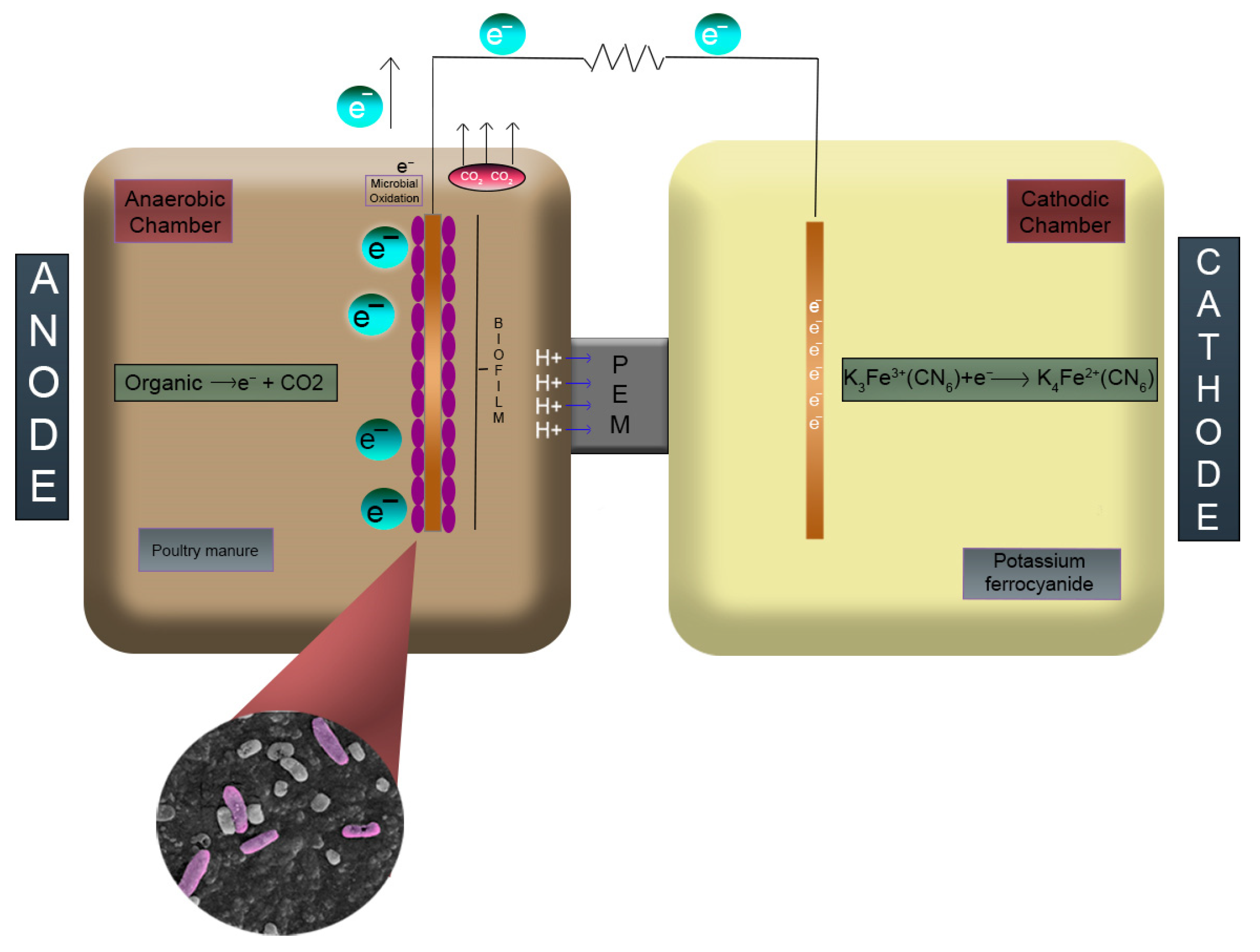
2.1. MFC Construction and Operation

The cells were fabricated of Polyvinyl Chloride (PVC). These two cells were 18 cm high × 11.4 cm diameter × 2 mm thick, with a capacity of 1.71 L. The cells were used as anode and cathode, both were connected through a removable system called Bush, this one was composed of a hermetic removable coupling and an O-ring of inert material inserted inside the coupling; and was sealed with a polytetrafluoroethylene material. For proton exchange, a Nafion® 117 membrane of 3 cm in diameter was used [20], this one was placed in the Bush system.
In the cathodic chamber, 50 mM of K3[Fe (CN)]6 electron acceptor solution was used [21,22,23,24]. The operation of the cells was by a Batch system; copper was used for the material of the electrodes, and they were placed inside the chambers. The working temperature of the cells was 20.92 ± 1.18 °C. In Figure 1 we can observe the configuration of the MFC in more detail.
In Figure 1, anodic and cathodic cells were linked through the Bush system. In Figure 1 we can observe that the Nafion® 117 exchange membrane is displayed, as well as the electrodes, the poultry substrate (in the anode), the potassium ferrocyanide (in the cathode), and the exchange of electrons through the circuit in a graphically way.

2.2. Electrochemical Configuration of MFC

Copper plates were used as electrodes, the working area of these plates (anode and cathode) was 0.0028 m2. The cells had an open circuit configuration connected to a Keysight Technologies (Santa Rosa, CA, USA) 3472A LX1 Data Acquisition/Switch Unit data collector to evaluate the circuit voltage for 240 h.

2.3. Substrate Obtention and Preparation

The raw material was collected from the local poultry industry area in Arequipa, Perú. The sampling was carried out according to the methodology outlined in the Standard Guide for Sampling of Waste and Soil for Volatile Organic Compounds ASTM D-4547:2009 [25] considering the initial temperature and humidity of each sample. The poultry substrate was collected and stored at 4 °C for 24 h, then dried in a Venticell ECOline brand convection oven at 80 °C. Each sample was then crushed and sifted through a 250 µm sifter.

2.4. Physico-Chemical Parameters

Physico-chemical parameters were taken from the anodic chamber with a multiparameter of the Hanna brand, models HI 5522 and HI 542, to determine pH, ORP, OD, conductivity, TDS, and average temperature of 20.92 ± 1.18 °C during 240 h of work with time intervals of 24 h, these results are shown in Table 1. To measure and control the relative humidity (%RH) a humidifier Emerson quiet kool at 33% was used. To ensure the best performance of the MFC and avoid cells polarization, temperature and pH were maintained constant.

2.5. Inoculum Development and Strain Isolation of Av_G1

Previous experiments were carried out in the MFC cells with native poultry bacteria, where known bacterial consortia were found, Citrobacter sp. was found on the surfaces of the copper electrodes with a particular behavior. To perform the isolation of the microorganisms (bacteria), after 240 h of cell operation, the copper electrode of the anodic chamber was scraped with the previously selected bacterial strain [26,27]. We can consider that Av_G1 was working into an MFC with a consortium during 30 days before it was isolated and characterized, thus this new bacterium was going through a previous conditioning stage in the same subtract and parameters that we exposed in Section 2.2 and Section 2.3.
This inoculum was transferred to sterile Eppendorf tubes containing 1 mL of triton × 100 at 0.01% [28], then the bacterial consortium was enriched with Brain–Heart Infusion Broth (BHI) for 24 h at 37 °C, after this time the culture was seeded on Luria–Bertani (LB) agar and incubated in an anaerobic chamber for 12 h with CO2 purge following the protocol of [29]. To carry out the isolation of the Av_G1 strain, 5 repetitions were carried out for its purification, then the pure strain was inoculated in Potassium Cyanide Broth (KCN) for 24 h at 33 °C to finally grow it again in LB agar for microscopic observation.

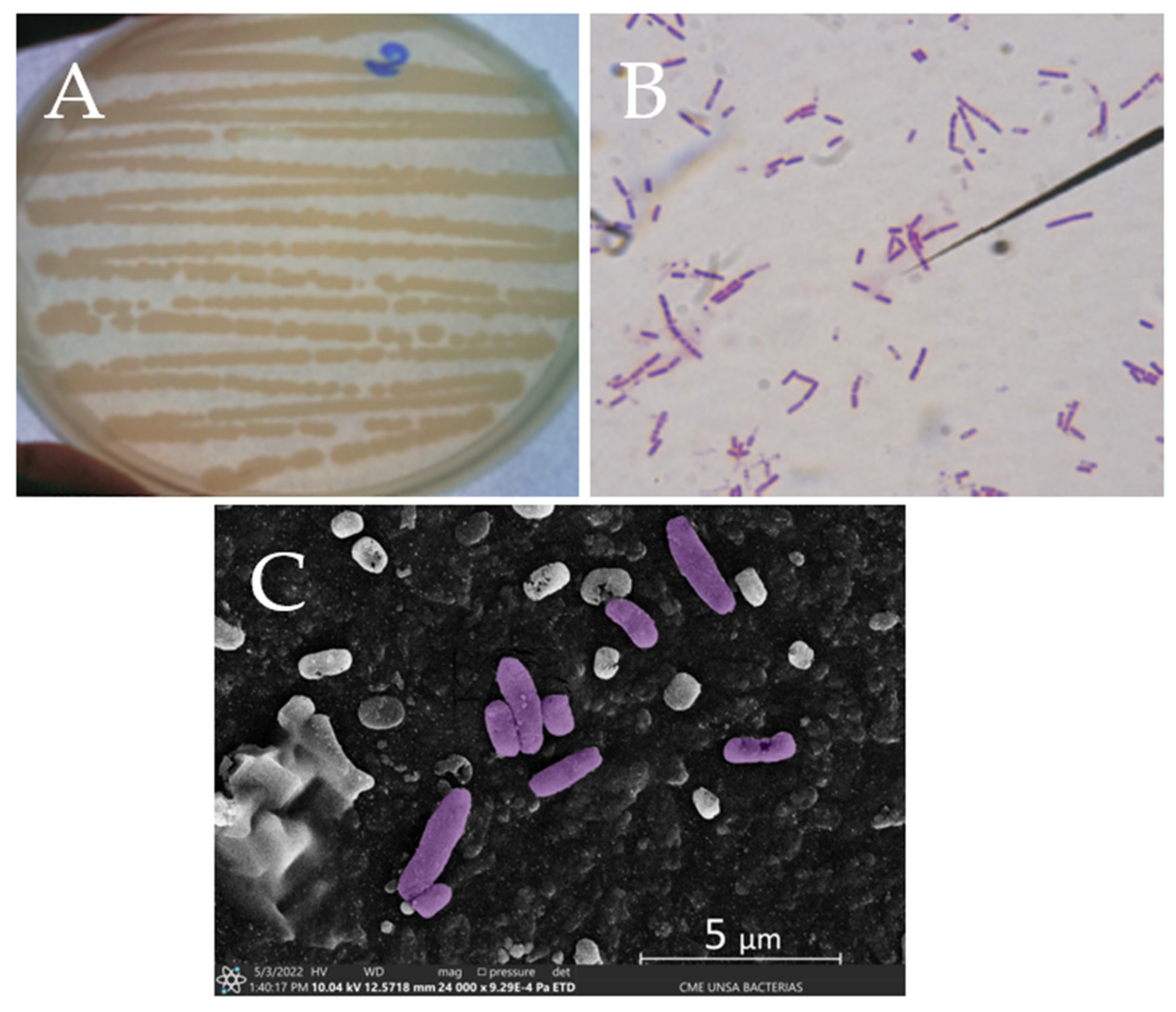
2.6. Morphological Characterization and Microscopic Analysis

The strain was identified by biochemical tests and propagation evaluation. Means were used to evaluate its biochemical activity to obtain the identity of the bacterium within the Enterobacteriaceae family. The inoculum from the KCN broth was used and brought to 0.5 on the McFarland scale. Tubes with hermetic lids were prepared in triplicate as well as an E. coli inoculum for quality testing.
Biochemical tests were prepared starting with Simmons Citrate (HI-Media, Einhausen, Germany), Lysine Iron Agar (L.I.A.) (HI-Medium), Motility/Ornithine (HI-Medium), Methyl Red indicator with Voges Proskauer (HI-Medium), Enzyme Oxidase (Merck, Rahway, NJ, USA), Enzyme Catalase (Merck), and finally Stain of Gram. The microscopy analysis was performed by a stereo microscopy with a Zeiss Model 508 Stereoscope, and through optical microscopy of the Euromex (Arnhem, The Netherlands) brand equipment, Image Focus Alpha and also through Scanning Electron Microscopy with the SEM Scanning Electron Microscope of the brand THERMO SCIENTIFIC (Waltham, MA, USA) Model SCIOS2.
The morphological observation and the characteristics of the colony in the Petri dishes were carried out after 24 h of incubation of the inoculated strain in LB medium by stereoscope. In addition, Gram stain was used for microscopic observation [30]. Finally, for the observation of the strain by SEM, the sample was prepared following the protocol pre-treatment by Kammoun, R. et al. [31].

2.7. Gene Sequencing for 16rDNA

For DNA extraction, the simplified Bazzicaculpo and Fancelli protocol was used [32].

2.7.1. Lysis and DNA Extraction

The DNA of the strains was isolated from colonies previously cultivated and purified from LB agar. With a nichrome loop, a roast of colonies was taken from the petri dish, which was re-suspended in 600 µL of Lysis Buffer 1X in an Eppendorf tube, and was incubated in a hot block for 45 min at 65 °C, after this period, 300 µL of Phenol-Chloroform was added, then the Eppendorf tube was shaken, and centrifuged for 5 min at 8000 rpm. The supernatant was collected and placed in a new Eppendorf tube, and 500 µL of isopropanol was added, this tube was centrifugated for 10 min at 11,000 rpm, then the supernatant was removed; 600 µL of absolute ethanol was added, it was taken to centrifugation for 2 min and the supernatant was discarded; the pellet containing the DNA was re-suspended with 100 µL of sterile distilled water.

2.7.2. DNA Amplification

As it is almost impossible to isolate pieces of DNA, the Polymerase Chain Reaction (PCR) technique was used for the amplification of the DNA trace, and a thermocycler was also used for a higher production of it. A total of 2 µL of DNA and 2 µL of the 27F (5′-AGAGTTTGATCCTGGCTCAG-3′) and 1392R (5′-GGTTACCTTGTTACGACTT-3′) primers solution were added to test tubes [27]. For 16S rRNA gene amplification, each test tube had a final concentration of 200 µM, and 30 µL of Platinum Blue PCR Supermix High Fidelity was added. The tubes were then placed in a BIO-RAD (Hercules, CA, USA) brand MyCycler™ Thermal Cycler System with Gradient Option thermocycler. The following steps were followed at certain configurations: (a) denaturation: 95 °C for 5 min, 25 cycles of 94 °C for 70 s; (b) hybridization or annealing: 55 °C for 70 s; (c) extension: and 72 °C for 70 s, and finally 1 cycle at 72 °C for 7 min.

2.7.3. Electrophoresis and DNA Purification

For electrophoresis, agarose gel methodology was used. The PCR product was separated on a 1.5% agarose gel, then the band, corresponding to the 16S RNAr gene, was cut from the gel for purification using the QIAquick PCR Purification Kit of the QIAGEN brand, columns of the same brand were used, and instructions provided by the company were followed [33]. For sequencing, 1 µL of Primer (16S RNAr for bacteria) was added to 20 µL of purified DNA, the 2 tubes were labeled and sent to DNA Sequencing|Functional Biosciences, Inc. (Madison, WI, USA).

2.8. Bioinformatic and Phylogeny Analysis

To perform the bioinformatic analysis, the RDP database was used, making use of the “classifier” tool to identify, at the taxonomic level, the genus of the strain [34]. Then, the 16s RNAr gene sequence of the new Av_ G1 strain was submitted to GenBank, a global gene bank, and the National Center for Biotechnology Information (NCBI) BLASTn Software was used to determine the similarity with other strains in the bank.
The sequences with the highest percentage of similarity and the lowest E-value were selected and downloaded [35]. The sequences were aligned in the bioinformatics tool Clustal Omega [36], the alignment was observed in Jal View and downloaded in FASTA format [37].
Phylogeny analysis was performed with MEGA XI software [38]. The initial trees for heuristic search were obtained automatically by applying the Neighbor-Join and BioNJ algorithms to a matrix of pairwise distances by pairs estimated, using the Tamura–Nei model and then selecting the topology with a higher logarithmic value of likelihood. A discrete Gamma distribution was used for this analysis. Finally, 15 nucleotide sequences from selected strains were involved and there were a total of 1541 positions in the final dataset.

3. Results

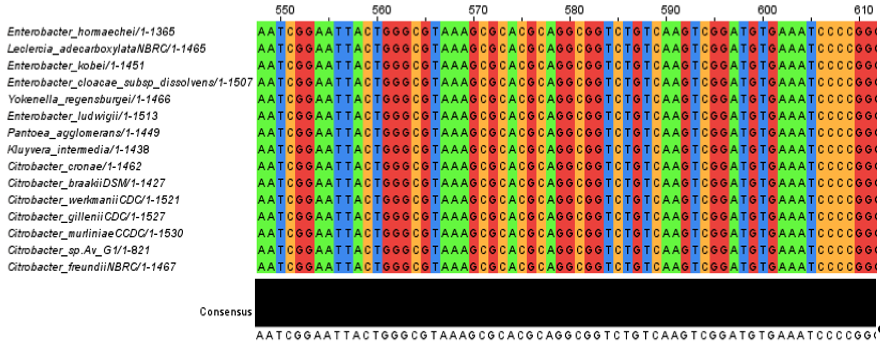
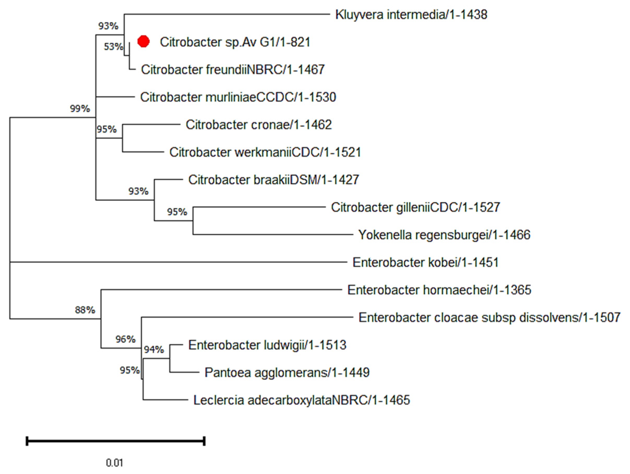
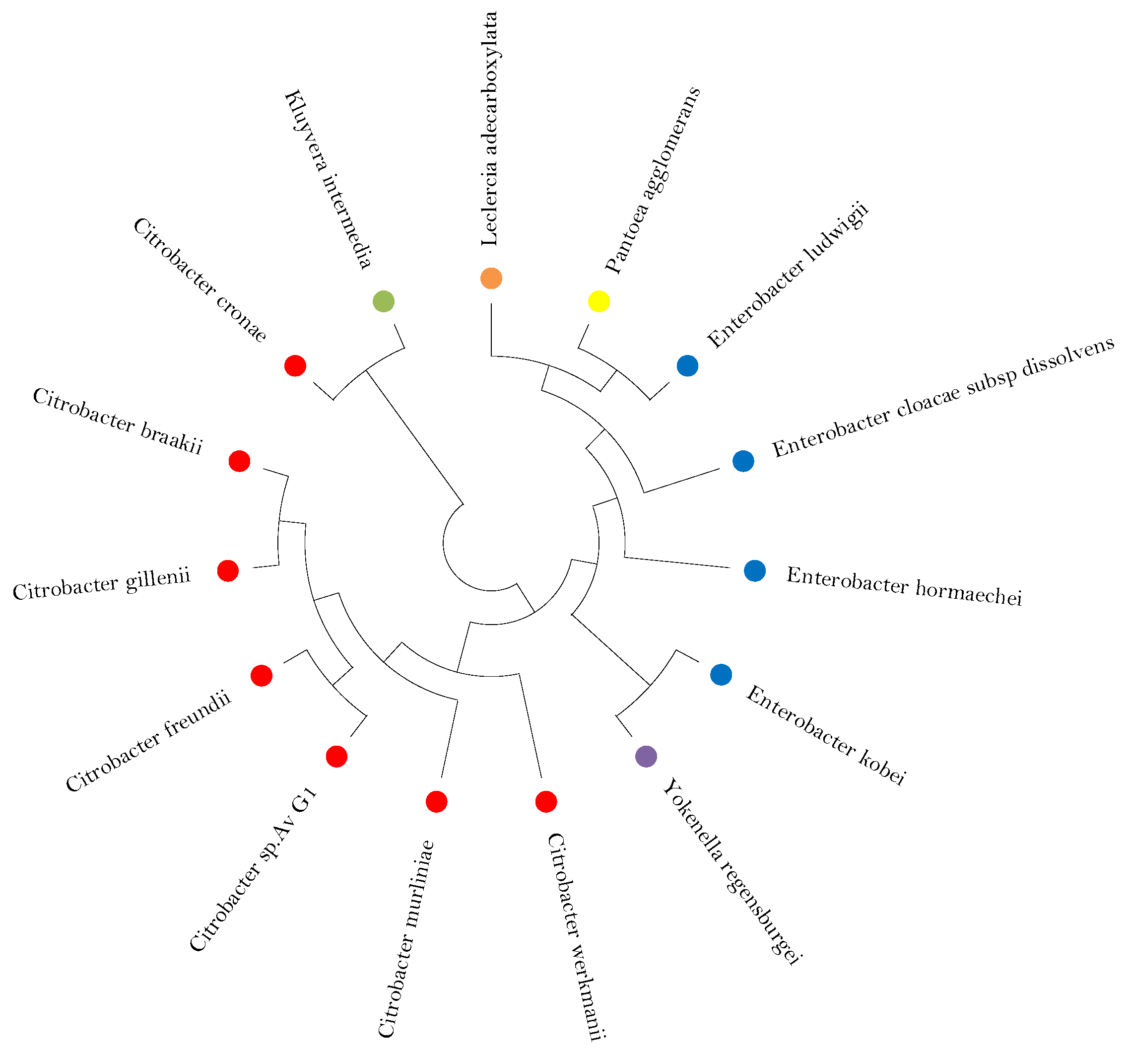
3.1. Phylogeny Analysis and Sequence of the Isolated Strain Av_G1

To evaluate the identity of the Av_G1 strain, the 16s region of rRNA with a size of 821 base pairs (bps) was sequenced. The sequence was uploaded to NCBI GenBank under accession number OM124933.1. The sequence was analyzed with the NCBI BLASTn tool to find the maximum likelihood, finding Citrobacter freundii with an identity percentage of 99.64% and an E-value of 0, Citrobacter brakii with 99.39%, and Citrobacter Murlinae with 99.15%. These were the organisms with the highest likelihood. Organisms with an identity percentage < 97% were not considered [39].
Figure 2 shows the alignment of sequences in JalView, showing the most conserved regions among the organisms that had the highest percentage of identity and ranging from 550 bp to 610 bp. In this sense, Figure 3 shows the nucleotide composition of every bacterial strain analyzed by JalView showing thymine T(U), cytosine (C), adenine (A) and guanine (G) frequencies content.

3.2. Characterization of Citrobacter sp. Av_G1

Table 2 reveals the negative Gram staining and the biochemical behavior, resulting positive for Simmons Citrate for the Av_G1 strain and negative for the E. coli strain passing the quality test. This showed a positive motility with the MIO medium and a negative Ornithine, this indicates that it does not present the enzyme Ornithine decarboxylase. For the test with methyl red, the color of the reagent did not turn/viro, which would indicate a negative test, confirming in this way the absence of acids from the fermentation of glucose.
The bacterium tested negative for LIA, but with the production of hydrogen sulfide in the background (SH2) due to the black coloration, this allows us to understand the use of sulfates as an electron acceptor in the degradation of organic matter.
Finally, positive catalase and oxidase tests demonstrate its facultative anaerobic behavior and the presence of a cytochrome C system. In Figure 4 you can see the morphology of the strain Av_G1 in three parts, Figure 4A shows the culture in a Petri dish with LB agar and after 24 h, Figure 4B shows the optical microscope and Gram-negative staining and Figure 4C a magnification with electron microscope.
Citrobacter sp. Av_G1 was characterized according to its colony, bacterial morphology, biochemical profile, molecular, and bioinformatic analysis. The observed colonies presented are oval-shaped and cream-colored; the bacteria observed are planktonic bacilli and negative for Gram stain, see Figure 4.
Figure 5 shows us the consensus tree made by the MEGA X tool from the alignments made. This inference is robust and highly reliable since all nodes pass 50% and even most have a percentage greater than 90%. The Av_G1 strain was assigned to the Citrobacter genus and forms a subcluster with Citrobacter freundii.
The phylogeny tree shown in a circle, with 500 bootstrap replicas, shows us an ordered panorama of the phylogeny inference of the new strain, confirming the relationship with Citrobacter freundii and placing the entire Citrobacter genus on one side, the genus Enterobacter on the other, and Leclercia adecarboxylata, Yokenella regensburgei, Pantoea agglomerans, and Kluyvera intermediate in different branches. See Figure 5 and Figure 6.

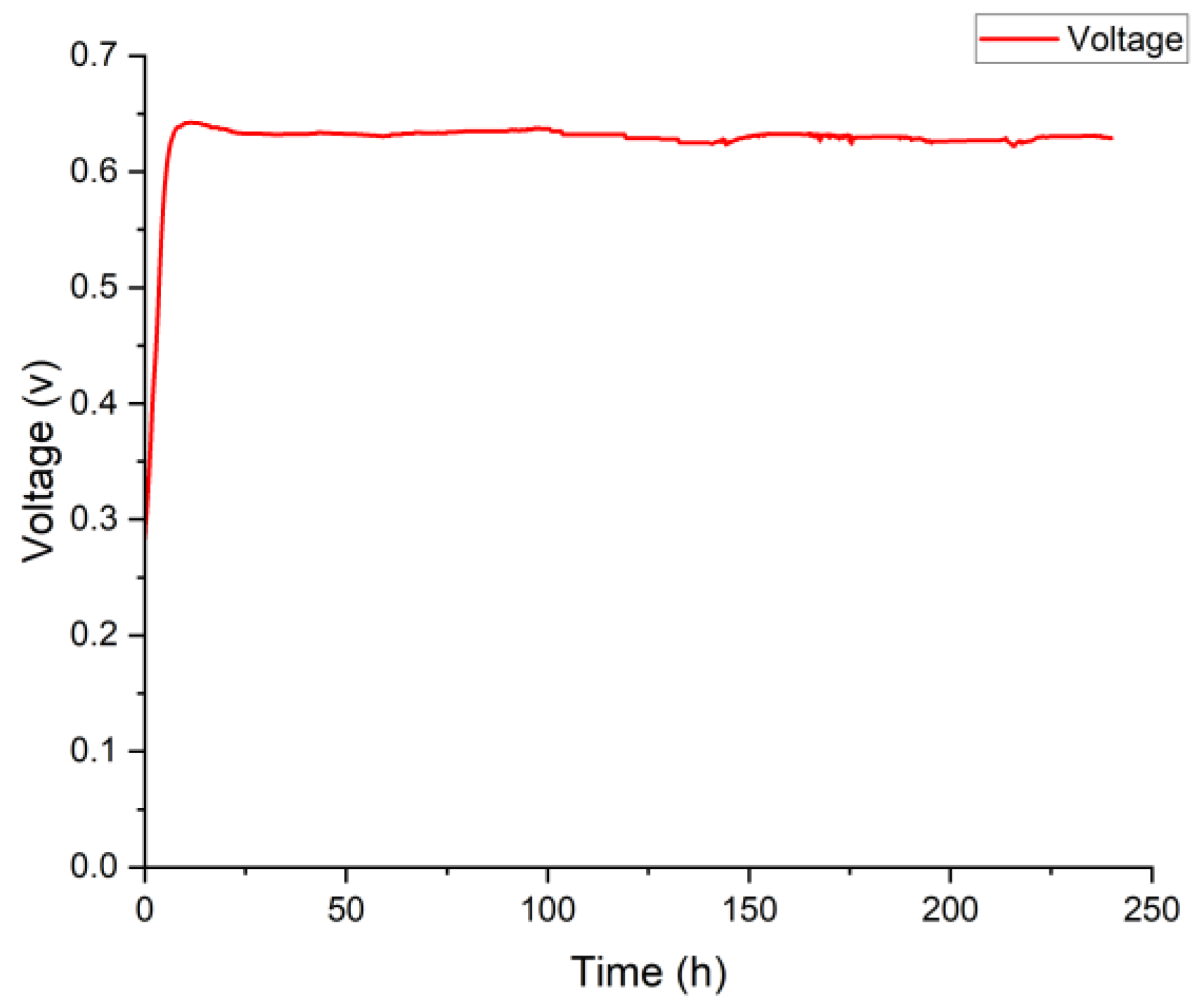
3.3. Current Generation of Citrobacter sp. Av_G1 in MFC

At the end of 240 h of working MCF, copper electrode was found to be free of any corrosion on its surface. Current generation depends on MFC and on the bacteria performance, thus, parameter monitoring such as pH or temperature is important. The results obtained from physicochemical parameters in anode showed us a pH between 6 and 7 that kept polarization low on this anolyte. Due to microbial oxidation, MFC tends to be acidic and triggers an acid shock, consequently, the current production is lost. To prevent imbalances and try to improve the MFC performance, the temperature was set at 20.92 ± 1.18 °C during the 240 h. The rest of the parameters are shown on Table 3.
In Figure 7 the output Open Circuit Voltages (OCVs) are shown during a period of 240 h, an evident increase is observed until 11 h with a maximum of 0.645 V; after 20 h the voltage production remained constant between the values of 0.612 V and 0.640 V, these values are within the ranges reported by other authors who established similar configurations [39], this initial stage of increasing trend until 11 h is due to the abundant availability of food that the bacteria received in their anodic behavior [40], for this reason the bacterial colony grew exponentially. Furthermore, the rapid activation observed is due to the previous conditioning stage that Av_G1 passed through making possible this exponential behavior. We can assign to this new strain the capability to withstand copper stress, to oxidize poultry manure, and the electrogenic capacity to the previous conditioning stage explained in part 2.5 of this research.
Electrogenic bacteria oxidize organic matter and release electrons that are transferred to the anode, this is evidenced by the generated electricity [41,42]. It is of great importance to consider the conditions of the substrate, its characteristics, and its components to generate electricity [43]. Poultry substrate waste is a powerful organic substrate within MFC, due to the amount of feed that generates a high voltage in the initial stage of operation as reported in [44,45,46] who obtained maximum power densities of 220 mW/m2 and 287.5 mW/m2 for poultry substrate waste and for different concentrations of wastewater from poultry manure.

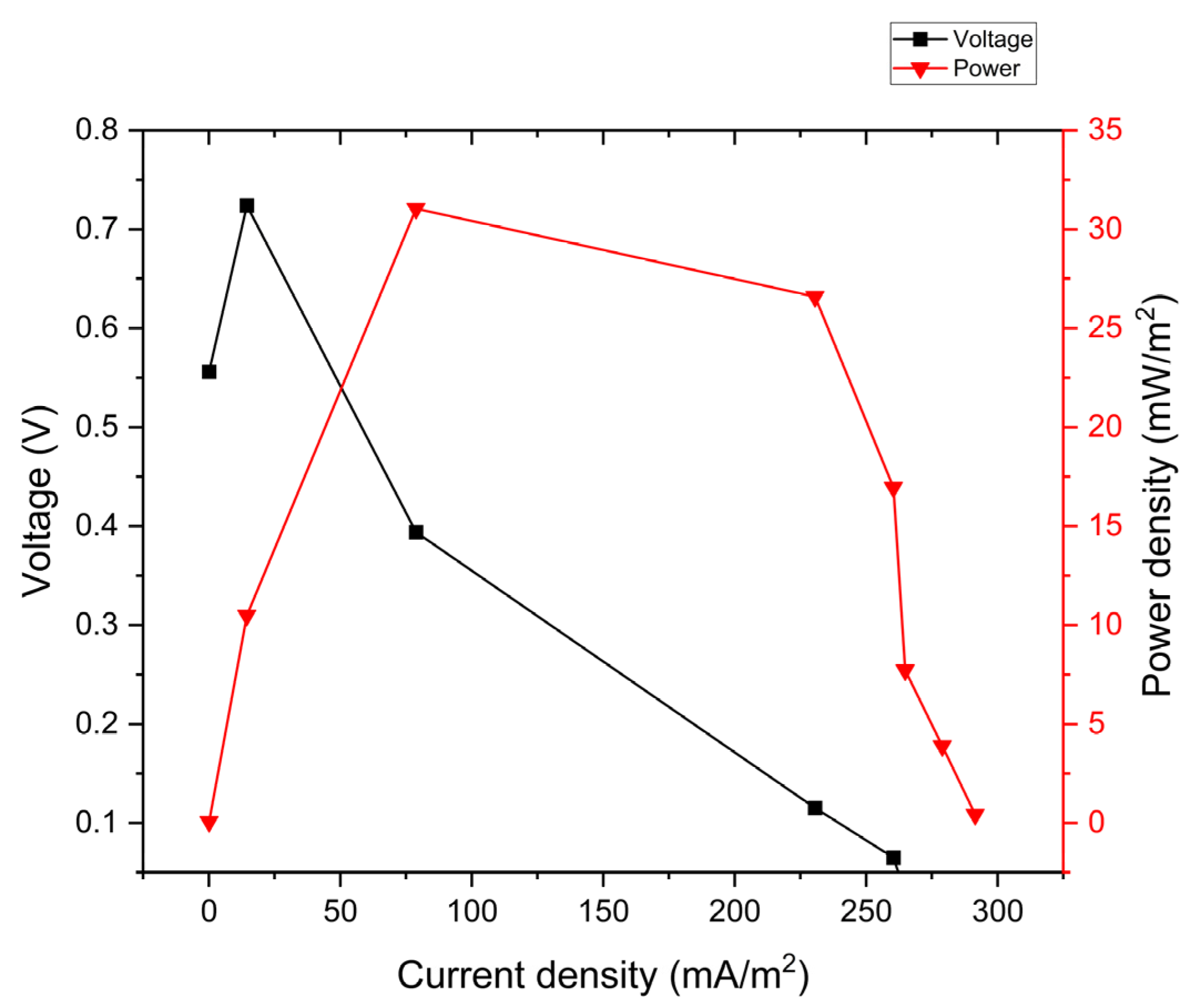
3.4. Polarization Curves and Power Density

In this study, the potential current properties were measured to know the optimal current in which cells can operate to maximize the power density of Citrobacter sp. Av_G1. Figure 8 shows the polarization curves assessed with eight external resistances from 10 to 10,000,000 Ω. The peak potential resulted of polarization assays was 18.67 mW/m2 using a 10,000 Ω resistance.
The low power density obtained in this research compared with those reported may be due to the different pretreatment conditions, substrate content, the adverse effect of the copper anode and MFC H type design. A similar study with copper anode but using avocado residues in electricity production [47] shows a similar behavior in electricity production, these results can confirm that bacterial growth was probably not inhibited due to the toxicity of the solubilized copper and allowing Av_G1 to thrive on this stressed condition regarding the copper bactericide effect. The influence of the MFC set up had a significant effect in the final performance and current production, as well the space between electrodes, the area/kind of membrane and the working substrate volume. Those have an influence on the internal resistance and biofilm formation, consequently, the abiotic stress generated in MFC allowed strain Av_G1 to be capable of biofilm development and survive on it. The electrodes presented a substantial distance between them, consequently rising the internal resistance. It can be verified by Singh, A. et al.; Cheng, S. et al.; and Clauwaert, P. et al. [48,49,50] who assessed the working volume and electrodes distance on H type MFC, showing high levels of power density as reactor volume and electrode distance decreases.
The substrate is an important factor in the production of electricity in the MFC. The present work shows the potential of the poultry substrate waste, establishing it as a low-cost substrate and establishing its importance within the circular economy. The copper anode presents bactericidal characteristics, where a reduction in the population of microorganisms is established; however, the new Av_G1 strain was not fully affected by this material. The biofilm, formed because of the stress submitted by the copper, was able to inhibit the antibacterial capacity of the material and thus prosper for the generation of bioelectricity. The biochemical profile of the strain provided us the production of hydrogen sulfide, from which it was deduced that the bacterium is sulfur-reducing (sulfate reduction). This property is found in anaerobic organisms; this adds information to the existing literature.
Finally, as we can observe in Figure 8, the polarization curve assay showed that the working resistance, where the H type cell and the bio electrogenic system can work at best (10,000 Ω), was really high. Therefore, we can confirm the elevated internal resistance of the cell despite all parameters on the anode and cathode were controlled.

4. Conclusions

In the present investigation and for the first time, we reported the bio electrochemical characterization of the Citrobacter sp. Av_G1 as an electroactive microorganism, possible exoelectrogenic, and facultative anaerobe. This strain was isolated, morphologically characterized, and analyzed with molecular and bioinformatic techniques to determine it as a new microorganism capable of generating electricity on a copper electrode, presenting a maximum current potential production of 0.645 V and a power density of 31.05 mW/m2 at a current density of 78.03 mA/m2.
The proposed electrode material with bactericidal capacity was a source of electricity generation and the development of this bacterium Av_G1 was not totally affected by this material. The generation of the biofilm found can inhibit the antibacterial capacity since the copper surface would make it impossible for the Av_G1 strain to produce electricity.
The biochemical profile of the strain showed us production of hydrogen sulfide, for this reason we state that the bacterium is a sulfur reducer, indicating that it was reducing sulfates, this mechanism is found in exceptional anaerobic organisms, contributing information to the literature.

Author Contributions

Conceptualization, A.A.P.B. and E.I.V.C.; methodology, V.L.A.T. and H.G.J.P.; investigation, A.A.P.B., E.I.V.C. and H.G.J.P.; writing—review and editing L.M.M.R., P.K.D.S., H.J.D.C.B. and H.G.J.P.; project administration H.J.D.C.B. and H.G.J.P.; funding acquisition L.M.M.R., V.L.A.T., H.J.D.C.B. and H.G.J.P.; writing—original draft preparation A.A.P.B., V.L.A.T., P.K.D.S. and H.G.J.P. All authors have read and agreed to the published version of the manuscript.

Funding

The article has not received funding.

Data Availability Statement

Data are available upon request to the correspondence author.

Acknowledgments

The authors thank the Universidad Católica de Santa María de Arequipa for the support of materials and facilities. We also thank Veronica Salinas Murillo for the support provided.

Conflicts of Interest

The authors declare no conflict of interest.

References

  1. Lorenczik, S.; Jorquera, J.; Hevia-Coch, P.; Hungerford, Z. Electricity Market Report Home Page. Available online: https://iea.blob.core.windows.net/assets/d75d928b-9448-4c9b-b13d6a92145af5a3/ElectricityMarketReport_January2022.pdf (accessed on 23 November 2022).
  2. Ochs, A.; Killeen, P.; Majano, A.M. Advancing Bioelectricity in Latin America and the Caribbean. Proceedings and Outcomes of the First Implementation Year of a Community of Practice of Leading Countries in the Region; Resilient and Low Emission Development Strategies: San José, Costa Rica, 2017. [Google Scholar]
  3. Bathia, S.K.; Joo, H.S.; Yang, Y.H. Biowaste-to-bioenergy using biomedical methods—A mini-review. Energy Convers. Manag. 2018, 177, 640–660. [Google Scholar] [CrossRef]
  4. Moqsud, M.A.; Bushra, Q.S.; Rahman, M.H. Composting barrel for sustainable organic waste management in Bangladesh. Waste Manag. Res. 2011, 29, 1286–1293. [Google Scholar] [CrossRef] [Green Version]
  5. Varanasi, J.L.; Das, D. Microbial Fuel Cell; Springer: Kharagpur, India, 2017; Chapter 2; pp. 43–62. ISBN 978-3-319-66793-5. [Google Scholar]
  6. Flimban, S.G.; Ismail, I.M.; Kim, T.; Oh, S. Overview of Recent Advancements in the Microbial Fuel Cell from Fundamentals to Applications: Design, Major Elements, and Scalability. Energies 2019, 12, 3390. [Google Scholar] [CrossRef] [Green Version]
  7. Santoro, C.; Arbizzani, C.; Erable, B.; Leropoulos, L. Microbial fuel cells: From fundamentals to applications. A review. J. Power Sources 2017, 356, 225–244. [Google Scholar] [CrossRef] [PubMed]
  8. Fadzli, F.; Bhawani, S.; Mohammad, R.E. Microbial Fuel Cell: Recent Developments in Organic Substrate Use and Bacterial Electrode Interaction. J. Chem. 2021, 2021, 4570388. [Google Scholar] [CrossRef]
  9. Logan, B.E.; Rabaey, K. Conversion of wastes into bioelectricity and chemicals by using microbial electrochemical technologies. Science 2012, 337, 686–690. [Google Scholar] [CrossRef] [Green Version]
  10. Saha, T.; Protity, A.; Zohora, F.; Shaha, M.; Ahmed, I.; Barua, E.; Sarker, P.; Mukherjee, S.; Barua, A.; Salimullah, M.; et al. Microbial Fuel Cell (MFC) Application for Generation of Electricity from Dumping Rubbish and Identification of Potential Electrogenic Bacteria. Adv. Ind. Biotechnol. 2019, 2, 1–8. [Google Scholar] [CrossRef]
  11. Chabert, N.; Ali, O.; Achouak, W. All ecosystems potentially host electrogenic bacteria. Bioelectrochemistry 2015, 106, 88–96. [Google Scholar] [CrossRef] [PubMed]
  12. Reguera, G.; Kashefi, K. The electrifying physiology of Geobacter bacteria, 30 years on. Adv. Microb. Physiol. 2019, 74, 1–96. [Google Scholar] [CrossRef] [PubMed]
  13. Yang, T.; Coppi, M.; Lovley, D.; Sun, J. Metabolic response of Geobacter sulfurreducens towards electron donor/acceptor variation. Microb. Cell Factories 2010, 9, 90. [Google Scholar] [CrossRef] [Green Version]
  14. Li, M.; Zhou, M.; Tian, X.; Tan, C.; McDaniel, C.; Hassett, D.; Gu, T. Microbial fuel cell (MFC) power performance improvement through enhanced microbial electrogenicity. Biotechnol. Adv. 2018, 36, 1316–1327. [Google Scholar] [CrossRef]
  15. Sacco, N.; Bonetto, M.; Cortón, E. Isolation and Characterization of a Novel Electrogenic Bacterium, Dietzia sp. RNV-4. PLoS ONE 2017, 12, e0169955. [Google Scholar] [CrossRef] [PubMed] [Green Version]
  16. Logan, B.; Rossi, R.; Ragab, A.; Saikaly, P. Electroactive microorganisms in bioelectrochemical systems. Nat. Rev. Microbiol. 2019, 17, 307–319. [Google Scholar] [CrossRef] [PubMed]
  17. Guang, L.; Koomson, D.; Jingyu, H.; Ewusi-Mensah, D.; Miwornunyuie, N. Performance of Exoelectrogenic Bacteria Used in Microbial Desalination Cell Technology. Int. J. Environ. Res. Public Health 2020, 17, 1121. [Google Scholar] [CrossRef] [Green Version]
  18. Karthikeyan, R.; Singh, R.; Bose, A. Microbial electron uptake in microbial electrosynthesis: A mini-review. J. Ind. Microbiol. Biotechnol. 2019, 46, 1419–1426. [Google Scholar] [CrossRef]
  19. Ng, C.; Xu, J.; Cai, Z.; Yang, L.; Thompson, I.; Huang, W.; Cao, B. Elevated intracellular cyclic-di-GMP level in Shewanella oneidensis increases expression of c-type cytochromes. Microb. Biotechnol. 2020, 13, 1904–1916. [Google Scholar] [CrossRef] [PubMed]
  20. Mostafa, G.; Ramli, W.D.; Ahmad, F.I.; Yaghoob, J.; Manal, I.; Alireza, M.; Othman, J. Simultaneous wastewater treatment and electricity generation by microbial fuel cell: Performance comparison and cost investigation of using Nafion 117 and SPEEK as separators. Desalination 2013, 325, 1–6. [Google Scholar] [CrossRef]
  21. Wei, L.; Han, H.; Shen, J. Effects of cathodic electron acceptors and potassium ferricyanide concentrations on the performance of microbial fuel cell. Int. J. Hydrog. Energy 2012, 37, 12980–12986. [Google Scholar] [CrossRef]
  22. Fraiwan, A.; Choi, S. A stackable, two-chambered, paper-based microbial fuel cell. Biosens. Bioelectron. 2016, 83, 27–32. [Google Scholar] [CrossRef] [Green Version]
  23. Yu, D.; Bai, L.; Zhai, J.; Wang, Y.; Dong, S. Toxicity detection in water containing heavy metal ions with a self-powered microbial fuel cell-based biosensor. Talanta 2017, 168, 210–216. [Google Scholar] [CrossRef] [PubMed]
  24. Naik, S.; Jujjavarappu, S.E. Simultaneous bioelectricity generation from cost-effective MFC and water treatment using various wastewater samples. Environ. Sci. Pollut. Res. 2020, 27, 27383–27393. [Google Scholar] [CrossRef]
  25. ASTM-D 4547: 2009; Standard Guide for Sampling Waste and Soils for Volatile Organic Compounds. American Society for Testing and Materials: Philadelphia, PA, USA, 2009. Available online: https://infostore.saiglobal.com/en-us/standards/astm-d-4547-2009-149061_saig_astm_astm_344953/ (accessed on 12 October 2022).
  26. Kim, J.R.; Min, B.; Logan, B.E. Evaluation of procedures to acclimate a microbial fuel cell for electricity production. Appl. Microbiol. Biotechnol. 2005, 68, 23–30. [Google Scholar] [CrossRef] [PubMed]
  27. Yuvraj, C.; Aranganathan, V. MFC—An Approach in Enhancing Electricity Generation Using Electroactive Biofilm of Dissimilatory Iron-Reducing (DIR) Bacteria. Arab. J. Sci. Eng. 2017, 42, 2341–23477. [Google Scholar] [CrossRef]
  28. Xiao, K.; Wang, K.; Liu, C.; Wang, M.; Dong, W.; Pan, T. Influence of Triton X-100 and β-cyclodextrin on the bioavailability and biodegradation of crystalline phenanthrene covered with biofilms. Process Biochem. 2021, in press. [CrossRef]
  29. Speers, A.M.; Colloggi, D.L.; Reguera, G. Anaerobic Cell Culture. Curr. Protoc. Microbiol. 2009, 12, A.4F.1–A.4F.16. [Google Scholar] [CrossRef]
  30. Smith, A.; Hussey, M. Gram Stain Protocols Home Page. Available online: https://asm.org/getattachment/5c95a063-326b-4b2f-98ce-001de9a5ece3/gram-stain-protocol-2886.pdf (accessed on 1 November 2022).
  31. Kammoun, R.; Zmantar, T.; Ghoul, S. Scanning electron microscopy approach to observe bacterial adhesion to dental surfaces. MethodsX 2020, 7, 101107. [Google Scholar] [CrossRef] [PubMed]
  32. Bazzicalupo, M.; Fancelli, S. DNA Extraction from Bacterial Cultures. In Fingerprinting Methods Based on Arbitrarily Primed PCR; Springer Lab Manuals; Micheli, M.R., Bova, R., Eds.; Springer: Berlin/Heidelberg, Germany, 1997. [Google Scholar] [CrossRef]
  33. Qiagen. QIAquick® Spin Handbook QIAGEN; Qiagen: Hilden, Germany, 2020; pp. 19–20. [Google Scholar]
  34. Wang, Q.; Garrity, G.M.; Tiedje, J.M.; Cole, J.R. Naïve Bayesian classifier for rapid assignment of rRNA sequences into the new bacterial taxonomy. Appl. Environ. Microbiol. 2007, 73, 5261–5267. [Google Scholar] [CrossRef] [Green Version]
  35. NCBI Resource Coordinators. Database resources of the National Center for Biotechnology Information. Nucleic Acids Res. 2016, 44, D7–D19. [Google Scholar] [CrossRef] [Green Version]
  36. McWilliam, H.; Li, W.; Uludag, M.; Squizzato, S.; Park, Y.M.; Buso, N.; Cowley, A.P.; Lopez, R. Analysis Tool Web Services from the EMBL-EBI. Nucleic Acids Res. 2013, 41, 597–600. [Google Scholar] [CrossRef] [Green Version]
  37. Clamp, M.; Cuff, J.; Searle, S.M.; Barton, G.J. The Jalview Java alignment editor. Bioinformatics 2004, 20, 426–427. [Google Scholar] [CrossRef] [PubMed] [Green Version]
  38. Tamura, K.; Stecher, G.; Kumar, S. MEGA11: Molecular Evolutionary Genetics Analysis Version 11. Mol. Biol. Evol. 2021, 38, 3022–3027. [Google Scholar] [CrossRef]
  39. Jenkins, C.; Ling, C.; Ciesielczuk, H.; Lockwood, J.; Hopkins, S.; McHugh, T.; Gillespie, S.; Kibbler, C. Detection and identification of bacteria in clinical samples by 16S rRNA gene sequencing: Comparison of two different approaches in clinical practice. J. Med. Microbiol. 2012, 61 Pt 4, 483–488. [Google Scholar] [CrossRef]
  40. Fazal, T.; Saif ur Rehman, M.; Mushtaq, A.; Hafeez, A.; Javed, F.; Aslam, M.; Fatima, M.; Faisal, A.; Iqbal, J.; Rehman, F.; et al. Simultaneous production of bioelectricity and biogas from chicken droppings and dairy industry wastewater employing bioelectrochemical system. Fuel 2019, 256, 115902. [Google Scholar] [CrossRef]
  41. Gazali, T.A.; Moqsud, M.A. The Effectiveness of Animal Dungs and Leaf Mold for Bioelectricity Generation Using Microbial Fuel Cell with Soils. J. Sustain. Bioenergy Syst. 2017, 4, 165–181. [Google Scholar] [CrossRef]
  42. Watanabe, K. Recent Developments in Microbial Fuel Cell Technologies for Sustainable Bioenergy. J. Biosci. Bioeng. 2008, 106, 528–536. [Google Scholar] [CrossRef]
  43. Pant, D.; van Bogaert, G.; Diels, L.; Vanbroekhoven, K. A review of the substrates used in microbial fuel cells (MFCs) for sustainable energy production. Bioresour. Technol. 2010, 101, 1533–1543. [Google Scholar] [CrossRef] [PubMed]
  44. El-Nahhal, Y.Z.; Al-Agha, M.R.; El-Nahhal, I.Y.; el Aila, N.A.; El-Nahal, F.I.; Alhalabi, R.A. Electricity generation from animal manure. Biomass Bioenergy 2020, 136, 105531. [Google Scholar] [CrossRef]
  45. Mohd Azmi, N.; Mohd Sabri, M.N.I.; Tajarudin, H.A.; Shoparwe, N.F.; Makhtar, M.M.Z.; Shukor, H.; Alam, M.; Siddiqui, M.R.; Rafatullah, M. The Effect of Different Pretreatment of Chicken Manure for Electricity Generation in Membrane-Less Microbial Fuel Cell. Catalysts 2022, 12, 810. [Google Scholar] [CrossRef]
  46. Oyiwona, G.E.; Ogbonna, J.C.; Anyanwu, C.U.; Okabe, S. Electricity generation potential of poultry droppings wastewater in microbial fuel cell using rice husk charcoal electrodes. Bioresour. Bioprocess. 2018, 5, 13. [Google Scholar] [CrossRef] [Green Version]
  47. Rojas-Flores, S.; de La Cruz-Noriega, M.; Nazario-Naveda, R.; Benites, S.M.; Delfín-Narciso, D.; Rojas-Villacorta, W.; Romero, C.V. Bioelectricity through microbial fuel cells using avocado waste. Energy Rep. 2022, 8, 376–382. [Google Scholar] [CrossRef]
  48. Singh, A.; Kaushik, A. Sustained energy production from wastewater in microbial fuel cell: Effect of inoculum sources, electrode spacing and working volume. 3 Biotech 2021, 11, 344. [Google Scholar] [CrossRef]
  49. Cheng, S.; Liu, H.; Logan, B.E. Increased Power Generation in a Continuous Flow MFC with Advective Flow through the Porous Anode and Reduced Electrode Spacing. Environ. Sci. Technol 2006, 40, 2426–2432. [Google Scholar] [CrossRef] [PubMed]
  50. Clauwaert, P.; Aelterman, P.; Pham, T.; De Schamphelaire, L.; Carballa, M.; Rabaey, K.; Verstraete, W. Minimizing losses in bio-electrochemical systems: The road to applications. Appl. Microbiol. Biotechnol. 2008, 79, 901–913. [Google Scholar] [CrossRef] [PubMed]
Figure 1. Microbial Fuel Cells (MFC) batch type.
Energies 16 03047 g001
Figure 2. Sequence alignment in JalView.
Energies 16 03047 g002
Figure 3. Nucleotide composition of 16S RNA sequences of bacterial strains.
Energies 16 03047 g003
Figure 4. Morphological characterization of the Av_G1 strain: (A) isolation of bacteria by LB agar after 24 h, (B) observation of the final replicate of the Av_G1 strain under an optical microscope and Gram-negative staining. (C) Citrobacter sp. Av_G1 from the biofilm under electron microscopy with 24,000× magnification with a reference of 5 µm in size and coloration of the strain to distinguish the microorganism.
Energies 16 03047 g004
Figure 5. Phylogeny tree that was inferred using the maximum likelihood method and the Tamura–Nei model. The tree with the highest logarithmic probability (−2938.23) is shown.
Energies 16 03047 g005
Figure 6. Circular phylogeny tree applying booststrap method with 500 replicas. In red is placed the genus Citrobacter, in blue the genus Enterobacter, orange Leclercia adecarboxylata, in purple Yokenella regensburgei, yellow Pantoea agglomerans, and in green Kluyvera intermedia.
Energies 16 03047 g006
Figure 7. Voltage variation with time obtained by Citrobacter sp. Av_G1.
Energies 16 03047 g007
Figure 8. Measurement of polarization curves of the MFC by Citrobacter sp. Av_G1.
Energies 16 03047 g008
Table 1. Initial and final physico-chemical parameters.
HourmV pHpHORP (mV)OD (ppm)Conductivity (µS/cm)TDS (ppm)
24106.84−229.21.3490444523
24024.16.59−227.95.2864803243
Table 2. Biochemical tests for identification of Citrobacter sp.
CEPASimmons Citrate AgarL.I.A.SH2MotilityOrnithineMethyl Red IndicatorOxidaseCatalaseGram Stain Test
Av_G1+++++
Table 3. Physicochemical parameters monitored during 240 h.
HourmV–pHpHORP mVOD ppmuS/cmTDS ppm
24106.84−229.21.3490444523
426.76.89−211.82.0363093156
74−3.27.06−182.21.9476153806
95−1.77.04−176.53.5781734088
119−10.17.18−163.52.7617,2308646
14321.16.64−142.63.0115,3507680
16422.46.63−179.62.4412,4706236
19623.96.61−186.26.1871496386
20824.26.57−213.45.8969174283
24024.16.59−227.95.2864803243
Disclaimer/Publisher’s Note: The statements, opinions and data contained in all publications are solely those of the individual author(s) and contributor(s) and not of MDPI and/or the editor(s). MDPI and/or the editor(s) disclaim responsibility for any injury to people or property resulting from any ideas, methods, instructions or products referred to in the content.

