Article

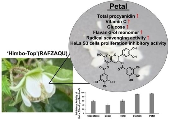
Potential of Raspberry Flower Petals as a Rich Source of Bioactive Flavan-3-ol Derivatives Revealed by Polyphenolic Profiling

1
Graduate School of Engineering, Osaka Electro-Communication University (OECU), 18-8 Hatsu-cho, Neyagawa 572-8530, Osaka, Japan
2
Faculty of Engineering, Osaka Electro-Communication University (OECU), 18-8 Hatsu-cho, Neyagawa 572-8530, Osaka, Japan
3
Research Institute for Sustainable Humanosphere, Kyoto University, Uji 611-0011, Kyoto, Japan
*
Authors to whom correspondence should be addressed.
These authors contributed equally to this work.
Nutraceuticals 2023, 3(2), 196-209; https://doi.org/10.3390/nutraceuticals3020015
Submission received: 4 February 2023 / Revised: 22 March 2023 / Accepted: 4 April 2023 / Published: 12 April 2023

Abstract

:
Inspired by the potential functional activity of polyphenol compounds contained in raspberry (Rubus idaeus), we previously explored the effects of the cultivation environment and maturity on the polyphenolic profiles of raspberry leaves and fruits. Herein, building on our previous studies, we used high-performance liquid chromatography and liquid chromatography–mass spectrometry to profile the polyphenol compounds contained in five parts of raspberry flowers (receptacles, sepals, pistils, stamens, and petals), revealing the presence of (+)-catechin, (−)-epicatechin, procyanidin B4, procyanidin C3, sanguiin H-6, and lambertianin C in all flower parts. Petals also contained (−)-epicatechin-3,5-di-O-gallate, kaempferol-7-O-glucoside, and naringenin-7-O-glucoside as well as other flavan-3-ol derivatives efficiently scavenging free radicals and inhibiting the growth of cancer (HeLa S3) cells. Thus, raspberry flower petals were concluded to be a good source of characteristic and highly functional flavan-3-ol derivatives.

Graphical Abstract

1. Introduction

Polyphenol compounds with multiple phenolic hydroxyl groups, are found in various foods and are believed to benefit human health by exerting antioxidant, cytotoxic, anti-inflammatory, antihypertensive, antidiabetic, and other effects [1]. Plants are a good source of structurally diverse polyphenol compounds [2], as exemplified by phenylpropanoids (e.g., caffeic [3] and chlorogenic [4] acids), stilbenoids (e.g., resveratrol [5]), hydroxybenzoic acid derivatives (e.g., gallic and ellagic acids [6]), and flavonoids (catechin, anthocyanins, and quercetin [7]). The isolation of polyphenol compounds from plants and the functionality of these compounds and foods containing them have attracted much attention [8]. In particular, our group is interested in the structure and functionality of flavonoids, namely flavan-3-ol derivatives, which include catechins and epigallocatechin gallate (EGCG), the main polyphenol contained in green tea [9,10]. The flavan-3-ol core features two chiral centers (second and third positions) and therefore exists in several stereoisomeric forms. Some plants produce highly functional oligomeric flavan-3-ol derivatives that are collectively denoted as proanthocyanidins, which are the main components of cocoa [11], wine [12], and apple [13] polyphenols, and exhibit health functionality [14].
Although flavan-3-ol is highly functional, it often exists as a mixture of compounds with different numbers of hydroxyl groups, stereotypes, and condensation numbers, which complicates individual functionality evaluation. In addition, the isolation of trace components is hindered by the fact that their structural isomers have similar chemical properties. Therefore, we have developed the syntheses of naturally occurring compounds such as flavan-3-ol derivatives [15,16] and confirmed their high functionality and three-dimensional structures by exploring their structure-activity-relationships, revealing that fine structural differences can significantly impact biological activity [17]. Specifically, the dimer of flavan-3-ol contained in beer as a minor component was shown to suppress the growth of yeast and human cancer cells, while a minor component contained in green tea was found to be more toxic to cancer cells than EGCG [18]. In addition, as a result of our synthetic research, we have established a library containing many types of both natural and non-natural polyphenol compounds. As these library entries can potentially be used as standard compounds, we defined our next challenge as the search for foods containing polyphenol compounds with skeletons suggested to be highly bioactive in our previous studies on structure-activity relationships.
Berry fruits are rich in polyphenol compounds and are therefore used to prepare numerous commercially available functional supplements [19]. Among the numerous berry fruits, raspberries (Rubus idaeus) contain complex polyphenol compounds with a wide variety of skeletons and have therefore drawn our attention [20]. In particular, we are interested in the effects of raspberry plant part, growth stage, and cultivation environment on the polyphenolic profile. Previously, we reported that the wavelength of light-emitting diode (LED) light influences the structure of polyphenol compounds produced in raspberry leaves and revealed that the abundance of flavan-3-ol derivatives increases upon blue LED irradiation [21]. In addition, we established eight levels of raspberry fruit maturity, examined its effect on the content of polyphenol compounds, and determined biosynthetic enzyme expression levels [22]. The results indicated that the polyphenolic profile depends on the part of the raspberry plant and the cultivation environment.
Inspired by the above, we herein used a combination of chromatographic and mass spectrometric techniques to profile the polyphenols contained in different parts of raspberry flowers. Petal extracts contained compounds inhibiting the growth of cancer cells and were richer in flavan-3-ol derivatives than other parts, thus exhibiting high biological activity.

2. Materials and Methods

2.1. Reagents and Materials

High-performance liquid chromatography (HPLC)-grade methanol and molecular biology-grade dimethyl sulfoxide (DMSO) were obtained from FUJIFILM Wako Pure Chemical Co., Ltd. (Osaka, Japan). Liquid chromatography–mass spectrometry (LC-MS)-grade acetonitrile and HPLC-grade formic acid were obtained from Honeywell International, Inc. (Charlotte, NJ, USA). All commercially available chemicals for crude extract solution were dissolved in DMSO and stored at −40 °C until use. The standard samples, (+)-catechin, (−)-epicatechin, kaempferol-7-O-glucoside, and naringenin-7-O-glucoside were obtained from Sigma-Aldrich Inc. (St. Louis, MO, USA). The procyanidin B2 standard was obtained from Extrasynthese (Genay, France). Folin-Ciocalteu reagent was obtained from Merck KGaA (Darmstadt, Germany), and the 1,1-diphenyl-2-picrylhydrazyl (DPPH) radical was obtained from Tokyo Chemical Industry Co., Ltd. (Tokyo, Japan). Potassium persulfate and 2,2′-azino-bis(3-ethylbenzothiazoline-6-sulfonic acid) diammonium salt (ABTS) were obtained from Tokyo Chemical Industry, Co., Ltd. (Tokyo, Japan) and FUJIFILM Wako Pure Chemical Co., Ltd. (Osaka, Japan), respectively. All other reagents and chemicals were of guaranteed grade unless stated otherwise.

2.2. Cultivation of Raspberries and Collection of Flower Samples

Himbo-Top® raspberry seedlings (registration name according to the Plant Variety Protection and Seed Act: RAFZAQU, Himbo Top®) were obtained from Tenkoen Co. (Yamagata, Japan). Raspberries were cultivated by open-field cultivation in Neyagawa, Osaka Prefecture (latitude 34°45′37″, longitude 135°37′37″). Seedlings of ~30 cm in size were planted in March, and the buds formed between June and November were harvested immediately after flowering.

2.3. Preparation of Polyphenol Crude Extract

The collected raspberry flowers were separated into five parts (receptacle (RE), sepal (SE), pistil (PI), stamen (ST), and petal (PE)), and each part was extracted with LC-grade methanol. Specifically, methanol (15 mL) and the desired flower part were placed in a mortar and thoroughly ground at room temperature for 5 min. The mixture was supplemented with additional methanol (15 mL), extracted for 24 h at room temperature, and filtered through Celite. The filtrate was concentrated using a rotary evaporator, and the residue was vacuum-dried using a rotary pump and dissolved in DMSO to a concentration of 100 mg/mL to afford the crude extract [21,22].

2.4. Determination of Total Polyphenol Content

Total polyphenol content was determined by the Folin–Ciocalteu method [23,24,25] partially modified for application to small samples. A 1.5 mL microtube was charged with the crude extract (2 μL) and Milli Q water (200 μL), and the resulting solution was alkalinized with 10% (w/v) aqueous Na2CO3 (25 μL), supplemented with the Folin reagent (25 μL), and incubated in the dark for 30 min. The concentration of the produced blue pigment was determined from the absorbance at 740 nm measured by a microplate reader (Multiskan FC, Thermo Fisher Scientific, Waltham, MA, USA), and the total polyphenol content was calculated from this absorbance using a calibration curve prepared with gallic acid (10 mg/mL~0.1 mg/mL). The measurements were performed with n = 6 and repeated three times.

2.5. Determination of Total Proanthocyanidin Content

Total proanthocyanidin content was determined by a previously reported method partially modified for application to small samples [21,26,27]. A 1.5 mL microtube was charged with the crude extract (5 μL), a solution of Rochelle salt (5 mg/mL, 125 μL; pH 3.3) in 10% ethanol, and aqueous bovine serum albumin (BSA) (1 mg/mL, 250 μL). The mixture was vortexed, incubated at 4 °C for 15 min, and centrifuged at 15,000 rpm for 5 min. The supernatant was removed to obtain a precipitate. The precipitate was supplemented with a solution of 50 μL of NaCl (170 mM, 50 μL) in 200 mM aqueous acetic acid (pH 4.9), the reaction mixture was vigorously stirred and centrifuged, and the supernatant was removed. The obtained precipitate was supplemented with a 10% (w/v) solution of sodium lauryl sulfate (SDS) in 5 vol% triethanolamine (TEA) (70 µL), and the mixture was stirred and supplemented with 10 mM FeCl3 (10 µL). The absorbance at 520 nm was measured by a microplate reader (Multiskan FC, Thermo Fisher Scientific, Waltham, MA, USA) after 10 min incubation and used to calculate the total proanthocyanidin content using a calibration curve prepared with procyanidin B2 (10 mg/mL~0.1 mg/mL). The measurements were performed with n = 6 and repeated three times.

2.6. Quantitation of Vitamin C Content

Vitamin C was quantified by the dinitrohydrazine method using HPLC [28]. A test tube containing 5% (w/v) metaphosphoric acid (1 mL) was supplemented with the crude extract (1 μL) upon vigorous stirring. The obtained solution was dropwise treated with the indophenol solution until the development of an unfading pink color, supplemented with thiourea-metaphosphoric acid solution (2% (w/v), 1.0 mL) and 2% (w/v) 2,4-dinitrophenylhydrazine–4.5 M sulfuric acid solution (250 μL), and stirred. After 90 min incubation in a constant-temperature bath (50 °C), the mixture was extracted with ethyl acetate, and the content of vitamin C was determined using a calibration curve created with the same from the area of the target HPLC peak (detection wavelength = 495 nm).

2.7. Quantitation of Total Glucose Content

Total glucose content was determined using the Enzytec™ Liquid D-Glucose kit (R-Biopharm; HE; Darmstadt, Germany). The amount of the employed enzyme solution was reduced so that the measurement could be performed for small samples. The 100 mg/mL of crude extract (1 µL), Milli Q water (10 µL), and buffer solution (200 µL) were placed in a 1.5 mL microtube, and the mixture was stirred by turning the microtube upside down. After 1 min incubation at 37 °C, the mix solution of hexokinase and glucose-6-phosphate dehydrogenase (50 µL) was added, and the mixture was stirred as above. After 10 min incubation at 37 °C, the absorbance at 340 nm was measured by a microplate reader (Multiskan FC, Thermo Fisher Scientific, Waltham, MA, USA) and used to calculate total glucose content based on a calibration curve prepared with glucose. The measurements were performed with n = 6 and repeated three times.

2.8. HPLC and LC-MS Analyses

HPLC analysis was performed using a Shimadzu LC-2020 system equipped with a degassing unit (DGU-20A3R), binary pump (LC-20A), autosampler (SIL-20AC), diode array detector (SPD-M20A), column oven (CTO-20AC), and communications bus module (CBM-20A) connected to an LC work station (Shimadzu Co., Ltd. Kyoto, Japan). A Shimadzu 2020 Quadrupole mass spectrometer (Shimadzu Co., Ltd. Kyoto, Japan) equipped with a positive/negative electrospray ionization source was used as a detector. A Wakopac® MS-5C18GT column (φ 150 mm × 2.0 mm, 5 μm, FUJIFILM Wako Pure Chemical Co., Ltd. Osaka, Japan) was also employed. Mobile phase A consisted of water with 0.05 vol% formic acid and mobile phase B was acetonitrile with 0.05 vol% formic acid. Raspberry extract analyses were carried out using the following program: 0 → 35% B over 0–90 min; 35 → 100% B over 90–95 min; 100% B over 95–100 min; and 0% B over 100–110 min. The flow rate was set at 0.2 mL/min. The injection volume was 10 μL, and the column oven was maintained at 40 °C. The mass spectrometer was operated in the negative selected-ion-monitoring (SIM) mode with a capillary voltage of 1.2 V for the identification of phenolic compounds, while the positive SIM mode was employed for the detection of betanin. The conditions for MS analysis were as follows: spray voltage = −3.5 V, dissolving line temperature = 250 °C, nebulizer gas flow = 1.5 L/min, heat block temperature = 200 °C, drying gas flow = 12.00 and 15.00 mL/min for the phenolics and betanin, respectively, and detector voltage = 1.2 V [20,21,22].

2.9. DPPH Radical Scavenging Activity

Radical scavenging activity was determined using a modification of a previously described DPPH assay [29]. Briefly, a solution of the DPPH radical in ethanol (30 μM, 1.0 mL) was added to 1 μL of the 100 mg/mL crude extract, and the mixture was incubated at 30 °C for 30 min). Scavenging activity was estimated from optical density (OD) at 515 nm determined by a microplate reader (Multiskan FC, Thermo Fisher Scientific, Waltham, MA, USA). The negative control corresponded to a solution of DMSO (1 μL) in ethanol (1.0 mL). Absorbance readings were converted into radical scavenging activity (%) as [(absorbance of control* − absorbance of sample)/absorbance of control*] × 100. The ‘absorbance of control’ is the absorbance of DPPH in wells without sample minus the absorbance in wells with DMSO but no DPPH. The results were reported as the means ± standard deviations of eight measurements (n = 8).

2.10. ABTS Radical Scavenging Activity

The ABTS radical scavenging activity was determined as previously mentioned [30]. ABTS solution (7 mM, 5 mL) was mixed with potassium peroxodisulfate solution (140 mM, 88 μL), and the mixture was allowed to stand in dark at room temperature for ~12 h, diluted with ethanol to an absorbance (740 nm) of 0.7 ± 0.02, and used as the ABTS solution for measurement. The latter ABTS solution (1 mL) was supplemented with the 100 mg/mL crude extract (1 μL), vigorously stirred for 10 s, and incubated at 30 °C for 4 min. The absorbance at 740 nm determined by a microplate reader (Multiskan FC, Thermo Fisher Scientific, Waltham, MA, USA) after 4 min was converted into radical scavenging activity (%) as [(absorbance of control − absorbance of sample)/absorbance of control] × 100. A 10 mM aqueous solution of (+)-catechin (1) was used as a positive control. DMSO in which the compound was dissolved was used as a negative control. The results were reported as the means ± standard deviations of eight measurements (n = 8).

2.11. Lipid Peroxidation Inhibitory Activity Using TBA Method

Antioxidant activity was quantified in terms of the lipid peroxide inhibitory activity determined by the thiobarbituric acid (TBA) method [31]. The 100 mg/mL of crude extract (1 µL) was mixed with 0.1% aqueous linoleic acid solution (100 µL) containing 0.8% (w/v) SDS, and the mixture was irradiated with UV light (254 nm) for 3 h upon cooling with ice. The solution was supplemented with 4.5% aqueous butylated hydroxytoluene (BHT) solution (2 µL) to stop the oxidation reaction, further supplemented with 20% aqueous acetic acid solution (150 µL) and 0.8% aqueous TBA solution (150 µL), and heated at 95 °C for 1 h. After cooling with ice, the mixture was treated with n-butanol solution containing 15% methanol (400 µL), vigorously stirred to extract the produced red pigment, and centrifuged at 15,000 rpm for 4 min at 4 °C. The absorbance of the upper n-butanol layer was measured at 530 nm by a microplate reader (Multiskan FC, Thermo Fisher Scientific, Waltham, MA, USA). Absorbance readings were converted to lipid peroxide inhibitory activity (%) as {1 − [(absorbance of the UV irradiated sample − absorbance of the non-UV irradiated sample)/(absorbance of the + UV control − absorbance of the − UV control)]} × 100. A 10 mM VE solution was used as a positive control. DMSO in which the compound was dissolved was used as a negative control. The results were reported as the means ± standard deviations of eight measurements (n = 8).

2.12. Inhibitory Activity against Cervical Cancer (HeLa S3) Cells

The human cervical adenocarcinoma cell line, HeLa S3, was provided by RIKEN BRC through the National Bio-Resource Project of MEXT (Tsukuba, Japan). Cells were maintained in high-glucose Dulbecco’s Modified Eagle’s Medium (Gibco Life Technology Co., Ltd. Grand Island, NY, USA) supplemented with 10% fetal bovine serum (Thermo Fisher Scientific, Waltham, MA, USA) and 1% Pen-Strep (Life Technology Co., Ltd. Grand Island, NY, USA). A total of 3000 cells in 100 µL of the medium were counted, added to each well of a 96-well plate, and incubated for 24 h at 37 °C in an incubator equilibrated with an atmosphere of 5% CO2 and 95% humidified air. Each well was supplemented with 100 mg/mL of crude extract (1 µL) and incubated for 24 or 48 h (final concentration = 0.1 mg/mL). Pure DMSO served as a negative control. The medium was removed, and fresh medium (90 µL) and 3-(4,5-dimethylthiazol-2-yl)-2,5-diphenyltetrazolium bromide solution (MTT; 5 mg/mL, 10 µL; FUJIFILM Wako Chemicals Co., Ltd. Osaka, Japan) was added to each well, with subsequent incubation performed for 2.5 h at 37 °C. The reaction medium was removed, and DMSO (100 µL) was added to each well and mixed. Viable cells were then assessed using a Filter Max F5 multi-mode microplate reader (Molecular Devices LLC, San Jose, CA, USA) to measure OD at 570 nm [32]. Absorbance readings were converted to cell growth inhibitory activity (%) as [1 − (absorbance of sample/absorbance of control)] × 100. The results were reported as the means ± standard deviations of eight measurements (n = 6).

2.13. Statistical Analysis

The results were obtained by MS Excel and analyzed using Student’s t-test and the analysis of variance (ANOVA) test. A p < 0.05 was set for statistical significance. Basic descriptive statistics were used to describe the data, including arithmetic means, standard deviations, and percentiles [33,34].

3. Results

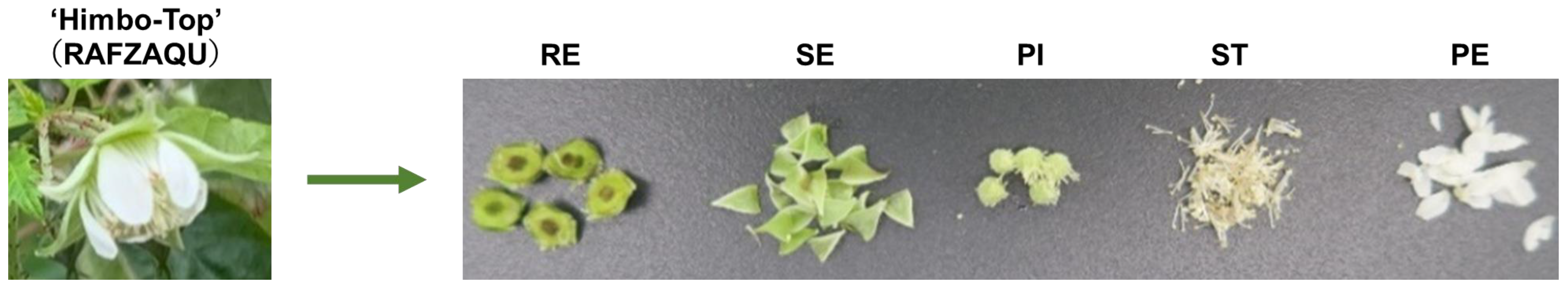
3.1. Separation of Flowers into Parts and Extraction

Previously, we showed that the amount and type of polyphenols in raspberry leaves depended on the wavelength of LED light used for cultivation [21] and that the polyphenol profile of raspberry fruits strongly depended on their maturity degree [22]. In particular, the total polyphenol content was highest for fruits that had just set and negatively correlated with the maturity degree. Inspired by these results, we hypothesized that raspberry flowers may also have a high polyphenol content. Although the natural compounds contained in raspberries have been well investigated [20,35,36], no reports have dealt with raspberry flowers, let alone with the analysis of individual flower parts. Therefore, we decided to collect raspberry flowers immediately after flowering and examine the compounds contained therein. As shown in Figure 1, the collected flowers were disassembled into five parts (RE, SE, PI, ST, and PE). Table 1 shows the wet weight of each part (i.e., the weights of parts determined immediately after flower decomposition without drying) and the dry weights of the corresponding methanolic extracts (i.e., the weights of the residues obtained after extract concentration in vacuo). The dry extract weights were close to 50% of the corresponding wet weight except for PE, in which case the dry extract was only 15% of the wet weight. Thus, many components were found to be soluble in methanol except in the case of PE.

3.2. Determination of Compounds Contained in Extracts

The extracts were quantified in terms of total polyphenol (Figure 2a), total proanthocyanidin (Figure 2b), total vitamin C (Figure 2c), and total glucose (Figure 2d) contents. The total polyphenol and proanthocyanidin contents of RE, SE, and PI were relatively low, while those of ST and PE were rather high. The vitamin C contents of RE and SE were lower than those of PI, ST, and PE, and the maximal value was observed for PE. Given that plants contain many glycosylated compounds and that many polyphenolics are glycated [37], we believe that total glucose content is an indicator of not only the amount of free glucose but also the amount of glycated compounds. Supplemental data are shown as the result of statistically evaluating whether the difference with other fractions in Figure 2 is superior compared to PE (Supplementary Materials Table S1).

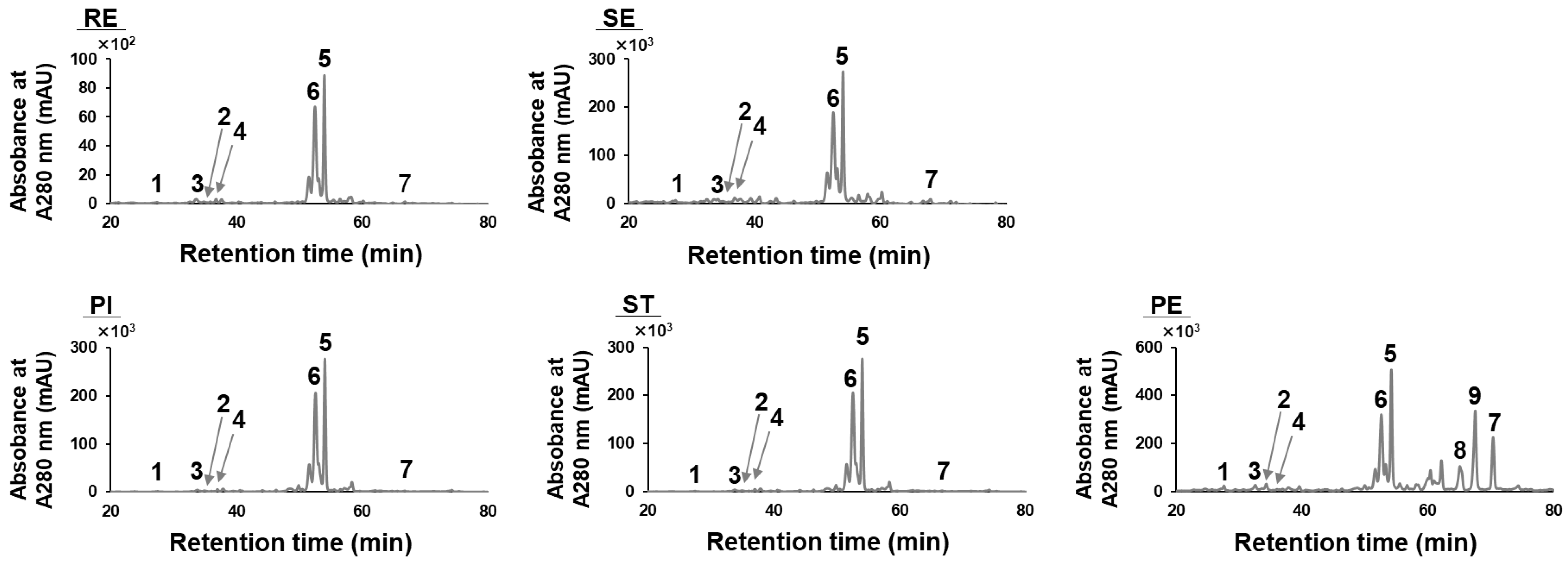
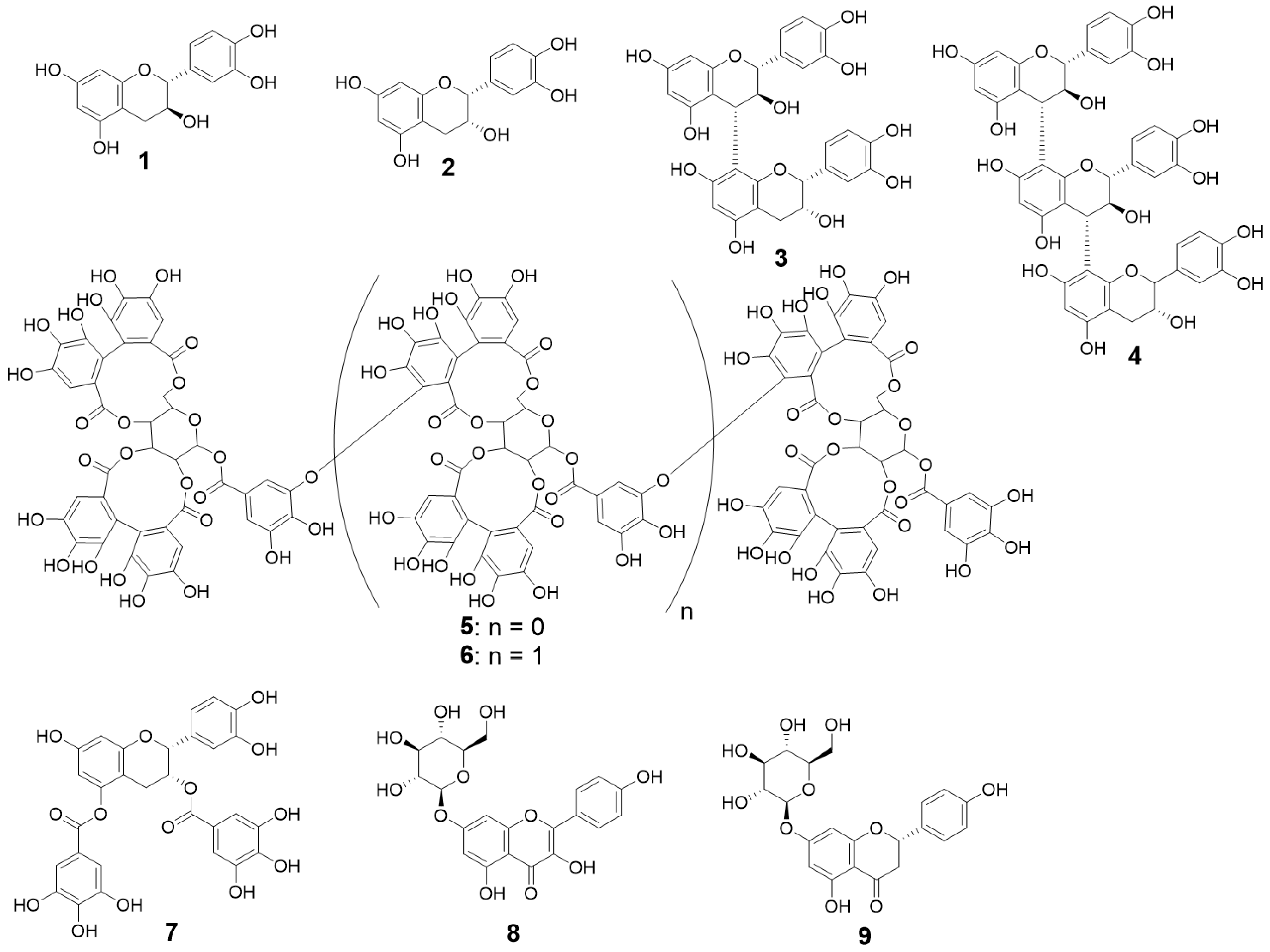
3.3. HPLC Analysis of Extracts and Identification of Compound Structures

Figure 3 shows the results of HPLC analysis for all extracts. The observed peak intensities suggest that the polyphenol contents of SE, PI, and ST exceed that of RE by a factor of three and are lower than that of PE by a factor of two. Detection was performed at a wavelength of 280 nm to secure the sensitive detection of flavan-3-ol derivatives and proanthocyanidins. The chromatograms obtained for RE, SE, PI, and ST were similar, showing only slight differences in individual peak intensities. In contrast, the PE chromatogram featured peaks not observed in other cases. Compounds 1 to 4 were identified as flavan-3-ol derivatives, namely (+)-catechin, (−)-epicatechin, procyanidin B4, and procyanidin C3, respectively, while compounds 5 and 6 were identified as ellagitannin derivatives, namely sanguiin H-6 and lambertianin C, respectively (Figure 4). These six compounds have been previously detected in raspberry leaves [21] and fruits [22] and have also been reported in the analytical studies of other research groups [20].
Surprisingly, multiple distinct peaks with retention times higher than those of ellagitannin derivatives were detected for PE. Based on their molecular weights determined by LC-MS and the comparison of their LC retention times with those of standard samples, compounds 8 and 9 were identified as kaempferol-7-O-glucoside and naringenin-7-O-glucoside, respectively. Although 8 has not been reported in raspberry analysis, it has been detected in the same Rosaceae plant [38], and 9 has been detected in raspberry fruit [39]. These glycated compounds contained in PE are thought to contribute to the high total glucose content of this flower part (Figure 2). Given that kaempferol and naringenin derivatives are known to be present in berry plants including raspberries, their detection was not unexpected, but the fact that they were contained at detectable levels only in PE was of great interest.
Although small amounts of 7 were detected in parts other than PE, PE remained the main repository of this species. As all compounds had different extinction coefficients at 280 nm, the corresponding absorbances could not be used for direct quantitation. Therefore, we decided to quantify the individual compounds based on HPLC peak heights and areas.

3.4. Quantitation of Polyphenolics in Raspberry Flowers

Figure 5 shows the concentrations of flavan-3-ol derivatives 14 and 79 in crude extracts. Given that we have developed a method for synthesizing flavan-3-ol derivatives, including 3, 4, and 7, we quantified these species with the help of calibration curves prepared using standard compounds. Compounds 3 and 4, which are proanthocyanidins, are known to be difficult to isolate, and the detection of rotamers in their NMR spectra further complicates structure determination for small isolated amounts. Moreover, the HPLC retention time of galloylated compounds such as 7 is often the same for species with different numbers and locations of galloylated sites, which complicates separation. In addition, the location of galloylated sites is difficult to determine. Therefore, we have developed synthetic protocols and regioselective galloylation methods for proanthocyanidins.
Monomers 1 and 2 were most abundant in PE. ST contained relatively large amounts of 2, while the content of 3, which is a dimer of 1 and 2, was relatively low in SE and PI but high in RE, ST, and PE. Compound 4, a trimer composed of one epicatechin and two catechin residues, was relatively abundant in RE and PE. These results show a weak correlation with total polyphenol content and total proanthocyanidin content in Figure 2a,b. However, RE and PE may contain proanthocyanidins other than those shown here, e.g., long-chain oligomers. Compound 7 was most abundant in PE, which agrees well with the HPLC data in Figure 3. Table 2 shows the content of flavan-3-ol derivatives in different flower parts, revealing that it was highest for PE. Supplemental data are shown as the result of statistically evaluating whether the difference with other fractions in Figure 5 and Table 2 is superior compared to PE (Supplementary Materials Table S2).

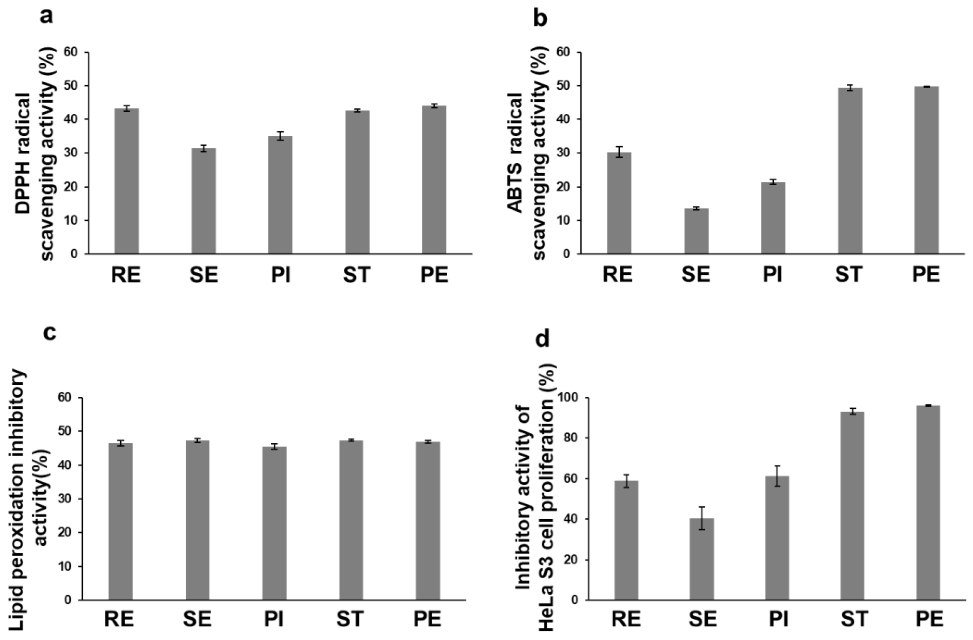
3.5. Biological Activities of Raspberry Flower Extracts

Next, the biological activity of the extracts was evaluated. Given that the scavenging of reactive oxygen species and antioxidant activity are the main biological activities of polyphenolics, two radical scavenging activity tests, DPPH (Figure 6a) and ABTS (Figure 6b), and an antioxidant activity test using the TBA method were performed (Figure 6c). Both DPPH and ABTS radical scavenging activities were low for SE and PI and high for ST and PE. Regarding the radical scavenging activity of RE, DPPH showed a high scavenging rate, while ABTS showed a low scavenging rate. This suggests that RE contains compounds with different properties that affect DPPH and ABTS. All extracts showed similar moderate antioxidant activities. Radical scavenging activity was suggested to be correlated with the content of polyphenolic compounds. The inhibitory activity against HeLa S3 (Figure 6d) showed the same trend as radical scavenging activity (Figure 6a,b) and was highest for ST and PE. These results suggest that radical scavenging activity is correlated with polyphenol content. Supplemental data are shown as the result of statistically evaluating whether the difference with other fractions in Figure 6 is superior compared to PE (Supplementary Materials Table S3).

4. Discussion

We have so far focused on polyphenols, especially flavan-3-ol derivatives, and conducted structure-activity relationship studies. In addition, using standard samples obtained through research on organic synthesis, we have studied changes in raspberry polyphenolics due to factors such as the cultivation environment and fruit maturity.
Table 3 summarizes the results of quantitative experiments and bioactivity tests performed herein. The HPLC data in Figure 3 show that PE contained (−)-epicatechin-3,5-di-O-gallate (7), kaempferol-7-O-glucoside (8), and naringenin-7-O-glucoside (9). Compound 7, in particular, has not been detected in berry plants such as raspberries and could not be detected in the raspberry fruits that we have thus far analyzed. As mentioned above, to the best of our knowledge, there has been no report in the literature that disassembled the raspberry flower into each part and analyzed each part.
Previously, we quantified flavan-3-ol derivatives in raspberry leaves grown under white fluorescent light in a plant factory, reporting values of 0.50, 0.10, 0.59, and 0.34 mg/100 mg dry extract for catechin (1), epicatechin (2), procyanidin B4 (3), and procyanidin C3 (4), respectively [21]. The content of flavan-3-ol derivatives in flowers was almost the same as that in leaves. Conversely, compounds 7, 8, and 9 were contained by detected exclusively in PE.
As Figure 4 shows that the 3- and 5-positions of (−)-epicatechin (1) in 7 are galloylated, and there is no reported example of flavan-3-ol digallate detection in raspberries. The above compound is a trace component of green tea polyphenols [40] and was reported to be more toxic to cancer cells than EGCG, the main component of green tea polyphenols [18]. Therefore, we believe that PE containing compound 7 is promising as a supplement.
The results of bioactivity tests (Figure 6) suggested that, unlike antioxidant activity, radical scavenging activity was correlated with the amount of polyphenols. Given that antioxidant activity was assessed as the ability to prevent the formation of peroxides due to UV irradiation, we believe that polyphenolics were partially decomposed by this irradiation and remained stable compounds, resulting in similar values. ST and PE extracts exhibited high cytostatic activity against HeLa S3. There are reports on the use of stamens as health foods, for example, Chinese peony stamens have a high polyphenol content and are highly functional, so they are used as medicinal teas and herbs [41]. Conversely, raspberry stamens are difficult to collect in large quantities, and it is considered difficult to use only the stamens. In addition, it is considered that the harvesting of stamens makes it difficult to bear fruit. Raspberry petals, however, detach soon after pollination. Since it is possible to collect petals while cultivating fruits, raspberry is considered to have a high potential as a material for petals.
Compound 7 contained in PE is known to actively inhibit the growth of cancer cells and to induce cell death through apoptosis by our very recent studies [42]. Thus, 7 may be used to develop new food products in the future because of its wealth of dietary experience. In addition, given the high anticancer activity of compound 8 [43], we thought that these PE-specific compounds exhibited high activity. The HPLC chromatogram of the ST extract was not much different from those of SE and PE extracts, which suggests that anticancer activity may be partly determined by compounds that cannot be detected by HPLC. The active compounds contained in ST are currently under investigation.

5. Conclusions

Raspberry flowers were divided into five parts, which were individually extracted. The extracts were characterized in terms of their polyphenolic profiles and bioactivities. The petals (PE) were identified as the most promising source of flavan-3-ol derivatives and were shown to exhibit high biological activity and contain a digallate form of (−)-epicatechin, which has not previously been detected in other flower parts. PE featured the smallest dry extract/wet flower part weight ratio. However, the PE extract exhibited a high dry-weight-basis content of functional polyphenols and high biological activity. Among the five flower parts, PE is easily detached after pollination and is not required for fruit growth. Thus, by collecting only the petals falling off after pollination, it may be possible to obtain a source of flavan-3-ol without damaging raspberry fruits.

Supplementary Materials

The following supporting information can be downloaded at: https://www.mdpi.com/article/10.3390/nutraceuticals3020015/s1, Table S1: Statistical results comparing fractions of each flower and petals (PE) in each analysis result in Figure 2; Table S2: Statistical results comparing fractions of each flower and petals (PE) for the amounts of compounds shown in Figure 5 and Table 2; Table S3: Statistical results comparing fractions of each flower and petals (PE) in the bioactivity test results shown in Figure 6.

Author Contributions

Conceptualization, R.K., R.D. and A.S.; synthesis, R.K.; methodology, R.K., R.D., T.K. and A.S.; investigation, R.K., A.A., M.T. and R.D.; writing—original draft preparation, R.D.; writing—review and editing, R.K., T.K. and A.S.; supervision, T.K. and A.S.; project administration, A.S.; funding acquisition, A.S. All authors have read and agreed to the published version of the manuscript.

Funding

This work was supported by BRAIN: Program for Promotion of Basic and Applied Research for Innovations in Bio-oriented Industry (GR104, A.S.), the Technology Research Promotion Program for Agriculture, Forestry, Fisheries, and Food Industry (A.S.), the Tojuro Iijima Foundation for Food and Technology (A.S.) and the Kurata Memorial Hitachi Science and Technology Foundation (A.S.).

Informed Consent Statement

Not applicable.

Data Availability Statement

Not applicable.

Conflicts of Interest

The authors declare no conflict of interest.

Abbreviations

ABTS2,2′-azino-bis(3-ethylbenzothiazoline-6-sulfonic acid) diammonium salt
BHTbutylated hydroxytoluene
DPPH1,1-diphenyl-2-picrylhydrazyl
EGCG(−)-Epigallocatechin-3-O-gallate
HPLCHigh-performance liquid chromatography
PEpetal
PIpistil
REreceptacle
SEsepal
SDSsodium Dodecyl Sulfate
STstamen
TEAtriethanolamine
TBAthiobarbituric acid
VEVitamin E

References

  1. Rana, A.; Samtiya, M.; Dhewa, T.; Mishra, V.; Aluko, V.E. Health benefits of polyphenols: A concise review. J. Food Biochem. 2022, 46, e14264. [Google Scholar] [CrossRef] [PubMed]
  2. Quideau, S.; Deffieux, D.; Douat-Casassus, C.; Pouységu, L. Plant polyphenols: Chemical properties, biological activities, and synthesis. Angew. Chem. Int. Ed. 2011, 50, 586–621. [Google Scholar] [CrossRef] [PubMed]
  3. Magnani, C.; Isaac, V.L.B.; Correa, M.A.; Salgado, H.R.N. Caffeic acid: A review of its potential use in medications and cosmetics. Anal. Methods 2014, 6, 3203–3210. [Google Scholar] [CrossRef]
  4. Naveed, M.; Hejazi, V.; Abbas, M.; Kamboh, A.A.; Khan, G.J.; Shumzai, M.; Ahmad, F.; Babazadeh, D.; Fang, X.F.; Modarresi-Ghazani, F.; et al. Chlorogenic acid (CGA): A pharmacological review and call for further research. Biomed. Pharmacother. 2018, 97, 67–74. [Google Scholar] [CrossRef]
  5. Zhang, L.X.; Li, C.X.; Kakar, M.U.; Khan, M.S.; Wu, P.F.; Amir, R.M.; Dai, D.F.; Naveed, M.; Li, Q.Y.; Saeed, M.; et al. Resveratrol (RV): A pharmacological review and call for further research. Biomed. Pharmacother. 2021, 143, 12164. [Google Scholar] [CrossRef] [PubMed]
  6. Vattem, D.A.; Shetty, K. Biological functionality of ellagic acid: A review. J. Food Biochem. 2005, 29, 234–266. [Google Scholar] [CrossRef]
  7. Panche, A.N.; Diwan, A.A.D.; Chandra, S.R. Flavonoids: An overview. J. Nutr. Sci. 2016, 5, e47. [Google Scholar] [CrossRef]
  8. Rasoulia, H.; Farzaeib, M.H.; Khodarahm, R. Polyphenols and their benefits: A review. Int. J. Food Prop. 2017, 20, S1700–S1741. [Google Scholar] [CrossRef]
  9. Negri, A.; Valeria Naponelli, V.; Rizzi, F.; Bettuzzi, S. Molecular targets of epigallocatechin—Gallate (EGCG): A special focus on signal transduction and cancer. Nutrients 2018, 10, 1936. [Google Scholar] [CrossRef]
  10. Prasanth, M.I.; Sivamaruthi, B.S.; Chaiyasut, C.; Tencomnao, T. A Review of the role of green tea (Camellia sinensis) in antiphotoaging, stress resistance, neuroprotection, and autophagy. Nutrients 2019, 11, 474. [Google Scholar] [CrossRef]
  11. Ángeles, M.; Ramos, M.S. Health beneficial effects of cocoa phenolic compounds: A mini-review. Curr. Opin. Food Sci. 2017, 14, 20–25. [Google Scholar]
  12. German, J.B.; Rosemary, L.; Walzem, R.L. The health benefits of wine. Annu. Rev. Nutr. 2000, 20, 561–593. [Google Scholar] [CrossRef] [PubMed]
  13. Boyer, J.; Liu, R.H. Apple phytochemicals and their health benefits. Nutr. J. 2004, 3, 5. [Google Scholar] [CrossRef] [PubMed]
  14. Rauf, A.; Imran, M.; Abu-Izneid, T.; Haq, I.U.; Patel, S.; Pan, X.; Naz, S.; Silva, A.S.; Saeed, F.; Suleria, H.A.R. Proanthocyanidins: A comprehensive review. Biomed. Pharmacother. 2019, 116, 108999. [Google Scholar] [CrossRef] [PubMed]
  15. Saito, A.; Mizushina, Y.; Tanaka, A.; Nakajima, N. Versatile synthesis of epicatechin series procyanidin oligomers, and their antioxidant and DNA polymerase inhibitory activity. Tetrahedron 2009, 65, 7422–7428. [Google Scholar] [CrossRef]
  16. Higashino, Y.; Okamoto, T.; Mori, K.; Kawasaki, T.; Hamada, M.; Nakajima, N.; Saito, A. Regioselective synthesis of procyanidin B6, a 4-6-condensed (+)-catechin dimer, by intramolecular condensation. Molecules 2018, 23, 205. [Google Scholar] [CrossRef]
  17. Hamada, Y.; Takano, S.; Ayano, Y.; Tokunaga, M.; Koashi, T.; Okamoto, S.; Doi, S.; Ishida, M.; Kawasaki, T.; Hamada, M.; et al. Structure–activity relationship of oligomeric flavan-3-ols: Importance of the upper-unit B-ring hydroxyl groups in the dimeric structure for strong activities. Molecules 2015, 20, 18870–18885. [Google Scholar] [CrossRef]
  18. Mori, K.; Ayano, Y.; Hamada, Y.; Hojima, T.; Tanaka, R.; Higashino, Y.; Izuno, M.; Okamoto, T.; Kawasaki, T.; Hamada, M.; et al. Role of 2,3-cis structure of (−)-epicatechin-3,5-O-digallate in inhibition of HeLa S3 cell proliferation. Nat. Prod. Chem. Res. 2015, 3, 172. [Google Scholar]
  19. Bonyadi, N.; Dolatkhah, N.; Salekzamani, Y.; Hashemian, M. Effect of berry-based supplements and foods on cognitive function: A systematic review. Sci. Rep. 2022, 12, 3239. [Google Scholar] [CrossRef]
  20. Wu, L.; Liu, Y.; Qin, Y.; Wang, L.; Wu, Z. HPLC-ESI-qTOF-MS/MS characterization, antioxidant activities and inhibitory ability of digestive enzymes with molecular docking analysis of various parts of raspberry (Rubus ideaus L.). Antioxidants 2019, 8, 274. [Google Scholar] [CrossRef]
  21. Kobori, R.; Hashimoto, S.; Koshimizu, H.; Yakami, S.; Hirai, M.; Noro, K.; Kawasaki, T.; Saito, A. Flavan-3-ols content in red raspberry leaves increases under blue LED-light irradiation. Metabolites 2019, 9, 56. [Google Scholar] [CrossRef] [PubMed]
  22. Kobori, R.; Yakami, S.; Kawasaki, T.; Saito, A. Changes in the polyphenol content of red raspberry fruits during ripening. Horticulturae 2021, 7, 569. [Google Scholar] [CrossRef]
  23. Folin, O.; Denis, W.A. Colorimetric method for the determination of phenols (and derivatives) in urine. J. Biol. Chem. 1915, 22, 305–308. [Google Scholar] [CrossRef]
  24. Tiitto, R.J. Phenolic constituents in the leaves of northern willows: Methods for the analysis of certain phenolics. J. Agric. Food Chem. 1985, 33, 213–217. [Google Scholar] [CrossRef]
  25. Geremu, M.; Tola, Y.B.; Sualeh, A. Extraction and determination of total polyphenols and antioxidant capacity of red coffee (Coffea arabica L.) pulp of wet processing plants. Chem. Biol. Technol. Agric. 2016, 3, 25. [Google Scholar] [CrossRef]
  26. Harbertson, J.F.; Kennedy, J.A.; Adams, D.O. Tannin in skin and seeds of cabernet sauvignon, syrah, and pinoy noir berries during ripening. Am. J. Enol. Vitic. 2002, 52, 54–59. [Google Scholar] [CrossRef]
  27. Mella, A.C.; Neira, A.P.; Bastias, J.N.; Campos, C.J.; Solis, R.L.; Canals, J.M. Comparison of analytical methods for measuring proanthocyanidins in wine and their relationship with perceived astringency. Int. J. Food Sci. Technol. 2013, 48, 2588–2594. [Google Scholar] [CrossRef]
  28. Kishida, E.; Nishimoto, Y.; Kojo, S. Specific determination of ascorbic acid with chemical derivatization and high-performance liquid chromatography. Anal. Chem. 1992, 64, 1505–1507. [Google Scholar] [CrossRef]
  29. Nanjo, F.; Goto, K.; Seto, R.; Suzuki, M.; Sakai, M.; Hara, Y. Scavenging effects of tea catechins and their derivatives on 1,1-diphenyl-2-picrylhydrazyl radical. Free Radic. Biol. Med. 1996, 21, 895–902. [Google Scholar] [CrossRef]
  30. Pellegrini, R.R.N.; Proteggente, A.; Pannala, A.; Yang, M.; Evans, C.R. Antioxidant activity applying an improved ABTS radical cation decolorization assay. Free Radic. Biol. Med. 1999, 26, 9–10. [Google Scholar]
  31. Saito, A.; Doi, Y.; Tanaka, A.; Matsuura, N.; Ubukata, M.; Nakajima, N. Systematic synthesis of four epicatechin series procyanidin trimers and their inhibitory activity on the Maillard reaction and antioxidant activity. Bioorg. Med. Chem. 2004, 12, 4783–4790. [Google Scholar] [CrossRef] [PubMed]
  32. Zhang, A.; Dong, Y.; Chen, X.; Tan, C.S.H.; Li, M.; Miao, K.; Lu, J.H. Toosendanin, a late-stage autophagy inhibitor, sensitizes triple-negative breast cancer to irinotecan chemotherapy. Clin. Med. 2022, 17, 55. [Google Scholar] [CrossRef] [PubMed]
  33. Begum, S.; Prabhu, V.; Mohanty, V.; Krishnaraj, U.; Abdulla, R. Unveiling the arcanum of formalin-fixed paraffin-embedded archival tissue blocks: A valuable resource for genomic DNA extraction. J. Oral Maxillofac. Pathol. 2022, 26, 289. [Google Scholar] [PubMed]
  34. Kušnierová, P.; Zeman, D.; Hradilic, P.; Čábal, M.; Zapletalová, O. Neurofilament levels in patients with neurological diseases: A comparison of neurofilament light and heavy chain levels. J. Clin. Lab. Anal. 2019, 33, e22948. [Google Scholar] [CrossRef]
  35. Yang, J.; Cui, J.; Chen, J.; Yao, J.; Hao, Y.; Fan, Y.; Liu, Y. Evaluation of physicochemical properties in three raspberries (Rubus ideaus) at five ripening stages in northern China. Sci. Hortic. 2020, 263, 109146. [Google Scholar] [CrossRef]
  36. Li, X.; Sun, J.; Chen, Z.; Jiang, J.; Jackson, A. Metabolite profile and genes/proteins expression in b-citraturin biosynthesis during fruit ripening in Chinese raspberry (Rubus chingi Hu). Plant Physiol. Biochem. 2021, 163, 76–86. [Google Scholar] [CrossRef]
  37. Mojzer, E.B.; Hrnčič, M.K.; Škerget, M.; Knez, Z.; Bren, U. Polyphenols: Extraction methods, antioxidative action, bioavailability and anticarcinogenic effects. Molecules 2016, 21, 901. [Google Scholar] [CrossRef]
  38. Wan, H.; Yu, C.; Han, Y.; Guo, X.; Ahmed, S.; Tang, A.; Wang, J.; Cheng, T.; Pen, H.; Shang, Q. Flavonols and carotenoids in yellow petals of rose cultivar (Rosa ‘Sun City’): A possible rich source of bioactive compounds. J. Agric. Food Chem. 2018, 66, 4171–4181. [Google Scholar] [CrossRef]
  39. Renai, L.; Scordo, C.V.A.; Chiuminatto, U.; Ulaszewska, M.; Giordani, E.; Petrucci, W.A.; Tozzi, F.; Nin, S.; Bubba, M.D. Liquid chromatographic quadrupole time-of-flight mass spectrometric untargeted profiling of (poly)phenolic compounds in Rubus idaeus L. and ubus occidentalis L. Fruits and their comparative evaluation. Antioxidants 2021, 10, 704. [Google Scholar] [CrossRef]
  40. Saijo, R.; Takeda, Y. HPLC analysis of catechins in various kinds of green teas produced in Japan and abroad. Nippo Shokuhin Kagaku Kogaku Kaishi 1999, 46, 138–147. [Google Scholar] [CrossRef]
  41. Xie, L.; Yan, Z.; Li, M.; Tian, Y.; Kilaru, A.; Niu, L.; Zhang, Y. Identification of phytochemical markers for quality evaluation of tree peony stamen using comprehensive HPLC-based analysis. Ind. Crops Prod. 2020, 154, 112711. [Google Scholar] [CrossRef]
  42. Doge, R.; Nishino, Y.; Saito, A. Fluorinated derivatives of digalloyl-flavan-3-ol induce autophagic cell death by forming granular aggregates containing mitochondria. BioChem, 2023; in press. [Google Scholar]
  43. Li, Y.; Gan, G.P.; Zhang, H.Z.; Wu, H.Z.; Li, C.L.; Huang, Y.P.; Liu, Y.W.; Liua, J.W. A flavonoid glycoside isolated from Smilax china L. rhizome in vitro anticancer effects on human cancer cell lines. J. Ethnopharmacol. 2007, 113, 115–124. [Google Scholar] [CrossRef] [PubMed]
Figure 1. Raspberry flowers were separated into five parts, namely receptacle (RE), sepal (SE), pistil (PI), stamen (ST), and petal (PE).
Nutraceuticals 03 00015 g001
Figure 2. (a) Total polyphenol content, (b) total proanthocyanidin content, (c) vitamin C, and (d) total glucose contents of different extracts. Total polyphenol contents are represented by Gallic Acid Equivalents (GAE) and total proanthocyanidin contents are represented by procyanidin B2 equivalents.
Nutraceuticals 03 00015 g002
Figure 3. HPLC chromatograms of extracts obtained from different parts of raspberry flowers. 1: (+)-catechin; 2: (−)-epicatechin; 3: procyanidin B4; 4: procyanidin C3; 5: sanguiin H-6; 6: lambertianin C and 7: (−)-epicatechin-3,5-di-O-gallate; 8: kaempferol-7-O-glucoside; 9: naringenin-7-O-glucoside. The vertical axis scale is not the same for all chromatograms to allow the main peaks to have the same visually perceived intensity for easy viewing.
Nutraceuticals 03 00015 g003
Figure 4. Structures of polyphenolics detected in raspberry flowers by HPLC: 1: (+)-catechin; 2: (−)-epicatechin; 3: procyanidin B4; 4: procyanidin C3; 5: sanguiin H-6; 6: lambertianin C; 7: (−)-epicatechin-3,5-di-O-gallate; 8: kaempferol-7-O-glucoside, 9: naringenin-7-O-glucoside.
Nutraceuticals 03 00015 g004
Figure 5. Contents of flavan-3-ol derivatives in crude extracts calculated from HPLC peak areas: (a) (+)-catechin (1); (b) (−)-epicatechin (2); (c) procyanidin B4 (3); (d) procyanidin C3 (4); (e) (−)-epicatechin-3,5-di-O-gallate (7). Compounds 1 and 2 were quantified with the help of a calibration curve prepared using commercially available compounds. Compounds 3, 4, and 7 have been calibrated using pure compounds prepared in our previous synthetic studies.
Nutraceuticals 03 00015 g005
Figure 6. (a) DPPH radical scavenging activity (final conc. 100 μg/mL), (b) ABTS radical scavenging activity (final conc. 100 μg/mL), (c) lipid peroxide inhibitory activity (TBA method) (final conc. 1 μg/mL), (d) HeLa S3 cell inhibitory activity of different extracts (final conc. 100 μg/mL).
Nutraceuticals 03 00015 g006
Table 1. Wet weights of flower portions and dry weights of corresponding methanolic extracts.
RESEPISTPE
Sample wet weight (g)1.030.600.08530.600.165
Extract dry weight (g)0.4820.3940.04310.3790.0245
Table 2. Amounts of different flavan-3-ol derivatives (mg/100 mg dry weight) in flower part extracts.
RESEPISTPE
Dry weight of extract (g) 10.480.390.0430.3800.025
(+)-Catechin (1) (mg) 20.130.130.220.140.83
(−)-Epicatechin (2) (mg) 20.060.080.060.150.19
Procyanidin B4 (3) (mg) 20.440.250.290.420.40
Procyanidin C3 (4) (mg) 20.190.120.130.110.15
(−)-Epicatechin-3,5-di-O-gallate (7) (mg) 20.020.020.020.030.08
Kaempferol-7-O-glucoside (8)nd 3ndndnd0.03
Naringenin-7-O-glucoside (9)ndndndnd0.03
1 Dry weight of each flower part shown in Table 1. 2 Amount of each compound per 100 mg dry weight calculated from HPLC peak areas. 3 nd: not detected.
Table 3. Summary of compositions and biological activities of different flower part extracts.
RESEPISTPE
Total polyphenol 1+++++++++++
Total proanthocyanidin 1++-++++++
Vitamin C 1+++++++++++++
Total glucose 1+++++++++++
Flavan-3-ol mononers 1--+++++
Procyanidins 1+++++++++++++
Radical scavenging activity 2+++++++++++++
Antioxidant activity 2+++++++++++++++
HeLa S3 cell proliferation inhibitory activity 2++++++++++++
1 ‘+’ indicates relative content; 2 ‘+’ indicates relative activity. For a 100 mg/mL extract solution, the maximum value of 100 was used for each quantitative experiment, and biological activity test was used. +++: ≥80, ++: ≥50, +: ≥20, -: ≤20.
Disclaimer/Publisher’s Note: The statements, opinions and data contained in all publications are solely those of the individual author(s) and contributor(s) and not of MDPI and/or the editor(s). MDPI and/or the editor(s) disclaim responsibility for any injury to people or property resulting from any ideas, methods, instructions or products referred to in the content.

