
Effects of Fatigue Induced by Repeated Sprints on Sprint Biomechanics in Football Players: Should We Look at the Group or the Individual?
 ,
,
Abstract
:1. Introduction
2. Materials and Methods
2.1. Subjects
2.2. Procedures
2.3. Sprint Kinetics
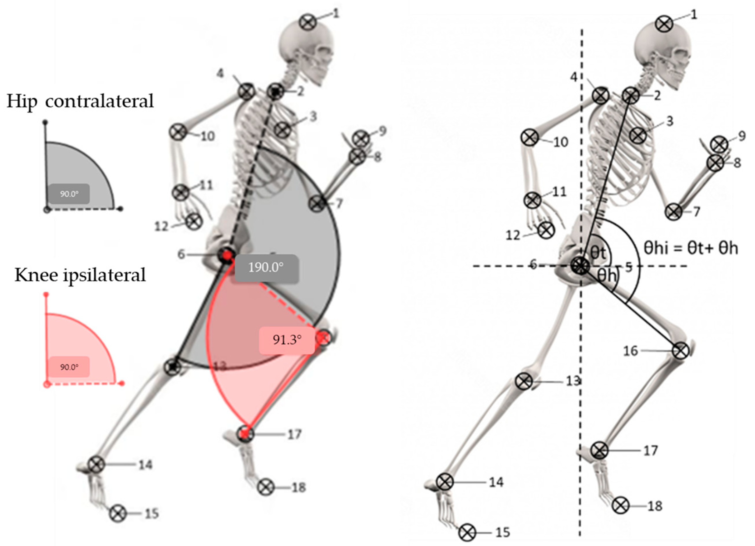
2.4. Sprint Kinematics
2.5. Statistical Analyses
3. Results
3.1. Kinetics
3.2. Kinematics
4. Discussion
5. Conclusions
Author Contributions
Funding
Institutional Review Board Statement
Informed Consent Statement
Data Availability Statement
Conflicts of Interest
References
- Dellal, A.; Chamari, K.; Wong, D.P.; Ahmaidi, S.; Keller, D.; Barros, R.; Bisciotti, G.N.; Carling, C. Comparison of Physical and Technical Performance in European Soccer Match-Play: FA Premier League and La Liga. Eur. J. Sport Sci. 2011, 11, 51–59. [Google Scholar] [CrossRef]
- Djaoui, L.; Wong, D.P.; Pialoux, V.; Hautier, C.; Da Silva, C.D.; Chamari, K.; Dellal, A. Physical Activity during a Prolonged Congested Period in a Top-Class European Football Team. Asian J. Sports Med. 2014, 5, 47–53. [Google Scholar] [CrossRef] [PubMed] [Green Version]
- Rampinini, E.; Coutts, A.J.; Castagna, C.; Sassi, R.; Impellizzeri, F.M. Variation in Top Level Soccer Match Performance. Int. J. Sports Med. 2007, 28, 1018–1024. [Google Scholar] [CrossRef] [Green Version]
- Faude, O.; Koch, T.; Meyer, T. Straight Sprinting Is the Most Frequent Action in Goal Situations in Professional Football. J. Sports Sci. 2012, 30, 625–631. [Google Scholar] [CrossRef] [PubMed]
- Morin, J.-B.; Gimenez, P.; Edouard, P.; Arnal, P.; Jiménez-Reyes, P.; Samozino, P.; Brughelli, M.; Mendiguchia, J. Sprint Acceleration Mechanics: The Major Role of Hamstrings in Horizontal Force Production. Front. Physiol. 2015, 6, 404. [Google Scholar] [CrossRef]
- Higashihara, A.; Nagano, Y.; Ono, T.; Fukubayashi, T. Differences in Hamstring Activation Characteristics between the Acceleration and Maximum-Speed Phases of Sprinting. J. Sports Sci. 2018, 36, 1313–1318. [Google Scholar] [CrossRef] [PubMed]
- Heiderscheit, B.C.; Hoerth, D.M.; Chumanov, E.S.; Swanson, S.C.; Thelen, B.J.; Thelen, D.G. Identifying the Time of Occurrence of a Hamstring Strain Injury during Treadmill Running: A Case Study. Clin. Biomech. 2005, 20, 1072–1078. [Google Scholar] [CrossRef]
- Schache, A.G.; Wrigley, T.V.; Baker, R.; Pandy, M.G. Biomechanical Response to Hamstring Muscle Strain Injury. Gait Posture 2009, 29, 332–338. [Google Scholar] [CrossRef]
- Liu, H.; Garrett, W.E.; Moorman, C.T.; Yu, B. Injury Rate, Mechanism, and Risk Factors of Hamstring Strain Injuries in Sports: A Review of the Literature. J. Sport Health Sci. 2012, 1, 92–101. [Google Scholar] [CrossRef] [Green Version]
- Yu, B.; Liu, H.; Garrett, W.E. Mechanism of Hamstring Muscle Strain Injury in Sprinting. J. Sport Health Sci. 2017, 6, 130–132. [Google Scholar] [CrossRef]
- Van Den Tillaar, R.; Solheim, J.A.B.; Bencke, J. Comparison of hamstring muscle activation during high-speed running and various hamstring strengthening exercises. Int. J. Sports Phys. Ther. 2017, 12, 718–727. [Google Scholar] [CrossRef] [PubMed] [Green Version]
- Hegyi, A.; Gonçalves, B.A.M.; Finni, T.; Cronin, N.J. Individual Region- and Muscle-Specific Hamstring Activity at Different Running Speeds. Med. Sci. Sports Exerc. 2019, 51, 2274–2285. [Google Scholar] [CrossRef] [PubMed]
- Prince, C.; Morin, J.-B.; Mendiguchia, J.; Lahti, J.; Guex, K.; Edouard, P.; Samozino, P. Sprint Specificity of Isolated Hamstring-Strengthening Exercises in Terms of Muscle Activity and Force Production. Front. Sports Act. Living 2020, 2, 609636. [Google Scholar] [CrossRef] [PubMed]
- Barnes, C.; Archer, D.T.; Hogg, B.; Bush, M.; Bradley, P.S. The Evolution of Physical and Technical Performance Parameters in the English Premier League. Int J. Sports Med. 2014, 35, 1095–1100. [Google Scholar] [CrossRef] [PubMed]
- Haugen, T.A.; Tønnessen, E.; Seiler, S. Anaerobic Performance Testing of Professional Soccer Players 1995-2010. Int J. Sports Physiol. Perform. 2013, 8, 148–156. [Google Scholar] [CrossRef]
- Woods, C.; Hawkins, R.D.; Maltby, S.; Hulse, M.; Thomas, A.; Hodson, A. Football Association Medical Research Programme The Football Association Medical Research Programme: An Audit of Injuries in Professional Football--Analysis of Hamstring Injuries. Br. J. Sports Med. 2004, 38, 36–41. [Google Scholar] [CrossRef] [Green Version]
- Orchard, J.; Best, T.M.; Verrall, G.M. Return to Play Following Muscle Strains. Clin. J. Sport Med. 2005, 15, 436–441. [Google Scholar] [CrossRef]
- Hägglund, M.; Waldén, M.; Ekstrand, J. Previous Injury as a Risk Factor for Injury in Elite Football: A Prospective Study over Two Consecutive Seasons. Br. J. Sports Med. 2006, 40, 767–772. [Google Scholar] [CrossRef] [Green Version]
- Hallén, A.; Ekstrand, J. Return to Play Following Muscle Injuries in Professional Footballers. J. Sports Sci. 2014, 32, 1229–1236. [Google Scholar] [CrossRef]
- Ekstrand, J.; Krutsch, W.; Spreco, A.; van Zoest, W.; Roberts, C.; Meyer, T.; Bengtsson, H. Time before Return to Play for the Most Common Injuries in Professional Football: A 16-Year Follow-up of the UEFA Elite Club Injury Study. Br. J. Sports Med. 2020, 54, 421–426. [Google Scholar] [CrossRef]
- Jones, A.; Jones, G.; Greig, N.; Bower, P.; Brown, J.; Hind, K.; Francis, P. Epidemiology of Injury in English Professional Football Players: A Cohort Study. Phys. Ther. Sport 2019, 35, 18–22. [Google Scholar] [CrossRef] [PubMed] [Green Version]
- Raya-González, J.; Suárez-Arrones, L.; Navandar, A.; Balsalobre-Fernández, C.; Sáez de Villarreal, E. Injury Profile of Elite Male Young Soccer Players in a Spanish Professional Soccer Club: A Prospective Study During 4 Consecutive Seasons. J. Sport Rehabil. 2020, 29, 801–807. [Google Scholar] [CrossRef] [PubMed]
- Schache, A.G.; Dorn, T.W.; Blanch, P.D.; Brown, N.A.T.; Pandy, M.G. Mechanics of the Human Hamstring Muscles during Sprinting. Med. Sci. Sports Exerc. 2012, 44, 647–658. [Google Scholar] [CrossRef] [PubMed] [Green Version]
- Chumanov, E.S.; Heiderscheit, B.C.; Thelen, D.G. Hamstring Musculotendon Dynamics during Stance and Swing Phases of High-Speed Running. Med. Sci. Sports Exerc. 2011, 43, 525–532. [Google Scholar] [CrossRef] [Green Version]
- Chumanov, E.S.; Schache, A.G.; Heiderscheit, B.C.; Thelen, D.G. Hamstrings Are Most Susceptible to Injury during the Late Swing Phase of Sprinting. Br. J. Sports Med. 2012, 46, 90. [Google Scholar] [CrossRef] [Green Version]
- Yu, B.; Queen, R.M.; Abbey, A.N.; Liu, Y.; Moorman, C.T.; Garrett, W.E. Hamstring Muscle Kinematics and Activation during Overground Sprinting. J Biomech. 2008, 41, 3121–3126. [Google Scholar] [CrossRef]
- Orchard, J.W. Hamstrings Are Most Susceptible to Injury during the Early Stance Phase of Sprinting. Br. J. Sports Med. 2012, 46, 88–89. [Google Scholar] [CrossRef] [Green Version]
- Ono, T.; Higashihara, A.; Shinohara, J.; Hirose, N.; Fukubayashi, T. Estimation of Tensile Force in the Hamstring Muscles during Overground Sprinting. Int. J. Sports Med. 2015, 36, 163–168. [Google Scholar]
- Mann, R.; Sprague, P. A Kinetic Analysis of the Ground Leg during Sprint Running. Res. Q Exerc. Sport 1980, 51, 334–348. [Google Scholar] [CrossRef]
- Sun, Y.; Wei, S.; Zhong, Y.; Fu, W.; Li, L.; Liu, Y. How Joint Torques Affect Hamstring Injury Risk in Sprinting Swing-Stance Transition. Med. Sci. Sports Exerc. 2015, 47, 373–380. [Google Scholar] [CrossRef] [Green Version]
- Liu, Y.; Sun, Y.; Zhu, W.; Yu, J. The Late Swing and Early Stance of Sprinting Are Most Hazardous for Hamstring Injuries. J. Sport Health Sci 2017, 6, 133–136. [Google Scholar] [CrossRef] [PubMed]
- Chapman, A.E.; Caldwell, G.E. Factors Determining Changes in Lower Limb Energy during Swing in Treadmill Running. J. Biomech. 1983, 16, 69–77. [Google Scholar] [CrossRef]
- Gregson, W.; Di Salvo, V.; Varley, M.C.; Modonutti, M.; Belli, A.; Chamari, K.; Weston, M.; Lolli, L.; Eirale, C. Harmful Association of Sprinting with Muscle Injury Occurrence in Professional Soccer Match-Play: A Two-Season, League Wide Exploratory Investigation from the Qatar Stars League. J. Sci. Med. Sport 2020, 23, 134–138. [Google Scholar] [CrossRef] [PubMed]
- Carling, C.; Gall, F.L.; Reilly, T.P. Effects of Physical Efforts on Injury in Elite Soccer. Int. J. Sports Med. 2010, 31, 180–185. [Google Scholar] [CrossRef] [PubMed]
- Mair, S.D.; Seaber, A.V.; Glisson, R.R.; Garrett, W.E. The Role of Fatigue in Susceptibility to Acute Muscle Strain Injury. Am. J. Sports Med. 1996, 24, 137–143. [Google Scholar] [CrossRef] [PubMed]
- Järvinen, T.A.; Kääriäinen, M.; Järvinen, M.; Kalimo, H. Muscle Strain Injuries. Curr. Opin. Rheumatol. 2000, 12, 155–161. [Google Scholar] [CrossRef]
- de Souza, J.; Gottfried, C. Muscle Injury: Review of Experimental Models. J. Electromyogr. Kinesiol. 2013, 23, 1253–1260. [Google Scholar] [CrossRef] [PubMed]
- Beltran, L.; Ghazikhanian, V.; Padron, M.; Beltran, J. The Proximal Hamstring Muscle-Tendon-Bone Unit: A Review of the Normal Anatomy, Biomechanics, and Pathophysiology. Eur. J. Radiol. 2012, 81, 3772–3779. [Google Scholar] [CrossRef]
- Morin, J.-B.; Samozino, P.; Edouard, P.; Tomazin, K. Effect of Fatigue on Force Production and Force Application Technique during Repeated Sprints. J. Biomech. 2011, 44, 2719–2723. [Google Scholar] [CrossRef]
- Edouard, P.; Mendiguchia, J.; Lahti, J.; Arnal, P.J.; Gimenez, P.; Jiménez-Reyes, P.; Brughelli, M.; Samozino, P.; Morin, J.-B. Sprint Acceleration Mechanics in Fatigue Conditions: Compensatory Role of Gluteal Muscles in Horizontal Force Production and Potential Protection of Hamstring Muscles. Front. Physiol. 2018, 9, 1706. [Google Scholar] [CrossRef] [Green Version]
- Jiménez-Reyes, P.; Cross, M.; Ross, A.; Samozino, P.; Brughelli, M.; Gill, N.; Morin, J.-B. Changes in Mechanical Properties of Sprinting during Repeated Sprint in Elite Rugby Sevens Athletes. Eur. J. Sport Sci. 2019, 19, 585–594. [Google Scholar] [CrossRef] [PubMed]
- Nagahara, R.; Morin, J.-B.; Koido, M. Impairment of Sprint Mechanical Properties in an Actual Soccer Match: A Pilot Study. Int. J. Sports Physiol. Perform. 2016, 11, 893–898. [Google Scholar] [CrossRef] [PubMed]
- Pinniger, G.J.; Steele, J.R.; Groeller, H. Does Fatigue Induced by Repeated Dynamic Efforts Affect Hamstring Muscle Function? Med. Sci. Sports Exerc. 2000, 32, 647–653. [Google Scholar] [CrossRef] [PubMed]
- Small, K.; McNaughton, L.R.; Greig, M.; Lohkamp, M.; Lovell, R. Soccer Fatigue, Sprinting and Hamstring Injury Risk. Int. J. Sports Med. 2009, 30, 573–578. [Google Scholar] [CrossRef] [PubMed]
- Schache, A.G.; Blanch, P.D.; Murphy, A.T. Relation of Anterior Pelvic Tilt during Running to Clinical and Kinematic Measures of Hip Extension. Br. J. Sports Med. 2000, 34, 279–283. [Google Scholar] [CrossRef] [Green Version]
- Thelen, D.G.; Chumanov, E.S.; Sherry, M.A.; Heiderscheit, B.C. Neuromusculoskeletal Models Provide Insights into the Mechanisms and Rehabilitation of Hamstring Strains. Exerc. Sport Sci. Rev. 2006, 34, 135–141. [Google Scholar] [CrossRef]
- Higashihara, A.; Nagano, Y.; Takahashi, K.; Fukubayashi, T. Effects of Forward Trunk Lean on Hamstring Muscle Kinematics during Sprinting. J. Sports Sci. 2015, 33, 1366–1375. [Google Scholar] [CrossRef]
- Schuermans, J.; Van Tiggelen, D.; Palmans, T.; Danneels, L.; Witvrouw, E. Deviating Running Kinematics and Hamstring Injury Susceptibility in Male Soccer Players: Cause or Consequence? Gait Posture 2017, 57, 270–277. [Google Scholar] [CrossRef]
- Kenneally-Dabrowski, C.J.B.; Brown, N.A.T.; Lai, A.K.M.; Perriman, D.; Spratford, W.; Serpell, B.G. Late Swing or Early Stance? A Narrative Review of Hamstring Injury Mechanisms during High-Speed Running. Scand. J. Med. Sci. Sports 2019, 29, 1083–1091. [Google Scholar] [CrossRef]
- Samozino, P.; Rabita, G.; Dorel, S.; Slawinski, J.; Peyrot, N.; de Villarreal, E.S.; Morin, J.-B. A Simple Method for Measuring Power, Force, Velocity Properties, and Mechanical Effectiveness in Sprint Running. Scand J. Med. Sci. Sports 2016, 26, 648–658. [Google Scholar] [CrossRef]
- Morin, J.-B.; Samozino, P.; Murata, M.; Cross, M.R.; Nagahara, R. A Simple Method for Computing Sprint Acceleration Kinetics from Running Velocity Data: Replication Study with Improved Design. J. Biomech. 2019, 94, 82–87. [Google Scholar] [CrossRef] [PubMed]
- Morin, J.-B.; Samozino, P. Interpreting Power-Force-Velocity Profiles for Individualized and Specific Training. Int. J. Sports Physiol. Perform. 2016, 11, 267–272. [Google Scholar] [CrossRef] [PubMed]
- Schwenzfeier, A.; Rhoades, J.L.; Fitzgerald, J.; Whitehead, J.; Short, M. Increased Sprint Performance with False Step in Collegiate Athletes Trained to Forward Step. Sports Biomech 2020, 21, 958–965. [Google Scholar] [CrossRef] [PubMed] [Green Version]
- Clark, K.P.; Rieger, R.H.; Bruno, R.F.; Stearne, D.J. The National Football League Combine 40-Yd Dash: How Important Is Maximum Velocity? J. Strength Cond Res. 2019, 33, 1542–1550. [Google Scholar] [CrossRef] [PubMed]
- Bezodis, N.E.; Salo, A.I.T.; Trewartha, G. Choice of Sprint Start Performance Measure Affects the Performance-Based Ranking within a Group of Sprinters: Which Is the Most Appropriate Measure? Sports Biomech. 2010, 9, 258–269. [Google Scholar] [CrossRef] [PubMed] [Green Version]
- Wild, J.J.; Bezodis, I.N.; North, J.S.; Bezodis, N.E. Differences in Step Characteristics and Linear Kinematics between Rugby Players and Sprinters during Initial Sprint Acceleration. Eur. J. Sport Sci. 2018, 18, 1327–1337. [Google Scholar] [CrossRef]
- Lahti, J.; Huuhka, T.; Romero, V.; Bezodis, I.; Morin, J.-B.; Häkkinen, K. Changes in Sprint Performance and Sagittal Plane Kinematics after Heavy Resisted Sprint Training in Professional Soccer Players. PeerJ 2020, 8, e10507. [Google Scholar] [CrossRef]
- Buchheit, M. Magnitudes Matter More than Beetroot Juice. Sport Performance Sci. Rep. 2018, 1, 1–3. [Google Scholar]
- M Khair, R.; Stenroth, L.; Péter, A.; Cronin, N.J.; Reito, A.; Paloneva, J.; Finni, T. Non-Uniform Displacement within Ruptured Achilles Tendon during Isometric Contraction. Scand J. Med. Sci. Sports 2021, 31, 1069–1077. [Google Scholar] [CrossRef]
- Morin, J.-B.; Capelo-Ramirez, F.; Rodriguez-Pérez, M.A.; Cross, M.R.; Jimenez-Reyes, P. Individual Adaptation Kinetics Following Heavy Resisted Sprint Training. J. Strength Cond. Res. 2020, 36, 1158–1161. [Google Scholar] [CrossRef]
- Welch, N.; Richter, C.; Moran, K.; Franklyn-Miller, A. Rehabilitation Interventions Need More than Methodological Standardisation: An Individualised Approach. BMJ Open Sport Exerc. Med. 2020, 6, e000899. [Google Scholar] [CrossRef] [PubMed]
- Sprague, P.; Mann, R.V. The Effects of Muscular Fatigue on the Kinetics of Sprint Running. Res. Q. Exerc. Sport 1983, 54, 60–66. [Google Scholar] [CrossRef]
- Silder, A.; Heiderscheit, B.C.; Thelen, D.G.; Enright, T.; Tuite, M.J. MR Observations of Long-Term Musculotendon Remodeling Following a Hamstring Strain Injury. Skeletal Radiol. 2008, 37, 1101–1109. [Google Scholar] [CrossRef] [PubMed] [Green Version]
- Girard, O.; Mendez-Villanueva, A.; Bishop, D. Repeated-Sprint Ability—Part I: Factors Contributing to Fatigue. Sports Med. 2011, 41, 673–694. [Google Scholar] [CrossRef] [PubMed]
- Garrett, W.E. Muscle Strain Injuries: Clinical and Basic Aspects. Med. Sci. Sports Exerc. 1990, 22, 436–443. [Google Scholar] [CrossRef]
- Small, K.; McNaughton, L.; Greig, M.; Lovell, R. The Effects of Multidirectional Soccer-Specific Fatigue on Markers of Hamstring Injury Risk. J. Sci. Med. Sport 2010, 13, 120–125. [Google Scholar] [CrossRef]
- Stanton, P.; Purdham, C. Hamstring Injuries in Sprinting—the Role of Eccentric Exercise. J. Orthop. Sports Phys. Ther. 1989, 10, 343–349. [Google Scholar] [CrossRef]
- Souza, R.B. An Evidence-Based Videotaped Running Biomechanics Analysis. Phys. Med. Rehabil. Clin. N. Am. 2016, 27, 217–236. [Google Scholar] [CrossRef] [Green Version]
- Simoni, L.; Pancani, S.; Vannetti, F.; Macchi, C.; Pasquini, G. Relationship between Lower Limb Kinematics and Upper Trunk Acceleration in Recreational Runners. J. Healthc. Eng. 2020, 2020, 8973010. [Google Scholar] [CrossRef] [Green Version]
- Sado, N.; Yoshioka, S.; Fukashiro, S. The Three-Dimensional Kinetic Behaviour of the Pelvic Rotation in Maximal Sprint Running. Sports Biomech. 2017, 16, 258–271. [Google Scholar] [CrossRef]
- Chumanov, E.S.; Heiderscheit, B.C.; Thelen, D.G. The Effect of Speed and Influence of Individual Muscles on Hamstring Mechanics during the Swing Phase of Sprinting. J. Biomech. 2007, 40, 3555–3562. [Google Scholar] [CrossRef] [PubMed]
- Wilmes, E.; de Ruiter, C.J.; Bastiaansen, B.J.C.; Goedhart, E.A.; Brink, M.S.; van der Helm, F.C.T.; Savelsbergh, G.J.P. Associations between Hamstring Fatigue and Sprint Kinematics during a Simulated Football (Soccer) Match. Med. Sci. Sports Exerc. 2021, 53, 2586–2595. [Google Scholar] [CrossRef] [PubMed]
- Baumert, P.; Temple, S.; Stanley, J.M.; Cocks, M.; Strauss, J.A.; Shepherd, S.O.; Drust, B.; Lake, M.J.; Stewart, C.E.; Erskine, R.M. Neuromuscular Fatigue and Recovery after Strenuous Exercise Depends on Skeletal Muscle Size and Stem Cell Characteristics. Sci. Rep. 2021, 11, 7733. [Google Scholar] [CrossRef] [PubMed]


| Variable | Pre x ± SD (CV) | Post x ± SD (CV) | %∆ ± SD | p-Value | Effect Size (Lower/Upper 95% CI) | Descriptor | Individual Response (Increase/No Change/Decrease) |
|---|---|---|---|---|---|---|---|
| Vmax theoretical V0 (m/s) | 8.53 ± 0.47 (5.53) | 7.43 ± 0.62 (8.39) | −12.89± 6.09 | <0.01 | −1.99 (−2.06/–1.92) | Large decrease | 0-1-50 |
| Fmax theoretical F0 (N/kg) | 7.50 ± 0.76 (10.08) | 6.98 ± 0.85 (12.18) | −6.98 ± 7.86 | <0.01 | −0.65 (−0.71/–0.60) | Moderate decrease | 6-9-36 |
| Pmax (W/kg) | 15.86 ± 1.88 (11.84) | 12.86 ± 1.95 (15.19) | −18.90 ± 8.41 | <0.01 | −1.56 (−1.63/–1.50) | Large decrease | 0-1-50 |
| RFpeak | 0.50 ± 0.03 (6.18) | 0.47 ± 0.04 (7.89) | −6.42 ± 6.36 | <0.01 | −0.94 (−1.00/–0.89) | Large decrease | 5-6-40 |
| DRF | −0.08 ± 0.01 (10.62) | −0.09 ± 0.01 (−14.46) | −9.64 ± 12.72 | <0.01 | −0.71 (−0.77/–0.66) | Moderate decrease | 11-4-36 |
| Time @ 5 m (s) | 1.39 ± 0.06 (4.60) | 1.48 ± 0.08 (5.30) | 6.33 ± 3.98 | <0.01 | 1.23 (1.17/1.29) | Large increase | 47-4-0 |
| Time @ 10 m (s) | 2.15 ± 0.09 (4.24) | 2.31 ± 0.12 (5.10) | 7.38 ± 3.86 | <0.01 | 1.51 (1.45/1.57) | Large increase | 50-1-0 |
| Time @ 20 m (s) | 3.48 ± 0.14 (4.12) | 3.80 ± 0.20 (5.29) | 9.00 ± 4.39 | <0.01 | 1.80 (1.73/1.86) | Large increase | 51-0-0 |
| Touchdown Parameters | ||||
|---|---|---|---|---|
| Group | Trunk Angle | Hip Angle | Knee Angle | Contralateral Hip Angle |
| Pre x ± SD (CV) | 75.22 ± 4.81 (6.39) | 135.18 ± 13.83 (5.47) | 149.07 ± 6.63 (4.45) | 170.24 ± 9.03 (5.30) |
| Post x ± SD (CV) | 77.74 ± 4.47 (5.76) | 135.23 ± 12.51 (4.51) | 151.63 ± 5.30 (3.50) | 180.02 ± 7.82 (4.35) |
| %∆ ± SD | 3.52 ± 5.44 | 0.28 ± 6.09 | 1.84 ± 4.18 | 5.98 ± 6.46 |
| p-value | <0.001 | 0.964 | 0.003 | <0.001 |
| Effect Size (Lower/Upper 95% CI) | 0.54 (0.49/0.60) | 0.01 (−0.05/0.06) | 0.43 (0.37/0.48) | 1.16 (1.10/1.22) |
| Descriptor | Moderate increase | Trivial | Small increase | Large increase |
| Individual Response (Increase/No change/Decrease) | 36-8-7 | 18-20-13 | 30-7-14 | 39-7-5 |
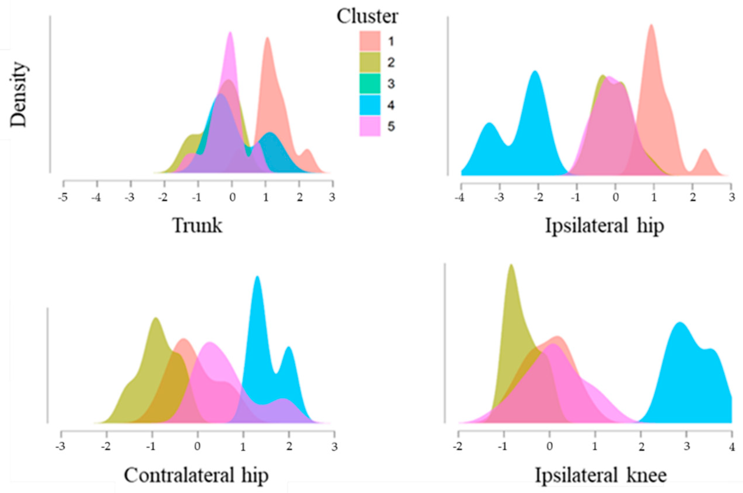
| Standard Score | ||||
| Cluster | Trunk Angle | Hip Angle | Knee Angle | Contralateral Hip Angle |
| C1 (n = 11) | 1.231 | 1.151 | −0.053 | −0.069 |
| C2 (n = 16) | −0.393 | −0.048 | −0.565 | −0.869 |
| C3 (n = 1) | −3.989 | −2.638 | −2.658 | −2.864 |
| C4 (n = 3) | 0.160 | −2.464 | 2.823 | 1.537 |
| C5 (n = 20) | −0.187 | −0.093 | 0.132 | 0.646 |
| RELIABILITY (SPRINT 1 vs. 2) | ||||
| Trunk Angle | Hip Angle | Knee Angle | Contralateral Hip Angle | |
| ICC (Lower/Upper 95% CI) | 0.97 (0.95/0.98) | 0.99 (0.98/0.99) | 0.98 (0.97/0.99) | 0.99 (0.98/1.00) |
| CV % (Lower/Upper 95% CI) | 0.65 (0.25/1.05) | 0.46 (0.30/0.62) | 0.47 (0.28/0.67) | 0.37 (0.21/0.52) |
| MDC | 2.32 | 2.16 | 2.66 | 2.38 |
| MDC (%) | 3.09 | 1.60 | 1.78 | 1.40 |
Publisher’s Note: MDPI stays neutral with regard to jurisdictional claims in published maps and institutional affiliations. |
© 2022 by the authors. Licensee MDPI, Basel, Switzerland. This article is an open access article distributed under the terms and conditions of the Creative Commons Attribution (CC BY) license (https://creativecommons.org/licenses/by/4.0/).
Share and Cite
Romero, V.; Lahti, J.; Castaño Zambudio, A.; Mendiguchia, J.; Jiménez Reyes, P.; Morin, J.-B. Effects of Fatigue Induced by Repeated Sprints on Sprint Biomechanics in Football Players: Should We Look at the Group or the Individual? Int. J. Environ. Res. Public Health 2022, 19, 14643. https://doi.org/10.3390/ijerph192214643
Romero V, Lahti J, Castaño Zambudio A, Mendiguchia J, Jiménez Reyes P, Morin J-B. Effects of Fatigue Induced by Repeated Sprints on Sprint Biomechanics in Football Players: Should We Look at the Group or the Individual? International Journal of Environmental Research and Public Health. 2022; 19(22):14643. https://doi.org/10.3390/ijerph192214643
Chicago/Turabian StyleRomero, Valentin, Johan Lahti, Adrián Castaño Zambudio, Jurdan Mendiguchia, Pedro Jiménez Reyes, and Jean-Benoît Morin. 2022. "Effects of Fatigue Induced by Repeated Sprints on Sprint Biomechanics in Football Players: Should We Look at the Group or the Individual?" International Journal of Environmental Research and Public Health 19, no. 22: 14643. https://doi.org/10.3390/ijerph192214643