Article

Use of the Monocyte-to-Lymphocyte Ratio to Predict Diabetic Retinopathy

1
Department of Ophthalmology, The First Affiliated Hospital of China Medical University, No.155 Nanjing North Street, Heping District, Shenyang 110001, China
2
Department of Endocrinology and Metabolism, Institute of Endocrinology, Liaoning Provincial Key Laboratory of Endocrine Diseases, The First Affiliated Hospital of China Medical University, Shenyang 110001, China
*
Authors to whom correspondence should be addressed.
These authors contributed equally to this work.
Int. J. Environ. Res. Public Health 2015, 12(8), 10009-10019; https://doi.org/10.3390/ijerph120810009
Submission received: 9 July 2015 / Revised: 13 August 2015 / Accepted: 18 August 2015 / Published: 21 August 2015
(This article belongs to the Collection Health Care and Diabetes)

Abstract

:
Background: Diabetic retinopathy (DR) is a common complication of type 2 diabetes mellitus (T2DM) and the leading cause of blindness in adults. DR pathogenesis has not been fully elucidated, but inflammation is widely accepted to play an important role. Emerging evidence suggests that the platelet-to-lymphocyte ratio (PLR), monocyte-to-lymphocyte ratio (MLR), and neutrophil-to-lymphocyte ratio (NLR) are novel potential markers of inflammatory responses. The present study aimed to evaluate the associations between DR and the PLR, MLR, and NLR. Patients and Methods: We performed a case-control study involving 247 patients with T2DM. The patients were divided into three groups: 125 control subjects with T2DM, 63 diabetic subjects with non-proliferative diabetic retinopathy (NPDR), and 59 patients with proliferative diabetic retinopathy (PDR). Results: The mean PLR and NLR were significantly higher in patients with DR compared with patients without DR (p < 0.01, p = 0.02, respectively). The mean MLR in the NPDR group was higher than that of patients without DR, but there were no significant differences among the three groups (p = 0.07). Logistic regression showed that the MLR was an independent risk factor for DR (odds ratio [OR]: 54.574, 95% confidence interval [CI]: 2.708–1099.907). Based on the receiver operating characteristic (ROC) curve, use of the MLR as an indicator for DR diagnosis was projected to be 2.25, and yielded a sensitivity and specificity of 47.1% and 69.6%, respectively, with an area under the curve of 0.581 (95% CI: 0.510–0.653). Conclusions: The PLR and NLR are significantly increased in the setting of DR. After correcting for possible confounding factors, the MLR was found to be a risk factor for DR. Although the MLR may be pathophysiologically and clinically relevant in DR, its predictive ability was limited.

1. Introduction

Many reports indicate that Asia will become the epicenter for diabetes mellitus (DM), particularly in developing countries [1,2]. As DM prevalence increases, so does the development of diabetic retinopathy (DR). DR is a serious complication of DM, and is considered a major cause of blindness in the working age population [3]. DR pathogenesis is complicated and related to many factors, but many groups have described the role of inflammatory factors in DR development [4,5,6,7].
The white blood cell (WBC) count and its subtypes are classic indicators of inflammation [8]. The platelet-to-lymphocyte ratio (PLR), monocyte-to-lymphocyte ratio (MLR), and neutrophil-to-lymphocyte ratio (NLR) are potential markers of inflammation in various conditions, including tumors [9,10,11], cardiovascular conditions [12,13] and other diseases [14].
To date, only a few articles have studied the relationship between diabetic microvascular complications and the PLR, NLR, and MLR [15,16,17,18]. Some studies have reported that the PLR and NLR are associated with diabetes and its complications [15,19]. Akbas et al., reported that the PLR and NLR were significantly elevated in diabetic nephropathy patients with albuminuria [15]. The associations of the PLR and MLR with DR have not been investigated to date. Although the relationship between DR and the NLR has been studied, the results were contradictory. Ulu et al. [17] reported a positive correlation between the NLR and different DR grades but Ciray et al. [16] failed to find an independent association between the NLR and DR. The present study aimed to evaluate the relationships between DR and the PLR, NLR, and MLR.

2. Patients and Methods

2.1. Study Population

The case-control study was conducted from August 2014 to April 2015 in the Fengyutan health care center, Shenhe District, Shenyang City, China. Additional details were described previously [20]. We assessed a total of 246 patients diagnosed with T2DM, 121 of whom had DR. Patients were excluded if they were younger than 18 years old or had type 1 diabetes mellitus, any acute inflammation, active infection, cancer, chronic liver diseases, or any diabetic microvascular complications except DR.

2.2. Clinical Examination and Biochemical Analysis

All the subjects underwent stereo fundus photography to detect DR using a 45° Non-Mydriatic Fundus Camera (CR6-45NM, Canon, Tokyo, Japan) [21,22,23] through undilated pupils. For every subject, two fundus images centered on the fovea and optic disc for each eye were taken in a darkened room. Each image was independently graded in a masked manner by two well-trained ophthalmologists from Liaoning Diabetic Eye Center for DR diagnosis. If the grades were different, a third ophthalmologist would reach the final decision. The DR grade for each eye was determined, and an individual’s classification was based on the worse eye.
Participants’ systolic and diastolic blood pressures (SBP and DBP) were measured after a 5-min rest using a sphygmomanometer. The mean values of two measurements were recorded. Venous blood samples were drawn after an overnight fast. All biochemical analyses were performed in our hospital, including fasting plasma glucose (FPG), total cholesterol (TC), triglyceride (TG), low-density lipoprotein cholesterol (LDL), serum creatinine (Scr), blood urea nitrogen (BUN), high-density lipoprotein cholesterol (HDL), and glycosylated hemoglobin A1c (HbA1c).

2.3. PLR, NLR, MLR, DM, and DR Definitions

The NLR, PLR, and MLR were calculated as the ratios of the neutrophils, platelets, and monocytes to lymphocytes, respectively. All counts were determined from the same automated blood sample measurement. DR was evaluated according to the International Clinical Diabetic Retinopathy Disease Severity Scale [24]. Diabetes was diagnosed according to the 1999 World Health Organization criteria [25].

2.4. Statistical Analysis
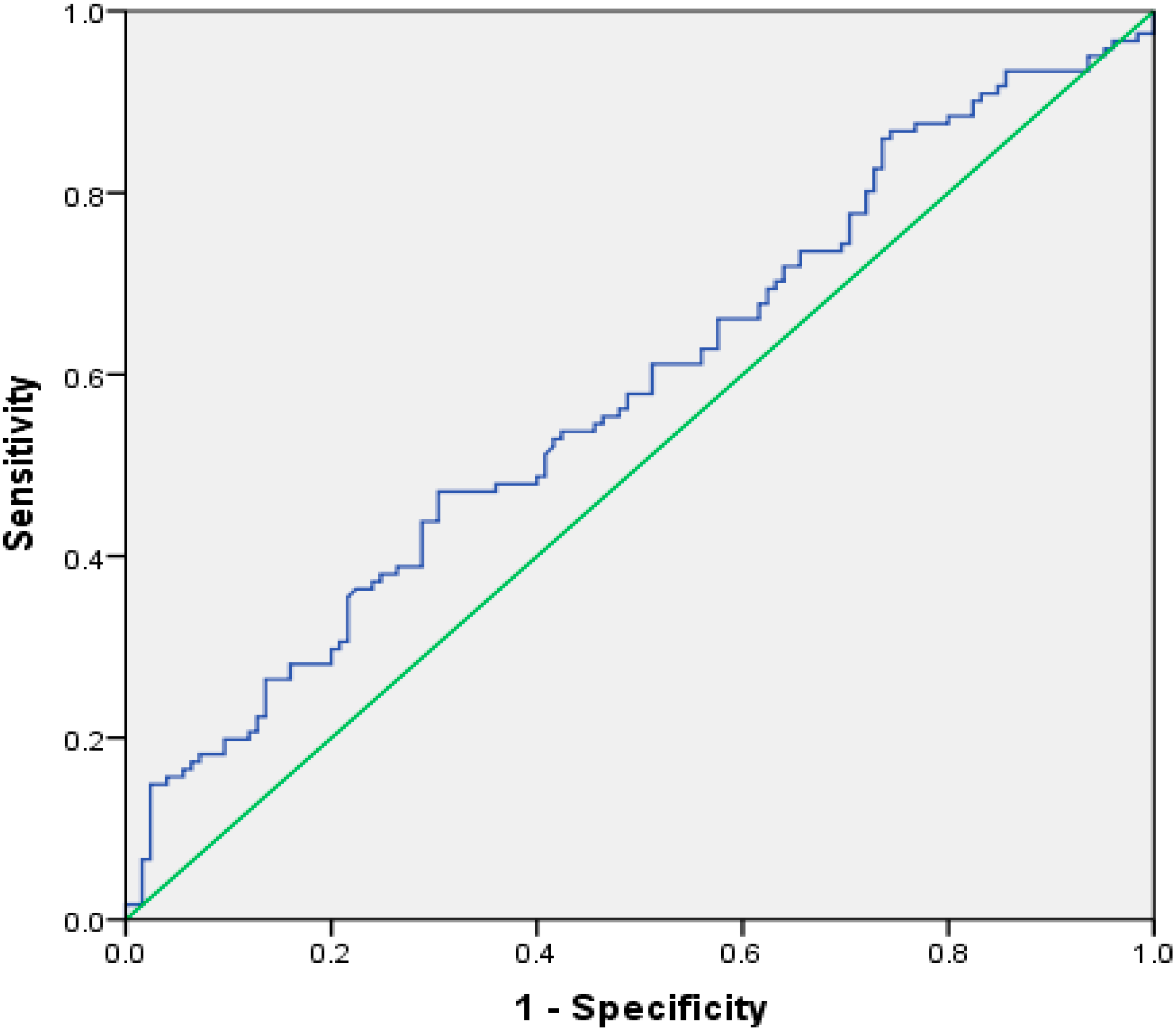
All data were expressed as a median and interquartile range (IQR) because of non-normally distributed numerical variables. Non-parametric analyses were used to compare the data. We performed chi-square, Kruskal-Wallis H and Mann Whitney U tests for dichotomous and continuous variables, respectively. Logistic regression was used to analyze DR risk factors. Receiver operating characteristic (ROC) curve analysis was used to compare the prognostic powers of the PLR and MLR for DR. The predictive validities were quantified as areas under the ROC curves. The positive predictive value (95%CI) and negative predictive value (95%CI) were calculated by MedCalc ver 15.2.1 (MedCalc Software, Mariakerke, Belgium). Others analyses were performed using SPSS 17.0 (SPSS for Windows, version 17.0; SPSS, Inc., Chicago, IL, USA), and p < 0.05 was considered statistically significant.

3. Results

The patients were divided into three groups: 125 control subjects with T2DM, 62 diabetic subjects with NPDR, and 59 patients with PDR. The baseline characteristics of all 246 subjects are shown in Table 1. We observed significant differences for DM duration, SBP, DBP, HbA1c, TC, HDL-C, LDL-C, PLT, NLR, PLR, and MLR (p < 0.05). DR patients were divided into two groups based on severity (NPDR and PDR). The results are shown in Table 2. The PLR and NLR were significantly higher in patients with DR compared with patients without DR (p < 0.01 and p = 0.02, respectively). The mean MLR of patients with NPDR were higher than those of patients without DR (median [IQR] 0.22[0.30–0.14] vs. 0.18[0.24–0.13]), but there were no statistical differences among the three groups (p = 0.07). In addition, retinopathy severity was not associated with increased NLR, PLR, or MLR. Logistic regression analysis revealed that independent risk factors for DR were DM duration, SBP, MLR, TG, HDL-C, and PLT (Table 3). Figure 1 shows that as an independent risk factor for DR, the cut-off value of MLR was 2.25, and the sensitivity and specificity of the MLR for DR diagnosis were 47.1% and 69.6%, respectively, with an area under the curve at 0.581 (95% confidence interval (CI): 0.510–0.653). The positive predictive value (95%CI), negative predictive value (95%CI) for MLR for DR diagnosis were 59.82% (49.25% to 69.75%) and 57.80% (49.5% to 65.78%), respectively.
Table 1. Patients’ baseline and clinical characteristics.
VariablesDM (n = 125)DR (n = 121)p
Age (years)56.00[63.00–48.00]55.00[61.00–47.00]0.23
Sex
Male (%)73 (58.4)62 (51.2)0.31
Duration of DM (years)5.00[10.00–1.50]10.00[16.00–7.00]<0.01
Family history of DM
Yes (%)47 (37.6)49 (40.5)0.69
SBP (mmHg)125.00[140.00–110.00]140.00[150.00–130.00]<0.01
DBP (mmHg)80.00[80.00–70.00]85[90–80]<0.01
FBG (mmol/L)8.00[11.00–6.00]9.20[12.00–6.50]0.09
HbA1c (%)7.00[9.00–6.00]8.00[10.00–7.20]<0.01
TG (mmol/L)2.00[2.50–1.00]1.43[2.17–1.00]0.93
TC (mmol/L)5.00[5.50–4.00]5.00[5.86–4.00]0.03
HDL-C (mmol/L)1.15[1.79–0.80]2.20[3.21–1.00]<0.01
LDL-C (mmol/L)3.00[4.00–2.00]1.53[3.00–1.00]<0.01
Scr (umol/L)59.00[70.00–51.00]58.00[74.00–46.00]0.69
BUN (mmol/L)6.00[7.00–5.00]6.09[7.90–5.00]0.18
WBC (×109/L)6.25[7.55–5.37]6.51[7.87–5.39]0.37
Lymphocytes (×109/L)2.10[2.65–1.61]2.00[1.53–2.41]0.13
Neutrophils (×109/L)3.52[4.57–2.90]3.88[4.85–3.0.2]0.13
Monocytes (×109/L)0.37[0.46–0.29]0.38[0.30–0.55]0.19
Platelets (×109/L)197.00[239.50–161.50]210.00[255.00–182.00]0.02
NLR1.74[2.29–1.31]1.99[2.62–1.47]0.02
PLR94.04[120.19–70.73]107.75[149.82–84.44]<0.01
MLR0.18[0.24–0.13]0.20[0.29–0.14]0.04
Data are expressed as median (inter-quartile range) or percentage; DBP: diastolic blood pressure; SBP: systolic blood pressure; FBG: Fasting blood glucose; HbA1c: Hemoglobin A1c; TG: triglycerides; TC: total cholesterol; HDL-C: high-density lipoprotein cholesterol; LDL-C: low-density lipoprotein cholesterol; Scr: serum creatinine; BUN: blood urea nitrogen; WBC: white blood cell; NLR: neutrophil-to-lymphocyte ratio; PLR: platelet-to-lymphocyte ratio; MLR: monocyte-to-lymphocyte ratio.
Table 2. Comparison of inflammatory and hematologic parameters in patients with and without retinopathy and its severity.
VariablesDM (n = 125)NPDR (n = 62)PDR (n = 59)p-Value
Age (years)56.00[63.00–48.00]53.50[60.25–46.00]56.00[62.00–50.00]0.17
Sex
Male (%)73 (58.4)34 (54.8)28 (47.5)0.38
Duration of DM (years)5.00[10.00–1.50] #,†10.00[15.00–6.75] *,†15.00[19.00–8.00] *,#<0.01
Family history of DM
Yes (%)47 (37.6)28 (45.2)21 (35.6)0.50
SBP (mmHg)125.00[140.00–110.00] #,†135.00[145.00–122.00] *,†145.00[150.00–130.00] *,#<0.01
DBP (mmHg)80.00[80.00–70.00] #,†84.00[90.00–80.00] †85.00[90.00–80.00] #<0.01
FBG (mmol/L)8.00[11.00–6.00]9.10[12.34–6.91]9.35[14.00–6.46]0.25
HbA1c (%)7.00[9.00–6.00] #8.00[10.00–7.00]8.20[10.00–7.40] #0.02
TG (mmol/L)2.00[2.50–1.00]1.70[2.16–1.00]1.41[2.21–1.00]0.53
TC (mmol/L)5.00[5.50–4.00]5.00[6.00–4.00]4.99[5.46–4.00]0.12
HDL-C (mmol/L)1.00[1.00–1.00] #,†1.00[2.49–1.00] *,†2.86[3.46–2.00] *,#<0.01
LDL-C (mmol/L)3.00[4.00–2.00] #,†2.50[4.00–1.09] *,†1.24[2.00–0.99] *,#<0.01
Scr (umol/L)59.00[70.00–51.00]59.00[71.25–46.00]57.00[78.75–45.75]0.99
BUN (mmol/L)6.00[7.00–5.00] #6.00[7.00–4.85] *6.75[9.99–5.06] *,#0.01
WBC (×109/L)6.25[7.55–5.37]6.67[8.11–5.41]6.43[7.50–5.34]0.42
Lymphocytes (×109/L)2.10[2.65–1.61]2.03[2.36–1.49]1.97[2.43–1.52]0.29
Neutrophils (×109/L)3.52[4.57–2.90]3.88[4.93–3.07]3.75[4.60–2.85]0.16
Monocytes (×109/L)0.37[0.46–0.29]0.39[0.56–0.31]0.38[0.53–0.28]0.27
Platelet (×109/L)197.00[239.50–161.50] #202.00[236.25–177.00]225.00[264.00–182.00] #0.03
NLR1.74[2.29–1.31] #,†2.05[2.75–1.55] †1.91[2.51–1.39] #0.03
PLR94.04[120.19–70.73] #,†105.07[151.42–81.53] †115.73[145.97–87.98] #<0.01
MLR0.18[0.24–0.13] †0.22[0.30–0.14] †0.20[0.28–0.15]0.07
Data are expressed as median (inter-quartile range) or percentage; DBP: diastolic blood pressure; SBP: systolic blood pressure; FBG: Fasting blood glucose; HbA1c: Hemoglobin A1c; TG: triglycerides; TC: total cholesterol; HDL-C: high-density lipoprotein cholesterol; LDL-C: low-density lipoprotein cholesterol; Scr: serum creatinine; BUN: blood urea nitrogen; WBC: white blood cell; NLR: neutrophil-to-lymphocyte ratio; PLR: platelet-to-lymphocyte ratio; MLR: monocyte-to-lymphocyte ratio; * Significant difference between PDR and NPDR; # Significant difference between DM and PDR; Significant difference between DM and NPDR.
Table 3. Logistic regression analysis showing independent predictors of retinopathy.
VariablesOR95%CIp-Value
Duration of DM1.1621.085–1.246<0.001
SBP1.0331.013–1.0540.001
MLR54.5742.708–1099.9070.009
TG1.6711.026–2.7220.039
HDL-C7.3573.004–18.017<0.001
LDL-C0.6250.359–1.0910.098
Platelet1.0071.000–1.0130.043
OR: Odds Ratio; 95% CI: 95% confidence interal; SBP: systolic blood pressure; MLR: monocyte-to-lymphocyte ratio; TG: triglycerides; HDL-C: high-density lipoprotein cholesterol; LDL-C: low-density lipoprotein cholesterol.
Figure 1. Receiver–operating characteristics (ROC) curve analysis for monocyte to lymphocyte ratio as a predictor of the severity of diabetic retinopathy.
Ijerph 12 10009 g001

4. Discussion

As a serious microvascular complication of DM [26], DR is a complex disease involving multiple events. Powell et al., previously reported that some anti-inflammatory drugs such as salicylates could prevent the occurrence of DR, suggesting that inflammation may play a role in DR pathogenesis [27]. Lutty et al., verified the association between white blood cells and DR occurrence [28]. Moreover, some epidemiological studies have indicated that DM and its microvascular complications are associated with chronic inflammation [29,30] and immune responses [31,32,33]. There are common sets of inflammatory cytokines and immune biomarkers including tumor necrosis factor (TNF)-a, interleukin (IL)-6, IL-1β, and intracellular adhesion molecule (ICAM)-1 that are up-regulated in both serum and vitreous fluid in subjects with DR. These findings suggest an interaction between inflammation and DR pathogenesis [34,35].
As important inflammation response markers, counts of WBCs and its subtypes are associated with cardiovascular disease [8]. Besides WBC counts, the PLR, MLR, and NLR are potential biomarkers reflecting inflammation and immune responses. Many studies have reported positive correlations of conventional inflammatory markers with the PLR and NLR. More importantly, a large number of studies found predictive effects of the PLR and NLR, particularly in DM, acute coronary syndromes, and various cancers [9,10,12,13,36]. However, as an immune indicator, the only study on MLR focused on malaria [14].
Our results suggest that DM patients with DR had higher NLRs compared with diabetic patients without DR. This is in accordance with the findings of Ulu et al. [17] and Wang et al. [18]; however, it was contrary to the results reported by Ciray et al. [16]. They found that the NLR was not significantly different in patients with or without DR. In addition, our results are different from those of Ulu et al., who found that DR severity was not associated with increasing NLR. The discrepancy between these two studies might be due to different sample sizes, as our cohort was slightly larger. Subject heterogeneity and lifestyle difference may be other reasons for the discrepancies. Notably, the NLR has been shown to reflect an immune microenvironment that favors vascular invasion by tumors [37]. Therefore, we have reason to believe that there is a correlation between the NLR and DR.
As stated above, PLR and MLR have been studied in many fields. This is the first investigation of the relationship between DR severity and the PLR and MLR, which are viewed as new inflammatory markers. Our results indicate that the PLR and MLR were significantly higher in patients with DR compared to those without. Some angiogenesis factors such as vascular endothelial growth factor (VEGF) are key protein modulators expressed by platelets [38]. Notably, high VEGF levels can stimulate the development of proliferative diabetic retinopathy (PDR). This would suggest that there is association between the PLR and DR progression.
Furthermore, there was no correlation of either the PLR or MLR with DR severity. Akbas et al. [15] showed that the PLR could predict inflammation and albuminuria in patients with diabetes. Hudzik et al., reported that the PLR was an independent risk factor for early and late mortality in patients with DM [36].
We only found one study investigating the relationship between the NLR and DR. The results suggested that higher NLR values may be a useful marker for DR [17]; however, they did not use ROC analysis to assess the NLR predictive performance. In our study, the NLR was not an independent risk factor for DR, so we did not perform ROC analysis to determine its predictive value for DR. The most important implication is that although the MLR correlated with the presence of DR and was an independent risk factor, it performed poorly as a screening tool for DR diagnosis. Monocytes are considered a biomarker for inflammation because their activation leads to the synthesis of inflammatory cytokines. A previous report suggested that monocytes may be relevant to angiogenic processes in atherosclerosis [39]. Nevertheless, the mechanisms underlying the association between the MLR and DR should be investigated in future studies.
As novel markers to predict DR, the PLR, NLR and MLR are superior to other cell parameters, e.g., neutrophil, lymphocyte, and total leukocyte counts. The advantage is that the PLR, NLR, and MLR are the absolute value counts and show good stability even when physiological, pathological, and physical factors of the WBC count vary. In addition, they may represent inflammatory and immune signaling in DR.
Our study has some potential limitations. First, the case-control study design did not allow us to investigate whether controlling the MLR may affect DR outcome. Second, we assessed a relatively small number of subjects. Third, we did not perform fundus fluorescein angiography (FFA) to detect DR.

5. Conclusions

In conclusion, the PLR and NLR are significantly increased in the setting of DR. Although the MLR is an independent risk factor for DR in Chinese patients with T2DM, it has a low performance for DR diagnosis. Additional studies are needed to identify mechanisms explaining the association between the MLR and DR.

Acknowledgments

We thank Hu-nan in Dalian University of Foreign Languages for our English language edition.
This study was supported by National Natural Science Foundation of China (81300783); Important Platform of Science and Technology for the University in Liaoning Province (16010).

Author Contributions

Song Yue and Lei Liu have contributed to the design of the study, analysis and interpretation of data, and prepared all figures and tables. Song Yue, Jingyang Wu, Lei Liu and Lei Chen drafted a part of manuscript. Song Yue, Jiahua Zhang and Jingyang Wu took part in analyzing data, and drafting a part of manuscript. All authors reviewed the manuscript.

Conflicts of Interest

The authors declare no conflict of interest.

References

  1. Chan, J.C.; Malik, V.; Jia, W.; Kadowaki, T.; Yajnik, C.S.; Yoon, K.H.; Hu, F.B. Diabetes in Asia: Epidemiology, risk factors, and pathophysiology. JAMA 2009, 301, 2129–2140. [Google Scholar] [CrossRef] [PubMed]
  2. Shaw, J.E.; Sicree, R.A.; Zimmet, P.Z. Global estimates of the prevalence of diabetes for 2010 and 2030. Diabetes Res. Clin. Pract. 2010, 87, 4–14. [Google Scholar] [CrossRef] [PubMed]
  3. Yamada, M.; Hiratsuka, Y.; Roberts, C.B.; Pezzullo, M.L.; Yates, K.; Takano, S.; Miyake, K.; Taylor, H.R. Prevalence of visual impairment in the adult Japanese population by cause and severity and future projections. Ophthalmic Epidemiol. 2010, 17, 50–57. [Google Scholar] [CrossRef] [PubMed]
  4. Joussen, A.M.; Poulaki, V.; Le, M.L.; Koizumi, K.; Esser, C.; Janicki, H.; Schraermeyer, U.; Kociok, N.; Fauser, S.; Kirchhof, B.; et al. A central role for inflammation in the pathogenesis of diabetic retinopathy. FASEB J. 2004, 18, 1450–1452. [Google Scholar] [PubMed]
  5. Tang, J.; Kern, T.S. Inflammation in diabetic retinopathy. Prog. Retin. Eye Res. 2011, 30, 343–358. [Google Scholar] [CrossRef] [PubMed]
  6. El-Asrar, A.M. Role of inflammation in the pathogenesis of diabetic retinopathy. Middle East Afr. J. Ophthalmol. 2012, 19, 70–74. [Google Scholar] [CrossRef] [PubMed]
  7. Tomić, M.; Ljubić, S.; Kaštelan, S.; Gverović Antunica, A.; Jazbec, A.; Poljičanin, T. Inflammation, haemostatic disturbance, and obesity: Possible link to pathogenesis of diabetic retinopathy in type 2 diabetes. Mediat. Inflamm. 2013, 2013, 818671. [Google Scholar] [CrossRef] [PubMed]
  8. Horne, B.D.; Anderson, J.L.; John, J.M. Which white blood cell subtypes predict increased cardiovascular risk? J. Am. Coll. Cardiol. 2005, 45, 1638–1643. [Google Scholar] [CrossRef] [PubMed]
  9. Gunduz, S.; Mutlu, H.; Tural, D.; Yıldız, Ö.; Uysal, M.; Coskun, H.S.; Bozcuk, H. Platelet to lymphocyte ratio as a new prognostic for patients with metastatic renal cell cancer. Asia Pac. J. Clin. Oncol. 2015. [Google Scholar] [CrossRef] [PubMed]
  10. Ozaksit, G.; Tokmak, A.; Kalkan, H.; Yesilyurt, H. Value of the platelet to lymphocyte ratio in the diagnosis of ovarian neoplasms in adolescents. Asian Pac. J. Cancer Prev. 2015, 16, 2037–2041. [Google Scholar] [PubMed]
  11. Liu, J.; Du, J.; Fan, J.; Liu, K.; Zhang, B.; Wang, S.; Wang, W.; Wang, Z.; Cai, Y.; Li, C.; et al. The Neutrophil-to-Lymphocyte Ratio Correlates with Age in Patients with Papillary Thyroid Carcinoma. ORL J. Otorhinolaryngol. Relat. Spec. 2015, 77, 109–116. [Google Scholar] [CrossRef] [PubMed]
  12. Akyel, A.; Yayla, Ç.; Erat, M.; Çimen, T.; Doğan, M.; Açıkel, S.; Aydoğdu, S.; Yeter, E. Neutrophil-to-lymphocyte ratio predicts hemodynamic significance of coronary artery stenosis. Anatol. J. Cardiol. 2015. [Google Scholar] [CrossRef] [PubMed]
  13. Oylumlu, M.; Yıldız, A.; Oylumlu, M.; Yuksel, M.; Polat, N.; Bilik, M.Z.; Akyuz, A.; Aydin, M.; Acet, H.; Soydinc, S. Platelet-to-lymphocyte ratio is a predictor of in-hospital mortality patients with acute coronary syndrome. Anatol. J. Cardiol. 2015, 15, 277–283. [Google Scholar] [CrossRef] [PubMed]
  14. Warimwe, G.M.; Fletcher, H.A.; Olotu, A.; Agnandji, S.T.; Hill, A.V.; Marsh, K.; Bejon, P. Peripheral blood monocyte-to-lymphocyte ratio at study enrollment predicts efficacy of the RTS, S malaria vaccine: Analysis of pooled phase II clinical trial data. BMC Med. 2013, 21, 184. [Google Scholar] [CrossRef] [PubMed]
  15. Akbas, E.M.; Demirtas, L.; Ozcicek, A.; Timuroglu, A.; Bakirci, E.M.; Hamur, H.; Ozcicek, F.; Turkmen, K. Association of epicardial adipose tissue, neutrophil-to-lymphocyte ratio and platelet-to-lymphocyte ratio with diabetic nephropathy. Int. J. Clin. Exp. Med. 2014, 7, 1794–1801. [Google Scholar] [PubMed]
  16. Ciray, H.; Aksoy, A.H.; Ulu, N.; Cizmecioglu, A.; Gaipov, A.; Solak, Y. Nephropathy, but not Angiographically Proven Retinopathy, is Associated with Neutrophil to Lymphocyte Ratio in Patients with Type 2 Diabetes. Exp. Clin. Endocrinol. Diabetes 2015, 123, 267–271. [Google Scholar] [CrossRef] [PubMed]
  17. Ulu, S.M.; Dogan, M.; Ahsen, A.; Altug, A.; Demir, K.; Acartürk, G.; Inan, S. Neutrophil-to-lymphocyte ratio as a quick and reliable predictive marker to diagnose the severity of diabetic retinopathy. Diabetes Technol. Ther. 2013, 15, 942–947. [Google Scholar] [CrossRef] [PubMed]
  18. Wang, R.T.; Zhang, J.R.; Li, Y.; Liu, T.; Yu, K.J. Neutrophil-Lymphocyte ratio is associated with arterial stiffness in diabetic retinopathy in type 2 diabetes. J. Diabetes Complicat. 2015, 29, 245–249. [Google Scholar] [CrossRef] [PubMed]
  19. Shiny, A.; Bibin, Y.S.; Shanthirani, C.S.; Regin, B.S.; Anjana, R.M.; Balasubramanyam, M.; Jebarani, S.; Mohan, V. Association of neutrophil-lymphocyte ratio with glucose intolerance: An indicator of systemic inflammation in patients with type 2 diabetes. Diabetes Technol. Ther. 2014, 16, 524–530. [Google Scholar] [CrossRef] [PubMed]
  20. Liu, L.; Geng, J.; Wu, J.; Yuan, Z.; Lian, J.; Desheng, H.; Chen, L. Prevalence of ocular fundus pathology with type 2 diabetes in a Chinese urban community as assessed by telescreening. BMJ Open 2013. [Google Scholar] [CrossRef] [PubMed]
  21. Romero, P.; Sagarra, R.; Ferrer, J.; Fernández-Ballart, J.; Baget, M. The incorporation of family physicians in the assessment of diabetic retinopathy by non-mydriatic fundus camera. Diabetes Res. Clin. Pract. 2010, 88, 184–188. [Google Scholar] [CrossRef] [PubMed]
  22. Szabó, D.; Fiedler, O.; Somogyi, A.; Somfai, G.M.; Bíró, Z.; Ölvedy, V.; Hargitai, Z.; Németh, J. Telemedical diabetic retinopathy screening in Hungary: A pilot programme. J. Telemed. Telecare 2015, 21, 167–173. [Google Scholar] [CrossRef] [PubMed]
  23. Massin, P.; Erginay, A.; Ben Mehidi, A.; Vicaut, E.; Quentel, G.; Victor, Z.; Marre, M.; Guillausseau, P.J.; Gaudric, A. Evaluation of a new non-mydriatic digital camera for detection of diabetic retinopathy. Diabet. Med. 2003, 20, 635–641. [Google Scholar] [CrossRef] [PubMed]
  24. Wilkinson, C.P.; Ferris, F.L.; Klein, R.E.; Lee, P.P.; Agardh, C.D.; Davis, M.; Dills, D.; Kampik, A.; Pararajasegaram, R.; Verdaguer, J.T. Global Diabetic Retinopathy Project Group: Proposed international clinical diabetic retinopathy and diabetic macular edema disease severity scales. Ophthalmology 2003, 110, 1677–1682. [Google Scholar] [CrossRef]
  25. Puavilai, G.; Chanprasertyotin, S.; Sriphrapradaeng, A. Diagnostic criteria for diabetes mellitus and other categories of glucose intolerance: 1997 Criteria by the Expert Committee on the Diagnosis and Classification of Diabetes Mellitus (ADA), 1998 WHO consultation criteria, and 1985 WHO criteria. World Health Organization. Diabetes Res. Clin. Pract. 1999, 44, 21–26. [Google Scholar] [PubMed]
  26. Varma, R.; Macias, G.L.; Torres, M.; Klein, R.; Pena, F.Y.; Azen, S.P. Biologic risk factors associated with diabetic retinopathy: The Los Angeles Latino Eye Study. Ophthalmology 2007, 114, 1332–1340. [Google Scholar] [CrossRef] [PubMed]
  27. Powell, E.D.; Field, R.A. Diabetic retinopathy and rheumatoid arthritis. Lancet. 1964, 734, 17–18. [Google Scholar] [CrossRef]
  28. Lutty, G.A.; Cao, J.T.; McLeod, D.S. Relationship of polymorphonuclear leukocytes to capillary dropout in the human diabetic choroid. Am. J. Pathol. 1997, 151, 707–714. [Google Scholar] [PubMed]
  29. Pitsavos, C.; Tampourlou, M.; Panagiotakos, D.B.; Skoumas, Y.; Chrysohoou, C.; Nomikos, T.; Stefanadis, C. Association between low-grade systemic inflammation and type 2 diabetes mellitus among men and women from the ATTICA Study. Rev. Diabet. Stud. 2007, 4, 98–104. [Google Scholar] [CrossRef] [PubMed]
  30. Fujita, T.; Hemmi, S.; Kajiwara, M.; Yabuki, M.; Fuke, Y.; Satomura, A.; Soma, M. Complement-mediated chronic inflammation is associated with diabetic microvascular complication. Diabetes Metab. Res. Rev. 2013, 29, 220–226. [Google Scholar] [CrossRef] [PubMed]
  31. Grossmann, V.; Schmitt, V.H.; Zeller, T.; Panova-Noeva, M.; Schulz, A.; Laubert-Reh, D.; Juenger, C.; Schnabel, R.B.; Abt, T.G.; Laskowski, R.; et al. Profile of the Immune and Inflammatory Response in Individuals with Prediabetes and Type 2 Diabetes. Diabetes Care 2015, 38, 1356–1364. [Google Scholar] [CrossRef] [PubMed]
  32. Afzal, N.; Zaman, S.; Shahzad, F.; Javaid, K.; Zafar, A.; Nagi, A.H. Immune mechanisms in type-2 diabetic retinopathy. J. Pak. Med. Assoc. 2015, 65, 159–163. [Google Scholar] [PubMed]
  33. Frostegard, J. Immune mechanisms in atherosclerosis, especially in diabetes type 2. Front. Endocrinol. (Lausanne) 2013, 29, 162. [Google Scholar] [CrossRef] [PubMed]
  34. Kaul, K.; Hodgkinson, A.; Tarr, J.M.; Kohner, E.M.; Chibber, R. Is inflammation a common retinal-renal-nerve pathogenic link in diabetes? Curr. Diabetes Rev. 2010, 6, 294–303. [Google Scholar] [CrossRef] [PubMed]
  35. Sasongko, M.B.; Wong, T.Y.; Jenkins, A.J.; Nguyen, T.T.; Shaw, J.E.; Wang, J.J. Circulating markers of inflammation and endothelial function, and their relationship to diabetic retinopathy. Diabet. Med. 2015, 32, 686–691. [Google Scholar] [CrossRef] [PubMed]
  36. Hudzik, B.; Szkodzinski, J.; Gorol, J.; Niedziela, J.; Lekston, A.; Gasior, M.; Polonski, L. Platelet-to-lymphocyte ratio is a marker of poor prognosis in patients with diabetes mellitus and ST-elevation myocardial infarction. Biomark. Med. 2015, 9, 199–207. [Google Scholar] [CrossRef] [PubMed]
  37. Xiao, W.K.; Chen, D.; Li, S.Q.; Fu, S.J.; Peng, B.G.; Liang, L.J. Prognostic significance of neutrophil-lymphocyte ratio in hepatocellular carcinoma: A meta-analysis. BMC Cancer 2014, 14, 117. [Google Scholar] [CrossRef] [PubMed]
  38. Bambace, N.M.; Levis, J.E.; Holmes, C.E. The effect of P2Y-mediated platelet activation on the release of VEGF and endostatin from platelets. Platelets 2010, 21, 85–93. [Google Scholar] [CrossRef] [PubMed]
  39. Jaipersad, A.S.; Lip, G.Y.; Silverman, S.; Shantsila, E. The role of monocytes in angiogenesis and atherosclerosis. J. Am. Coll. Cardiol. 2014, 63, 1–11. [Google Scholar] [CrossRef] [PubMed]

