Article

Thiolactones and Δ8,9-Pregnene Steroids from the Marine-Derived Fungus Meira sp. 1210CH-42 and Their α-Glucosidase Inhibitory Activity

1
Marine Natural Products Chemistry Laboratory, Korea Institute of Ocean Science and Technology, 385 Haeyang-ro, Yeongdo-gu, Busan 49111, Republic of Korea
2
Department of Marine Biotechnology, University of Science and Technology (UST), 217 Gajungro, Yuseong-gu, Daejeon 34113, Republic of Korea
3
Department of Chemistry, Pukyong National University, Busan 48513, Republic of Korea
*
Author to whom correspondence should be addressed.
Mar. Drugs 2023, 21(4), 246; https://doi.org/10.3390/md21040246
Submission received: 5 April 2023 / Revised: 13 April 2023 / Accepted: 14 April 2023 / Published: 16 April 2023
(This article belongs to the Special Issue Bioactive Secondary Metabolites of Marine Fungi)

Abstract

:
The fungal genus Meira was first reported in 2003 and has mostly been found on land. This is the first report of second metabolites from the marine-derived yeast-like fungus Meira sp. One new thiolactone (1), along with one revised thiolactone (2), two new Δ8,9-steroids (4, 5), and one known Δ8,9-steroid (3), were isolated from the Meira sp. 1210CH-42. Their structures were elucidated based on the comprehensive spectroscopic data analysis of 1D, 2D NMR, HR-ESIMS, ECD calculations, and the pyridine-induced deshielding effect. The structure of 5 was confirmed by oxidation of 4 to semisynthetic 5. In the α-glucosidase inhibition assay, compounds 24 showed potent in vitro inhibitory activity with IC50 values of 148.4, 279.7, and 86.0 μM, respectively. Compounds 24 exhibited superior activity as compared to acarbose (IC50 = 418.9 μM).

1. Introduction

Fungi constitute one of the largest groups of organisms. Fungal-derived natural products (NPs) are pharmaceutically abundant, with several important biological applications ranging from highly potent toxins to approved drugs [1]. In particular, secondary metabolites obtained from marine fungi have garnered significant interest due to their unique chemical structures and potential biomedical applications [1,2]. While the number of cultivable marine fungi is extremely low (1% or less) compared to their global biodiversity [1,3], more than 1000 molecules have been reported and characterized from marine fungi, including alkaloids, lipids, peptides, polyketides, prenylated polyketides, and terpenoids [4,5,6,7]. Most research on secondary metabolites produced by marine fungi has primarily focused on a few genera, including Penicillium, Aspergillus, Fusarium, and Cladosporium [8,9]. Research into natural products derived from marine fungi is continually expanding, and as a result, a broader range of genera is now being investigated, with a particular focus on those associated with unique substrates and previously unexplored habitats [10,11,12].
In 2003, the genus Meira was first reported, namely M. geulakonigii and M. argovae, as a novel basidiomycetous [13]. M. geulakonigii was isolated from the citrus rust mite on pummelo (Citrus grandis), and M. argovae originated from a carmine spider mite on the leaves of castor bean (Ricinus communis) [8]. These Meira species have a similar morphology to yeast-like fungi. Nonetheless, the phylogenetic analysis of rDNA sequence data has identified Meira as a member of the Brachybasidiaceae family within the Exobasidiales, which is classified under the Ustilaginomycetes (Basidiomycota) in the Exobasidiomycetidae group [14]. M. geulakonigii has been used successfully as a biological control agent against citrus and other phytophagous mites, as well as powdery mildew fungi [13,15,16,17]. A potential biocontrol agent against five mite species has been demonstrated for M. argovae [18]. Recently, M. nicotianae came from the rhizosphere of tobacco root, and that strain has the capability to promote plant growth possible in similar ways as plant growth-promoting fungi and arbuscular mycorrhizal fungi [19].
In this study, we isolated a yeast-like fungal species from a seawater sample. Phylogenetic analysis of ITS rDNA indicated that strain 1210CH-42 is closely related to other Meira species: Meira sp. M40, M. nashicola CY-1, and M. miltonrushii NIOSN-SK46-S121. So far, there are only a few reports on the isolation of Meira strains, but natural products from the genus Meira have not been investigated. This is the first report on the secondary metabolites from the marine-derived yeast-like fungus Meira. Herein, we report the isolation, structure elucidation, α-glucosidase inhibitory activity of 15, and the structure revision of 2 isolated from the Meira strain 1210CH-42 (Figure 1).

2. Results and Discussion

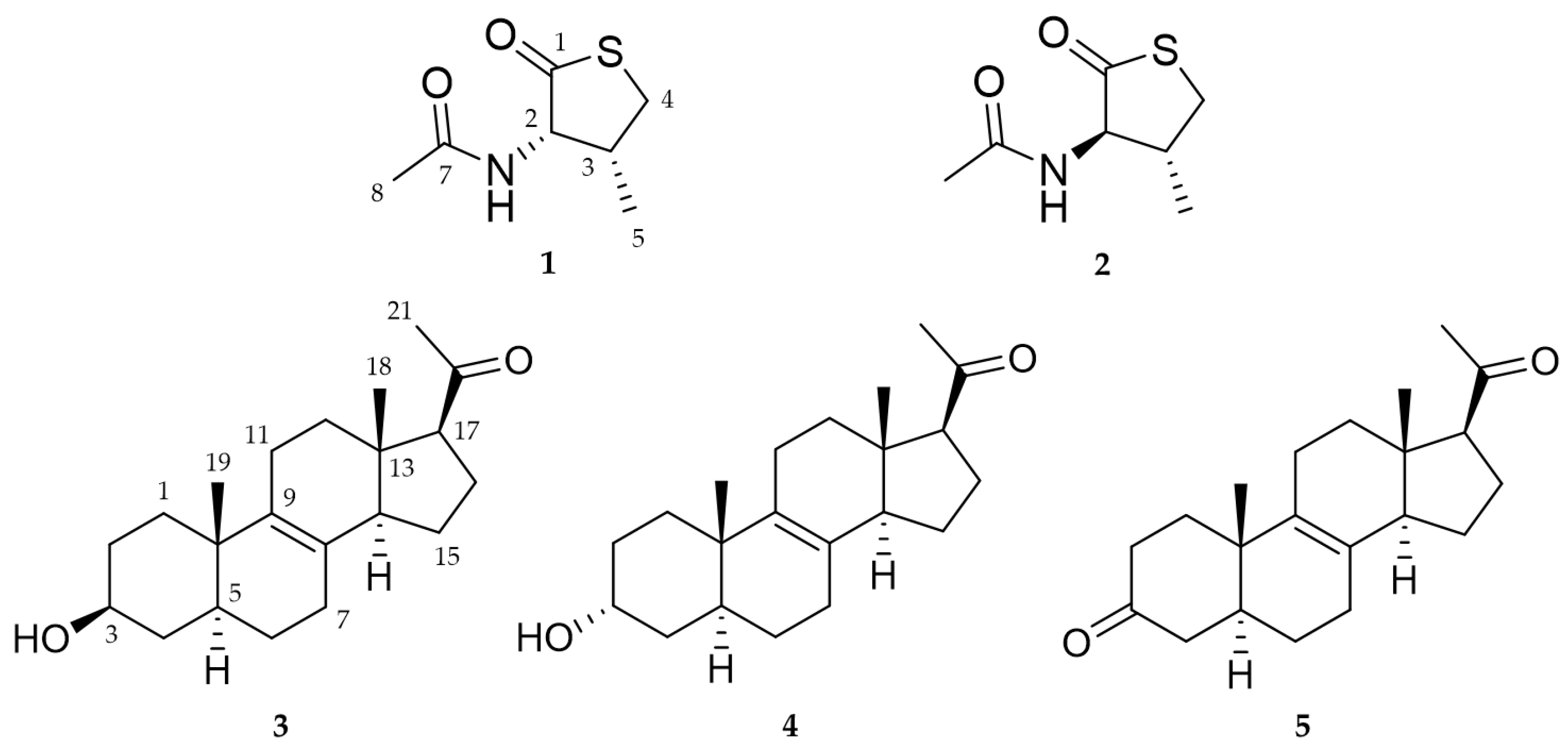
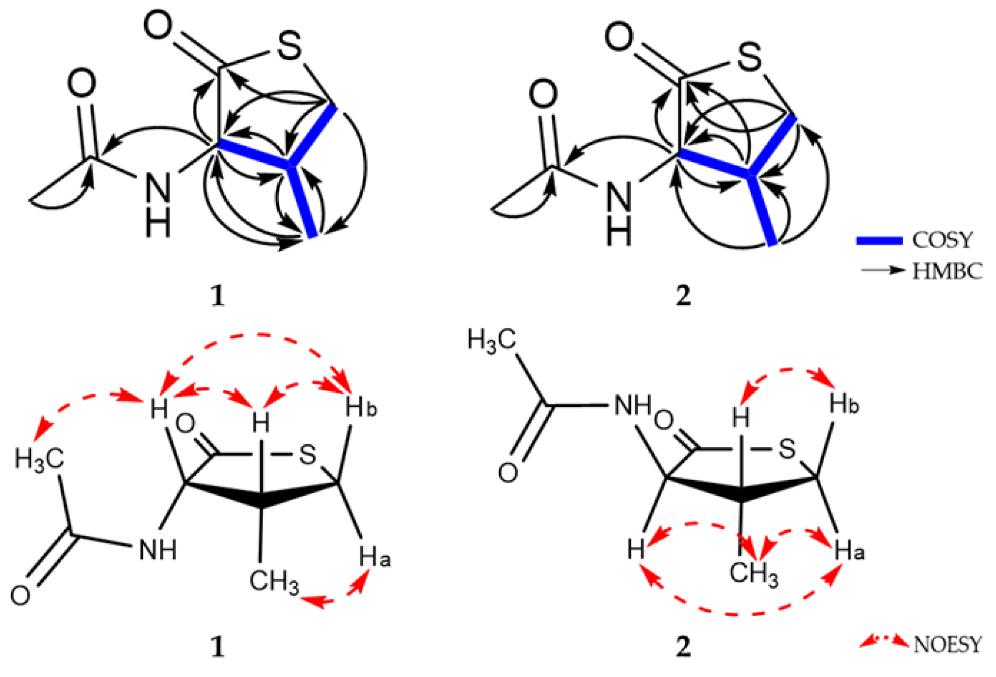
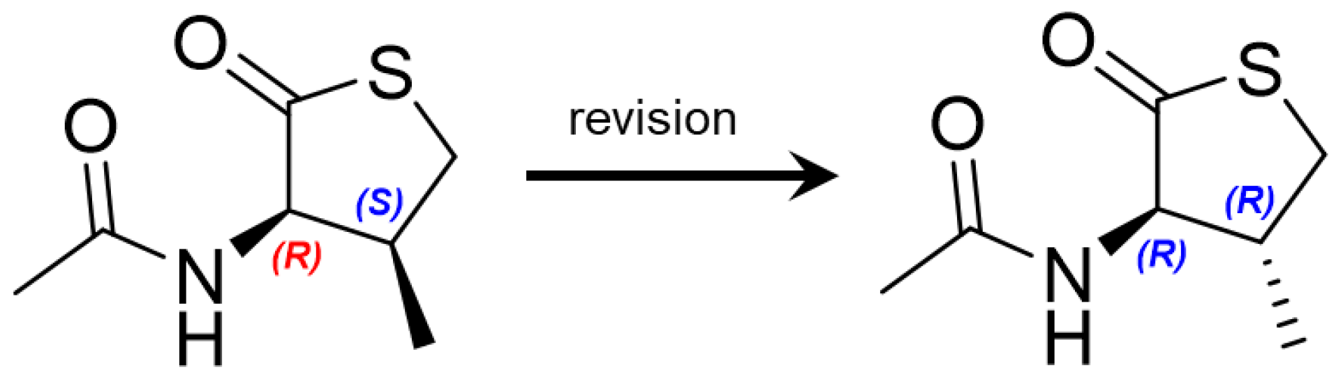
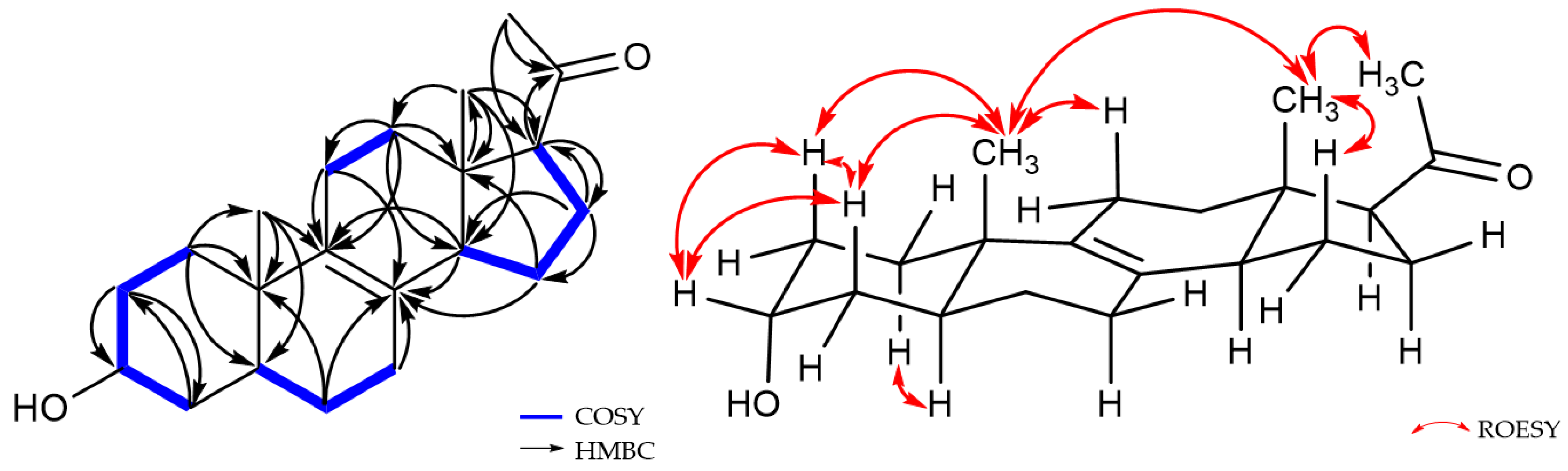
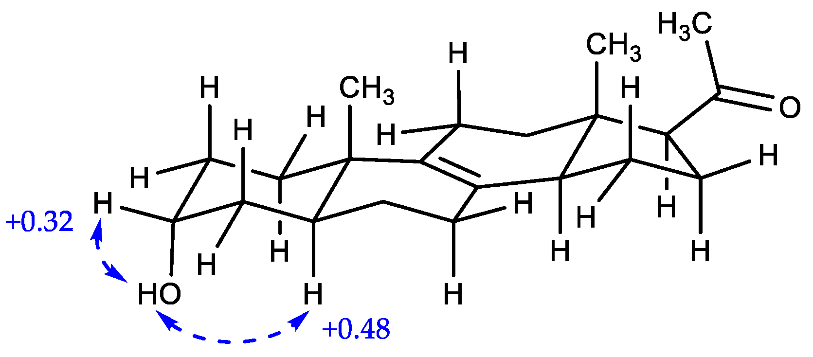
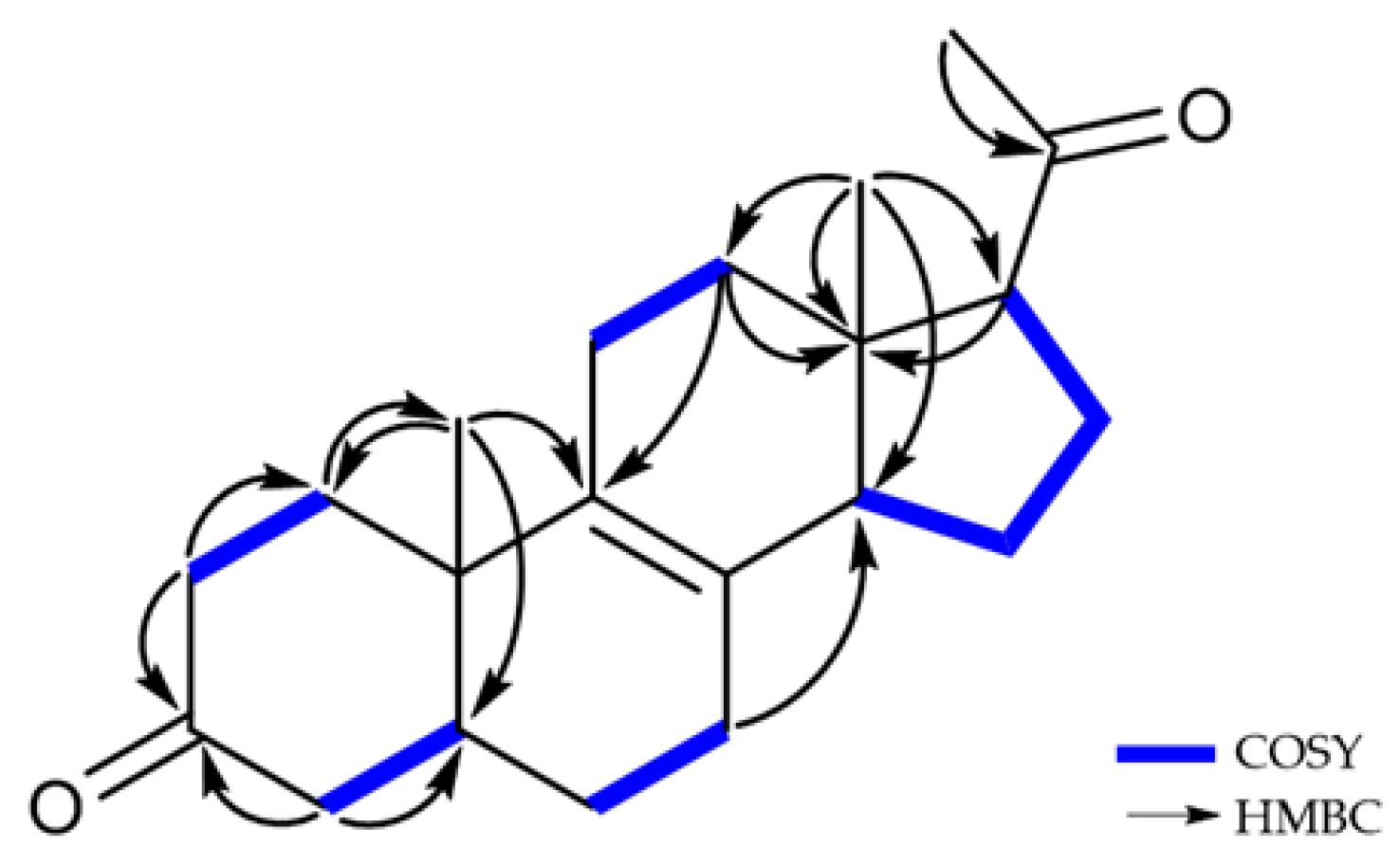
2.1. Structure Elucidation of New Compounds

Compound 1 was obtained as a white amorphous powder, and its molecular formula was determined to be C7H11NO2S by HR-ESIMS, with three degrees of unsaturation. The 1H and 13C NMR data of 1 are summarized in Table 1. The 1H NMR spectrum of 1 in CD3OD revealed two methine protons (δH 4.79 and 2.86), one methylene proton (δH 3.64 and 3.10), and two methyl protons (δH 2.03 and 1.04). The 13C NMR and HSQC spectra showed the presence of seven signals, including two carbonyl carbons (δC 206.5 and 173.8), two methines (δC 63.9 and 36.0), one methylene (δC 35.9) and two methyl carbons (δC 22.4 and 13.0). The planar structure of 1 was elucidated by analysis of 1H-1H COSY and HMBC correlations (Figure 2). The COSY correlations from H-2 (δH 4.79)/H-3 (δH 2.86), H-3 (δH 2.86)/H-4 (δH 3.64), and H-3 (δH 2.86)/H-5 (δH 1.04) were observed. In addition, the HMBC correlations from H-2 (δH 4.79) to C-1 (δC 206.5)/C-3 (δC 36.0)/C-5 (δC 13.0)/C-7 (δC 173.8), H-4 (δH 3.10 and 3.64) to C-1 (δC 206.5)/C-2 (δC 63.9)/C-5 (δC 13.0) and H-8 (δH 2.03) to C-7 (δC 206.5) suggested that 1 has a ring system, and confirmed the planar structure of 1.
Detailed analysis of 3JH,H coupling constants and 1D NOESY data determined the relative configuration of 1. The relative stereochemistry of C-2 could be established by the observation of strong selective 1D NOESY correlations between H-2 and H-3/H-4b, between H-4b and H-2/H-3, and between H-5 and H-4a (Figure 2). These correlations suggested that the relative configurations of C-2 and C-3 must be cis rather than trans-configuration in 1. Thus, the relative configuration of 1 could be assigned as 2S*, 3R*. To determine the absolute configuration of 1, the theoretical electronic circular dichroism (ECD) spectra of 1 and its enantiomer were calculated. The experimental ECD spectrum of 1 showed a good agreement with the calculated ECD spectrum of the 2S, 3R-isomer (Figure 3). Therefore, the structure of 1 was elucidated to be a 2S-acetamide-3R-methyl-thiolactone.
Compound 2 was isolated as a white amorphous powder. The molecular formula of 2 was the same as that of 1 (C7H11NO2S) based on the HR-ESIMS data. Furthermore, the 1D NMR data of 2 were also similar but not identical to those of 1 (Table 1). The planar structure of 2 was determined to be the same as 1 by analysis of 1H-1H COSY and HMBC data (Figure 2). However, the 1H and 13C chemical shifts of 2 were different from 1, especially those for the chiral centers, suggesting that the stereochemistry of 2 might be different from 1. The relative configuration of 2 was also determined by analysis of 3JH,H coupling constants and selective 1D NOESY data. The relative stereochemistry of C-2 could be established through the observation of strong NOESY contacts between H-2 and H-4a/H-5, between H-4a and H-2/H-5, and between H-4b and H-3. A relatively large coupling constant was observed between H-2 and H-3 (3JH,H = 12.5 Hz). Thus, the relative configurations of H-2 and H-3 had a trans-configuration in 2 (Figure 2). The J-based configurational analysis and NOESY measurements could not discriminate the possible relative configurations for (2S*, 3S*) or (2R*, 3R*). To solve this issue and to determine the absolute configuration of 2, the ECD spectra of 2 and its enantiomer were calculated. The experimental ECD spectrum of 2 showed a good agreement with the calculated ECD spectrum of the 2R, 3R-isomer (Figure 3). Therefore, the structure of 2 was elucidated as an epimer of 1 and to be a 2R-acetamide-3R-methyl-thiolactone.
Notably, the 1H and 13C NMR data in CDCl3 of 2 were almost the same as those of the previously reported thiolactone with 2R, 3S-configuration isolated from a Penicillium chrysogenum (Table S1 and Figure S15) [20]. The reported compound with 2R, 3S-configuration possesses the same planar structure as 2 in this study. In the original paper for the compound with 2R, 3S-configuration, by the NOE correlation between H-3 (δH 2.24) and H-2 (δH 4.45), the authors insisted that the two protons were oriented on the same side of the ring system. However, its 1D NOE spectrum for the reported compound showed signals from H-3 (δH 2.24) to H-2/H-4/H-5/H-6 and NH, making it unclear to determine the orientation of H-3 to the same side of H-2 or not (Figures S15 and S16). Moreover, if the reported configuration is correct, H-2 and H-3 are in syn relation, and they should have a small scalar coupling constant, but H-2 in the reported thiolactone had a large coupling constant (12.5 Hz) as in the revised structure (Table S1). In this study, we carefully compared and checked the selective 1D NOESY data of 2 with those for the reported compound. As noted above, 2 exhibited strong NOE correlations from H-2 to H-5/ H-4a and from H-4b to H-3 but not from H-4b to H-2, suggesting that H-2 and H-5 are on the same face. Furthermore, the reported compound with 2R, 3S-configuration and 1 (2S, 3R-configuration) are enantiomers and should have the same but opposite-in-sign specific rotation values. However, the optical rotation values of the reported thiolactone and 1 were [α] D 25 +1.5 (c 0.1, MeOH) and [α] D 25 +60.0 (c 0.1, MeOH), respectively. Considering all these results, the structure of the reported compound (2R, 3S-configuration) should be revised to 2R-acetamide-3R-methyl-thiolactone (Figure 4).
Compound 3 was isolated as a white amorphous powder, and its molecular formula was determined to be C21H32O2. By the comparison of the 1H and 13C NMR (Table 2), HR-ESIMS, and optical rotation data of 3 with those reported previously in the literature, 3 was identified as a known compound, (+)-03219A, Δ8,9-3β-hydroxy-5α-17-acetyl steroid [21,22,23].
Compound 4 was purified as a white amorphous powder, and its molecular formula was determined to be C21H32O2 by HR-ESIMS, which is identical to that of 3, with 6 degrees of unsaturation. The 1H and 13C NMR data of 4 are summarized in Table 2. The 1H NMR data for 4 revealed the signals of three methyl groups (δH 0.57, 0.94, and 2.13), one oxymethine (δH 3.97), nine methylenes, and three sp3 methines. The 13C NMR and HSQC data of 4 exhibited 21 carbon signals containing three methyls (δC 13.2, 17.3, and 31.7), one oxymethine (δC 67.2), nine methylenes, two olefinic quaternary carbons (δC 129.0 and 137.2), two sp3 quarternary carbons (δC 37.6 and 45.1), and one ketone carbonyl carbon (δC 212.5). The planar structure of 4 was elucidated by 1H-1H COSY and HMBC data (Figure 5). The 1H-1H COSY correlations suggested the presence of four 1H-1H spin systems: from H-1 to H-4, from H-5 to H-7, from H-11 to H-12, and from H-14 to H-17. The HMBC correlations from H-6/H-7/H-11/H-14/H-15 to C-8 (δC 129.0) and from H-11/H-12/H-14/H-19 to C-9 (δC 137.2) indicated a double bond was located at C-8 and C-9. Additionally, the HMBC correlations from H-21 to C-17 (δC 63.5)/C-20 (δC 212.5) supported the assignment of an acetyl moiety connected to C-17 of the five-membered ring. The planar structure of 4 was the same as that of 3, (+)-03219A [23], except for the difference in the chemical shifts around the oxymethine (δH 3.97 and δC 67.2) at C-3, suggesting that the stereochemistry of C-3 might be different from 3 (Figure 1 and Table 2). The stereochemistry of 4 was determined by analysis of the ROESY spectrum, 1D NOESY data, coupling constants, and the pyridine-induced deshielding effect. The relative configuration of 4 was confirmed by the ROESY correlations from H-3 to H-2a/H-2b/H-4, from H-19 to H-2b/H-4/H-11/H-18, and from H-18 to H-15/H-21 (Figure 5). The selective 1D NOE correlations were observed from H-3 to H-2a/H-2b/H-4/H-19 (Figure S27). Furthermore, the small coupling constant of H-3 at δH 3.97 (t, J = 2.8) was indicative of the C-3 hydroxyl group being axial from an examination of the Dreiding model (Table 2 and Figure 5) [24]. The significant deshielded chemical shifts of Heq-3 (ΔδH = +0.32) and Hax-5 (ΔδH = +0.48) in pyridine-d5 compared with those in CD3OD indicated that OH-3 and H-5 adopted α-orientation, supporting the identified orientation (Figure 6 and Figure S29) [25,26,27,28]. Consequently, the structure of 4 was determined as a new epimer of 3, Δ8,9-3α-hydroxy-5α-17-acetyl steroid.
Compound 5 was obtained as a white amorphous powder. The NMR data of 5 were similar to those of 4, except for the absence of signals for the oxymethine at C-3 (δH 3.97 and δC 67.2) in 4 and the appearance of a ketone signal at C-3 (δC 214.6) in 5 (Table 2), revealing that 5 would be an oxidized form of 4. The 1H and 13C NMR spectra, compared to those of 3 and 4, showed the significantly deshielded chemical shifts of C-2 (δH 2.31/2.53 and δC 39.1) and C-4 (δH 2.11/2.40 and δC 45.7). Additionally, the HMBC correlations between H-2b (δH 2.53)/H-4 (δH 2.11/2.40) and C-3 (δC 214.6) determined the position of the ketone at C-3 (Figure 7). To clearly confirm the structure of 5, 4 was oxidized to obtain the semisynthetic 5. Both 5 and semisynthetic 5 exhibited identical 1H NMR, HSQC, and HMBC spectra (Figures S35, S36 and S37). The molecular formula of semisynthetic 5 was determined to be C21H30O2 by HR-ESIMS (m/z 337.2134 [M + Na]+, calcd. for C21H30O2Na, 337.2138). Based on these results, the structure of 5 was determined as a 3-keto derivative of 4, with 7 degrees of unsaturation. Therefore, the structures of 5 and semisynthetic 5 were designated as Δ8,9-5α-3,20-dione-17-acetyl steroids.

2.2. α-Glucosidase Inhibitory Activities of Compounds

Compounds 14 were evaluated for α-glucosidase inhibitory activities (Table 3). Compound 4 exhibited the most significant inhibitory effect with an IC50 value of 86.0 μM, while 2 and 3 showed moderate activities with IC50 values of 148.4 and 279.7 μM, respectively. Further, 1 exhibited weak inhibitory activity at a concentration of 400 μM. The change in the stereochemistry of the compounds remarkably altered the α-glucosidase inhibitory activities. Compounds 1 and 2, as well as 3 and 4, are stereoisomers of each other. Compounds 2 and 4 showed stronger α-glucosidase inhibitory effects than 1 and 3. It could be noted herein that the stereochemistry was important for α-glucosidase inhibitory activity.

3. Materials and Methods

3.1. General Experimental Procedures and Reagents

NMR spectra were acquired with a Bruker AVANCE III 600 spectrometer (Bruker BioSpin GmbH, Rheinstetten, Germany) with a 3 mm probe operating at 600 MHz (1H) and 150 MHz (13C). Chemical shifts were expressed in ppm with reference to the solvent peaks (δH 3.31 and δC 49.15 ppm for CD3OD, δH 7.26 and δC 77.26 ppm for CDCl3). UV spectra were recorded with a Shimadzu UV-1650PC spectrophotometer (Shimadzu Corporation, Kyoto, Japan). IR spectra were obtained on a JASCO FT/IR-4100 spectrophotometer (JASCO Corporation, Tokyo, Japan). Optical rotations were measured with a Rudolph analytical Autopol III S2 polarimeter (Rudolph Research Analytical, Hackettstown, NJ, USA). LR-ESIMS data were obtained with an ISQ EM mass spectrometer (Thermo Fisher Scientific Korea Ltd., Seoul, Republic of Korea). HR-ESIMS data were obtained with a Waters SYNPT G2 Q-TOF mass spectrometer (Waters Corporation, Milford, CT, USA) at Korea Basic Science Institute (KBSI) in Cheongju, Republic of Korea and a Sciex X500R Q-TOF spectrometer (Framingham, MA, USA). ECD spectra were recorded with a JASCO J-1500 polarimeter at the Center for Research Facilities, Changwon National University, Changwon, Republic of Korea. HPLC was performed using a BLS-Class pump (Teledyne SSI, Inc., State College, PA 16803, USA) with Shodex RI-201H refractive index detector (Shoko Scientific Co., Ltd., Yokohama, Japan). Columns for HPLC were YMC-ODS-A (250 mm × 10 mm, 5 μm; and 250 mm × 10 mm, 5 μm) and YMC-Triart (250 mm × 10 mm, 5 μm; and 250 mm × 10 mm 5 μm). C18-reversed-phase silica gel (YMC-Gel ODS-A, 12 nm, S-75 μm) was used for open-column chromatography. Organic solvents were purchased as HPLC grade, and ultrapure waters were obtained from the Milipore Mili-Q Direct 8 system (Milipore S.A.S. Molsheim, France). The reagents used in the bioassay were purchased from Sigma-Aldrich (Merck Korea, Seoul, Republic of Korea) and Tokyo Chemical Industry (TCI Co., Ltd., Tokyo, Japan).

3.2. Fungal Strain and Fermentation

The strain 1210CH-42 was isolated from a seawater sample collected at Chuuk Islands, Federated States of Micronesia, in 2010. The seawater sample was filtered, concentrated, and diluted (10−1 and 10−2) with sterile seawater under aseptic conditions. Then the diluted sample was spread on Bennett’s agar plates (1% D-glucose, 0.2% tryptone, 0.1% yeast extract, 0.1% beef extract, 0.5% glycerol, 1.7% agar, sea salt 32 g/L, pH 7.0). The plates were incubated for 7 days at 28 °C, and the single colony of the strain 1210CH-42 was collected. The fungus was identified as Meira sp. (GenBank accession number OQ693946) by DNA amplification and sequencing of the ITS region of the rRNA gene. The used primers were ITS4 (TCCTCCGCTTATTGATATGC) and ITS5 (GGAAGTAAAAGTCGTAACAAG G). The cultures of the strain 1210CH-42 were performed in modified Bennett’s broth medium (1% D-glucose, 0.2% tryptone, 0.1% yeast extract, 0.1% beef extract, 0.5% glycerol, sea salt 10 g/L, pH 7.0). A seed culture was prepared from a spore suspension of the strain 1210CH-42 by inoculating into 1 L flasks and incubating it at 28 °C for 5 days on a rotary shaker at 120 rpm. The seed culture was inoculated aseptically into 2 L flasks (total 32 flasks) containing 1.0 L of medium and a 20 L fermenter containing 18 L of sterilized culture medium (0.1% v/v), respectively. The large-scale fermentation was done under the same conditions as the seed culture for 8 days and then harvested.

3.3. Extraction and Isolation of Compounds 15

The culture broth (total 50 L) of the strain 1210CH-42 was harvested by high-speed centrifugation (60,000 rpm), and then the supernatant was extracted two times with ethyl acetate (100 L). The EtOAc extract was evaporated to afford a crude extract (3.05 g). The crude extract was subjected to ODS open column chromatography (YMC Gel ODS-A, 12 nm, S75 μm) followed by stepwise gradient elution with MeOH/H2O (v/v) (20:80, 40:60, 60:40, 80:20, and 100:0) as eluent. The 20% MeOH fraction was purified by a reversed-phase HPLC (YMC ODS-A column, 250 × 10 mm i.d., 5 μm; 10% MeOH in H2O; flow rate: 1.5 mL/min; detector: RI) to yield 1 (2.9 mg, tR 44.0 min). Peak 10 from the 20% MeOH fraction was further purified by a reversed-phase HPLC (YMC ODS-A column, 250 × 10 mm i.d., 5 μm; 5% MeOH in H2O; flow rate: 1.5 mL/min; detector: RI) to yield 2 (0.6 mg, tR 64.0 min). The 80% MeOH fraction was purified by a reversed-phase HPLC (YMC ODS-A column, 250 × 10 mm i.d., 5 μm; 70% MeOH in H2O; flow rate: 1.5 mL/min; detector: RI) to yield 3 (0.6 mg, tR 84.0 min), 4 (2.1 mg, tR 95.5 min), and 5 (0.3 mg, tR 79.0 min).
Compound 1: White amorphous powder; [α ] D 25 +60.0 (c 0.1, MeOH); UV (MeOH) λmax (log ε) 204 (3.64), 235 (3.33) nm; IR (MeOH) νmax 3296, 2940, 1667, 1548, 1448, 1021 cm−1; 1H and 13C NMR data (CD3OD), see Table 1; HR-ESIMS m/z 196.0408 [M + Na]+, calcd. for C7H11NO2NaS, 196.0408.
Compound 2: White amorphous powder; [α ] D 25 +10.0 (c 0.1, MeOH); UV (MeOH) λmax (log ε) 206 (3.88), 234 (3.69) nm; IR (MeOH) νmax 3275, 2933, 1700, 1650, 1548, 1448, 1021 cm−1; 1H and 13C NMR data (CD3OD), see Table 1; HR-ESIMS m/z 196.0406 [M + Na]+, calcd. for C7H11NO2NaS, 196.0408.
Compound 3: White crystalline needles; [α ] D 25 +86.0 (c 0.1, MeOH); UV (MeOH) λmax (log ε) 202 (4.10) nm; IR (MeOH) νmax 3371, 2925, 2855, 1703, 1452, 1357, 1032 cm−1; 1H and 13C NMR data (CD3OD), see Table 2; HR-ESIMS m/z 339.2297 [M + Na]+, calcd. for C21H32O2Na, 339.2300.
Compound 4: White crystalline needles; [α ] D 25 +97.3 (c 0.1, MeOH); UV (MeOH) λmax (log ε) 202 (3.96) nm; IR (MeOH) νmax 3286, 2925, 2870, 1703, 1452, 1353, 1025 cm−1; 1H and 13C NMR data (CD3OD), see Table 2; HR-ESIMS m/z 339.2301 [M + Na]+, calcd. for C21H32O2Na, 339.2300.
Compound 5: White amorphous; [α ] D 25 +63.3 (c 0.1, MeOH); UV (MeOH) λmax (log ε) 204 (3.86) nm; IR (MeOH) νmax 3378, 2933, 2866, 1707, 1456, 1367, 1036 cm−1; 1H and 13C NMR data (CD3OD), see Table 2.
Oxidation of 4. To a compound 4 (2.0 mg, 6.32 μmol) in anhydrous CH2Cl2 (0.5 mL) was added Dess-Martin reagent (8.04 mg, 18.96 μmol) at 0 °C. The mixture was stirred at r.t. for 24 h under N2 gas. The solution was washed with 5% NaHCO3 and brine and concentrated under reduced pressure [29,30]. Then the reactant was partitioned with EtOAc and H2O. The EtOAc layer was concentrated, and subjected to a reversed-phase HPLC (YMC-Triart C18 column, 250 × 10 mm i.d., 5 μm; 70% MeOH in H2O; flow rate: 2.0 mL/min; detector: RI) to yield semisynthetic 5 (0.5 mg): white amorphous solid; 1H NMR (600 MHz, CD3OD, representative signals) δH 2.71 (t, J = 8.7 Hz, 1H), 2.53–2.31 (m, 2H), 2.40–2.08 (t, J = 14.6, o. l, 2H), 2.30 (o. l, H), 2.23 (o. l, 2H), 2.14 (s, 3H), 2.21–1.71 (o. l, 2H), 2.07 (o. l, 2H), 1.81 (m, 2H), 1.70–1.47 (o. l, 2H), 1.56 (o. l, 2H), 1.45 (o. l, 2H), 1.18 (s, 3H), 0.60 (s, 3H); 13C NMR data from HMBC spectrum (CD3OD, representative signals) δC 214.5, 212.3, 135.6, 130.4, 63.5, 53.2, 45.7, 45.0, 44.4, 39.1, 38.1, 37.2, 31.7, 28.4, 26.7, 25.3, 24.3, 24.1, 23.7, 17.5, 13.1; HR-ESIMS m/z 327.2134 [M + Na]+, calcd. for C21H30O2Na, 327.2138.

3.4. Computational Analysis

The initial geometry optimization and conformational searches were generated using the Conflex 8 (Rev. B, Conflex Corp., Tokyo, Japan). The optimization and calculation for ECD were carried out using the Gaussian 16 program (rev. B.01, Gaussian Corp., Wallingford, C.T., USA). Conformational searches were performed using MMFF94s force field calculations with a 10 kcal/mol search limit. The conformers were optimized using the ground state method at the B3LYP/6-311+G (d, p) level in MeOH with a default model for ECD. The theoretical calculations of ECD spectra were performed using TD-SCF at the B3LYP /6-311+G (d, p) level in the gas phase. The ECD spectra were simulated by SpecDis (v. 1.71) using σ = 0.30–0.50 eV. All calculated curves were shifted to +10 nm to simulate experimental spectra better.

3.5. Measurement of α-Glucosidase Inhibitory Activity

The evaluation of α-glucosidase inhibitory activity was performed with reference to previously reported literature [31,32]. All the assays were carried out under 0.1 M PBS buffer (pH 7.4, Sigma). The samples (10 mM) were dissolved with DMSO (Sigma) and diluted into gradient concentrations with PBS buffer. The pre-reaction mixture consisted of the 130 μL sample with 30 μL α-glucosidase solution (0.2 U/mL, Sigma) and shaken well, then added to a 96-well plate and placed at 37 °C for 10 min in an incubator. Subsequently, 40 μL of 5 mM p-nitrophenyl-α-D-glucopyranoside (pNPG, TCI) was added and further incubated at 37 °C for 20 min. Finally, the α-glucosidase inhibitory activity was determined by measuring the release of pNPG at 405 nm of the microplate reader. The negative control was prepared by adding PBS buffer instead of the sample in the same way as the test. The blank was prepared by adding PBS buffer instead of pNPG using the same method. Acarbose was used as the positive control, and experiments were carried out in triplicate.

4. Conclusions

In summary, one new thiolactone (1), along with one revised thiolactone (2), two new Δ8,9-steroids (4, 5), and one known Δ8,9-steroid (3), were isolated from the marine-derived fungus Meira sp. 1210CH-42. The absolute configurations of 1 and 2 were determined by analysis of the selective 1D NOESY and ECD data. Compounds 1 and 2 were identified as a pair of acetamide epimers at C-2. While compounds 3 and 4 were identified as epimers for the hydroxyl group at C-3, which was confirmed by analysis of 1H NMR, ROESY, 1D NOESY, coupling constants, and the pyridine-induced deshielding effect. In addition, the structure of 5 was obtained as the 3-keto derivative of 3. Compounds 14 were screened for their α-glucosidase inhibitory activity preliminarily. Compound 4 exhibited intense activity with an IC50 value of 86.0 μM. Furthermore, compounds 2 (IC50 = 148.4 μM) and 3 (IC50 = 279.7 μM) demonstrated superior activity as compared to acarbose (IC50 = 418.9 μM). To the best of our knowledge, this is the first report of new bioactive metabolites with potent α-glucosidase inhibitory activity from the yeast-like fungus Meira. These results show that Meira sp. 1210CH-42 produces unique and diverse metabolites which have the potential for an anti-diabetic agent. The genus Meira is mostly found on land, and secondary metabolites from the marine-derived genus have not yet been reported. Therefore, further research is needed for the marine-derived fungus Meira sp. 1210CH-42 to discover novel secondary metabolites and investigate their biological properties.

Supplementary Materials

The following are available online at: https://www.mdpi.com/article/10.3390/md21040246/s1, Figures S1–S14: 1H, 13C NMR, HSQC, COSY, HMBC, selective 1D NOESY, and HR-ESIMS data of 1 and 2, Table S1 and Figures S15–S16: 1H, 13C NMR data, and 1D NOESY data of the reported compound, Figures S17–S20: 1H, 13C NMR, HSQC, and HR-ESIMS data of 3, Figures S21–S28: 1H, 13C NMR, HSQC, COSY, HMBC, ROESY, 1D NOESY, and HR-ESIMS data of 4, Figure S29: Comparison of 1H data of 4 in pyridine-d5 and in CD3OD, Figures S30–S34: 1H, 13C NMR, HSQC, COSY, and HMBC data of 5, Figures S35–S38: 1H MMR, HSQC, HMBC, and HR-ESIMS data of semisynthetic 5.

Author Contributions

Conceptualization, H.J.S.; investigation, M.A.L., H.-S.L. and C.-S.H.; resources, M.A.L.; writing—original draft preparation, M.A.L.; writing—review and editing, H.J.S.; project administration, H.J.S.; funding acquisition, H.J.S. All authors have read and agreed to the published version of the manuscript.

Funding

This research was supported by the Korea Institute of Marine Science & Technology Promotion (KIMST) grant funded by the Ministry of Oceans and Fisheries, Korea (Grant no. 20220027) and the Korea Institute of Ocean Science and Technology (PEA0121).

Institutional Review Board Statement

Not applicable.

Data Availability Statement

The data presented in the article are available in the Supplementary Materials.

Acknowledgments

The authors express gratitude to Jung Hoon Choi, Korea Basic Science Institute, Ochang, Korea, for providing mass data.

Conflicts of Interest

The authors declare no conflict of interest.

References

  1. Schueffler, A.; Anke, T. Fungal natural products in research and development. Nat. Prod. Rep. 2014, 31, 1425–1448. [Google Scholar] [CrossRef] [PubMed]
  2. Duraes, F.; Szemeredi, N.; Kumla, D.; Pinto, M.; Kijjoa, A.; Spengler, G.; Sousa, E. Metabolites from Marine-Derived Fungi as Potential Antimicrobial Adjuvants. Mar. Drugs 2021, 19, 475. [Google Scholar] [CrossRef] [PubMed]
  3. Arrieche, D.; Cabrera-Pardo, J.R.; San-Martin, A.; Carrasco, H.; Taborga, L. Natural Products from Chilean and Antarctic Marine Fungi and Their Biomedical Relevance. Mar. Drugs 2023, 21, 98. [Google Scholar] [CrossRef] [PubMed]
  4. El-Demerdash, A.; Kumla, D.; Kijjoa, A. Chemical Diversity and Biological Activities of Meroterpenoids from Marine Derived-Fungi: A Comprehensive Update. Mar. Drugs 2020, 18, 317. [Google Scholar] [CrossRef]
  5. Hafez Ghoran, S.; Kijjoa, A. Marine-Derived Compounds with Anti-Alzheimer’s Disease Activities. Mar. Drugs 2021, 19, 410. [Google Scholar] [CrossRef]
  6. Jiang, M.; Wu, Z.; Guo, H.; Liu, L.; Chen, S. A Review of Terpenes from Marine-Derived Fungi: 2015–2019. Mar. Drugs 2020, 18, 321. [Google Scholar] [CrossRef] [PubMed]
  7. Rateb, M.E.; Ebel, R. Secondary metabolites of fungi from marine habitats. Nat. Prod. Rep. 2011, 28, 290–344. [Google Scholar] [CrossRef]
  8. Imhoff, J.F. Natural Products from Marine Fungi-Still an Underrepresented Resource. Mar. Drugs 2016, 14, 19. [Google Scholar] [CrossRef]
  9. Shin, H.J. Natural Products from Marine Fungi. Mar. Drugs 2020, 18, 230. [Google Scholar] [CrossRef]
  10. El-Bondkly, E.A.M.; El-Bondkly, A.A.M.; El-Bondkly, A.A.M. Marine endophytic fungal metabolites: A whole new world of pharmaceutical therapy exploration. Heliyon 2021, 7, e06362. [Google Scholar] [CrossRef] [PubMed]
  11. Gonçalves, M.F.M.; Esteves, A.C.; Alves, A. Marine Fungi: Opportunities and Challenges. Encyclopedia 2022, 2, 559–577. [Google Scholar] [CrossRef]
  12. Julianti, E.; Abrian, I.A.; Wibowo, M.S.; Azhari, M.; Tsurayya, N.; Izzati, F.; Juanssilfero, A.B.; Bayu, A.; Rahmawati, S.I.; Putra, M.Y. Secondary Metabolites from Marine-Derived Fungi and Actinobacteria as Potential Sources of Novel Colorectal Cancer Drugs. Mar. Drugs 2022, 20, 67. [Google Scholar] [CrossRef]
  13. Boekhout, T.; Theelen, B.; Houbraken, J.; Robert, V.; Scorzetti, G.; Gafni, A.; Gerson, U.; Sztejnberg, A. Novel anamorphic mite-associated fungi belonging to the Ustilaginomycetes: Meira geulakonigii gen. nov., sp nov., Meira argovae sp nov and Acaromyces ingoldii gen. nov., sp nov. Int. J. Syst. Evol. Micr. 2003, 53, 1655–1664. [Google Scholar] [CrossRef] [PubMed]
  14. Rush, T.A.; Aime, M.C. The genus Meira: Phylogenetic placement and description of a new species. Anton. Leeuw. Int. J. G. 2013, 103, 1097–1106. [Google Scholar] [CrossRef]
  15. Gerson, U.; Gafni, A.; Paz, Z.; Sztejnberg, A. A tale of three acaropathogenic fungi in Israel: Hirsutella, Meira and Acaromyces. Exp. Appl. Acarol. 2008, 46, 183–194. [Google Scholar] [CrossRef] [PubMed]
  16. Paz, Z.; Burdman, S.; Gerson, U.; Sztejnberg, A. Antagonistic effects of the endophytic fungus Meira geulakonigii on the citrus rust mite Phyllocoptruta oleivora. J. Appl. Microbiol. 2007, 103, 2570–2579. [Google Scholar] [CrossRef]
  17. Sztejnberg, A.; Paz, Z.; Boekhout, T.; Gafni, A.; Gerson, U. A new fungus with dual biocontrol capabilities: Reducing the numbers of phytophagous mites and powdery mildew disease damage. Crop. Prot. 2004, 23, 1125–1129. [Google Scholar] [CrossRef]
  18. Paz, Z.; Gerson, U.; Sztejnberg, A. Assaying three new fungi against citrus mites in the laboratory, and a field trial. Biocontrol 2007, 52, 855–862. [Google Scholar] [CrossRef]
  19. Cao, Y.; Li, P.-D.; Zhao, J.; Wang, H.-K.; Jeewon, R.; Bhoyroo, V.; Aruna, B.; Lin, F.-C.; Wang, Q. Morph-molecular characterization of Meira nicotianae sp. nov., a novel basidiomycetous, anamorphic yeast-like fungus associated with growth improvement in tobacco plant. Phytotaxa 2018, 365, 169–181. [Google Scholar] [CrossRef]
  20. Han, X.; Li, P.; Luo, X.; Qiao, D.; Tang, X.; Li, G. Two new compounds from the marine sponge derived fungus Penicillium chrysogenum. Nat. Prod. Res. 2020, 34, 2926–2930. [Google Scholar] [CrossRef]
  21. Dos Santos, A.; Rodrigues-Filho, E. NewΔ8,9-pregnene steroids isolated from the extremophile fungus Exophiala oligosperma. Nat. Prod. Res. 2021, 35, 2598–2601. [Google Scholar] [CrossRef]
  22. Shalit, Z.A.; Valdes, L.C.; Kim, W.S.; Micalizio, G.C. From an ent-Estrane, through a nat-Androstane, to the Total Synthesis of the Marine-Derived Δ8,9-Pregnene (+)-03219A. Org. Lett. 2021, 23, 2248–2252. [Google Scholar] [CrossRef]
  23. Zhang, Y.; Zhou, X.; Huang, H.; Tian, X.; Song, Y.; Zhang, S.; Ju, J. 03219A, a new Δ8,9-pregnene isolated from Streptomyces sp. SCSIO 03219 obtained from a South China Sea sediment. J. Antibiot. 2013, 66, 327–331. [Google Scholar] [CrossRef]
  24. Qiu, S.X.; van Hung, N.; Xuan, L.T.; Gu, J.Q.; Lobkovsky, E.; Khanh, T.C.; Soejarto, D.D.; Clardy, J.; Pezzuto, J.M.; Dong, Y.M.; et al. A pregnane steroid from Aglaia lawii and structure confirmation of cabraleadiol monoacetate by X-ray crystallography. Phytochemistry 2001, 56, 775–780. [Google Scholar] [CrossRef]
  25. Cao, V.A.; Kwon, J.H.; Kang, J.S.; Lee, H.S.; Heo, C.S.; Shin, H.J. Aspersterols A-D, Ergostane-Type Sterols with an Unusual Unsaturated Side Chain from the Deep-Sea-Derived Fungus Aspergillus unguis. J. Nat. Prod. 2022, 85, 2177–2183. [Google Scholar] [CrossRef] [PubMed]
  26. Demarco, P.V.; Farkas, E.; Doddrell, D.; Mylari, B.L.; Wenkert, E. Pyridine-induced solvent shifts in the nuclear magnetic resonance spectra of hydroxylic compounds. J. Am. Chem. Soc. 1968, 90, 5480–5486. [Google Scholar] [CrossRef]
  27. Luo, X.; Li, F.; Shinde, P.B.; Hong, J.; Lee, C.O.; Im, K.S.; Jung, J.H. 26,27-cyclosterols and other polyoxygenated sterols from a marine sponge Topsentia sp. J. Nat. Prod. 2006, 69, 1760–1768. [Google Scholar] [CrossRef] [PubMed]
  28. Shi, Q.; Huang, Y.; Su, H.; Gao, Y.; Peng, X.; Zhou, L.; Li, X.; Qiu, M. C28 steroids from the fruiting bodies of Ganoderma resinaceum with potential anti-inflammatory activity. Phytochemistry 2019, 168, 112109. [Google Scholar] [CrossRef] [PubMed]
  29. Han, Y.; Cheng, Y.; Tian, L.W. Semisynthesis of 22,25-Epoxylanostane Triterpenoids: Structure Revision and Protective Effects against Oxygen-Glucose Deprivation/Reoxygenation Injury in H9c2 Cells. J. Nat. Prod. 2023, 86, 406–415. [Google Scholar] [CrossRef] [PubMed]
  30. Lee, H.S.; Kang, J.S.; Cho, D.Y.; Choi, D.K.; Shin, H.J. Isolation, Structure Determination, and Semisynthesis of Diphenazine Compounds from a Deep-Sea-Derived Strain of the Fungus Cystobasidium laryngis and Their Biological Activities. J. Nat. Prod. 2022, 85, 857–865. [Google Scholar] [CrossRef]
  31. Li, M.; Li, S.; Hu, J.; Gao, X.; Wang, Y.; Liu, Z.; Zhang, W. Thioester-Containing Benzoate Derivatives with alpha-Glucosidase Inhibitory Activity from the Deep-Sea-Derived Fungus Talaromyces indigoticus FS688. Mar. Drugs 2021, 20, 33. [Google Scholar] [CrossRef] [PubMed]
  32. Zhao, W.; Zeng, Y.; Chang, W.; Chen, H.; Wang, H.; Dai, H.; Lv, F. Potential α-Glucosidase Inhibitors from the Deep-Sea Sediment-Derived Fungus Aspergillus insulicola. Mar. Drugs 2023, 21, 157. [Google Scholar] [CrossRef] [PubMed]
Figure 1. Structures of 15 from the marine fungus strain Meira sp. 1210CH-42.
Marinedrugs 21 00246 g001
Figure 2. 1H-1H COSY and key 2D NMR correlations of 1 and 2.
Marinedrugs 21 00246 g002
Figure 3. Experimental CD spectra and the calculated ECD spectra of 1 and 2.
Marinedrugs 21 00246 g003
Figure 4. Reported and revised structures of 2.
Marinedrugs 21 00246 g004
Figure 5. 1H-1H COSY and key 2D NMR correlations of 4.
Marinedrugs 21 00246 g005
Figure 6. Pyridine-induced deshielding effects of 4 (Δδ = δH in C5D5N-δH in CD3OD).
Marinedrugs 21 00246 g006
Figure 7. 1H-1H COSY and key HMBC correlations of 5.
Marinedrugs 21 00246 g007
Table 1. 1H and 13C NMR data of 1 and 2 (600 MHz for 1H and 150 MHz for 13C, in CD3OD).
Position12
δC, TypeδH, Mult. (J in Hz)δC, TypeδH, Mult. (J in Hz)
1206.5, C 206.5, C
263.9, CH4.79, d (6.6)65.7, CH4.29, d (12.5)
336.0, CH2.86, m40.2, CH2.37, m
4a35.9, CH23.10, dd (11.4, 2.2)34.7, CH23.08, t (11.2)
4b3.64, dd (11.4, 5.4)3.34, d (11.2)
513.0, CH31.04, d (6.9)17.5, CH31.20, d (6.5)
6-NH
7173.8, C 174.0, C
822.4, CH32.03, s22.6, CH32.02, s
Table 2. 1H and 13C NMR data of 35 (600 MHz for 1H and 150 MHz for 13C, in CD3OD).
Position345
δC, TypeδH, Mult. (J in Hz)δC, Type δH, Mult. (J in Hz)δC, Type δH, Mult. (J in Hz)
1a36.6, CH21.22, td (16.2, 5.2)32.0, CH21.55, m38.0, CH21.55, m
1b 1.80, o.l 1
2a32.4, CH21.42, o.l37.1, CH21.48, o.l39.1, CH22.31, o.l
2b 1.80, o.l 1.54, o.l 2.53, m
371.8, CH3.53, m67.2, CH3.97, t (2.8)214.6, C
4a39.2, CH21.31, o.l30.0, CH21.68, m45.7, CH22.11, o.l
4b 1.61, m 2.40, t (14.6)
542.3, CH1.40, o.l36.4, CH1.86, m44.4, CH1.80, m
6a26.8, CH21.39, o.l26.7, CH21.32, m26.7, CH21.47, o.l
6b 1.52, m 1.46, o.l 1.60, m
728.5, CH22.02, m28.4, CH22.02, m28.4, CH22.04, m
8129.2, C 129.0, C 130.1, C
9136.3, C 137.2, C 135.6, C
1037.1, CH 37.6, CH 37.3, CH
11a24.0, CH22.15, m23.6, CH22.13, o.l24.1, CH22.20, m
11b 2.27, o.l 2.28, o.l 2.25, o.l
12a37.3, CH21.69, m37.3, CH21.70, o.l37.2, CH21.70, o.l
12b 2.07, m 2.07, o.l 2.08, o.l
1345.0, C 45.1, C 45.0, C
1453.3, CH2.27, o.l53.4, CH2.30, o.l53.2, CH2.29, m
15a25.3, CH21.42, o.l25.3, CH21.42, o.l25.3, CH21.45, o.l
15b 1.72, m 1.72, o.l
16a24.3, CH21.72, o.l24.2, CH21.72, o.l24.3, CH21.72, o.l
16b 2.21, m 2.21, m 2.21, m
1763.5, CH2.69, t (8.7)63.5, CH2.70, t (8.6)63.5, CH2.70, t (8.7)
1813.2, CH30.57, s13.2, CH30.57, s13.2, CH30.60, s
1918.3, CH30.97, s17.3, CH30.94, s17.3, CH31.18, s
20212.4, C 212.5, C 212.3, C
2131.7, CH32.13, s31.7, CH32.13, s31.7, CH32.14, s
1 Signals were overlapped with other signals.
Table 3. α-Glucosidase inhibitory activities of 14.
CompoundsIC50 (μM) 1
1>400
2148.4
3279.7
486.0
Acarbose 2418.9
1 The 50% inhibitory concentration (μM). 2 Acarbose is used as a positive control.
Disclaimer/Publisher’s Note: The statements, opinions and data contained in all publications are solely those of the individual author(s) and contributor(s) and not of MDPI and/or the editor(s). MDPI and/or the editor(s) disclaim responsibility for any injury to people or property resulting from any ideas, methods, instructions or products referred to in the content.

