Article

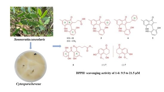
Antioxidative Indenone and Benzophenone Derivatives from the Mangrove-Derived Fungus Cytospora heveae NSHSJ-2

1
School of Chemistry, Sun Yat-Sen University, Guangzhou 510275, China
2
Guangdong Key Laboratory of Animal Conservation and Resource Utilization, Guangdong Public Laboratory of Wild Animal Conservation and Utilization, Institute of Zoology, Guangdong Academy of Sciences, Guangzhou 510260, China
*
Authors to whom correspondence should be addressed.
Mar. Drugs 2023, 21(3), 181; https://doi.org/10.3390/md21030181
Submission received: 23 February 2023 / Revised: 13 March 2023 / Accepted: 14 March 2023 / Published: 14 March 2023
(This article belongs to the Special Issue Pharmacological Potential of Marine Natural Products)

Abstract

:
Seven new polyketides, including four indenone derivatives, cytoindenones A–C (1, 34), 3′-methoxycytoindenone A (2), a benzophenone derivative, cytorhizophin J (6), and a pair of tetralone enantiomers, (±)-4,6-dihydroxy-5-methoxy-α-tetralone (7), together with a known compound (5) were obtained from the endophytic fungus Cytospora heveae NSHSJ-2 isolated from the fresh stem of the mangrove plant Sonneratia caseolaris. Compound 3 represented the first natural indenone monomer substituted by two benzene moieties at C-2 and C-3. Their structures were determined by the analysis of 1D and 2D NMR, as well as mass spectroscopic data, and the absolute configurations of (±)-7 were determined on the basis of the observed specific rotation value compared with those of the tetralone derivatives previously reported. In bioactivity assays, compounds 1, 46 showed potent DPPH· scavenging activities, with EC50 values ranging from 9.5 to 16.6 µM, better than the positive control ascorbic acid (21.9 µM); compounds 23 also exhibited DPPH· scavenging activities comparable to ascorbic acid.

Graphical Abstract

1. Introduction

Indenones are characterized by a cyclopentenone ring fused with an aromatic benzene ring, providing a rigid bicyclic ring framework which enables the extensive evaluation of structure–activity relationship analysis of target therapeutic molecules [1], and indenone derivatives have been synthesized extensively for drug discovery [2,3,4,5]. The indenone moiety usually exists in natural products as a structural fragment or a small independent molecule [6,7,8,9,10,11], and 2,3-diaryl indenone analogues are rarely reported [12,13,14]. These compounds were considered to be dimers of benzophenone, xanthone, diphenyl ether moieties and indanone moieties, and there was no natural 2,3-diphenyl indenone monomer reported previously. Indenones have multiple bioactivities, including cytotoxicity, DPPH· scavenging activity, anti-inflammatory activity, anti-osteoporosis activity, human DNA dealkylation repair enzyme AlkBH3 inhibitory activity, and PPAR γ agonistic activity [2,3,4,5,8,13,14,15].
Mangrove-associated fungi are known to be an essential source of natural products for the discovery of new drug leads [16,17]. In our continuing search for structurally diverse and biologically active metabolites from mangrove endophytic fungi [18,19,20,21,22], a chemical investigation for new secondary metabolites from mangrove endophytic fungus Cytospora heveae NSHSJ-2, which was isolated from the fresh stem of the mangrove plant Sonneratia caseolaris, led to the isolation and characterization of seven new polyketides (Figure 1), including four new indenone derivatives, cytoindenones A–C (1, 34), 3′-methoxycytoindenone A (2), a new benzophenone derivative, cytorhizophin J (6), and a pair of undescribed tetralone enantiomers, (±)-4,6-dihydroxy-5-methoxy-α-tetralone (7), together with a known compound, cytosporaphenones E (5) [23]. Among them, compound 3 represented the first natural indenone monomer substituted by two benzene moieties at C-2 and C-3. Herein, the isolation, structure elucidation, and DPPH· radical scavenging activities of these compounds are described.

2. Results

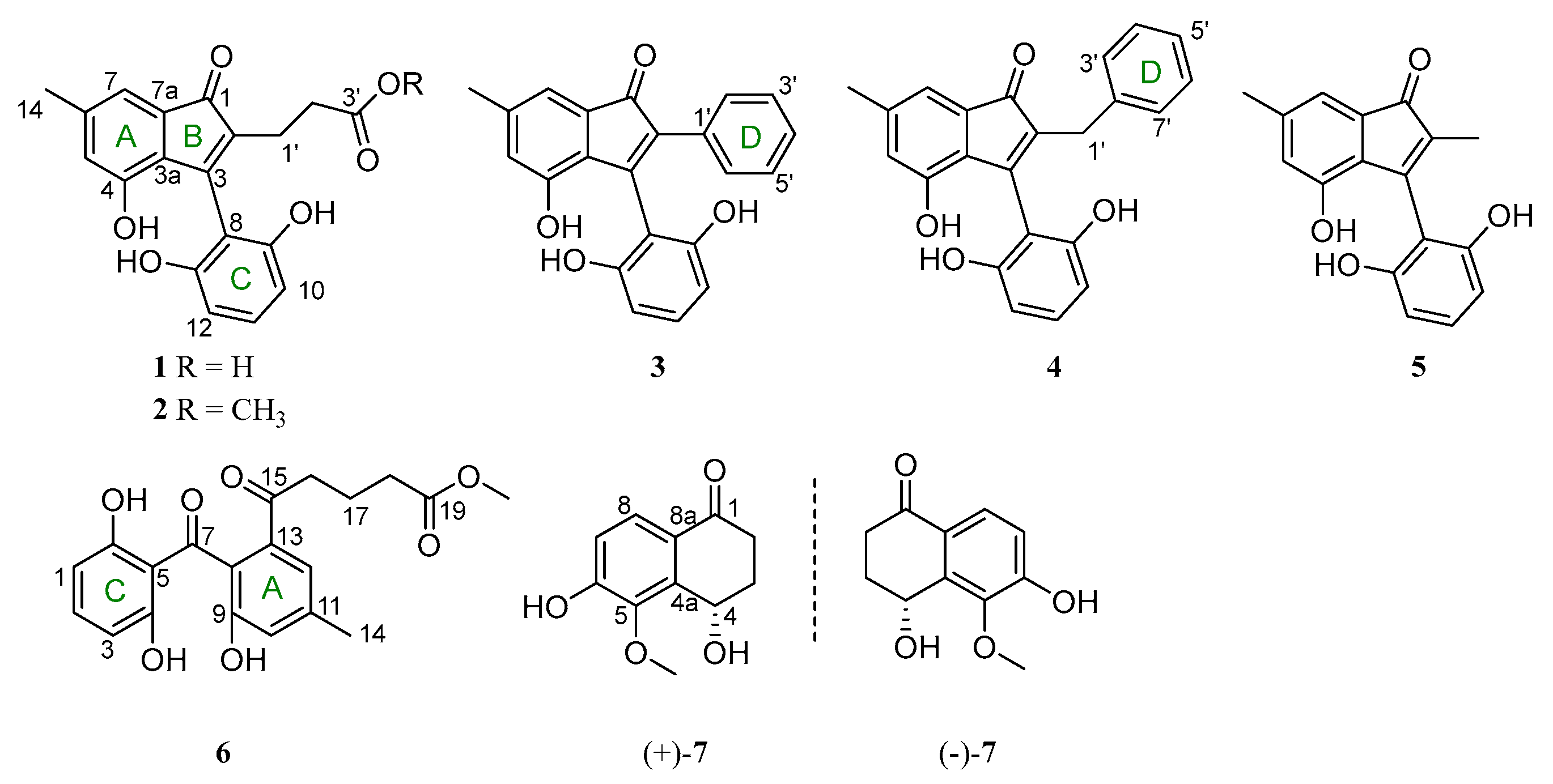
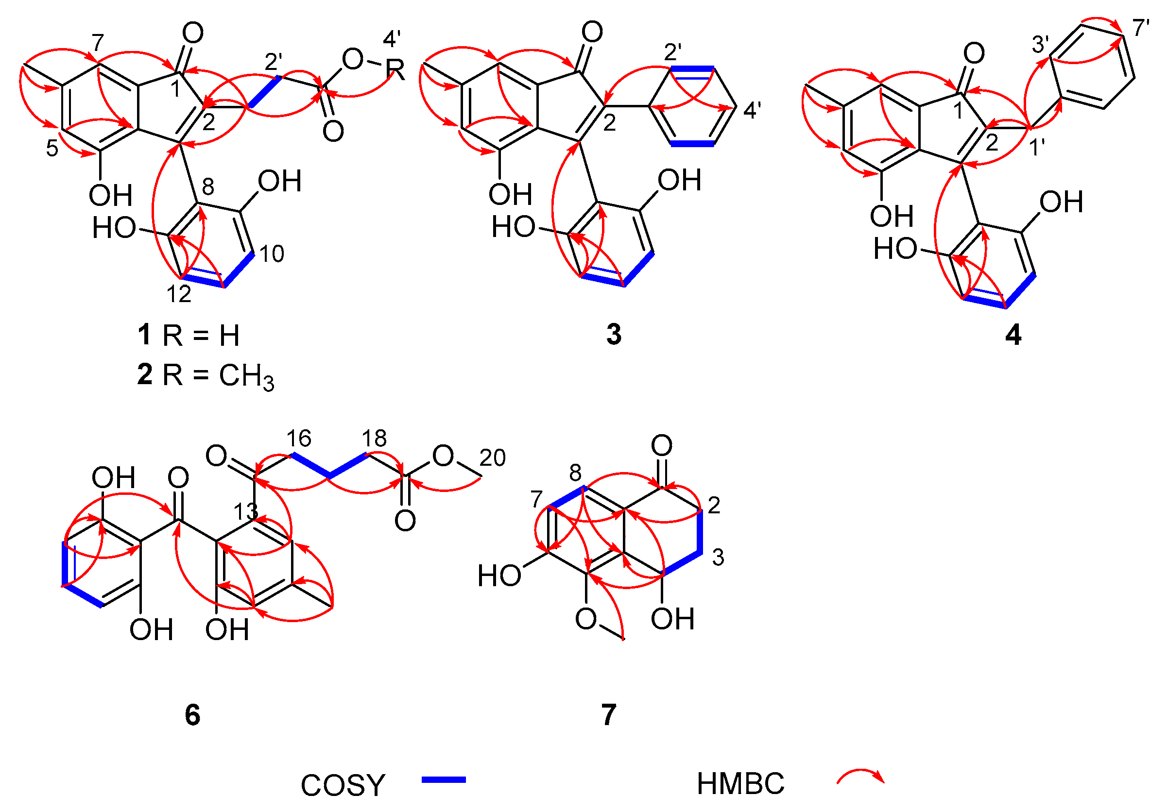
2.1. Structure Elucidation

Compound 1 was obtained as brown oil. Its molecular formula was assigned as C19H16O6 on the basis of HRESIMS analysis at m/z 363.08383 [M + Na]+ (calcd. For C19H16O6Na, 363.08391), which was determined to possess 12 degrees of unsaturation. In the 1H NMR spectrum (Table 1), the signals for five olefinic protons (δH 7.06, 6.79, 6.65, 6.50 and 6.50), two methylenes (δH 2.49 and 2.42) and one methyl (δH 2.24) were observed. The 13C NMR data (Table 2) exhibited 19 carbon resonances, including two carbonyls (δC 198.2 and 174.3), two aromatic rings (A and C) (δC 156.2, 156.2, 151.7, 140.7, 134.3, 130.7, 127.1, 124.5, 116.4, 110.6, 108.0, 108.0), two olefinic carbons for one double bond (δC 152.1, 134.4), two methylenes (δC 32.4 and 20.4) and one methyl (δC 21.0).
The HMBC correlations from H-1′, to C-1, C-2, C-3, from H-14 to C-5, C-6, C-7, from H-5 to C-3a, C-4, and from H-7 to C-1, C-3a suggested the presence of an indenone fragment (rings A and B) (Figure 2). Additionally, the 1H-1H COSY correlations of H-10/H-11/H-12, together with the HMBC correlations from H-12 to C-3, C-8 and C-13, and from H-11 to C-13, completed the 2,6-dihydroxybenzoyl fragment (ring C), which connected to the indenone ring at C-3. The structures of ring A, B and C were further confirmed by comparison of 1H and 13C NMR spectra between 1 and 5 [23]. Furthermore, the 1H-1H COSY correlation of H-1′/H-2′ and the HMBC correlations from H-1′, H-2′ to C-3′, from H-1′ to C-1, C-2, C-3, and from H-2′, to C-2 indicated the presence of the 2-carboxyethyl group, which was assigned to be connected to the indenone ring at C-2. Thus, the structure of 1 was deduced, named cytoindenone A.
Compound 2 was isolated as brown oil. Its molecular formula was determined as C20H18O6 (12 degrees of unsaturation) in terms of HREIMS analysis at m/z 377.09985 [M + Na]+ (calcd. for C20H18O6Na, 377.09956). Analysis of the 1H and 13C NMR spectroscopic data of 2 (Table 1 and Table 2) revealed mostly similarities with that of 1, except that the hydroxyl group was substituted with the methoxy group (δH 3.58, δC 52.2) at C-3′. Combined with the HMBC from H-4′ to C-3′ (Figure 2), the structure of compound 2 was clearly confirmed, named 3′-methoxycytoindenone A.
Compound 3 was acquired as brown oil and had a molecular formula of C22H16O4, determined by HRESIMS data m/z 367.09424 [M + Na]+ (calcd. 367.09408) with 15 degrees of unsaturation. The 1H NMR spectrum of 3 displayed the signal for ten olefinic protons (δH 7.30, 7.30, 7.15, 7.15, 7.13, 6.99, 6.87, 6.63, 6.33 and 6.33) and one methyl (δH 2.27). The 13C NMR data exhibited one carbonyl (δC 199.1), three aromatic rings (δC 156.5, 156.5, 153.1, 141.9, 134.4, 133.7, 130.7, 129.9, 129.9, 128.5, 128.5, 127.9, 127.1, 125.1, 116.9, 112.1, 107.9 and 107.9), two olefinic carbons for one double bond (δC 154.0, 133.6) and one methyl (δC 21.2) (Table 1 and Table 2). According to 1D NMR and 2D NMR data, the rings A, B and C of 2 were similar to that of 1. The obvious difference was the absence of the 2-carboxyethyl group at the C-2 position of compound 1 and the presence of a phenyl group (ring D) at the C-2 position of compound 3. Meanwhile, the 1H-1H COSY correlations of H-3′/H-4′/H-5′ failed to be identified because the chemical shifts of H-3′, H-4′ and H-5′ were overlapped; the 1H-1H COSY correlations of H-2′/H-3′, the HMBC from H-2′ to C-2, C-5′, and from H-3′ to C-1′ also indicated that ring D was formed and connected to the indenone ring at C-2, and the structure of compound 3 was determined, named cytoindenone B.
Compound 4 was obtained as brown oil. The molecular formula was determined as C23H18O4 on the basis of HRESIMS data at m/z 381.10980 [M + Na]+ (calcd. for C23H18O4Na, 381.10973), which was thus determined to possess 15 degrees of unsaturation. The 1H and 13C NMR spectroscopic data were listed in Table 1 and Table 2, which suggested that the structure of compound 4 was similar to compound 3, except the presence of methylenes (δH 3.41, δC 30.5). Similarly, the 1H-1H COSY correlations of H-3′/H-4′/H-5′/H-6′/H-7′ failed to be identified because of the overlapping chemical shifts. Combined with the HMBC from H-1′ to C-1, C-2, C-3, C-2′, C-3′, and from H-3′, H-4′ to C-5′ (Figure 2), ring D was formed and C-1′ was connected to the indenone ring and ring D at C-2 and C-2′, and the structure of compound 5 was clearly confirmed, named cytoindenone C.
Compound 6 was isolated as white powder and assigned an HRESIMS ion peak at m/z 395.11005 ([M + Na]+, calcd. for C20H20O7Na, 395.11012), which perfectly matched the molecular formula of C20H20O7 with 11 degrees of unsaturation. The 1H NMR spectrum of 6 displayed the signal for five olefinic protons (δH 7.25, 7.18, 6.87, 6.27 and 6.27), one methoxyl (δH 3.63), three methylenes (δH 2.96, 2.32 and 1.85) and one methyl (δH 2.36). The 13C NMR data revealed 20 carbon resonances, involving two carbonyls (δC 204.1 and 202.2), one ester carbonyl (dC 175.5), two aromatic rings (δC 163.2, 163.2, 155.0, 140.8, 137.8, 137.0, 130.0, 121.6, 121.6, 112.7, 108.1, 108.1), one methoxyl (δC 52.0), three methylenes (δC 38.9, 33.7 and 20.6) and one methyl (δC 21.3) (Table 3). According to 1D NMR and 2D NMR data, the benzophenone moiety of 6 was similar to cytorhizophin C [24]. The only difference between them were that the popionyl group at the C-13 position of cytorhizophin C was replaced by the 5-methoxy-5-oxopentanoyl group of compound 6. The 1H-1H COSY correlations of H-16/H-17/H-18, together with the HMBC correlations from H-16 to C-13 and C-15, from H-17 to C-15, and from H-18, H-20 to C-19 indicated that the 5-methoxy-5-oxopentanoyl group was located at C-13. Therefore, the structure of 6 was deduced and named cytorhizophin J.
Compound 7 was acquired as colorless oil. Its molecular formula C11H12O4 (six degrees of unsaturation) was established on the basis of HREIMS analysis at m/z 209.08093 [M + H]+ (calcd. For C11H13O4, 209.08084). Analysis of the 1H and 13C NMR spectroscopic data of 7 (Table 4) revealed mostly similarities to 3,4-dihydro-4β,6-dihydroxy-5-methoxy-2α-methyl-1(2H)-naphthalenone [25]. The main difference between them were the absence of one methine at δH 2.98 (1H, m, H-2β) and one methyl at δH 1.11 (3H, d, J = 6.8 Hz, 2-Me) in 3,4-dihydro-4β,6-dihydroxy-5-methoxy-2α-methyl-1(2H)-naphthalenone and the presence of one methylene at δH 2.99 (1H, m, Ha-2) and 2.43 (1H, dt, J = 17.2, 3.6, Hb-2) in 7, which was confirmed by the 1H-1H COSY correlations of Ha, b-2/Ha, b-3/H-4, and the HMBC correlations (Figure 2) from Ha, b-2 to C-1, C-8a. Thus, compound 7 was assigned as shown in Figure 1, and named 4,6-dihydroxy-5-methoxy-α-tetralone. However, chiral HPLC analysis of 7 showed two peaks (tR 21.3 min and 24.6 min), and subsequent chiral HPLC purification of (±)-7 led to the separation of the two enantiomers (+)-7 and (−)-7. The absolute configurations of (+)-7 and (−)-7 were determined as 4S and 4R by the comparison of the observed specific rotation value [(+)-7: [ α ] D 25 + 31.3, (+)-7: [ α ] D 25 − 31.5)] of compounds (±)-7 with those for (4S)-4,8-dihydroxy-α-tetralone ( [ α ] D 27 + 24.5), (4S)-5-hydroxy-4-methoxy-α-tetralone ( [ α ] D 27 + 50.0), (4R)-4,8-dihydroxy-α-tetralone ( [ α ] D 27 − 26.0) and (4R)-5-hydroxy-4-methoxy-α-tetralone ( [ α ] D 27 − 50.0) (Figure S39) [26].

2.2. Biological Evaluation

Compounds 17 were tested for their DPPH· scavenging activity. As seen in Table 5, the results indicated that compounds 1, 46 showed significant DPPH· scavenging activities with EC50 values ranging from 9.5 to 16.6 µM, better than the positive control ascorbic acid (21.9 µM) [27,28]; compounds 23 also exhibited DPPH· scavenging activities comparable to ascorbic acid.
The antioxidant activities of phenolic compounds were widely investigated and the phenolic content and the side chain functional groups had significant influences on DPPH· scavenging activities [29,30]. Comparing the activities of compounds 12, when the carboxyl group at C-3′ was esterified by the methyl group, the antioxidant activity of 2 decreases significantly. Comparing the activities of compounds 25, the higher activity of compound 5 was due to the accessibility of the phenolic OH group by DPPH·. The activities of compounds 24 were due to the presence of bulky groups at C-2 obstructing DPPH· access to the phenolic OH group. Compound 6 could be regarded as a precursor of compound 2, which formed ring B through C7−C16 aldol-type cyclization. Compound 6 exhibited the strongest antioxidant activity due to the disconnection of ring B and the smallest steric hindrance of phenolic ring C. Compounds (+)-7 and (–)-7 showed no antioxidant activities due to the reduction of the phenolic content.

3. Experimental Section

3.1. General Experimental Procedures

Optical rotations were performed on an MCP300 (Anton Paar, Shanghai, China). UV data were measured on a Shimadzu UV-2600 spectrophotometer (Shimadzu, Kyoto, Japan). The ECD experiment data were conducted with a J-810 spectropolarimeter (JASCO, Tokyo, Japan). IR spectra were measured on an IR Affinity-1 spectrometer (Shimadzu, Kyoto, Japan). Melting points were recorded on a Fisher-Johns hot-stage apparatus. The NMR spectra were tested on a Bruker Avance spectrometer (Bruker, Beijing, China) (Compounds 34: 600 MHz for 1H and 150 MHz for 13C; compounds 12 and 57: 400 MHz for 1H and 100 MHz for 13C, respectively). HRESIMS data were conducted on a ThermoFisher LTQ-Orbitrap-LC-MS spectrometer (Palo Alto, CA, USA). Column chromatography (CC) was performed on silica gel (200–300 mesh, Marine Chemical Factory, Qingdao, China) and Sephadex LH-20 (Amersham Pharmacia, Piscataway, NJ, USA). Semi-preparative HPLC (Ultimate 3000 BioRS, Thermo Scientific, Germany) were conducted using the Chiral INA column (5 μm, 4.6 × 250 mm, Phenomenex, Piscataway, NJ, USA), and the Chiralcel ODH column (5 μm, 4.6 × 250 mm, Daicel, Tokyo, Japan) for chiral separation.

3.2. Fungal Material

The fungal strain NSHSJ-2 used in this study was isolated from the fresh stem of mangrove plant Sonneratia caseolaris, which was collected in June 2020 from the Nansha Mangrove National Nature Reserve in Guangdong Province, China. The strain was identified as Cytospora heveae (compared to no. OQ423127) upon the analysis of ITS sequence data of the rDNA gene. The ITS sequence data obtained from the fungal strain has been submitted to GenBank with accession no. OL780505.1. A voucher strain was deposited in our laboratory.

3.3. Fermentation, Extraction and Isolation

The fungus Cytospora heveae NSHSJ-2 was fermented on solid cultured medium (sixty 1000 mL Erlenmeyer flasks, each containing 50 g of rice and 50 mL of distilled water with 3% sea salt) for 30 days at 25 °C. The cultures were extracted three times with MeOH to yield 22.9 g of residue. Then, the crude extract was eluted by using a gradient elution with petroleum ether/EtOAc from 9:1 to 0:10 (v/v) on silica gel CC to get six fractions (Fr. A–F). Fr. D (297 mg) was subjected to silica gel CC (CH2Cl2/MeOH, v/v, 800:1 to 200:1) to obtain three subfractions (Fr. D1–D3). Fr. D2 (9.4 mg) was separated by normal phase HPLC on a chiral column (INA), using hexane/isopropanol (80:20, v/v, flow rate: 1.0 mL/min) as the solvent system, to yield compounds 3 (1.6 mg, tR 14.0 min) and 4 (4.3 mg, tR 21.2 min). Fr. D3 (83.4 mg) was applied to Sephadex LH-20 CC (CH2Cl2/MeOH, v/v, 1:1) to yield compound 5 (26 mg). Fr. E (749 mg) was subjected to silica gel CC (CH2Cl2/MeOH, v/v, 100:1 to 20:1) to afford four fractions (Fr. E1−E4). Fr. E2 (204 mg) was subjected to silica gel CC (petroleum ether/EtOAc, v/v, 7:3) to yield compounds 2 (46.5 mg). Fr. E3 (56.4 mg) was subjected to silica gel CC (petroleum ether/EtOAc, v/v, 6:4) to yield compounds 6 (15.4 mg) and (±)-7 (9.4 mg). The chiral HPLC separation of (±)-7 was accomplished over a chiral column (ODH) (column size: 4.6 × 250 mm, 5 μm; flow rate: 1.0 mL/min; solvent: n-hexane-isopropanol = 90:10) to yield (+)-7 (1.4 mg, tR 21.3 min) and (−)-7 (7.3 mg, tR 24.6 min). Fr. E4 (103 mg) was purified by Sephadex LH-20 CC and eluted with MeOH to obtain compound 1 (27.9 mg).
Cytoindenone A (1): brown oil; UV (MeOH) λmax (log ε): 205 (1.24), 247 (0.53) nm; IR υmax 3282, 2949, 2835, 1695, 1435, 1276, 1010, 781 cm−1; HRESIMS m/z 363.08383 [M + Na]+ (calcd. for C19H16O6Na, 363.08391); 1H NMR (400 MHz, Actone-d6) data and 13C NMR (100 MHz, Actone-d6) data (see Table 1 and Table 2).
3′-methoxycytoindenone A (2): brown oil; UV (MeOH) λmax (log ε): 204 (0.90), 248 (0.42) nm; IR υmax 3360, 2954, 2920, 1697, 1622, 1462, 1278, 1012, 783 cm−1; HRESIMS m/z 377.09985 [M + Na]+ (calcd. for C20H18O6Na, 377.09956); 1H NMR (400 MHz, CDCl3) data and 13C NMR (100 MHz, CDCl3) data (see Table 1 and Table 2).
Cytoindenone B (3): brown oil; UV (MeOH) λmax (log ε): 203 (0.32), 272 (0.15) nm; IR υmax 3365, 2949, 2850, 1689, 1618, 1462, 1280, 1014, 792 cm−1; HRESIMS m/z 367.09424 [M + Na]+ (calcd. for C22H16O4Na, 367.09408); 1H NMR (600 MHz, CD3OD) data and 13C NMR (150 MHz, CD3OD) data (see Table 1 and Table 2).
Cytoindenone C (4): brown oil; UV (MeOH) λmax (log ε): 205 (0.80), 249 (0.35) nm; IR υmax 3358, 2922, 2852, 1683, 1618, 1462, 1276, 1012, 700 cm−1; HRESIMS m/z 381.10980 [M + Na]+ (calcd. for C23H18O4Na, 381.10973); 1H NMR (600 MHz, CD3OD) data and 13C NMR (150 MHz, CD3OD) data (see Table 1 and Table 2).
Cytorhizophin J (6): white powder, mp 190.2−191.6 ◦C; UV (MeOH) λmax (log ε): 216 (1.43), 270 (0.67) nm; IR υmax 3342, 2924, 1716, 1627, 1456, 1338, 1226, 1031, 925, cm−1; HRESIMS m/z 395.11005 [M + Na]+ (calcd. for C20H20O7Na, 395.11012); 1H NMR (400 MHz, CD3OD) and 13C NMR (100 MHz, CD3OD) data (see Table 3).
(±)-4,6-dihydroxy-5-methoxy-α-tetralone (7): colorless oil; UV (MeOH) λmax (log ε): 205 (1.63), 230 (1.29), 282 (1.01) nm; IR νmax 3261, 2943, 2839, 1660, 1578, 1305, 1290, 1190, 1012 cm−1; 1H NMR (400 MHz, CD3OD) data and 13C NMR (100 MHz, CD3OD) data (see Table 4); HRESIMS m/z 209.08093 [M + H]+ (calcd for C11H13O4, 209.08084). (+)-7, [ α ] D 25 + 31.3 (c 0.1 MeOH); ECD (c = 0.18 mg/mL, MeOH) λmaxε) 205 (+13.5), 230 (+8.4), 284 (+7.0), 327 (−6.0). (−)-7, [ α ] D 25 − 31.5 (c 0.1 MeOH); ECD (c = 0.17 mg/mL, MeOH) λmaxε) 205 (−14.9), 225 (−6.1), 277 (−5.4), 320 (+5.8).

3.4. Biological Assays

The DPPH·radical scavenging activities of compounds 17 were determined according to the reported method [14,28]. The DPPH· radical scavenging test was performed in 96-well microplates. Testing materials (compounds 17) were added to 100 µL (0.16 mmol/L) DPPH solution in MeOH at a range of 100 µL solutions of different concentrations (6.25−100 µM). Ascorbic acid was prepared as positive control at the same concentrations (Table 5). Absorbance was recorded at λ = 517 nm after 45 min of incubation in the dark. The DPPH·radical scavenging activity was calculated using the formula:
DPPH radical scavenging activity (%) = [(Abscontrol − Abssample)/Abscontrol] × 100

4. Conclusions

In summary, seven new polyketides including four indenone derivatives, cytoindenones A–C (1, 34), 3′-methoxycytoindenone A (2), a new benzophenone derivative, cytorhizophin J (6) and a pair of undescribed tetralone enantiomers, (±)-4,6-dihydroxy-5-methoxy-1-tetralone (7), together with a known compound (5), were isolated from the endophytic fungus Cytospora heveae NSHSJ-2. Compound 3 represented the first natural indenone monomer substituted by two benzene moieties at C-2 and C-3. Their structures were confirmed by the analysis of NMR, HR-MS and ECD spectra. All of the compounds were tested for their antioxidative activities. Compounds 1, 46 showed potent DPPH· scavenging activities with EC50 values ranging from 9.5 to 16.6 µM, better than the positive control ascorbic acid (21.9 µM); compounds 23 also exhibited DPPH· scavenging activities comparable to ascorbic acid.

Supplementary Materials

The following are available online at https://www.mdpi.com/article/10.3390/md21030181/s1, Figure S1: HRESIMS spectrum of compound 1; Figure S2: 1H NMR spectrum of compound 1 (400 MHz, Actone-d6); Figure S3: 13C NMR spectrum of compound 1 (100 MHz, Actone-d6); Figure S4: 1H-1H COSY spectrum of compound 1; Figure S5: HSQC spectrum of compound 1; Figure S6: HMBC spectrum of compound 1; Figure S7: HRESIMS spectrum of compound 2; Figure S8: 1H NMR spectrum of compound 2 (400 MHz, CDCl3); Figure S9: 13C NMR spectrum of compound 2 (100 MHz, CDCl3); Figure S10: 1H-1H COSY spectrum of compound 2; Figure S11: HSQC spectrum of compound 2; Figure S12: HMBC spectrum of compound 2; Figure S13: HRESIMS spectrum of compound 3; Figure S14: 1H NMR spectrum of compound 3 (600 MHz, CD3OD); Figure S15: 13C NMR spectrum of compound 3 (150 MHz, CD3OD); Figure S16: 1H-1H COSY spectrum of compound 3; Figure S17: HSQC spectrum of compound 3; Figure S18: HMBC spectrum of compound 3; Figure S19: HRESIMS spectrum of compound 4; Figure S20: 1H NMR spectrum of compound 4 (600 MHz, CD3OD); Figure S21: 13C NMR spectrum of compound 4 (150 MHz, CD3OD); Figure S22: 1H-1H COSY spectrum of compound 4; Figure S23: HSQC spectrum of compound 4; Figure S24: HMBC spectrum of compound 4; Figure S25: HRESIMS spectrum of compound 6; Figure S26: 1H NMR spectrum of compound 6 (400 MHz, CD3OD); Figure S27: 13C NMR spectrum of compound 6 (100 MHz, CD3OD); Figure S28: 1H-1H COSY spectrum of compound 6; Figure S29: HSQC spectrum of compound 6; Figure S30: HMBC spectrum of compound 6; Figure S31: HRESIMS spectrum of compound 7; Figure S32: 1H NMR spectrum of compound 7 (400 MHz, CD3OD); Figure S33: 13C NMR spectrum of compound 7 (100 MHz, CD3OD); Figure S34: 1H-1H COSY spectrum of compound 7; Figure S35: HSQC spectrum of compound 7; Figure S36: HMBC spectrum of compound 7; Figure S37: ECD spectrum of compound (+)-7; Figure S38: ECD spectrum of compound (–)-7; Figure S39: Structure of compounds 3,4-dihydro-4β,6-dihydroxy-5-methoxy-2α-methyl-1(2H)-naphthalenone, (4S)-4,8-dihydroxy-α-tetralone, (4R)-4,8-dihydroxy-α-tetralone, (4S)-5-hydroxy-4-methoxy-α-tetralone and (4R)- 5-hydroxy-4-methoxy-α-tetralone.

Author Contributions

G.Z. performed the experiments and wrote the paper; T.L., W.Y. and B.S. participated in the experiments; Y.C., B.W. and Y.O. analyzed the data and discussed the results; H.Y. and Z.S. reviewed the manuscript; Z.S. designed and supervised the experiments. All authors have read and agreed to the published version of the manuscript.

Funding

We thank the National Natural Science Foundation of China (U20A2001, 42276114, 82104228), the Key-Area Research and Development Program of Guangdong Province (2020B1111030005), and the GDAS Special Project of Science and Technology Development (Grant No. 2021GDASYL–20210103057) for the generous support.

Data Availability Statement

Data are contained within the article and Supplementary Material.

Conflicts of Interest

The authors declare no conflict of interest.

References

  1. Prasher, P.; Sharma, M. Medicinal chemistry of indane and its analogues: A mini review. ChemistrySelect 2021, 6, 2658–2677. [Google Scholar] [CrossRef]
  2. Nigam, R.; Babu, K.R.; Ghosh, T.; Kumari, B.; Akula, D.; Rath, S.N.; Das, P.; Anindya, R.; Khan, F.A. Indenone derivatives as inhibitor of human DNA dealkylation repair enzyme AlkBH3. Bioorgan. Med. Chem. 2018, 26, 4100–4112. [Google Scholar] [CrossRef] [PubMed]
  3. Hao, X.D.; Chang, J.; Qin, B.Y.; Zhong, C.; Chu, Z.B.; Huang, J.; Zhou, W.J.; Sun, X. Synthesis, estrogenic activity, and anti-osteoporosis effects in ovariectomized rats of resveratrol oligomer derivatives. Eur. J. Med. Chem. 2015, 102, 26–38. [Google Scholar] [CrossRef] [PubMed]
  4. Ahn, J.H.; Shin, M.S.; Jung, S.H.; Kang, S.K.; Kim, K.R.; Rhee, S.D.; Jung, W.H.; Yang, S.D.; Kim, S.J.; Woo, J.R.; et al. Indenone Derivatives: A Novel Template for Peroxisome Proliferator-Activated Receptor γ (PPARγ) Agonists. J. Med. Chem. 2006, 49, 4781–4784. [Google Scholar] [CrossRef]
  5. Liu, J.; Liu, L.; Zheng, L.; Feng, K.W.; Wang, H.T.; Xu, J.P.; Zhou, Z.Z. Discovery of novel 2,3-dihydro-1H-inden-1-ones as dual PDE4/AChE inhibitors with more potency against neuroinflammation for the treatment of Alzheimer’s disease. Eur. J. Med. Chem. 2022, 238, 114503. [Google Scholar] [CrossRef] [PubMed]
  6. Ernst-Russell, M.A.; Chai, C.L.L.; Wardlaw, J.H.; Elix, J.A. Euplectin and Coneuplectin, New Naphthopyrones from the Lichen Flavoparmelia euplecta. J. Nat. Prod. 2000, 63, 129–131. [Google Scholar] [CrossRef] [PubMed]
  7. Li, X.L.; Ru, T.; Navarro-Vázquez, A.; Lindemann, P.; Nazaré, M.; Li, X.W.; Guo, Y.W.; Sun, H. Weizhouochrones: Gorgonian-Derived Symmetric Dimers and Their Structure Elucidation Using Anisotropic NMR Combined with DP4+ Probability and CASE-3D. J. Nat. Prod. 2022, 85, 1730–1737. [Google Scholar] [CrossRef]
  8. Du, Y.E.; Byun, W.S.; Lee, S.B.; Hwang, S.; Shin, Y.H.; Shin, B.; Jang, Y.J.; Hong, S.; Shin, J.; Lee, S.K.; et al. Formicins, N-Acetylcysteamine-Bearing Indenone Thioesters from a Wood Ant-Associated Bacterium. Org. Lett. 2020, 22, 5337–5341. [Google Scholar] [CrossRef]
  9. Luo, H.F.; Zhang, L.P.; Hu, C.Q. Five novel oligostilbenes from the roots of Caragana sinica. Tetrahedron 2001, 57, 4849–4854. [Google Scholar] [CrossRef]
  10. Sema, D.K.; Lannang, A.M.; Tatsimo, S.J.N.; Rehman, M.; Yousuf, S.; Zoufou, D.; Iqbal, U.; Wansi, J.D.; Sewald, N.; Choudhary, M.I. New indane and naphthalene derivatives from the rhizomes of Kniphofia reflexa Hutchinson ex Codd. Phytochem. Lett. 2018, 26, 78–82. [Google Scholar] [CrossRef]
  11. Jaki, B.; Heilmann, J.; Sticher, O. New Antibacterial Metabolites from the Cyanobacterium Nostoc commune (EAWAG 122b). J. Nat. Prod. 2000, 63, 1283–1285. [Google Scholar] [CrossRef]
  12. Kim, H.; Yang, I.; Ryu, S.Y.; Won, D.H.; Giri, A.G.; Wang, W.H.; Choi, H.; Chin, J.; Hahn, D.; Kim, E.; et al. Acredinones A and B, Voltage-Dependent Potassium Channel Inhibitors from the Sponge-Derived Fungus Acremonium sp. F9A015. J. Nat. Prod. 2015, 78, 363–367. [Google Scholar] [CrossRef]
  13. Liu, Z.M.; Qiu, P.; Li, J.; Chen, G.Y.; Chen, Y.; Liu, H.J.; She, Z.G. Anti-inflammatory polyketides from the mangrove-derived fungus Ascomycota sp. SK2YWS-L. Tetrahedron 2018, 74, 746–751. [Google Scholar] [CrossRef]
  14. Tan, C.B.; Liu, Z.M.; Chen, S.H.; Huang, X.S.; Cui, H.; Long, Y.H.; Lu, Y.J.; She, Z.G. Antioxidative Polyketones from the Mangrove-Derived Fungus Ascomycota sp. SK2YWS-L. Sci. Rep. 2016, 6, 36609. [Google Scholar] [CrossRef] [PubMed] [Green Version]
  15. Zhong, C.; Liu, X.H.; Chang, J.; Yu, J.M.; Sun, X. Inhibitory effect of resveratrol dimerized derivatives on nitric oxide production in lipopolysaccharide-induced RAW 264.7 cells. Bioorg. Med. Chem. Lett. 2013, 23, 4413–4418. [Google Scholar] [CrossRef] [PubMed]
  16. Chen, S.H.; Cai, R.L.; Liu, Z.M.; Cui, H.; She, Z.G. Secondary metabolites from mangrove-associated fungi: Source, chemistry and bioactivities. Nat. Prod. Rep. 2022, 39, 560–595. [Google Scholar] [CrossRef] [PubMed]
  17. Xu, J. Bioactive natural products derived from mangrove-associated microbes. RSC Adv. 2015, 5, 841–892. [Google Scholar] [CrossRef]
  18. Tan, Q.; Yang, W.C.; Zhu, G.; Chen, T.; Wu, J.; Zhu, Y.J.; Wang, B.; Yuan, J.; She, Z.G. A Pair of Chromone Epimers and an Acetophenone Glucoside from the Mangrove Endophytic Fungus Mycosphaerella sp. L3A1. Chem. Biodivers. 2022, 19, e202200998. [Google Scholar] [CrossRef]
  19. Chen, T.; Yang, W.C.; Li, T.B.; Yin, Y.H.; Liu, Y.F.; Wang, B.; She, Z.G. Hemiacetalmeroterpenoids A–C and Astellolide Q with Antimicrobial Activity from the Marine-Derived Fungus Penicillium sp. N-5. Mar. Drugs 2022, 20, 514. [Google Scholar] [CrossRef]
  20. Chen, Y.; Yang, W.C.; Zou, G.; Wang, G.S.; Kang, W.Y.; Yuan, J.; She, Z.G. Cytotoxic Bromine- and Iodine-Containing Cytochalasins Produced by the Mangrove Endophytic Fungus Phomopsis sp. QYM-13 Using the OSMAC Approach. J. Nat. Prod. 2022, 85, 1229–1238. [Google Scholar] [CrossRef]
  21. Zang, Z.M.; Yang, W.C.; Cui, H.; Cai, R.L.; Li, C.Y.; Zou, G.; Wang, B.; She, Z.G. Two Antimicrobial Heterodimeric Tetrahydroxanthones with a 7,7′-Linkage from Mangrove Endophytic Fungus Aspergillus flavus QQYZ. Molecules 2022, 27, 2691. [Google Scholar] [CrossRef] [PubMed]
  22. Jiang, H.M.; Cai, R.L.; Zang, Z.M.; Yang, W.C.; Wang, B.; Zhu, G.; Yuan, J.; She, Z.G. Azaphilone derivatives with anti-inflammatory activity from the mangrove endophytic fungus Penicillium sclerotiorum ZJHJJ-18. Bioorg. Chem. 2022, 122, 105721. [Google Scholar] [CrossRef] [PubMed]
  23. Liu, H.X.; Liu, Z.M.; Chen, Y.C.; Tan, H.B.; Zhang, W.G.; Zhang, W.M. Polyketones from the endophytic fungus Cytospora rhizophorae. Nat. Prod. Res. 2021, 37, 1053–1059. [Google Scholar] [CrossRef] [PubMed]
  24. Liu, H.X.; Tan, H.B.; Wang, W.X.; Zhang, W.G.; Chen, Y.C.; Liu, S.N.; Liu, Z.M.; Li, H.H.; Zhang, W.M. Cytorhizophins A and B, benzophenone-hemiterpene adducts from the endophytic fungus Cytospora rhizophorae†. Org. Chem. Front. 2019, 6, 591–596. [Google Scholar] [CrossRef]
  25. Auamcharoen, W.; Kijjoa, A.; Chandrapatya, A.; Pinto, M.M.; Silva, A.M.S.; Naengchomnong, W.; Herz, W. A new tetralone from Diospyros cauliflora. Biochem. Syst. Ecol. 2009, 37, 690–692. [Google Scholar] [CrossRef]
  26. Machida, K.; Matsuoka, E.; Kasahara, T.; Kikuchi, M. Studies on the Constituents of Juglans Species. I. Structural Determination of (4S)- and (4R)-4-Hydroxy-α-tetralone Derivatives from the Fruit of Juglans mandshurica MAXIM. var. sieboldiana Makino. Chem. Pharm. Bull. 2005, 53, 934–937. [Google Scholar] [CrossRef] [Green Version]
  27. Chen, Y.; Yang, W.C.; Zou, G.; Chen, S.Y.; Pang, J.Y.; She, Z.G. Bioactive polyketides from the mangrove endophytic fungi Phoma sp. SYSUSK-7. Fitoterapia 2019, 139, 10436. [Google Scholar] [CrossRef]
  28. Qiu, P.; Liu, Z.M.; Chen, Y.; Cai, R.L.; Chen, G.Y.; She, Z.G. Secondary Metabolites with α-Glucosidase Inhibitory Activity from the Mangrove Fungus Mycosphaerella sp. SYSU-DZG01. Mar. Drugs 2019, 17, 483. [Google Scholar] [CrossRef] [Green Version]
  29. Ichikawa, K.; Sasada, R.; Chiba, K.; Gotoh, H. Effect of Side Chain Functional Groups on the DPPH Radical Scavenging Activity of Bisabolane-Type Phenols. Antioxidants 2019, 8, 65. [Google Scholar] [CrossRef] [Green Version]
  30. Odame, F.; Hosten, E.C.; Betz, R.; Krause, J.; Frost, C.L.; Lobb, K.; Tshentu, Z.R. Synthesis, characterization, computational studies and DPPH scavenging activity of some triazatetracyclic derivatives. J. Iran. Chem. Soc. 2021, 18, 1979–1995. [Google Scholar] [CrossRef]
Figure 1. Structure of compounds 17.
Marinedrugs 21 00181 g001
Figure 2. Key COSY and HMBC correlations of 14 and 67.
Marinedrugs 21 00181 g002
Table 1. 1H NMR data of 14 (J in Hz).
No.1 a2 b3 c4 c
56.65, s6.54, s6.63, s6.58, s
76.79, s6.82, s6.87, s6.75, s
106.50, d (8.1)6.57, d (8.3)6.33, d (8.1)6.40, d (8.2)
117.06, t, (8.1)7.15, t (8.3)6.99, t (8.1)7.02, t (8.2)
126.50, d (8.1)6.57, d (8.3)6.33, d (8.1)6.40, d (8.2)
142.24, s2.17, s2.27, s2.22, s
1′2.42, m2.42, t (7.1) 3.41, s
2′2.49, m2.66, t (7.1)7.30, m
3′ 7.15, m7.09, m
4′ 3.58, s7.13, m7.07, m
5′ 7.15, m7.02, t (8.2)
6′ 7.30, m7.07, m
7′ 7.09, m
a Data were recorded in Actone-d6 at 400 MHz for 1H NMR. b Data were recorded in CDCl3 at 400 MHz for 1H NMR. c Data were recorded in CD3OD at 600 MHz for 1H NMR.
Table 2. 13C NMR data of 14.
No.1 a2 b3 c4 c
1198.2, C197.5, C199.1, C200.2, C
2134.4, C135.2, C133.6, C134.8, C
3152.1, C148.3, C154.0, C154.8, C
3a127.1, C123.7, C127.1, C127.4, C
4151.7, C150.0, C153.1, C152.3, C
5124.5, CH124.7, CH125.1, CH125.0, CH
6140.7, C141.5, C141.9, C141.1, C
7116.4, CH118.2, CH116.9, CH116.6, CH
7a134.3, C132.5, C134.4, C134.6, C
8110.6, C108.4, C112.1, C111.4, C
9156.2, C153.6, C156.5, C156.6, C
10108.0, CH109.4, CH107.9, CH107.8, CH
11130.7, CH131.9, CH130.7, CH130.8, CH
12108.0, CH109.4, CH107.9, CH107.8, CH
13156.2, C153.6, C156.5, C156.6, C
1421.0, CH321.3, CH321.2, CH321.1, CH3
1′20.4, CH219.6, CH2133.7, C30.5, CH2
2′32.4, CH231.0, CH2129.9, CH140.9, C
3′174.3, C175.1, C128.5, CH129.7, CH
4′ 52.2, CH3127.9, CH128.9, CH
5′ 128.5, CH126.5, CH
6′ 129.9, CH128.9, CH
7′ 129.7, CH
a Data were recorded in Actone-d6 at 100 MHz for 13C NMR. b Data were recorded in CDCl3 at 100 MHz for 13C NMR. c Data were recorded in CD3OD at 150 MHz for 13C NMR.
Table 3. 1H and 13C NMR data for 6.
6 a
No.δC, TypeδH Mult (J in Hz)
1108.1, CH6.27, d (8.2)
2137.0, CH7.18, t (8.2)
3108.1, CH6.27, d (8.2)
4163.2, C
5112.7, C
6163.2, C
7204.1, C
8130.0, C
9155.0, C
10121.6, CH6.87, s
11140.8, C
12121.6, CH7.25, s
13137.8, C
1421.3, CH32.36, s
15202.2, C
1638.9, CH22.96, t (7.5)
1720.6, CH21.85, m
1833.7, CH22.32, t (7.3)
19175.5, C
2052.0, CH33.63, s
a Data were recorded in CD3OD at 400 MHz for 1H NMR and 100 MHz for 13C NMR.
Table 4. 1H and 13C NMR data for 7.
7 a
No.δC, TypeδH Mult (J in Hz)
1199.6, C
233.0, CH22.99, m
2.43, dt (17.2, 3.6)
331.6, CH22.26, m
2.17, m
461.7, CH5.26, t (3.1)
4a139.9, C
5146.2, C
6157.5, C
7117.8, C6.92, d (8.6)
8125.4, CH7.67, d (8.6)
8a125.6, C
961.9, CH3
a Data were recorded in CD3OD at 400 MHz for 1H NMR and 100 MHz for 13C NMR.
Table 5. DPPH· scavenging activities of compounds 19.
Compound% Inhibition (100 µM)EC50 (µM)
190.811.5 ± 0.1
272.521.5 ± 1.0
369.019.7 ± 1.8
478.216.6 ± 0.4
581.012.4 ± 0.5
687.39.5 ± 0.1
(+)-712.0
(–)-74.2
ascorbic acid a91.421.9 ± 0.3
a positive control.
Disclaimer/Publisher’s Note: The statements, opinions and data contained in all publications are solely those of the individual author(s) and contributor(s) and not of MDPI and/or the editor(s). MDPI and/or the editor(s) disclaim responsibility for any injury to people or property resulting from any ideas, methods, instructions or products referred to in the content.

