Communication

Potential α-Glucosidase Inhibitors from the Deep-Sea Sediment-Derived Fungus Aspergillus insulicola

1
Beijing Key Laboratory for Separation and Analysis in Biomedicine and Pharmaceuticals, School of Life Science, Beijing Institute of Technology, Beijing 100081, China
2
Hainan Provincial Key Laboratory for Functional Components Research and Utilization of Marine Bio-Resources, Institute of Tropical Bioscience and Biotechnology, Chinese Academy of Tropical Agricultural Sciences & Key Laboratory for Biology and Genetic Resources of Tropical Crops of Hainan Province, Hainan Institute for Tropical Agricultural Resources, Haikou 571101, China
3
Zhanjiang Experimental Station of Chinese Academy of Tropical Agricultural Sciences, Zhanjiang 524013, China
*
Author to whom correspondence should be addressed.
These authors contributed equally to this work.
Mar. Drugs 2023, 21(3), 157; https://doi.org/10.3390/md21030157
Submission received: 4 February 2023 / Revised: 23 February 2023 / Accepted: 24 February 2023 / Published: 26 February 2023

Abstract

:
Three new phenolic compounds, epicocconigrones C–D (12) and flavimycin C (3), together with six known phenolic compounds: epicocconigrone A (4); 2-(10-formyl-11,13-dihydroxy-12-methoxy-14-methyl)-6,7-dihydroxy-5-methyl-4-benzofurancarboxaldehyde (5); epicoccolide B (6); eleganketal A (7); 1,3-dihydro-5-methoxy-7-methylisobenzofuran (8); and 2,3,4-trihydroxy-6-(hydroxymethyl)-5-methylbenzyl-alcohol (9), were isolated from fermentation cultures of a deep-sea sediment-derived fungus, Aspergillus insulicola. Their planar structures were elucidated based on the 1D and 2D NMR spectra and HRESIMS data. The absolute configurations of compounds 13 were determined by ECD calculations. Compound 3 represented a rare fully symmetrical isobenzofuran dimer. All compounds were evaluated for their α-glucosidase inhibitory activity, and compounds 1, 47, and 9 exhibited more potent α-glucosidase inhibitory effect with IC50 values ranging from 17.04 to 292.47 μM than positive control acarbose with IC50 value of 822.97 μM, indicating that these phenolic compounds could be promising lead compounds of new hypoglycemic drugs.

1. Introduction

According to the International Diabetes Federation, 537 million people worldwide were diagnosed with diabetes mellitus in 2021, and about 90 percent of them were type 2 diabetes mellitus (T2DM) [1,2]. T2DM is a chronic metabolic disease that is characterized by postprandial hyperglycemia in the case of insulin resistance and relative lack of insulin [3]. The inhibition of α-glucosidase can reduce the cleavage of glucose from disaccharides or oligosaccharides to inhibit postprandial hyperglycemia [4]. Therefore, α-glucosidase is a common therapeutic target for the treatment of T2DM [5]. Currently available α-glucosidase inhibitors, such as acarbose, voglibose and miglitol, have been used to treat T2DM patients. Nevertheless, the use of these drugs has been associated with serious side effects, such as abdominal distension and diarrhea [6,7]. For this reason, the search for natural, efficient and non-toxic α-glucosidase inhibitors provides an attractive strategy for the development of new hypoglycemic drugs.
Phenolic compounds have been proved to be effective α-glucosidase inhibitors [8,9,10,11]. Marine phenolic compounds are far less researched than those from terrestrial sources, which could suggest great potential in the ocean to develop novel diabetes drugs [12]. Some marine phenolic compounds isolated from seaweed [13,14] and seagrass [15] have been confirmed to have wonderful α-glucosidase inhibitory activity. In order to find more marine phenolic compounds with α-glucosidase inhibitory activity, our team studied marine fungi from the South China Sea. Aspergillus insulicola, a fungi previously not extensively studied, had great development and utilization value. Previous chemical studies of A. insulicola have discovered many peptides [16,17,18] and nitrobenzoyl sesquiterpenoids [19,20], which showed significant biological activities, including anti-bacteria [16] and cytotoxic [19,20]. During our ongoing research in finding new compounds with potential bioactivities [21,22,23], a chemical investigation of the deep-sea sediment-derived fungus A. insulicola led to the isolation and identification of three new phenolic compounds, epicocconigrones C–D (12) and flavimycin C (3), together with six known phenolic compounds (49) (Figure 1). All compounds were investigated for their α-glucosidase inhibitory activity. Herein, we describe the structure elucidation of the new metabolites as well as the α-glucosidase inhibitory activity of the isolated compounds.

2. Results and Discussion

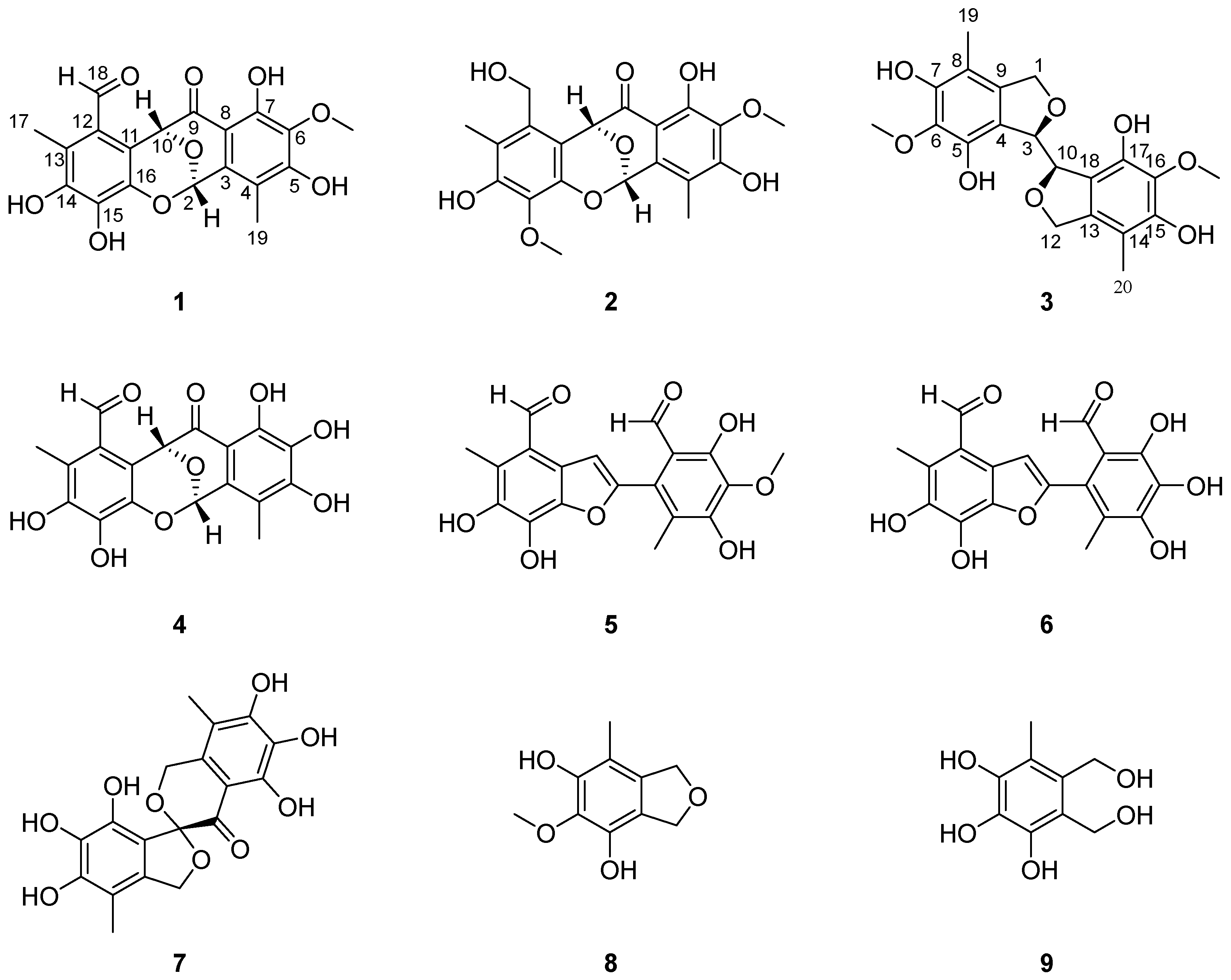
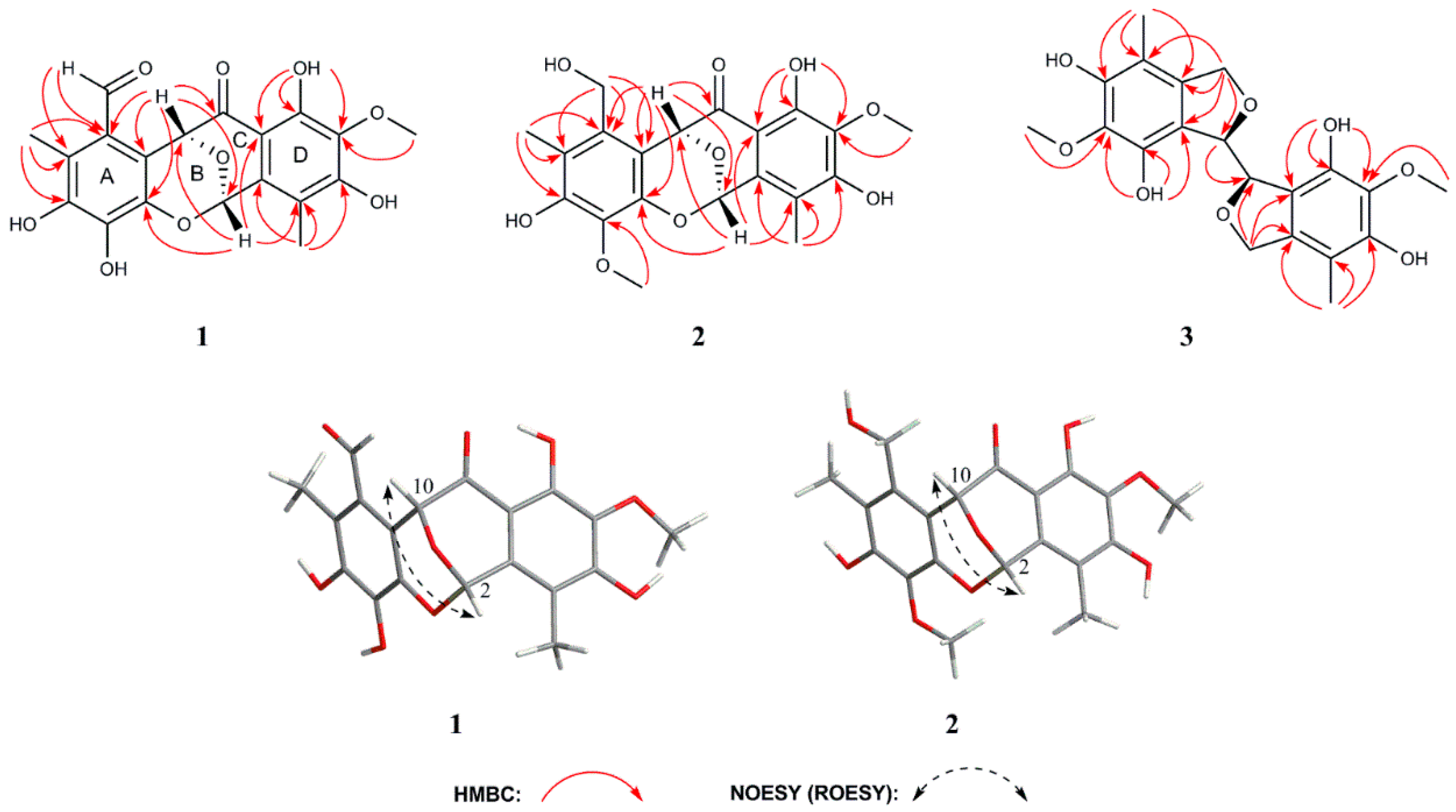
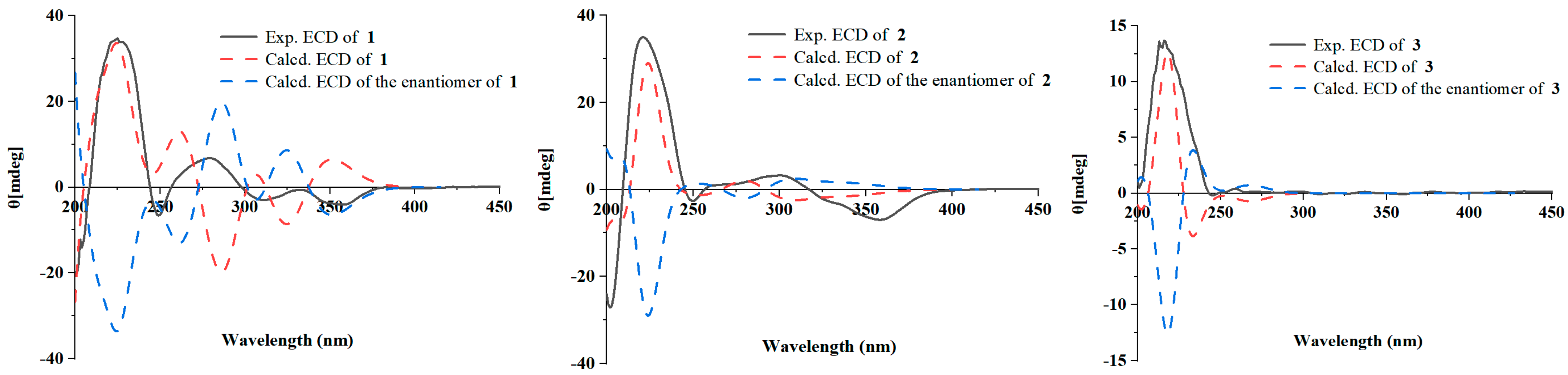
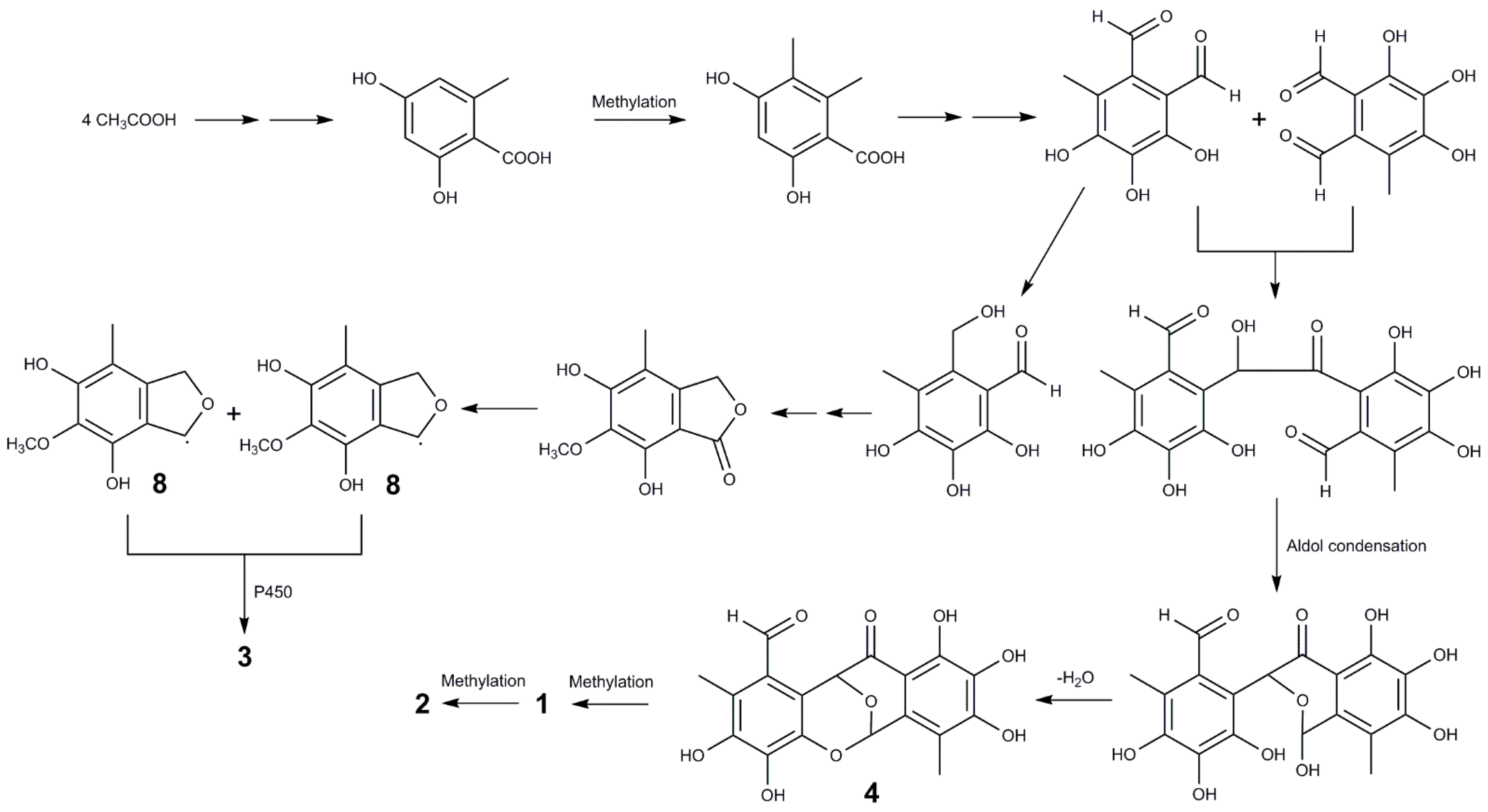
2.1. Structure Elucidation of New Compounds 13

Epicocconigrone C (1) was isolated as a yellow solid, and its molecular formula was determined to be C19H16O9 with 12 degrees of unsaturation by HRESIMS data at m/z 411.0698 (calcd. 411.0687 for C19H16O9Na, [M + Na]+), which was supported by the 13C NMR and DEPT spectral data. The IR spectrum of 1 featured typical absorption bands for hydroxyl (3413 cm−1) and conjugated ketone (1670 cm−1). The 1H NMR spectrum (Table 1) of 1 revealed two methyls (δH 2.26 and δH 2.31), one methoxy (δH 3.70), two oxymethines (δH 6.38 and δH 6.83), one aldehyde proton (δH 10.34), and one hydroxyl proton (δH 11.33). The 13C NMR (Table 2) and DEPT spectra showed 19 well-resolved carbon atom signals, including one ketone carbonyl (δC 196.9), one aldehydic carbonyl (δC 191.2), two oxygenated tertiary carbons (δC 89.8 and δC 68.6), one methoxy carbon (δC 60.2), two methyls (δC 11.8 and δC 10.2), and twelve olefinic quaternary carbons at δC 156.9−104.5, accounting for 8 degrees of unsaturation. Thus, compound 1 was thought to possess a tetracyclic skeleton. The strong heteronuclear multiple-bond correlation (HMBC) correlations from H-17 (δH 2.31) to C-12 (δC 121.7), C-13 (δC 121.9), and C-14 (δC 144.3), from H-18 (δH 10.34) to C-11 (δC 112.6), C-12, C-13, and C-14, as well as the weak signals from H-17 to C-11, C-15 (δC 138.4), and C-16 (δC 135.8) confirmed the existence of ring A (Figure 2). The HMBC correlations from H-19 (δH 2.26) to C-3 (δC 130.8), C-4 (δC 115.8), and C-5 (δC 156.9), as well as the HMBC correlations from 7-OH (δH 11.33) to C-6, C-7 (δC 153.6), and C-8 (δC 104.5) established the substitution of the aromatic ring D. Furthermore, the HMBC correlations from H-2 (δH 6.83) to C-10 (δC 68.6) and C-16, from H-10 (δH 6.38) to C-2 (δC 89.8), C-11, C-12 and C-16 suggested the presence of two oxygen bridges between C-16/C-2 and C-2/C-10 in ring B, which could be confirmed by the low field chemical shift signal of CH-2 (δC 89.8, δH 6.83). Ring C was established by the HMBC correlations from H-2 to C-4 and C-8, and from H-10 to C-8 and C-9 (δC 196.9). The comprehensive NMR analysis indicated that 1 shared the same oxygen-bridged skeleton with epicocconigrone A (4) [24], with the exception that the appearance of 6-OCH3 in 1 replaced 6-OH in 4, which was supported by the HMBC correlation from 6-OCH3 (δH 3.70) to C-6 (δC 134.7). Thus, the planar structure of 1 was elucidated as shown (Figure 1), named epicocconigrone C. In the nuclear Overhauser effect spectroscopy (NOESY) spectrum of 1, the correlation between H-2 and H-10 was indicative of their cis relationship (Figure 2). The absolute configuration of 1 was confirmed by the ECD calculation. Its experimental ECD curve for the absolute configurations of 2S and 10R was consistent with the calculated ECD curve of (2S, 10R) (Figure 3).
Epicocconigrone D (2) was obtained as a yellow solid. The molecular formula of 2 was determined as C20H20O9 with 11 unsaturated degrees by HRESIMS data at m/z 427.1004 (calcd. 427.1000 for C20H20O9Na, [M + Na]+), which was supported by the 13C NMR and DEPT spectral data. The IR spectrum of 2 featured typical absorption bands for hydroxyl (3446 cm−1) and conjugated ketone (1626 cm−1). The 1H NMR spectrum (Table 1) of 2 indicated two methyl groups (δH 2.09 and δH 2.18), two methoxy groups (δH 3.57 and δH 3.69), one methylene (δH 4.32, d, J = 12.1 Hz; 4.81 d, J = 12.1 Hz), two oxymethines (δH 5.65 and δH 6.76), and two hydroxyl protons (δH 8.92 and δH 11.46). The 13C NMR (Table 2) and DEPT spectra revealed 20 carbon atom signals, including one ketone carbonyl (δC 196.8), two oxygenated tertiary carbons (δC 89.9 and δC 70.3), two methoxy carbons (δC 60.3 and δC 60.0), one methylene (δC 55.8), two methyls (δC 11.0 and δC 10.3), and twelve olefinic quaternary carbons. Detailed analysis of 2D NMR spectra of 2 revealed that it had a similar structure to 1. The major differences in 2 were a hydroxymethylene group and a methoxy group substituted at C-12 and C-15, instead of the aldehyde group and the hydroxyl group, respectively, when compared to 1 (Figure 2), which were further confirmed by the HMBC correlations from H2-18 (δH 4.32, 4.81) to C-11 (δC 108.3), C-12 (δC 132.1), and C-13 (δC 118.1), and from 15-OCH3 (δH 3.69) to C-15 (δC 134.6). Thus, the planar structure of 2 was elucidated as shown (Figure 1), named epicocconigrone D. The ROESY correlation between H-2 and H-10 indicated their cis orientation (Figure 2). The absolute configuration of 2 was understood to be 2S, 10R by comparing the experimental and simulated ECD curves (Figure 3).
Flavimycin C (3) was isolated as a white solid. It had a molecular formula of C20H22O8 with 10 degrees of unsaturation, as determined by HRESIMS data at m/z 391.1385 (calcd. 391.1387 for C20H23O8, [M + H]+), which was supported by the 13C NMR and DEPT spectral data. The IR spectrum of 3 featured typical absorption bands for hydroxyl (3449 cm−1). The 1H NMR spectrum (Table 1) of 3 exhibited one methyl (δH 1.88), one methoxy (δH 3.64), one methine (δH 4.30), one methylene (δH 4.54 d, J = 15.0 Hz; 4.65 d, J = 15.0 Hz), and two hydroxyl protons (δH 8.55, δH 8.68). The 13C NMR (Table 2) and DEPT spectra displayed 10 well-resolved carbon atom signals, dividing into six quaternary carbons that were assigned to one benzene ring, one methylene (δC 65.8), one methine (δC 66.1), one methoxy carbon (δC 60.2), and one methyl (δC 9.5). The NMR data of 3 were very similar to those of 8 except for the absence of the methylene signal, and instead, the presence of the methine signal of C-3 (δH 4.30/δC 66.1) in 3. Combined with molecular formula, 3 was deduced to be a symmetrical dimeric derivative. The above data suggested 3 was a symmetrical dimer of 8, connecting at C-3/C-10 between the two units (Figure 2), which was further confirmed by the HMBC correlation from H-3 to C-10. Thus, the planar structure of 3 was confirmed as shown (Figure 1), and named flavimycin C. The 1H and 13C NMR spectra (Table 1 and Table 2) of this aromatic polyketide dimer only exhibited a set of signals of aromatic polyketide monomer. There were three possible absolute configurations of two chiral carbons C-3 and C-10 in 3. The obvious negative optical activity ( α D 20   = −70.0) and the Cotton effect indicated that compound 3 was not a mesomer, which implied the possibility of 3R, 10S was excluded. Consequently, the absolute configurations of C-3 and C-10 were the same (3S, 10S or 3R, 10R). The absolute configuration of 3 was understood to be 3R, 10R by comparing the experimental and simulated ECD curves (Figure 3).
The known compounds: epicocconigrone A (4) [24]; 2-(10-formyl-11,13-dihydroxy-12-methoxy-14-methyl)-6,7-dihydroxy-5-methyl-4-benzofurancarboxaldehyde (5) [25]; epicoccolide B (6) [26]; eleganketal A (7) [27]; 1,3-dihydro-5-methoxy-7-methylisobenzofuran (8) [28]; and 2,3,4-trihydroxy-6-(hydroxymethyl)-5-methylbenzyl-alcohol (9) [29] were identified by comparing their NMR data with those reported in the literature.
The new compounds 13 are all aromatic polyketide dimers, particularly compounds 1 and 2 simultaneously featuring consistent 6/6/6/6 heterotetracyclic ring cores and compounds 13 co-occurrence in the same marine-derived fungus suggest that they should originate from the same biogenetic pathway. A plausible biosynthetic pathway toward the formation of compounds 13 can be proposed by detailed analysis of their structures (Scheme 1).

2.2. In Vitro Evaluation of α-Glucosidase Inhibitory Activity

All compounds were tested for their α-glucosidase inhibitory activities using a reported method [30], with acarbose as the positive control. The results revealed that compounds 1, 47, and 9 showed more potent inhibitory activity (IC50 values ranging from 17.04 ± 0.28 to 292.47 ± 5.87 μM) than acarbose (IC50, 822.97 ± 7.10 μM) (Table 3). The potent α-glucosidase inhibitory activity of epicocconigrone A (4) and epicoccolide B (6) has been already reported [31]. It could be noted herein that the number of hydroxyl groups of polyhydroxy phenolic compounds was important for α-glucosidase inhibitory activity, as reflected by the low IC50 values of compounds 4 and 6, while structures with fewer hydroxyl groups (compounds 1 and 5) exhibited little activity.

3. Materials and Methods

3.1. Fungal Material and Fermentation

The fungal strain A. insulicola was isolated from deep-sea sediments, which were collected from the South China Sea at the depth of 2500 m. After grinding, the sample (1.0 g) was diluted to 10−2 g/mL with sterile H2O, 100 μL of which was spread on potato dextrose agar medium (200.0 g potato, 20.0 g glucose, and 20.0 g agar per liter of seawater) plates containing chloramphenicol as a bacterial inhibitor. It was identified by its morphological characteristics and ITS gene sequences (GenBank accessing No. ON413861), the used primers of which were ITS1 (TCCGTAGGTGAACCTGCGG) and ITS4 (TCCTCCGCTTATTGATATGC). A reference culture of A. insulicola was deposited at the Hainan Provincial Key Laboratory for Functional Components Research and Utilization of Marine Bio-resources, Haikou, China.

3.2. Culture Conditions

The fungal strain A. insulicola was cultured in potato dextrose broth medium (consisting of 200.0 g/L potato, 20.0 g/L glucose, and 1000.0 mL deionized water), and incubated on a rotary shaker (150 rpm) for 72 h at 28 °C. Thereafter, 3 mL of seed broth was transferred to fifty 1000 mL Erlenmeyer flasks containing solid rice medium (each flask contained 80 g rice and 120 mL seawater), used for fermentation. The flasks were incubated under static conditions at room temperature for 30 days.

3.3. General Experimental Procedures

Optical rotation was measured using a Modular Circular Polarimeter 5100 polarimeter (Anton Paar, Austria). The NMR spectra were measured on Bruker Avance 500 NMR spectrometer (Bruker, Bremen, Germany) and Bruker DRX-600 spectrometer (Bruker Biospin AG, Fällanden, Germany) using TMS as an internal standard. HRESIMS were determined with an API QSTAR Pulsar mass spectrometer (Bruker, Bremen, Germany). ECD and UV spectra were recorded on a MOS-500 spectrometer (Biologic, France). IR data were measured on a Nicolet 380 infrared spectrometer (Thermo Electron Corporation, Madison, WI, USA). Analytic HPLC was performed with an Agilent Technologies 1260 Infinity II equipped with an Agilent DAD G1315D detector (Agilent, Palo Alto, CA, USA), the separation columns were (COSMOSIL-packed C18, 5 mm, 4.6 mm × 250 mm). Semi-preparative HPLC was performed on reversed-phased columns (COSMOSIL-packed C18, 5 mm, 10 mm × 250 mm). Silica gel (60–80, 200–300 and 300–400 mesh, Qingdao Marine Chemical Co. Ltd., Qingdao, China) and Sephadex LH-20 (Merck, Germany) were used for column chromatography. TLC was conducted on precoated silica gel GF254 plates (Qingdao Marine Chemical Co. Ltd., Qingdao, China), and spots were detected by spraying with 10% H2SO4 in EtOH followed by heating.

3.4. Extraction and Isolation

After the fermentation of the strain, the cultures were extracted with EtOAc, then filtered with filter paper. After repeating the procedure three times, the EtOAc extract was evaporated under a reduced pressure to obtain a crude extract (124.0 g). The crude extract was dispersed in water and extracted with petroleum ether, ethyl acetate and n-butanol three times, respectively. After vacuum concentration, the petroleum ether extract (11.3 g), ethyl acetate extract (34.0 g) and n-butanol extract (20.0 g) were obtained, respectively. Then, the EtOAc extract (34.0 g) was subjected to silica gel vacuum liquid chromatography using step gradient elution with CHCl3/MeOH (1:0, 200:1, 150:1, 100:1, 80:1, 50:1, 20:1, 10:1, 0:1, v/v) to obtain 13 fractions (Fr.1–Fr.13). Fr.4 (425.0 mg) was applied to Sephadex LH-20 gel chromatography eluted with CHCl3/MeOH (1:1, v/v) to give six subfractions (Fr.4.1–Fr.4.6). Fr.4.3 (150.5 mg) was subjected to silica gel column chromatography (petroleum ether/EtOAc, 10:1, v/v) to afford nine subfractions (Fr.4.3.1–Fr.4.3.9). Fr.4.3.9 (40.5 mg) was separated by semi-preparative HPLC, eluting with 45% MeOH/H2O to yield compound 2 (tR 11.5 min, 4.5 mg), and Fr.4.3.7 (29.8 mg) was separated by semi-preparative HPLC, eluting with 35% MeOH/H2O to give compound 8 (tR 15.3 min, 4.2 mg). Fr.6 (1.15 g) was applied to ODS chromatography eluting with MeOH/H2O (10%–100%) to give thirteen subfractions (Fr.6.1–Fr.6.13). Fr.6.11 (91.5 mg) was subjected to Sephadex LH-20 (eluted with 100% MeOH) and then purified by semi-preparative HPLC (eluted with 48% MeOH/H2O) to give compound 1 (tR 21.9 min, 11.1 mg). Fr.6.12 (58.9 mg) was subjected to Sephadex LH-20 (eluted with 100% MeOH) and then purified by semi-preparative HPLC (eluted with 65% MeOH/H2O) to give compound 5 (tR 10.0 min, 4.2 mg). Fr.6.6 (72.3 mg) was purified on silica gel (petroleum ether/EtOAc, 3:2, v/v) to yield compound 3 (7.5 mg). Fr.9 (10.0 g) was subjected to Sephadex LH-20 gel chromatography eluted with MeOH to give ten subfractions (Fr.9.1–Fr.9.10). Fr.9.7 (2.1 g) was subjected to silica gel column chromatography (CH2Cl2/MeOH, 100:1, v/v), and subsequently purified by semi-preparative HPLC, eluting with 50 % MeOH/H2O to yield compounds 4 (tR 12.0 min, 5.1 mg) and 6 (tR 18.5 min, 2.7 mg). Fr.9.6 (1.67 g) was separated by Sephadex LH-20 column chromatography eluted with MeOH and then purified by silica gel column chromatography eluting with petroleum ether/EtOAc (3:1; v/v) to obtain compound 9 (5.1 mg). Fr.9.8 (0.8 g) was subjected to silica gel column chromatography (CH2Cl2/MeOH, 35:1, v/v), and subsequently purified by semi-preparative HPLC, eluting with 55 % MeOH/H2O to yield compound 7 (tR 6.8 min, 8.1 mg).
Epicocconigrone C (1): Yellow film. α D 20   = +98.0 (c 0.10, MeOH); UV (MeOH) λmax (logε): 237 (4.31) nm; 261 (3.91) nm; 309 (4.27) nm; 359 (4.06) nm; IR (KBr) vmax (cm−1): 3413, 1669, 1466, 1395, 1355, 1296, 1117. 1H and 13C NMR data see Table 1 and Table 2; HRESIMS [M + Na]+ m/z 411.0698 (calcd. for C19H16O9Na, 411.0687).
Epicocconigrone D (2): Yellow film. α D 20   = +57.0 (c 0.10, MeOH); UV (MeOH) λmax (logε): 234 (4.26) nm; 260 (4.04) nm; 309 (4.21) nm; 365 (4.19) nm; IR (KBr) vmax (cm−1): 3446, 2931, 1626, 1469, 1359, 1226, 1154, 1115. 1H and 13C NMR data see Table 1 and Table 2; HRESIMS [M + Na]+ m/z 427.1004 (calcd. for C20H20O9Na, 427.1000).
Flavimycin C (3): White film. α D 20   = −70.0 (c 0.10, MeOH); UV (MeOH) λmax (logε): 232 (4.15) nm; 284 (3.56) nm; IR (KBr) vmax (cm−1): 3449, 2928, 1606, 1478, 1376, 1264, 1110, 1027. 1H and 13C NMR data see Table 1 and Table 2; HRESIMS [M + H]+ m/z 391.1385 (calcd. for C20H23O8, 391.1387).

3.5. ECD Calculation

The conformers of compounds were generated using the Confab [32] program ebbed in the Openbabel 3.1.1 software, and further optimized with xtb at GFN2 level [33]. The conformers with population over 1% were subjected to geometry optimization using the Gaussian 16 package [34] at B3LYP/6-31G(d) level and proceeded to calculation of excitation energies, oscillator strength, and rotatory strength at B3LYP/TZVP level in the polarizable continuum model (PCM, methanol). The ECD spectra were Boltzmann-weighted and generated using SpecDis 1.71 software [35].

3.6. α-Glucosidase Inhibitory Activity

All the assays were carried out under 0.1 M sodium phosphate buffer (PH = 6.8). The samples were dissolved with DMSO and diluted into a series of gradient concentrations (final concentrations of 6.25, 12.5, 25, 50, 100, 200, 400, and 800 μM). The 10 μL sample was mixed with 100 μL α-glucosidase solution (0.2 U/mL, Sigma) and shaken well, then added to a 96-well plate and placed at 37 °C for 15 min. Subsequently, 40 μL of 2.5 mM 4-nitrophenyl-α-D-glucopyranoside was added and further incubated at 37 °C for 15 min. Finally, the OD value of each well was detected at 405 nm wavelength of microplate reader. Acarbose was used as a positive control. The control was prepared by adding DMSO instead of the sample in the same way as the test. The blank was prepared by adding sodium phosphate buffer instead of 4-nitrophenyl-α-D-glucopyranoside using the same method. The percentage inhibition was calculated using the following equation:
% inhibition= [(ODcontrol − ODsample)/(ODcontrol − ODblank)] × 100

4. Conclusions

In summary, two new tetracyclic cores of integrastatins, named epicocconigrones C–D (12), one new dimeric isobenzofuran, named flavimycin C (3), and six known compounds (49) were isolated from fermentation cultures of the deep-sea sediment-derived fungus A. insulicola. The biological evaluation revealed compounds 1, 47, 9 exhibited significant α-glucosidase inhibitory with IC50 values ranging from 17.04 ± 0.28 to 292.47 ± 5.87 μM, among which compound 6 was the most potent α-glucosidase inhibitor, with an IC50 value 48-fold stronger than positive control acarbose. Comparing the structure of compounds 1, 4, 5 and 6 revealed the α-glucosidase inhibitory activity was greatly enhanced after the hydroxyl group replaced the methoxy group, which further confirmed that polyhydroxy phenolic compounds were efficient α-glucosidase inhibitors, and provided a reference value for the synthesis of novel α-glucosidase inhibitors. In conclusion, the study has enriched the structural diversity of phenolic compounds and provided a promising lead toward the development of novel α-glucosidase inhibitors.

Supplementary Materials

The following supporting information can be downloaded at: https://www.mdpi.com/article/10.3390/md21030157/s1, Figures S1–S30: 1D, 2D NMR, MS, UV, and IR spectra of compounds 13.

Author Contributions

Conceptualization, Y.Z. and F.L.; methodology, Y.Z. and F.L.; software, Y.Z., F.L. and H.D.; validation, W.Z., Y.Z., W.C., F.L. and H.D.; formal analysis, Y.Z. and H.W.; investigation, W.Z. and W.C.; resources, Y.Z., F.L. and H.D.; data curation, W.Z. and W.C.; writing—original draft preparation, W.Z., Y.Z., H.W. and F.L.; writing—review and editing, Y.Z., F.L., H.W. and H.C.; visualization, W.Z., Y.Z. and F.L.; supervision, Y.Z., F.L. and H.D.; project administration, Y.Z., F.L. and H.D.; funding acquisition, Y.Z. and F.L. All authors have read and agreed to the published version of the manuscript.

Funding

This work was financially supported by Natural Science Foundation of Hainan (322MS131, 220RC702), National Natural Science Foundation of China (41776093), and Financial Fund of the Ministry of Agriculture and Rural Affairs, P. R. China (NFZX2023).

Institutional Review Board Statement

Not applicable.

Informed Consent Statement

Not applicable.

Data Availability Statement

The authors declare that all relevant data supporting the results of this study are available within the article and its Supplementary Materials file, or from the corresponding authors upon request.

Conflicts of Interest

The authors declare no conflict of interest.

References

  1. Kashtoh, H.; Baek, K.H. Recent updates on phytoconstituent alpha-glucosidase inhibitors: An approach towards the treatment of type two diabetes. Plants 2022, 11, 2722. [Google Scholar] [CrossRef] [PubMed]
  2. Malik, A.; Ardalani, H.; Anam, S.; McNair, L.M.; Kromphardt, K.J.K.; Frandsen, R.J.N.; Franzyk, H.; Staerk, D.; Kongstad, K.T. Antidiabetic xanthones with α-glucosidase inhibitory activities from an endophytic Penicillium canescens. Fitoterapia 2020, 142, 104522. [Google Scholar] [CrossRef]
  3. Du, X.P.; Wang, X.; Yan, X.; Yang, Y.F.; Li, Z.P.; Jiang, Z.D.; Ni, H. Hypoglycaemic effect of all-trans astaxanthin through inhibiting α-glucosidase. J. Funct. Foods 2020, 74, 104168. [Google Scholar] [CrossRef]
  4. Jiang, L.L.; Wang, Z.; Wang, X.Y.; Wang, S.J.; Cao, J.; Liu, Y. Exploring the inhibitory mechanism of piceatannol on α-glucosidase relevant to diabetes mellitus. RSC Adv. 2020, 10, 4529–4537. [Google Scholar] [CrossRef] [PubMed]
  5. Attjioui, M.; Ryan, S.; Ristic, A.K.; Higgins, T.; Goni, O.; Gibney, E.; Tierney, J.; O’Connell, S. Kinetics and mechanism of α-glucosidase inhibition by edible brown algae in the management of type 2 diabetes. P. Nutr. Soc. 2020, 79, E633. [Google Scholar] [CrossRef]
  6. Fallah, Z.; Tajbakhsh, M.; Alikhani, M.; Larijani, B.; Faramarzi, M.A.; Hamedifar, H.; Mohammadi-Khanaposhtani, M.; Mahdavi, M. A review on synthesis, mechanism of action, and structure-activity relationships of 1,2,3-triazole-based α-glucosidase inhibitors as promising anti-diabetic agents. J. Mol. Struct. 2022, 1255, 132469. [Google Scholar] [CrossRef]
  7. Shah, M.; Bashir, S.; Jaan, S.; Nawaz, H.; Nishan, U.; Abbasi, S.W.; Jamal, S.B.; Khan, A.; Afridi, S.G.; Iqbal, A. Computational analysis of plant-derived terpenes as α-glucosidase inhibitors for the discovery of therapeutic agents against type 2 diabetes mellitus. S. Afr. J. Bot. 2021, 143, 462–473. [Google Scholar] [CrossRef]
  8. Zahid, H.F.; Ali, A.; Ranadheera, C.S.; Fang, Z.X.; Dunshea, F.R.; Ajlouni, S. In vitro bioaccessibility of phenolic compounds and alpha-glucosidase inhibition activity in yoghurts enriched with mango peel powder. Food Biosci. 2022, 50, 102011. [Google Scholar] [CrossRef]
  9. Abdelli, I.; Benariba, N.; Adjdir, S.; Fekhikher, Z.; Daoud, I.; Terki, M.; Benramdane, H.; Ghalem, S. In silico evaluation of phenolic compounds as inhibitors of A-amylase and A-glucosidase. J. Biomol. Struct. Dyn. 2020, 39, 816–822. [Google Scholar] [CrossRef]
  10. Machida, S.; Mukai, S.; Kono, R.; Funato, M.; Saito, H.; Uchiyama, T. Synthesis and comparative structure–activity study of carbohydrate-based phenolic compounds as α-glucosidase inhibitors and antioxidants. Molecules 2019, 24, 4340. [Google Scholar] [CrossRef] [Green Version]
  11. Catarino, M.D.; Silva, A.M.S.; Mateus, N.; Cardoso, S.M. Optimization of phlorotannins extraction from fucus vesiculosus and evaluation of their potential to prevent metabolic disorders. Mar. Drugs 2019, 17, 162. [Google Scholar] [CrossRef] [PubMed] [Green Version]
  12. Mateos, R.; Pérez-Correa, J.R.; Domínguez, H. Bioactive properties of marine phenolics. Mar. Drugs 2020, 18, 501. [Google Scholar] [CrossRef] [PubMed]
  13. Zhu, Y.X.; Chen, W.Q.; Kong, L.; Zhou, B.X.; Hua, Y.; Han, Y.; Li, J.J.; Ji, J.; Fu, M.; Liu, W.W.; et al. Optimum conditions of ultrasound-assisted extraction and pharmacological activity study for phenolic compounds of the alga Chondrus ocellatus. J. Food Process. Preserv. 2022, 46, E16400. [Google Scholar] [CrossRef]
  14. Naveen, J.; Baskaran, R.; Baskarana, V. Profiling of bioactives and in vitro evaluation of antioxidant and antidiabetic property of polyphenols of marine algae Padina tetrastromatica. Algal Res. 2021, 55, 102250. [Google Scholar] [CrossRef]
  15. Rengasamy, K.R.R.; Sadeer, N.B.; Zengin, G.; Mahomoodally, M.F.; Cziáky, Z.; Jekőd, J.; Diuzheva, A.; Abdallah, H.H.; Kim, D.H. Biopharmaceutical potential, chemical profile and in silico study of the seagrass–Syringodium isoetifolium (Asch.) Dandy. S. Afr. J. Bot. 2019, 127, 167–175. [Google Scholar] [CrossRef]
  16. Sun, C.X.; Zhang, Z.P.; Ren, Z.L.; Yu, L.; Zhou, H.; Han, Y.X.; Shah, M.; Che, Q.; Zhang, G.J.; Li, D.H.; et al. Antibacterial cyclic tripeptides from Antarctica-sponge-derived fungus Aspergillus insulicola HDN151418. Mar. Drugs 2020, 18, 532. [Google Scholar] [CrossRef]
  17. Wu, Q.X.; Jin, X.J.; Draskovic, M.; Crews, M.S.; Tenney, K.; Valeriote, F.A.; Yao, X.J.; Crews, P. Unraveling the numerous biosynthetic products of the marine sediment-derived fungus, Aspergillus insulicola. Phytochem. Lett. 2012, 5, 114–117. [Google Scholar] [CrossRef] [Green Version]
  18. Wu, Q.X.; Crews, M.S.; Draskovic, M.; Sohn, J.; Johnson, T.A.; Tenney, K.; Valerick, F.A.; Yao, X.J.; Bjeldanes, L.F.; Crerws, P. Azonazine, a novel dipeptide from a Hawaiian marine sediment-derived fungus, Aspergillus insulicola. Org. Lett. 2010, 12, 4458–4461. [Google Scholar] [CrossRef] [Green Version]
  19. Sun, C.X.; Liu, X.Y.; Sun, N.; Zhang, X.M.; Shah, M.; Zhang, G.J.; Che, Q.; Zhu, T.J.; Li, J.; Li, D.H. Cytotoxic nitrobenzoyl sesquiterpenoids from an Antarctica sponge-derived Aspergillus insulicola. J. Nat. Prod. 2022, 85, 987–996. [Google Scholar] [CrossRef]
  20. Zhao, H.Y.; Anbuchezhian, R.; Sun, W.; Shao, C.L.; Zhang, F.L.; Yin, Y.; Yu, Z.S.; Li, Z.Y.; Wang, C.Y. Cytotoxic nitrobenzoyloxy-substituted sesquiterpenes from sponge-derived endozoic fungus Aspergillus insulicola MD10-2. Curr. Pharm. Biotechno. 2016, 17, 271–274. [Google Scholar] [CrossRef]
  21. Wang, S.; Zeng, Y.B.; Yin, J.J.; Chang, W.J.; Zhao, X.L.; Mao, Y. Two new azaphilones from the marine-derived fungus Penicillium sclerotiorum E23Y-1A. Phytochem. Lett. 2022, 47, 76–80. [Google Scholar] [CrossRef]
  22. Wang, Z.; Zeng, Y.B.; Zhao, W.B.; Dai, F.H.; Chang, W.J.; Lv, F. Structures and biological activities of brominated azaphilones produced by Penicillium sclerotiorum E23Y-1A. Phytochem. Lett. 2022, 52, 138–142. [Google Scholar] [CrossRef]
  23. Zeng, Y.B.; Wang, Z.; Chang, W.J.; Zhao, W.B.; Wang, H.; Chen, H.Q.; Dai, F.H.; Lv, F. New azaphilones from the marine-derived fungus Penicillium sclerotiorum E23Y-1A with their anti-inflammatory and antitumor activities. Mar. Drugs 2023, 21, 75. [Google Scholar] [CrossRef]
  24. El Amrani, M.; Lai, D.; Debbab, A.; Aly, A.H.; Siems, K.; Seidel, C.; Schnekenburger, M.; Gaigneaux, A.; Diederich, M.; Feger, D.; et al. Protein Kinase and HDAC Inhibitors from the Endophytic Fungus Epicoccum nigrum. J. Nat. Prod. 2014, 77, 49–56. [Google Scholar] [CrossRef]
  25. Laszlo, V.; Michael, K.; Astrid, M.E.; Luigi, T. 2-Phenyl-benzofuran derivatives, method for the production thereof and their use. D.E. Patent 10,351,315, 2005. [Google Scholar]
  26. Talontsi, F.M.; Dittrich, B.; Schüffler, A.; Sun, H.; Laatsch, H. Epicoccolides: Antimicrobial and antifungal polyketides from an endophytic fungus Epicoccum sp. associated with Theobroma cacao. Eur. J. Org. Chem. 2013, 2013, 3174–3180. [Google Scholar] [CrossRef]
  27. Luan, Y.P.; Wei, H.J.; Zhang, Z.P.; Che, Q.; Liu, Y.K.; Zhu, T.J.; Mándi, A.; Kurtán, T.; Gu, Q.Q.; Li, D.H. Eleganketal A, a highly oxygenated dibenzospiroketal from the marine-derived fungus Spicaria elegans KLA03. J. Nat. Prod. 2014, 77, 1718–1723. [Google Scholar] [CrossRef] [PubMed]
  28. Lee, N.H.; Gloer, J.B.; Wicklow, D.T. Isolation of chromanone and isobenzofuran derivatives from a fungicolous isolate of Epicoccum purpurascens. Bull. Korean Chem. Soc. 2007, 28, 877–879. [Google Scholar] [CrossRef]
  29. Nishihara, Y.; Takase, S.; Tsujii, E.; Hatanaka, H.; Hashimot, S. New anti-influenza agents, FR198248 and its derivatives. II. Characterization of FR198248, its related compounds and some derivatives. J. Antibiot. 2001, 24, 297–303. [Google Scholar] [CrossRef] [PubMed] [Green Version]
  30. Yang, L.; Yang, Y.L.; Dong, W.H.; Li, W.; Wang, P.; Cao, X.; Yuan, J.Z.; Chen, H.Q.; Mei, W.L.; Dai, H.F. Sesquiterpenoids and 2-(2-phenylethyl) chromones respectively acting as α-glucosidase and tyrosinase inhibitors from agarwood of an Aquilaria plant. J. Enzym. Inhib. Med. CH. 2019, 34, 853–862. [Google Scholar] [CrossRef] [PubMed] [Green Version]
  31. Yan, Z.Y.; Huang, C.Y.; Guo, H.X.; Zheng, S.Y.; He, J.R.; Lin, J.; Long, Y.H. Isobenzofuranone monomer and dimer derivatives from the mangrove endophytic fungus Epicoccum nigrum SCNU-F0002 possess α-glucosidase inhibitory and antioxidant activity. Bioorg. Chem. 2020, 94, 103407. [Google Scholar] [CrossRef] [PubMed]
  32. O’Boyle, N.M.; Vandermeersch, T.; Flynn, C.J.; Maguire, A.R.; Hutchison, G.R. Confab-systematic generation of diverse low-energy conformers. J. Cheminform. 2011, 3, 8. [Google Scholar] [CrossRef] [PubMed] [Green Version]
  33. Bannwarth, C.; Ehlert, S.; Grimme, S.J. GFN2-xTB an accurate and broadly parametrized self-consistent tight-binding quantum chemical method with multipole electrostatics and density-dependent dispersion contributions. Chem. Theory Comput. 2019, 15, 1652–1671. [Google Scholar] [CrossRef] [PubMed] [Green Version]
  34. Frisch, M.J.; Trucks, G.W.; Schlegel, H.B.; Scuseria, G.E.; Robb, M.A.; Cheeseman, J.R.; Scalmani, G.; Barone, V.; Petersson, G.A.; Nakatsuji, H.; et al. Gaussian 16, Revision, C.01; Gaussian, Inc.: Wallingford, CT, USA, 2019. [Google Scholar]
  35. Bruhn, T.; Schaumloffel, A.; Hemberger, Y.; Bringmann, G. SpecDis: Quantifying the comparison of calculated and experimental electronic circular dichroism spectra. Chirality 2013, 25, 243–249. [Google Scholar] [CrossRef] [PubMed]
Figure 1. Structures of compounds 19 from Aspergillus insulicola: epicocconigrones C–D (12); flavimycin C (3); epicocconigrone A (4); 2-(10-formyl-11,13-dihydroxy-12-methoxy-14-methyl)-6,7-dihydroxy-5-methyl-4-benzofurancarboxaldehyde (5); epicoccolide B (6); eleganketal A (7); 1,3-dihydro-5-methoxy-7-methylisobenzofuran (8); and 2,3,4-trihydroxy-6-(hydroxymethyl)-5-methylbenzyl-alcohol (9).
Marinedrugs 21 00157 g001
Figure 2. Key HMBC correlations of compounds 13 and key NOESY/ROESY correlations of compounds 12.
Marinedrugs 21 00157 g002
Figure 3. Experimental and calculated ECD spectra of compounds 13.
Marinedrugs 21 00157 g003
Scheme 1. Putative biosynthetic pathways toward the formation of compounds 13.
Marinedrugs 21 00157 sch001
Table 1. 1H NMR data of epicocconigrones C–D (12) and flavimycin C (3) (δ in ppm, J in Hz) in DMSO-d6.
Position1 a2 a3 b
1 4.54 (d, 15.0)
4.65 (d, 15.0)
26.83, s6.76, s
3 4.30, s
106.38, s5.65, s4.30, s
12 4.54 (d, 15.0)
4.65 (d, 15.0)
172.31, s2.09, s
1810.34, s4.81 (d, 12.1)
4.32 (d, 12.1)
192.26, s2.18, s1.88, s
20 1.88, s
6-OCH33.70, s3.57, s3.64, s
14-OCH3 3.64, s
15-OCH3 3.69, s
5-OH 8.55, s
7-OH11.33, s11.46, s8.68, s
14-OH 8.92, s
15-OH 8.68, s
17-OH 8.55, s
a Recorded at 500 MHz; b Recorded at 600 MHz.
Table 2. 13C NMR (125 MHz) data of epicocconigrones C-D (12) and flavimycin C (3) in DMSO-d6.
Position123
1 65.8, CH2
289.8, CH89.9, CH
3130.8, C131.0, C66.1, CH
4115.8, C115.8, C112.2, C
5156.9, C158.9, C147.0, C
6134.7, C134.6, C134.3, C
7153.6, C153.6, C147.8, C
8104.5, C103.4, C109.7, C
9196.9, C196.8, C129.7, C
1068.6, CH70.3, CH66.1, CH
11112.6, C108.3, C
12121.7, C132.1, C65.8, CH2
13121.9, C118.1, C129.7, C
14144.3, C148.5, C109.7, C
15138.4, C134.6, C147.8, C
16135.8, C140.3, C134.3, C
1711.8, CH311.0, CH3147.0, C
18191.2, CH55.8, CH2112.2, C
1910.2, CH310.3, CH39.5, CH3
20 9.5, CH3
6-OCH360.2, CH360.3, CH360.2, CH3
14-OCH3 60.2, CH3
15-OCH3 60.0, CH3
Table 3. α-Glucosidase inhibitory activities of compounds 19.
CompoundsIC50 ± SD (μM) a
1292.47 ± 5.87
2
3
425.69 ± 0.30
540.07 ± 4.64
617.04 ± 0.28
749.53 ± 2.45
8
9130.63 ± 2.87
Acarbose b822.97 ± 7.10
a Values represent means ± SD based on three parallel experiments. b Positive control.–no activity at a concentration of 200 μM.
Disclaimer/Publisher’s Note: The statements, opinions and data contained in all publications are solely those of the individual author(s) and contributor(s) and not of MDPI and/or the editor(s). MDPI and/or the editor(s) disclaim responsibility for any injury to people or property resulting from any ideas, methods, instructions or products referred to in the content.

