Article

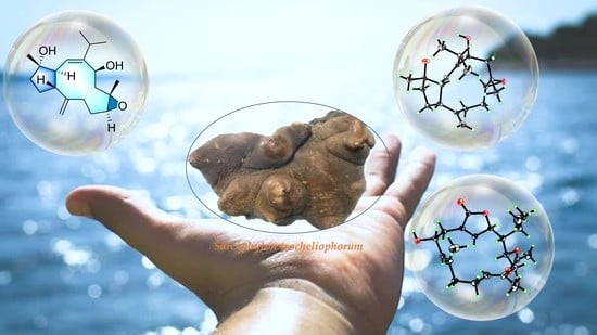
Structurally Diverse Diterpenes from the South China Sea Soft Coral Sarcophyton trocheliophorum

1
State Key Laboratory of Drug Research, Shanghai Institute of Materia Medica, Chinese Academy of Sciences, 555 Zu Chong Zhi Road, Zhangjiang Hi-Tech Park, Shanghai 201203, China
2
College of Life Sciences, Shanghai Normal University, 100 Guilin Road, Shanghai 200234, China
3
College of Materials Science and Engineering, Central South University of Forestry and Technology, 498 South Shaoshan Road, Changsha 410004, China
4
Shandong Laboratory of Yantai Drug Discovery, Bohai rim Advanced Research Institute for Drug Discovery, Yantai 264117, China
5
Key Laboratory of Zhanjiang for Research and Development Marine Microbial Resources in the Beibu Guif Rim, Marine Biomedical Research Institute, Guangdong Medical University, Zhanjiang 524023, China
*
Authors to whom correspondence should be addressed.
Mar. Drugs 2023, 21(2), 69; https://doi.org/10.3390/md21020069
Submission received: 15 December 2022 / Revised: 17 January 2023 / Accepted: 17 January 2023 / Published: 20 January 2023

Abstract

:
The present investigation of the South China Sea soft coral Sarcophyton trocheliophorum resulted in the discovery of six new polyoxygenated diterpenes, namely sartrocheliols A–E (1, 3, 58) along with four known ones, 2, 4, 9, and 10. Based on extensive spectroscopic data analysis, sartrocheliol A (1) was identified as an uncommon capnosane diterpene, while sartrocheliols B–E (3, 58) were established as cembrane diterpenes. They displayed diverse structural features not only at the distinctly different carbon frameworks but also at the various types of heterocycles, including the epoxide, γ-lactone, furan, and pyran rings. Moreover, their absolute configurations were determined by a combination of quantum mechanical-nuclear magnetic resonance (QM-NMR) approach, modified Mosher’s method, and X-ray diffraction analysis. In the anti-tumor bioassay, compound 4 exhibited moderate cytotoxic activities against A549, H1975, MDA-MB-231, and H1299 cells with the IC50 values ranging from 26.3 to 47.9 μM.

Graphical Abstract

1. Introduction

A fairly large variety of bioactive secondary metabolites have been found in the soft corals of the genus Sarcophyton [1,2,3,4]. Over the past few decades, about 20 Sarcophyton species have been chemically investigated and more than 500 secondary metabolites have been identified. Based on their structural types, these metabolites could be classified into terpenes [2], steroids [3,5,6,7], quinones [8,9], prostaglandins [10,11], ceramides [12,13], and other miscellaneous compounds [8,14,15]. As reported in the literature, terpenes have dominated the chemical profile of the genus Sarcophyton [2,16]. Moreover, terpenes display rich structural diversities, which could be further categorized as sesquiterpenes [17,18,19], diterpenes [20,21,22,23,24], and biscembranoids [21,25,26,27,28]. Moreover, these metabolites exhibit a wide spectrum of biological activities, including anti-angiogenic [17], antimicrobial [27,29], cytotoxic [29,30], anti-inflammatory [27,31], immunomodulatory [25], antifouling [8,32], and neuroprotective [33,34] effects. The intriguing chemical and biological properties of terpenes have led to extensive attention from global researchers [35].
In the last decades, we systematically carried out chemical and biological studies on South China Sea marine fauna and flora [36]. As a result, numerous terpenes with complex structures, some of which possessed unprecedented carbon frameworks, were found from the genus Sarcophyton [20,37,38]. During our continuous research, we frequently encountered the soft coral Sarcophyton trocheliophorum, one productive species belonging to the above-mentioned genus. Previously, our group disclosed a vast array of diterpenes with four skeletons, and these metabolites showed a broad spectrum of pharmacological activities such as protein tyrosine phosphatase 1B (PTP1B) inhibitory, antitumor, antibacterial and neuroprotective activities [39]. It was interesting to notice that the chemical profiles of diterpenes from the title soft corals varied upon temporal variations and geographical distributions. The samples collected in Yalong Bay, Hainan Island, South China Sea in February 2006 yielded diterpenes with an unprecedented carbon framework [37] together with sarsolenane, capnosane, and cembrane skeletons [39]. However, the specimen collected in the same water but in May 2006 only yielded cembrane diterpenes [40]. Of more interest, the soft corals collected from another different region, Ximao Island, South China Sea, merely afforded the cembranoids [41]. These findings probably reflected the existence of different metabolic processes in different seasons and inhabiting environments, which needs to be further investigated.
In order to obtain more evidence to disclose that the diterpenoid profile of the title animals was influenced by temporal variations, we made a new collection of S. trocheliophorum from Ximao Island. In the current study, six new polyoxygenated diterpenes, namely sartrocheliols A–E (1, 3, 58), and four known related analogs 2, 4, 9, and 10 were obtained (Figure 1). These diterpenes displayed two distinctly different carbon frameworks: capnosane and cembrane, the former of which has rarely been found in soft corals. Meanwhile, various types of heterocycles, including the epoxide, γ-lactone, furan, and pyran rings were incorporated in their macrocyclic skeletons. Herein, we report the detailed structural elucidation of these isolates from the title soft corals, especially the challenging stereochemistry determination of the new compounds, which was dissolved by a combination of quantum mechanical-nuclear magnetic resonance (QM-NMR) approach, modified Mosher’s method, and X-ray diffraction analysis. In addition, their biological evaluations including cytotoxicity against a panel of cancel cells and antibacterial against an array of bacteria are described.

2. Results and Discussion

The frozen animals were cut into pieces and extracted exhaustively with acetone. Then an Et2O-soluble portion of the acetone extract was repeatedly chromatographed over silica gel, Sephadex LH-20, and RP-HPLC to yield six new compounds 1, 3, 58, and four known analogs 2, 4, 9, and 10 (Figure 1). The known compounds were readily identified as sarcophyolide D (2) [42], sarcophytonolide H (4) [43], sarcophytrol O (9) [44], sinulaflexiolide I (10) [45], respectively, based on the comparison of their NMR spectral and specific optical data with those reported in the literature. In our previous work [43], the absolute configuration of sarcophytonolide H (4) was established by the modified Mosher’s method. In the present study, its crystals were obtained, which were suitable for X-ray diffraction experiment with Cu Kα (λ = 1.54178 Å) radiation. The X-ray diffraction analysis allowed the assignment of the absolute configuration of 4 as 1R,2R,6R,14S (Flack parameter: –0.03 (9)) (Figure 2, Table S2, CCDC 2205721), which was consistent with our previous study.
Sartrocheliol A (1) was obtained as optically active colorless crystals. Its molecular formula C20H32O3 was deduced from the protonated molecule peak at m/z 343.2243 ([M + Na]+, calcd. for C20H32O3Na, 343.2244) in the HRESIMS spectrum, implying five degrees of unsaturation. The IR spectrum indicated the presence of hydroxyl (νmax 3441 cm−1) and olefinic (νmax 1660, 910 cm−1) groups. The 1H and 13C NMR spectra displayed the signals of a trisubstituted double bond (δH 5.14 (1H, d, J = 12.6 Hz, H-2), δC 149.90 (qC, C-1), 125.85 (CH, C-2)), a terminal double bond (δH 4.88 (1H, s, H-19a), 4.69 (1H, s, H-19b), δC 148.54 (qC, C-8), 112.26 (CH2, C-19)), an epoxide (δH 2.89 (1H, dd, J = 10.6, 2.9 Hz, H-11), δC 62.28 (CH, C-11), 59.06 (qC, C-12)), an oxygenated methine (δH 4.88 (1H, dd, J = 11.2, 5.1 Hz, H-14), δC 69.34 (CH, C-14)), and an oxygenated carbon (δC 81.64 (C, C-4)) (Table 1 and Table 2). As revealed by the 1H and 13C NMR data, there were two double bonds and one epoxide, accounting for three degrees of unsaturation. The remaining two degrees of unsaturation were due to the presence of two rings in the molecule. Considering the co-isolated secondary metabolite sarcophyolide D (2) [42], compound 1 was likely a capnosane-type diterpene. Indeed, the NMR data of 1 was almost identical to those of 2, except for the chemical shift of C-11 (δC 62.28 for 1 vs. δC 59.54 for 2). The interpretation of 1H–1H COSY and HMBC spectra (Figure 3) indicated they shared the same gross structure. Analysis of the NOESY spectrum (Figure S6) revealed that there was lack of correlation between H-11 (δH 2.89) and H3-20 (δH 1.12), which revealed the trans-orientation of H-11 and H3-20 in 1, the orientation of which was different from that of the isolate 2. In order to check the proposed structure as well as establish the absolute configuration of 1, a suitable single crystal was obtained in MeOH after many attempts. A successful performance of X-ray crystallography study using Cu Kα (λ = 1.54178 Å) radiation firmly confirmed the structure of 1 and determined its absolute configuration ambiguously as 3S,4S,7R,11S,12S,14S (Flack parameter: 0.05 (6)) (Figure 2, Table S1, CCDC 2196207). Hereto, the structure of 1 was established, as shown in Figure 1.
Sartrocheliol B (3) was isolated as optically active colorless oil. Its molecular formula C22H34O5 was established by the HRESIMS protonated molecule peak at m/z 379.2482 ([M + H]+, calcd. for C22H35O5, 379.2479), which was indicative of six degrees of unsaturation. The IR spectrum of 3 suggested the presence of lactone (νmax 1759 cm−1), ester (νmax 1736 cm−1) and hydroxyl (νmax 3442 cm−1) groups, while a strong UV absorption at λmax 231 nm (logε 3.83) suggested the presence of α,β-unsaturated γ-lactone group [43]. According to the 1H and 13C NMR data of 3 (Table 1 and Table 2), five degrees of unsaturation were attributed to one α,β-unsaturated γ-lactone group (δH 4.95 (1H, d, J = 10.7 Hz, H-2), 7.44 (1H, t, J = 1.8 Hz, H-3), δC 81.19 (CH, C-2), 151.06 (CH, C-3), 131.39 (qC, C-4), 172.97 (qC, C-18)), one trisubstituted double bond (δH 5.05 (1H, t, J = 7.7 Hz, H-11), δC 129.20 (CH, C-11), 130.80 (qC, C-12)) and one acetyl group (δH 2.09 (3H, s), δC 21.35 (CH3), 171.13 (qC)). The remaining one degree of unsaturation implied the monocyclic nature of this molecule. Considering the co-isolated cembranoids 4, 9 and 10, metabolite 3 was likely a cembrane-type diterpene. Through detailed literature reviews on diterpenes from the genus Sarcophyton, the above-mentioned structural features were reminiscent of previously reported sarcophytonolide H (11) [43], a cembrane from South China Sea soft coral Sarcophyton latum. Our interpretation of the 2D NMR spectra (Figure 3) suggested they differed by the lack of the double bond Δ7, which was supported by the significantly up-field shifted chemical shifts of C-7 and C-8 (ΔδC ca. 80 and 110 ppm, respectively). Similar patterns of NOE correlations in the NOESY spectra of 3 and 11, especially for the key NOE cross-peaks of H-2 (δH 4.95)/H-14 (δH 5.02), H-2/H3-16 (δH 1.11), H-3 (δH 7.44)/H-5 (δH 2.45), and H-11 (δH 5.05)/H-13 (δH 2.20), indicated they shared the same configurations for the olefinic bonds Δ3 and Δ11 and the chiral centers C-1, C-2, and C-14 (Figure 4). The clear NOE correlation of H-6 (δH 4.22)/H3-19 (δH 0.77) suggested that H-6 and H3-19 possessed the same orientation. Due to the lack of NOE correlations between the two sets: a. H-6 and H3-19 and b. H-1, H-,2 and H-14, the absolute configurations of these chiral centers corresponding to the above-mentioned two sets of protons were determined by the modified Mosher’s method and CD spectrum, respectively. Treatment of 3 with (R)- and (S)-α-methoxy-α-trifluoromethylphenyl acetyl chlorides (MTPA-Cl) in dry pyridine successfully afforded the (S)- and (R)-MTPA ester derivatives 3s and 3r, respectively. The distribution pattern of observed ΔδH(S-R) values (Figure 5) established the absolute configuration S for C-6 in 3. Considering the correlation between H-6 and H3-19, the absolute configuration of C-8 could be determined as S. It might be worth pointing out that the absolute configuration of sarcophytonolide H (4) had already been ambiguously confirmed as 1R,2R,6R,14S by the single crystal X-ray diffraction experiment in this study (Figure 2). The CD spectrum of 3 displayed the Cotton effect resembling that of co-isolated 4 (Figure 6), suggesting they shared the same absolute configuration R for the chrial carbon C-2 of the chromophore α,β-unsaturated γ-lactone. With the relationships of H-1, H-2, and H-14 in hand, the absolute configurations of C-1 and C-14 could be assigned as R and S, respectively. Consequently, the absolute configuration of 3 could be established as 1R,2R,6S,8S,14S.
The protonated molecule peak at m/z 343.2237 ([M + Na]+, calcd. for C20H32O3Na, 343.2244) displayed in the HRESIMS spectrum of sartrocheliol C (5) revealed that compound 5 had the molecular formula C20H32O3, demonstrating the presence of an additional oxygen atom with respect to that of a dihydrofuran cembranoid 12 [46]. The IR spectrum of 5 showed the presence of olefinic (νmax 3726 cm−1) and hydroxyl (νmax 3441 cm−1) groups. A careful analysis of its NMR spectra revealed that the NMR spectroscopic features of 5 (Table 1 and Table 2) highly resembled those of 12. In fact, the main difference between compounds 5 and 12 was that the CH2-13 in 12 was hydroxylated in 5. The presence of a hydroxyl group at C-13 was supported by the dramatically down-field shifted carbon chemical shift (ΔδC 56 ppm), and further confirmed by the diagnostic HMBC correlations of from H-11 (δH 5.32) to C-13 (δC 79.67) and from H3-20 (δH 1.56) to C-13 (Figure 3). The cross-peak of H3-18 (δH 1.00)/H-2 (δH 5.51) observed in the NOESY spectrum of 5 (Figure 4) together with the small coupling constant (1.4 Hz) between H-2 and H-3 (δH 4.66) suggested the same orientation of H3-18 and H-3. Moreover, the coupling constant (4.6 Hz) between H-13 (δH 4.29) and H-14 (δH 4.84) indicated these two protons were cis-orientated. Whereas the lack of the NOE correlation between H-3 and H-14 revealed the trans-orientation of these two protons of the 2,5-dihydrofuran ring. Thus, the relative configuration of 5 could be assigned as 3R*,4R*,13S*,14S* based on the extensive analysis of the NOESY spectrum. Meanwhile, the relative configuration of 5 could also be elucidated via QM-NMR protocol by using the DP4+ method, which has become one of the most popular and reliable methods to find the most likely structure from a set of putative candidates [34,47]. Four possible isomers (2R*,3R*,13R*,14S*)-5a, (2R*,3R*,13S*,14S*)-5b, (2R*,3S*,13R*,14S*)-5c and (2R*,3S*,13S*,14R*)-5d (Table S3) were subjected to QM-NMR calculations. As a result, the experimental NMR data of compound 5 gave the best match for 5b, with 100% probability (Figure 7, Table S4). Herein, the assigned relative configuration 3R*,4R*,13S*,14S* was consistent with the observations deduced from the NOESY spectrum. As there was a secondary hydroxyl group at C-13, the modified Mosher’s method was applied. The resulting distribution pattern of observed ΔδH(S-R) values (Figure 5) established the absolute configuration R for C-13 in 5. Subsequently, the absolute configuration of 5 could be assigned as 3S,4S,13R,14R.
The HRESIMS spectrum of sartrocheliol D (6) displayed a protonated molecule peak at m/z 403.2459 ([M + Na]+, calcd. for C22H36O5Na, 403.2455), suggesting that 6 possessed the molecular formula C22H36O5. Thus, five degrees of unsaturation were determined for 6. The NMR data (Table 1 and Table 2) revealed the presence of two trisubstituted double bonds (δH 5.42 (1H, m, H-2), δC 146.95 (qC, C-1), 121.34 (CH, C-2); δH 5.24 (1H, t, J = 6.3 Hz, H-7), δC 127.40 (CH, C-7), 134.03 (qC, C-8)), one acetyl group (δH 2.08 (3H, s), δC 21.75 (CH3), 170.47 (qC)), two oxygenated methines (δH 4.40 (1H, br s, H-3), 5.04 (1H, d, J = 10.3 Hz, H-11), δC 72.70 (CH, C-11), 73.16 (CH, C-3)), and three oxygenated quaternary carbons (δC 75.86 (qC, C-4), 74.15 (qC, C-12), 74.60 (qC, C-15)), which accounted for three degrees of unsaturation. The remaining two degrees of unsaturation strongly indicated one macrocyclic carbon skeleton fused with an oxacycle. Careful analysis of the 2D NMR spectrum (Figure 3) of 6 revealed this compound had almost the same gross bicyclic framework of sarcophytrol R (13) [44] except the hydroxyl group at C-11 in 13 was acetylated in 6. Due to the acetylation, the chemical shifts of H-11 and C-11 shifted down-field (ΔδH 1.5 ppm, ΔδC 3.2 ppm, respectively). It might be worth pointing out that the lists of 1H and 13C NMR data of sarcophytrols R and S in the reference [44] were exchanged inadvertently by the authors. The high similarity of the 1H and 13C NMR data as well as similar patterns of NOE correlations of compounds 6 and 13 suggested they shared the same relative configuration 3S*,4R*,11S*,12R*. With the relative configuration in hand, we tried to use the Mosher’s method to establish the absolute configuration of this compound. However, to our disappointment, the left compound after bioassay was degraded although kept in a fridge.
Sartrocheliol E (7) was isolated as colorless oil, and its molecular formula was assigned as C20H34O2 by protonated molecule peak at m/z 329.2448 ([M + Na]+, calcd. for C20H34O2Na, 329.2451) in the HRESIMS spectrum. Its 13C NMR data (Table 2), in combination with the DEPT and HSQC spectra, allowed the identification of 20 carbon resonances, involving six olefinic carbons (δC 141.04, 132.94, 130.05, 128.47, 127.31, 122.10), two oxygenated carbons (δC 74.20, 68.82), and five methyl carbons (δC 29.17, 21.85, 21.15, 15.52, 15.09). The presence of six olefinic carbons was attributed to three double bonds, which accounted for three degrees of unsaturation, indicating that 7 had a monocyclic carbon framework. In fact, the NMR spectroscopic characters of 7 were reminiscent of those of cembrendiol (14) [48]. Careful comparison of their NMR data disclosed their structures differed at the position of the secondary hydroxyl substituent. The secondary hydroxyl group substituted at C-10 in 7 was supported by the consecutive proton system extending from H2-9 to H2-11 through H-10 in the 1H−1H COSY spectrum (Figure 3). Due to the lack of the evidence regarding the orientations of H-10 and H3-18, it was hard to figure out the relative configurations of C-4 and C-10. In order to solve this problem, the QM-NMR method was applied (Table S5). By means of this approach, the experimentally observed NMR data for compound 7 gave the best match with the 1S*,4R*,10S* isomer (>90% probability) (Figure 8, Table S6). Thus, the relative configuration of compound 7 was determined as 1S*,4R*,10S*. Unfortunately, the application of Mosher’s reaction failed, probably due to the insufficient amounts left after an array of bioassays.
The protonated molecule peak at m/z 403.2446 ([M + Na]+, calcd. for C22H36O5Na, 403.2455) in the HRESIMS spectrum of sartrocheliol F (8) suggested compound 8 and co-isolate 9 [44] had the same molecular formula C22H36O5. Moreover, the NMR data of 8 (Table 1 and Table 2) highly resembled those of 9, implying that they shared the same gross structure. The distinct difference was found as the chemical shift of C-4, which was δC 72.47 ppm in 8 whereas δC 74.49 ppm in 9, disclosing the reverse configuration of the hydroxyl group at C-4. This reversion was further deduced from the NOE interactions between H3-18 and H-2 and between H-2 and H3-16 (Figure 4). As the absolute configuration of the deacetylation derivative of 9 was determined as 1S,4S,11S,12R by the Mosher’s method [44], the absolute configuration of 8 was consequently established as 1S,4R,11S,12R.
Although chemical investigations of the soft coral S. trocheliophorum have been well documented in the literature, the present study of this species collected from Ximao Island provided further intriguing results. In the current study, six new cembranoids diterpenoids, sartrocheliols A–E (1, 3, 58), along with four known related ones (2, 4, 9, and 10) were obtained. Among them, compound 1 was a capnosane diterpene, while others were cembrane diterpenes. They displayed diverse structural features not only at the distinctly different carbon frameworks but also at the various types of heterocycles, including the epoxide, γ-lactone, furan, and pyran rings. Compared with the previous research of the Ximao Island specimen [41], the major difference was the discovery of a capnosane diterpene in this study. This observation further indicated the possible impact of temporal variations on the different metabolic processes for the title soft corals.
The anti-tumor effects of all the ten secondary metabolites were evaluated against a list of tumor cells including A549 (human lung cancer cell), H1975 (human lung adenocarcinoma cell), MDA-MB-231 (human breast cancer cell) and H1299 (human non-small cell lung cancer cell). The results showed compound 4 displayed moderate cytotoxic activities against these four cells with the IC50 values of 47.9, 26.3, 44.7, 33.1 μM, respectively, while 6 only showed moderate cytotoxicity against H1975 (IC50 = 40.4 μM). Further, we also conducted molecular interaction experiments on all the compounds, looking for compounds that have the potential to bind to BRD4 and ROR1 anti-tumor targets, respectively. Unfortunately, we did not obtain satisfactory results. In addition, these compounds were tested for their antibacterial activities against a vast array of bacteria including the human pathogens Staphylococcus aureus ATCC27154, Enterococcus faecium, Escherichia coli ATCC25922, Enterobacter cloacae ZR042, Enterobacter hormaechei 2R043, Pseudomonas aeruginosa ATCC10145, methicillin-resistant Staphylococcus aureus (MRSA), and Candida albicans ATCC76485 and the marine strains Streptococcus parauberis KSP28, Streptococcus parauberis SPOF3K, Lactococcus garvieae MP5245, Aeromonas salmonicida AS42, Phoyobacterium damselae FP2244, Pseudomonas fulva ZXM181, Photobacterium halotolerans LMG22194T. To our disappointment, all of them were judged as inactive. Other bioassays such as neuroprotective and anti-inflammatory are currently on the way.

3. Materials and Methods

3.1. Subsection

Melting points were measured on an X-4 digital micromelting point apparatus. The X-ray measurements were made on a Bruker D8 Venture X-ray diffractometer with Cu Kα radiation (Bruker Biospin AG, Fällanden, Germany). IR spectra were recorded on a Nicolet iS50 spectrometer (Thermo Fisher Scientific, Madison, WI, USA). Optical rotations were measured on a PerkinElmer 241MC polarimeter (PerkinElmer, Fremont, CA, USA). CD & UV spectra were measured on a JASCO J-810 instrument (JASCO Corporation, Tokyo, Japan). 1H and 13C NMR spectra were acquired on a Bruker AVANCE III 400 and 600 MHz spectrometer (Bruker Biospin AG, Fällanden, Germany). Chemical shifts were reported with the residual CHCl3 (δH 7.26; δC 77.16) as the internal standard for 1H and 13C NMR spectra. The LREIMS and HREIMS data were recorded on a Finnigan-MAT-95 mass spectrometer (Finnigan-MAT, San Jose, CA, USA). HRESIMS spectra were recorded on Agilent G6250 Q-TOF (Agilent, Santa Clara, CA, USA). Commercial silica gel (Qingdao Haiyang Chemical Co., Ltd., Qingdao, China, 200–300 mesh, 300–400 mesh) was used for column chromatography, and precoated silica gel GF254 plates (Sinopharm Chemical Reagent Co., Shanghai, China) were used for analytical TLC. Sephadex LH-20 (Pharmacia, Piscataway, NJ, USA) was also used for column chromatography. Reversed-phase (RP) HPLC was performed on an Agilent 1260 series liquid chromatography equipped with a DAD G1315D detector at 210 nm (Agilent, Santa Clara, CA, USA). An Agilent semi-preparative XDB-C18 column (5 μm, 250 × 9.4 mm) was employed for the purification. All solvents used for column chromatography and HPLC were of analytical grade (Shanghai Chemical Reagents Co., Ltd., Shanghai, China) and chromatographic grade (Dikma Technologies Inc., Foothill Ranch, CA, USA), respectively.

3.2. Animal Material

The soft coral Sarcophyton trocheliophorum was collected by scuba at a depth of 15 m in May 2018 in Ximao Island, Hainan Province, China. The animal material was identified by Prof. Xiu-Bao Li from Hainan University. A voucher specimen (No. 18XD-19) is available for inspection at the Shanghai Institute of Materia Medica, CAS.

3.3. Extraction and Isolation

The frozen animals (551 g, dry weight) were cut into pieces and extracted exhaustively with acetone at room temperature (3 × 3 L, 30 min in ultrasonic bath). The organic extract was evaporated to give a brown residue (80 g), which was partitioned between Et2O and H2O. The Et2O solution was concentrated under reduced pressure to give a dark brown residue (55.3 g), which was fractionated by gradient silica gel (200–300 mesh) column chromatography (0 → 100% Et2O in petroleum ether (PE)), yielding seven fractions (A–G). Fractions E and F were subjected to a column of Sephadex LH-20 eluted with CH2Cl2 and PE/CH2Cl2/MeOH (2:1:1) to remove the fatty acids and give ten subfractions (EA–EF and FA–FF), respectively. The subfraction EE was purified by semi-preparative HPLC (60% → 100% MeCN in 20 min, 2.5 mL/min), yielding compounds 5 (3.6 mg; tR 10.0 min) and 10 (1.5 mg; tR 12.0 min). FCA-FCC was got from subfraction FC by silica gel column chromatography (300—400 mesh, PE/Et2O (100:1 → 70:1)). The subfraction FCA afforded compounds 2 (1.0 mg; tR 7.1 min), 6 (0.9 mg; tR 17.0 min), 8 (0.9 mg; tR 18.9 min) and 9 (2.0 mg; tR 12.0 min) through semi-preparative HPLC (70% MeCN, 2.5 mL/min). While subfraction FCB gave compounds 1 (1.3 mg; tR 7.0 min) and 7 (1.6 mg; tR 16.0 min), through semi-preparative HPLC (70% MeCN, 2.5 mL/min) as well. The subfraction FD afforded compounds 3 (3.2 mg; tR 6.2 min) and 4 (8.0 mg; tR 5.5 min) through semi-preparative HPLC (70% MeCN, 2.5 mL/min).

3.4. Spectroscopic Data of Compounds

Sartrocheliol A (1): colorless crystals; [α ] D 20 +250.0 (c 0.05, MeOH); IR (KBr) νmax: 3441, 2925, 1384 cm−1; 1H and 13C NMR data, see Table 1 and Table 2; HRESIMS m/z 343.2243 [M + Na]+ (calcd. for C20H32NaO3, 343.2244).
Sartrocheliol B (3): colorless oil; [α ] D 20 +12.7 (c 0.05, MeOH); UV (MeOH) λmax (log ε) 240 (3.23) nm; CD (MeOH) λmax (∆ε) 240 (+4.06), 285 (−1.92) nm; IR (KBr) νmax: 3442, 2932, 1759, 1736, 1234, 1047, 1023 cm−1; 1H and 13C NMR data, see Table 1 and Table 2; HRESIMS m/z 379.2479 [M + H]+ (calcd. for C22H34O5, 379.2482).
Sartrocheliol C (5): colorless oil; [α ] D 20 -12.7 (c 0.1, MeOH); IR (KBr) νmax: 3726, 3624, 3441, 2960, 1384, 1087, 1032 cm−1; 1H and 13C NMR data, see Table 1 and Table 2; HRESIMS m/z 343.2237 [M + Na]+ (calcd. for C20H32NaO3, 343.2244).
Sartrocheliol D (6): colorless oil; [α ] D 20 -20.0 (c 0.05, MeOH); IR (KBr) νmax: 3443, 2927, 1384, 1038 cm−1; 1H and 13C NMR data, see Table 1 and Table 2; HRESIMS m/z 403.2459 [M + Na]+ (calcd. for C22H36NaO5, 403.2455).
Sartrocheliol E (7): colorless oil; [α ] D 20 +120.0 (c 0.01, MeOH); IR (KBr) νmax: 3446, 2922, 1384, 1142, 1044 cm−1; 1H and 13C NMR data, see Table 1 and Table 2; HR-ESIMS m/z 329.2448 [M + Na]+ (calcd. for C20H34NaO, 329.2451).
Sartrocheliol F (8): colorless oil; [α ] D 20 +40.0 (c 0.05, MeOH); IR (KBr) νmax: 3442, 1384 cm−1; 1H and 13C NMR data, see Table 1 and Table 2; HR-ESIMS m/z 403.2446 [M + Na]+ (calcd. for C22H36NaO5, 403.2455).

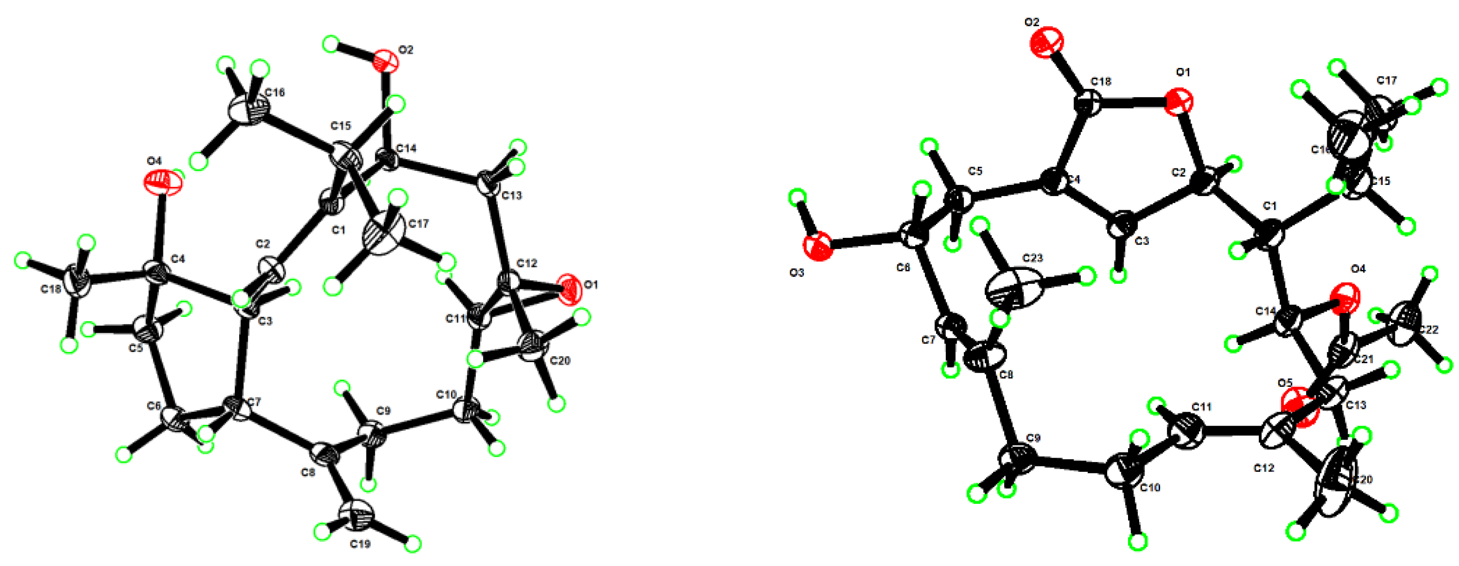
3.5. X-ray Crystallographic Analysis for Compounds 1 and 4

The crystals of 1 and 4 were both recrystallized from methanol at 4 °C. X-ray analysis of 1 and 4 were carried out on a Bruker D8 Venture diffractometer with Cu Kα radiation (λ = 1.54178 Å) at 170 K, respectively. The acquisition parameters for 1 and 4 are provided in the Supplementary Materials, and crystallographic data for compounds 1 and 4 (deposition no. CCDC 2,196,207 and CCDC 2205721) have been deposited at the Cambridge Crystallographic Data Center. Copies of the data can be obtained free of charge via www.ccdc.cam.ac.uk/conts/retrieving.html (accessed on 11 August 2022).

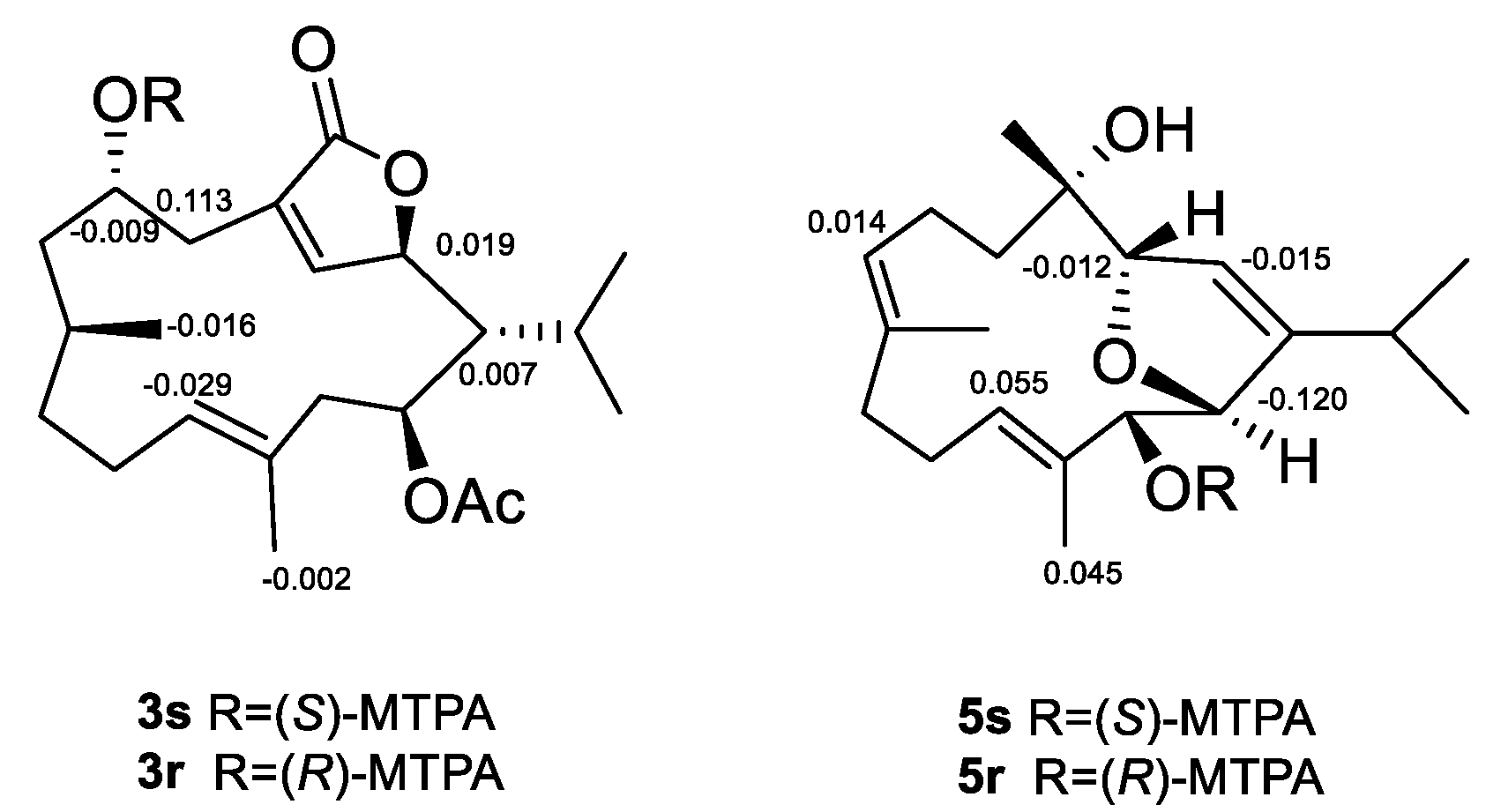
3.6. Esterification of Compounds 3 and 5 with MTPA Chlorides

Compounds 3 and 5 (2.0 mg each) were dissolved in dry pyridine (1.2 mL), divided them into two sets (0.6 mL each), then treated with (R)-(–)-2-methoxy-2-(trifluoromethyl) phenylacetyl chloride ((R)-(–)-MTPA-Cl) (10 μL) and (S)-(+)-2-methoxy-2-(trifluoromethyl) phenylacetyl chloride ((S)-(+)-MTPA-Cl) (10 μL), respectively. After stirring overnight at room temperature, the solutions were evaporated in vacuo and the residues were purified by silica gel column chromatography (PE/Et2O = 90:10) to obtain the S-MTPA ester 3r (0.3 mg), R-MTPA ester 3s (0.3 mg), S-MTPA ester 5r (0.3 mg), and R-MTPA ester 5s (0.3 mg), respectively. The obtained products were then subjected to the 1H NMR experiment.

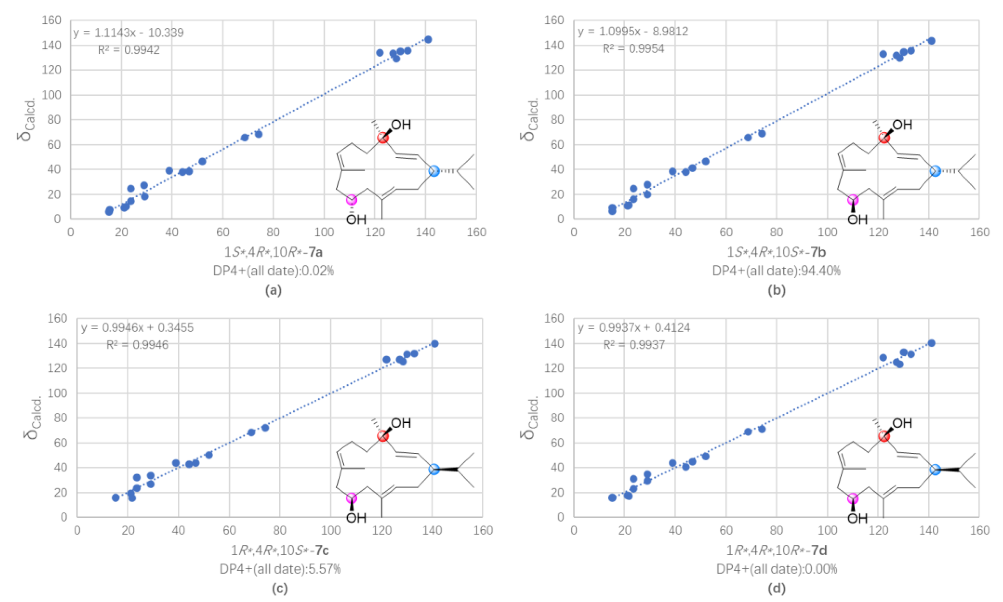
3.7. QM-NMR Calculation of Compounds 5 and 7

Theoretical calculations of all theoretical stereoisomers were carried out to determine the relative configuration of 5 and 7, based on the alignment of its 1D NMR chemical shifts (13C NMR chemical shifts herein) and calculation-generated chemical shifts. Confab was used to search the conformational space of 5a5d and 7a7d. Conformational searches were carried out using the torsional sampling (MCMM) method and OPLS_2005 force field in the Macromodel 9.9.223 software applying an energy window of 21 kJ/mol. Conformers above 1% population were re-optimized with Gaussian 09 at the B3LYP/6-311G(d,p) level with IEFPCM (Polarizable Continuum Model using the Integral Equation Formalism variant) solvent model for acetonitrile. The Boltzmann populations of the conformers were obtained based on the potential energy provided by the OPLS_2005 force field, leading to 11, 9, 13 and 24 conformers for 5a5d; 11, 9, 13 and 24 conformers for 7a7d above 1% population for further re-optimization, respectively. The obtained conformers were subjected to optimization and frequency calculations on B3LYP/6-311G(d,p) level of theory. GIAO DFT 13C NMR calculations were calculated on mPW1PW91/6-31G* (CHCl3) level of theory, and the calculated shielding tensors were Boltzmann averaged according to Gibbs free energy and then converted into chemical shifts following MSTD protocol. The experimental 13C NMR data of 5 and 7 were compared with the calculated NMR chemical shifts of 5a5d and 7a7d using the mean absolute error (MAE) values, maximum deviation (MD) values, correlation coefficient (R2), and DP4+ probability analysis. XYZ data for all conformations and detailed data for DP4+ analysis are provided in the Supplementary Materials.

3.8. Cytotoxic Bioassays

H1975, MDA-MB-231, A549, and H1299 cancer cell lines were purchased from the Procell Life Science & Technology Co., Ltd. Cells were cultured at 37 °C in a 5% CO2 humidified incubator and maintained in high glucose Dulbecco’s Modified Eagle Medium (DMEM, Nissui, Tokyo, Japan) containing 100 mg/mL streptomycin, 2.5 mg/L amphotericin B and 10% heat-inactivated fetal bovine serum (FBS). The cells were inoculated in 96-well culture plates for 12 h and then treated with different concentrations of compounds for 72 h. Water-soluble tezole (WST) reagent was added to each (10 µL) well and cultured at 37 °C for 2 h to assess cell viability. The absorbance was read with a microplate reader at 450 nm. Adriamycin (DOX) was the positive control.

4. Conclusions

In summary, the present investigation of the soft coral S. trocheliophorum from Ximao Island provided intriguing results including six new cembranoids diterpenoids, sartrocheliols A–E (1, 3, 58), along with four known related ones (2, 4, 9, and 10). This study not only extended the members of capnosane and cembrane diterpenes but also enriched the chemical diversity of the title species. In addition, a whole set of NMR computations, modified Mosher’s method, and X-ray diffraction analysis were applied to assign the absolute configurations of compounds 1, 3, 4, and 5. However, the absolute configurations of other new metabolites remain undefined. To achieve it, a collection of the biological materials and accumulation of these new compounds should be conducted in future, which could supply sufficient amounts of metabolites for either the subsequent chemical transformations or recrystallization experiments. Anti-tumor and antibacterial bioassays were carried out. Among these secondary metabolites, compound 4 displayed moderate cytotoxicity against A549, H1975, MDA-MB-231, and H1299 cells (IC50 = 47.9, 26.3, 44.7, 33.1 μM, respectively), while 6 only exhibited moderate cytotoxicity against H1975 (IC50 = 40.4 μM). However, none of them were active in the antibacterial bioassay. Moreover, the bioactivities of these compounds such as anti-virus, will be evaluated in future.

Supplementary Materials

The following supporting information can be downloaded at: https://www.mdpi.com/article/10.3390/md21020069/s1, Figures S1–S48: NMR, HRESIMS and IR data of compounds 1, 3, 58; Figures S49 and S50: Regression analysis of experimental vs. calculated 13C NMR chemical shifts of different isomers of compounds 5 and 7 at the PCM/mPW1PW91/6-31 + G** level using DP4+ method; Tables S1 and S2: X-ray crystallographic data for compounds 1 and 4; Tables S3 and S5: Cartesian coordinates of all conformers of isomers 5a5b and 7a7b used after optimization at the B3LYP/6-311G (d,p) level of theory as required for DP4+ analysis; Tables S4 and S6: DP4+ results obtained using experimental data of compounds 5 versus isomers 5a5b and 7 versus isomers 7a7b.

Author Contributions

Conceptualization, L.-F.L., F.Y. and Y.-W.G.; methodology, L.-F.L., F.Y. and Y.-W.G.; validation, Y.-T.S., M.-Z.S., H.L. and J.-G.C.; formal analysis, Y.-T.S. and D.-D.Y.; investigation, Y.-T.S. and D.-D.Y.; data curation, Y.-T.S. and D.-D.Y.; writing—original draft preparation, Y.-T.S.; writing—review and editing, L.-F.L. and Y.-W.G.; supervision, Y.-W.G.; project administration, Y.-W.G.; funding acquisition, L.-F.L. and Y.-W.G. All authors have read and agreed to the published version of the manuscript.

Funding

This research work was financially supported by the National Natural Science Foundation of China (NSFC) (Nos. 81991521 and 41876194), the National Key Research and Development Program of China (No. 2022YFC2804100), and the SKLDR/SIMM Project (No. SIMM2103ZZ-06).

Institutional Review Board Statement

Not applicable.

Informed Consent Statement

Not applicable.

Data Availability Statement

The data presented in this study are available on request from the corresponding author.

Acknowledgments

We thank X.-B. Li from Hainan University for the taxonomic identification of the soft coral material.

Conflicts of Interest

The authors declare no conflict of interest.

References

  1. Anjaneyulu, A.S.R.; Rao, G.V. Chemical constituents of the soft coral species of Sarcophyton genus: A review. J. Indian Chem. Soc. 1997, 74, 272–278. [Google Scholar]
  2. Liang, L.-F.; Guo, Y.-W. Terpenes from the soft corals of the genus Sarcophyton: Chemistry and biological activities. Chem. Biodivers. 2013, 10, 2161–2196. [Google Scholar] [CrossRef]
  3. Zubair, M.S.; Al-Footy, K.O.; Ayyad, S.-E.N.; Al-Lihaibi, S.S.; Alarif, W.M. A review of steroids from Sarcophyton species. Nat. Prod. Res. 2016, 30, 869–879. [Google Scholar] [CrossRef]
  4. Elkhawas, Y.A.; Elissawy, A.M.; Elnaggar, M.S.; Mostafa, N.M.; Al-Sayed, E.; Bishr, M.M.; Singab, A.N.B.; Salama, O.M. Chemical diversity in species belonging to soft coral genus Sacrophyton and its impact on biological activity: A review. Mar. Drugs 2020, 18, 41. [Google Scholar] [CrossRef] [Green Version]
  5. Wang, Z.; Tang, H.; Wang, P.; Gong, W.; Xue, M.; Zhang, H.; Liu, T.; Liu, B.; Yi, Y.; Zhang, W. Bioactive polyoxygenated steroids from the South China Sea soft coral, Sarcophyton sp. Mar. Drugs 2013, 11, 775–787. [Google Scholar] [CrossRef] [Green Version]
  6. Chen, W.-T.; Liu, H.-L.; Yao, L.-G.; Guo, Y.-W. 9,11-Secosteroids and polyhydroxylated steroids from two South China Sea soft corals Sarcophyton trocheliophorum and Sinularia flexibilis. Steroids 2014, 92, 56–61. [Google Scholar] [CrossRef]
  7. Ngoc, N.T.; Hanh, T.T.H.; Quang, T.H.; Cuong, N.X.; Nam, N.H.; Thao, D.T.; Thung, D.C.; Kiem, P.V.; Minh, C.V. Polyhydroxylated steroids from the Vietnamese soft coral Sarcophyton ehrenbergi. Steroids 2021, 176, 108932. [Google Scholar] [CrossRef]
  8. Wang, C.-Y.; Chen, A.-N.; Shao, C.-L.; Li, L.; Xu, Y.; Qian, P.-Y. Chemical constituents of soft coral Sarcophyton infundibuliforme from the South China Sea. Biochem. Syst. Ecol. 2011, 39, 853–856. [Google Scholar] [CrossRef]
  9. Huang, T.-Y.; Huang, C.-Y.; Chen, S.-R.; Weng, J.-R.; Tu, T.-H.; Cheng, Y.-B.; Wu, S.-H.; Sheu, J.-H. New hydroquinone monoterpenoid and cembranoid-related metabolites from the soft coral Sarcophyton tenuispiculatum. Mar. Drugs 2021, 19, 8. [Google Scholar] [CrossRef]
  10. Anjaneyulu, A.S.R.; Murthy, M.V.R.K.; Gowri, P.M.; Venugopal, M.; Laatsch, H. A rare prostaglandin from the soft coral Sarcophyton crassocaule of the Indian Ocean. J. Nat. Prod. 2000, 63, 1425–1426. [Google Scholar] [CrossRef]
  11. Cheng, Z.-B.; Deng, Y.-L.; Fan, C.-Q.; Han, Q.-H.; Lin, S.-L.; Tang, G.-H.; Luo, H.-B.; Yin, S. Prostaglandin derivatives: Nonaromatic phosphodiesterase-4 inhibitors from the soft coral Sarcophyton ehrenbergi. J. Nat. Prod. 2014, 77, 1928–1936. [Google Scholar] [CrossRef] [PubMed]
  12. Cheng, S.-Y.; Wen, Z.-H.; Chiou, S.-F.; Tsai, C.-W.; Wang, S.-K.; Hsu, C.-H.; Dai, C.-F.; Chiang, M.Y.; Wang, W.-H.; Duh, C.-Y. Ceramide and cerebrosides from the octocoral Sarcophyton ehrenbergi. J. Nat. Prod. 2009, 72, 465–468. [Google Scholar] [CrossRef] [PubMed]
  13. Eltahawy, N.A.; Ibrahim, A.K.; Radwan, M.M.; Zaitone, S.A.; Gomaa, M.; ElSohly, M.A.; Hassanean, H.A.; Ahmed, S.A. Mechanism of action of antiepileptic ceramide from Red Sea soft coral Sarcophyton auritum. Bioorg. Med. Chem. Lett. 2015, 25, 5819–5824. [Google Scholar] [CrossRef] [PubMed]
  14. Řezanka, T.; Dembitsky, V.M. γ-Lactones from the soft corals Sarcophyton trocheliophorum and Lithophyton arboreum. Tetrahedron 2001, 57, 8743–8749. [Google Scholar] [CrossRef]
  15. Shaaban, M.; Ghani, M.A.; Issa, M.Y. New naturally occurring compounds from Sarcophyton trocheliophorum. Biointerface Res. App. Chem. 2022, 12, 2285–2331. [Google Scholar] [CrossRef]
  16. Rodrigues, I.G.; Miguel, M.G.; Mnif, W. A brief review on new naturally occurring cembranoid diterpene derivatives from the soft corals of the genera Sarcophyton, Sinularia, and Lobophytum since 2016. Molecules 2019, 24, 781. [Google Scholar] [CrossRef] [Green Version]
  17. Shaaban, M.; Yassin, F.Y.; Soltan, M.M. Calamusins J-K: New anti-angiogenic sesquiterpenes from Sarcophyton glaucum. Nat. Prod. Res. 2021, 35, 5720–5731. [Google Scholar] [CrossRef]
  18. Sawant, S.S.; Youssef, D.T.A.; Sylvester, P.W.; Wali, V.; Sayed, K.A.E. Antiproliferative sesquiterpenes from the Red Sea soft coral Sarcophyton glaucum. Nat. Prod. Commun. 2007, 2, 117–119. [Google Scholar] [CrossRef] [Green Version]
  19. Anjaneyulu, A.S.R.; Rao, V.L.; Sastry, V.G.; Rao, D.V. Trocheliophorin: A novel rearranged sesquiterpenoid from the Indian Ocean soft coral Sarcophyton trocheliophorum. J. Asian Nat. Prod. Res. 2008, 10, 597–601. [Google Scholar] [CrossRef]
  20. Ye, F.; Li, J.; Wu, Y.; Zhu, Z.-D.; Mollo, E.; Gavagnin, M.; Gu, Y.-C.; Zhu, W.-L.; Li, X.-W.; Guo, Y.-W. Sarinfacetamides A and B, nitrogenous diterpenoids with tricyclo [6.3.1.01,5] dodecane scaffold from the South China Sea soft coral Sarcophyton infundibuliforme. Org. Lett. 2018, 20, 2637–2640. [Google Scholar] [CrossRef]
  21. Lin, K.-H.; Lin, Y.-C.; Huang, C.-Y.; Tseng, Y.-J.; Chen, S.-R.; Cheng, Y.-B.; Hwang, T.-L.; Wang, S.-Y.; Chen, H.-Y.; Dai, C.-F.; et al. Cembranoid-related diterpenes, novel secoditerpenes, and an unusual bisditerpene from a Formosan soft coral Sarcophyton tortuosum. Bull. Chem. Soc. Jpn. 2021, 94, 2774–2783. [Google Scholar] [CrossRef]
  22. Bu, Q.; Yang, M.; Yan, X.-Y.; Yao, L.-G.; Guo, Y.-W.; Liang, L.-F. New flexible cembrane-type macrocyclic diterpenes as TNF-α inhibitors from the South China Sea soft coral Sarcophyton mililatensis. Int. J. Biol. Macromol. 2022, 222, 880–886. [Google Scholar] [CrossRef] [PubMed]
  23. Wang, C.; Zhang, J.; Shi, X.; Li, K.; Li, F.; Tang, X.; Li, G.; Li, P. Sarcoeleganolides C–G, five new cembranes from the South China Sea soft coral Sarcophyton elegans. Mar. Drugs 2022, 20, 574. [Google Scholar] [CrossRef] [PubMed]
  24. Mohamed, T.A.; Elshamy, A.I.; Abd El-Razek, M.H.; Abdel-Tawab, A.M.; Ali, S.K.; Aboelmagd, M.; Suenaga, M.; Pare, P.W.; Umeyama, A.; Hegazy, M.-E.F. Sarcoconvolutums F and G: Polyoxygenated cembrane-type diterpenoids from Sarcophyton convolutum, a Red Sea soft coral. Molecules 2022, 27, 5835. [Google Scholar] [CrossRef]
  25. Sun, P.; Cai, F.-Y.; Lauro, G.; Tang, H.; Su, L.; Wang, H.-L.; Li, H.H.; Mándi, A.; Kurtán, T.; Riccio, R.; et al. Immunomodulatory biscembranoids and assignment of their relative and absolute configurations: Data set modulation in the density functional theory/nuclear magnetic resonance approach. J. Nat. Prod. 2019, 82, 1264–1273. [Google Scholar] [CrossRef] [PubMed]
  26. Li, Y.; Li, S.; Cuadrado, C.; Gao, C.; Wu, Q.; Li, X.; Pang, T.; Daranas, A.H.; Guo, Y.; Li, X. Polyoxygenated anti-inflammatory biscembranoids from the soft coral Sarcophyton tortuosum and their stereochemistry. Chin. Chem. Lett. 2021, 32, 271–276. [Google Scholar] [CrossRef]
  27. Yan, X.; Liu, J.; Huang, J.; Wang, Y.; Leng, X.; Li, T.; Ouyang, H.; Yan, X.; He, S. Bistrochelides H−L: Biscembranoids from the South China Sea soft coral Sarcophyton serenei. Phytochemistry 2022, 204, 113438. [Google Scholar] [CrossRef]
  28. Nguyen, N.B.; Chen, L.-Y.; Chen, P.-J.; El-Shazly, M.; Hwang, T.-L.; Su, J.-H.; Su, C.-H.; Yen, P.-T.; Peng, B.-R.; Lai, K.-H. MS/MS molecular networking unveils the chemical diversity of biscembranoid derivatives, neutrophilic inflammatory mediators from the cultured soft coral Sarcophyton trocheliophorum. Int. J. Mol. Sci. 2022, 23, 15464. [Google Scholar] [CrossRef]
  29. Al-Footy, K.O.; Alarif, W.M.; Asiri, F.; Aly, M.M.; Ayyad, S.-E.N. Rare pyrane-based cembranoids from the Red Sea soft coral Sarcophyton trocheliophorum as potential antimicrobial–antitumor agents. Med. Chem. Res. 2015, 24, 505–512. [Google Scholar] [CrossRef]
  30. Mohamed, T.A.; Elshamy, A.I.; Abdel-Tawab, A.M.; AbdelMohsen, M.M.; Ohta, S.; Pare, P.W.; Hegazy, M.-E.F. Oxygenated cembrene diterpenes from Sarcophyton convolutum: Cytotoxic sarcoconvolutum A–E. Mar. Drugs 2021, 19, 519. [Google Scholar] [CrossRef]
  31. Li, J.-F.; Zeng, Y.-B.; Li, W.-S.; Luo, H.; Zhang, H.-Y.; Guo, Y.-W. Xishaglaucumins A—J, new cembranoids with anti-inflammatory activities from the South China Sea soft coral Sarcophyton glaucum. Chin. J. Chem. 2022, 40, 79–90. [Google Scholar] [CrossRef]
  32. Zhang, J.; Tang, X.; Han, X.; Feng, D.; Luo, X.; Ofwegen, L.v.; Li, P.; Li, G. Sarcoglaucins A-I, new antifouling cembrane-type diterpenes from the South China Sea soft coral Sarcophyton glaucum. Org. Chem. Front. 2019, 6, 2004–2013. [Google Scholar] [CrossRef]
  33. Badria, F.A.; Guirguis, A.N.; Perovic, S.; Steffen, R.; Müller, W.E.G.; Schröder, H.C. Sarcophytolide: A new neuroprotective compound from the soft coral Sarcophyton glaucum. Toxicology 1998, 131, 133–143. [Google Scholar] [CrossRef] [PubMed]
  34. Du, Y.-Q.; Chen, J.; Wu, M.-J.; Zhang, H.-Y.; Liang, L.-F.; Guo, Y.-W. Uncommon capnosane diterpenes with neuroprotective potential from South China Sea soft coral Sarcophyton boettgeri. Mar. Drugs 2022, 20, 602. [Google Scholar] [CrossRef] [PubMed]
  35. Carroll, A.R.; Copp, B.R.; Davis, R.A.; Keyzers, R.A.; Prinsep, M.R. Marine natural products. Nat. Prod. Rep. 2022, 39, 1122–1171. [Google Scholar] [CrossRef]
  36. Liu, J.; Gu, Y.-c.; Su, M.-z.; Guo, Y.-w. Chemistry and bioactivity of secondary metabolites from South China Sea marine fauna and flora: Recent research advances and perspective. Acta Pharmacol. Sin. 2022, 43, 3062–3079. [Google Scholar] [CrossRef]
  37. Liang, L.-F.; Kurtán, T.; Mándi, A.; Yao, L.-G.; Li, J.; Zhang, W.; Guo, Y.-W. Unprecedented diterpenoids as a PTP1B inhibitor from the Hainan soft coral Sarcophyton trocheliophorum Marenzeller. Org. Lett. 2013, 15, 274–277. [Google Scholar] [CrossRef]
  38. Yang, M.; Li, X.-L.; Wang, J.-R.; Lei, X.; Tang, W.; Li, X.-W.; Sun, H.; Guo, Y.-W. Sarcomililate A, an unusual diterpenoid with tricyclo [11.3.0.02,16] hexadecane carbon skeleton, and its potential biogenetic precursors from the Hainan soft coral Sarcophyton mililatensis. J. Org. Chem. 2019, 84, 2568–2576. [Google Scholar] [CrossRef]
  39. Yang, Q.-B.; Wu, Q.; Chen, J.-K.; Liang, L.-F. The soft coral Sarcophyton trocheliophorum: A warehouse of terpenoids with structural and pharmacological diversity. Mar. Drugs 2023, 21, 30. [Google Scholar] [CrossRef]
  40. Yao, L.-G.; Zhang, H.-Y.; Liang, L.-F.; Guo, X.-J.; Mao, S.-C.; Guo, Y.-W. Yalongenes A and B, two new cembranoids with cytoprotective effects from the Hainan soft coral Sarcophyton trocheliophorum Marenzeller. Helv. Chim. Acta 2012, 95, 235–239. [Google Scholar] [CrossRef]
  41. Chen, Z.-H.; Gao, T.-R.; Yang, M.; Yao, L.-G.; Guo, Y.-W. Further new cembranoids from the South China Sea soft coral Sarcophyton trocheliophorum. Fitoterapia 2021, 151, 104902. [Google Scholar] [CrossRef] [PubMed]
  42. Xi, Z.; Bie, W.; Chen, W.; Liu, D.; van Ofwegen, L.; Proksch, P.; Lin, W. Sarcophyolides B–E, new cembranoids from the soft coral Sarcophyton elegans. Mar. Drugs 2013, 11, 3186–3196. [Google Scholar] [CrossRef] [PubMed]
  43. Jia, R.; Guo, Y.-W.; Mollo, E.; Gavagnin, M.; Cimino, G. Sarcophytonolides E−H, cembranolides from the Hainan soft coral Sarcophyton latum. J. Nat. Prod. 2006, 69, 819–822. [Google Scholar] [CrossRef] [PubMed]
  44. Liang, L.-F.; Chen, W.-T.; Li, X.-W.; Wang, H.-Y.; Guo, Y.-W. New bicyclic cembranoids from the South China Sea soft coral Sarcophyton trocheliophorum. Sci. Rep. 2017, 7, 46584. [Google Scholar] [CrossRef] [Green Version]
  45. Wen, T.; Ding, Y.; Deng, Z.; van Ofwegen, L.; Proksch, P.; Lin, W. Sinulaflexiolides A-K, cembrane-type diterpenoids from the Chinese soft coral Sinularia flexibilis. J. Nat. Prod. 2008, 71, 1133–1140. [Google Scholar] [CrossRef]
  46. Kobayashi, M.; Kondo, K.; Osabe, K.; Mitsuhashi, H. Marine terpenes and terpenoids. V. Oxidation of sarcophytol A, a potent anti-tumor-promoter from the soft coral Sarcophyton glaucum. Chem. Pharm. Bull. 1988, 36, 2331–2341. [Google Scholar] [CrossRef] [Green Version]
  47. Li, S.-W.; Cuadrado, C.; Yao, L.-G.; Daranas, A.H.; Guo, Y.-W. Quantum mechanical–NMR-aided configuration and conformation of two unreported macrocycles isolated from the soft coral Lobophytum sp.: Energy calculations versus coupling constants. Org. Lett. 2020, 22, 4093–4096. [Google Scholar] [CrossRef]
  48. Raldugin, V.A.; Pleshkov, I.G.; Gatilov, Y.V.; Yaroshenko, N.I.; Salenko, V.L.; Shevtsov, S.A.; Pentegova, V.A. Photosensitized oxidation of isocembrol. VII. Products of reaction at the C11 double bond. Chem. Nat. Compd. 1984, 20, 45–52. [Google Scholar] [CrossRef]
Figure 1. The chemical structures of compounds 114.
Marinedrugs 21 00069 g001
Figure 2. Perspective ORTEP drawing of the X-ray structures of compounds 1 (left) and 4 (right) (displacement ellipsoids are drawn at the 50% probability level).
Marinedrugs 21 00069 g002
Figure 3. The selected key 1H–1H COSY (red lines) and HMBC (blue arrows, from 1H to 13C) correlations of compounds 1, 3, 58.
Marinedrugs 21 00069 g003
Figure 4. The selected key NOESY (pink dashed lines, from 1H to 1H) correlations of compounds 3, 58.
Marinedrugs 21 00069 g004
Figure 5. Δδ values (δS–δR) (ppm) for (S)-and (R)-MTPA esters of 3 and 5.
Marinedrugs 21 00069 g005
Figure 6. ECD curves of compounds 3 (up) and 4 (down).
Marinedrugs 21 00069 g006
Figure 7. Regression analysis of experimental vs. calculated 13C NMR chemical shifts of (a) (2R*,3R*,13R*,14S*)-5a, (b) (2R*,3R*,13S*,14S*)-5b, (c) (2R*,3S*,13R*,14S*)-5c, and (d) (2R*,3S*,13S*,14R*)-5d at the PCM/mPW1PW91/6-31 + G** level, using the DP4+ method.
Marinedrugs 21 00069 g007
Figure 8. Regression analysis of experimental vs. calculated 13C NMR chemical shifts of (a) (1S*,4R*,10R*)-7a, (b) (1S*,4R*,10S*)-7b, (c) (1R*,4R*,10S*)-7c, and (d) (1R*,4R*,10R*)-7d at the PCM/mPW1PW91/6-31 + G** level, using the DP4+ method.
Marinedrugs 21 00069 g008
Table 1. 1H NMR spectroscopic data of sartrocheliols A–E (1, 3, 58) in CDCl3.
No.1 a3 b5 b6 b7 b8 a
1 1.55 (d, 10.7) 1.54 (br s)
25.14 (d, 10.6)4.95 (d, 10.7)5.51 (d, 1.4)5.42 (m)5.51 (dd, 15.6, 9.6)5.56 (d, 15.6)
32.84 (t, 10.6)7.44 (t, 1.8)4.66 (dq, 5.5, 1.4)4.40 (br s)5.63 (d, 15.6)6.13 (d, 15.9)
4
51.84 (m)2.75 (dt, 13.2, 1.2)1.83 (m)1.94 (m)1.89 (ddd, 13.4, 8.6, 1.9)1.87 (m)
1.78 (m)2.45 (dd, 13.3, 10.5)1.53 (m)1.76 (m)1.58 (m)1.69 (m)
61.66 (m)4.22 (t, 9.8)2.34 (m)2.06 (m)2.24 (m)2.64 (m)
1.92 (m)
72.48 (t, 7.1)1.63 (m)5.22 (m)5.24 (t, 6.3)5.11 (d, 7.8)5.43 (dd, 10.6, 4.0)
1.35 (m)
8 1.29 (m)
92.33 (m)1.36 (m)2.10 (m)1.96 (m)2.25 (m)2.00 (m)
2.00 (t, 13.5)1.26 (m) 1.67 (m)2.12 (m)
102.21 (m)2.08 (m)2.26 (m)1.86 (m)4.07 (d, 10,9)1.90 (m)
1.46 (m) 2.11 (m)1.43 (m) 1.55 (m)
112.89 (dd, 10.6, 2.9)5.05 (t, 7.7)5.32 (m)5.04 (d, 10.3)2.09 (m)5.08 (d, 9.4)
12
132.25 (m)2.26 (dd, 13.5, 3.0)4.29 (t, 4.3)1.94 (m)5.24 (t, 7.7)1.78 (m)
1.65 (m)2.20 (dd, 13.5, 10.9) 1.61 (m) 1.59 (m)
144.88 (dd, 11.2, 5.1)5.02 (ddd, 10.8, 4.2, 1.1)4.84 (td, 5.0, 1.6)3.04 (m)2.07 (m)2.33 (td, 11.8, 7.4)
2.38 (m) 1.68 (m)
152.48 (m)2.22 (m)2.65 (m) 1.70 (m)
161.12 (d, 6.5)1.11 (d, 6.6)1.16 (d, 6.8)1.37 (s)0.96 (d, 6.6)1.11 (s)
171.16 (d, 6.5)1.12 (d, 6.6)1.08 (d, 6.9)1.30 (s)0.84 (d, 6.7)1.13 (s)
181.16 (s) 1.00 (s)1.20 (s)1.30 (s)1.35 (s)
194.88 (s)0.77 (d, 6.0)1.56 (s)1.56 (s)1.54 (s)1.71 (s)
4.69 (s)
201.12 (s)1.67 (s)1.56 (s)1.10 (s)1.57 (m)1.17 (s)
OAc 2.09 (s) 2.08 (s) 2.05 (s)
a 500 MHz. b 600 MHz.
Table 2. 13C NMR spectroscopic data (125 MHz, CDCl3) of sartrocheliols A–E (1, 3, 58).
No.135678
1149.90, qC49.67, CH150.34, qC146.95, qC51.96, CH91.52, qC
2125.85, CH81.19, CH121.20, CH121.34, CH122.10, CH129.84, CH
351.08, CH151.06, CH87.63, CH73.16, CH141.04, CH137.41, CH
481.64, qC131.39, qC74.93, qC75.86, qC74.20, qC72.47, qC
541.04, CH237.42, CH239.37, CH239.18, CH244.21, CH241.64, CH2
625.90, CH266.40, CH22.57, CH221.75, CH223.66, CH222.90, CH2
754.09, CH47.19, CH2128.74, CH127.40, CH127.31, CH130.06, CH
8148.54, qC28.31, CH133.16, qC134.03, qC130.05, qC132.11, qC
924.65, CH237.25, CH239.64, CH233.90, CH246.88, CH234.86, CH2
1028.07, CH224.31, CH225.00, CH225.03, CH268.82, CH27.37, CH2
1162.28, CH129.20, CH130.08, CH72.70, CH38.96, CH277.36, CH
1259.06, qC130.80, qC132.19, qC74.15, qC132.94, qC84.58, qC
1345.46, CH241.43, CH279.67, CH29.33, CH2128.47, CH35.80, CH2
1469.34, CH73.92, CH86.05, CH20.95, CH223.68, CH230.76, CH2
1527.09, CH25.81, CH27.43, CH74.60, qC29.13, CH72.84, qC
1621.23, CH324.93, CH321.11, CH330.42, CH321.15, CH326.01, CH3
1723.55, CH318.74, CH322.57, CH328.85, CH321.85, CH324.62, CH3
1827.70, CH3172.97, qC23.36, CH321.72, CH329.17, CH329.00, CH3
19112.26, CH218.25, CH315.84, CH317.41, CH315.52, CH316.33, CH3
2023.05, CH318.47, CH312.89, CH323.63, CH315.09, CH320.69, CH3
OAc 171.13, qC 170.47, qC 170.17, qC
21.35, CH3 21.75, CH3 21.42, CH3
Disclaimer/Publisher’s Note: The statements, opinions and data contained in all publications are solely those of the individual author(s) and contributor(s) and not of MDPI and/or the editor(s). MDPI and/or the editor(s) disclaim responsibility for any injury to people or property resulting from any ideas, methods, instructions or products referred to in the content.

