Article

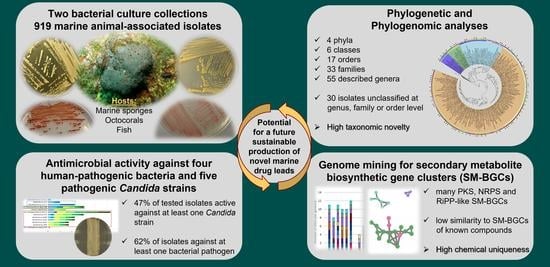
Marine Sponge and Octocoral-Associated Bacteria Show Versatile Secondary Metabolite Biosynthesis Potential and Antimicrobial Activities against Human Pathogens

1
iBB—Institute for Bioengineering and Biosciences and i4HB—Institute for Health and Bioeconomy, Instituto Superior Técnico, Av. Rovisco Pais, 1049-001 Lisbon, Portugal
2
Department of Bioengineering, Instituto Superior Técnico, Av. Rovisco Pais, 1049-001 Lisbon, Portugal
3
Department of Biology and Centre for Environmental and Marine Studies (CESAM), University of Aveiro, 3810-193 Aveiro, Portugal
4
Center for Neuroscience and Cell Biology (CNC), Rua Larga—Faculdade de Medicina, University of Coimbra, 3004-504 Coimbra, Portugal
5
Biocant—Transfer Technology Association, BiocantPark, 3060-197 Cantanhede, Portugal
6
Department of Marine Recreation, National Penghu University of Science and Technology, Magong City 880-011, Taiwan
7
Centre of Marine Sciences (CCMAR/CIMAR LA), University of Algarve, Campus de Gambelas, 8005-139 Faro, Portugal
*
Authors to whom correspondence should be addressed.
Mar. Drugs 2023, 21(1), 34; https://doi.org/10.3390/md21010034
Submission received: 14 November 2022 / Revised: 15 December 2022 / Accepted: 27 December 2022 / Published: 30 December 2022
(This article belongs to the Special Issue Reef Ecology and Marine Drug Discovery)

Abstract

:
Marine microbiomes are prolific sources of bioactive natural products of potential pharmaceutical value. This study inspected two culture collections comprising 919 host-associated marine bacteria belonging to 55 genera and several thus-far unclassified lineages to identify isolates with potentially rich secondary metabolism and antimicrobial activities. Seventy representative isolates had their genomes mined for secondary metabolite biosynthetic gene clusters (SM-BGCs) and were screened for antimicrobial activities against four pathogenic bacteria and five pathogenic Candida strains. In total, 466 SM-BGCs were identified, with antimicrobial peptide- and polyketide synthase-related SM-BGCs being frequently detected. Only 38 SM-BGCs had similarities greater than 70% to SM-BGCs encoding known compounds, highlighting the potential biosynthetic novelty encoded by these genomes. Cross-streak assays showed that 33 of the 70 genome-sequenced isolates were active against at least one Candida species, while 44 isolates showed activity against at least one bacterial pathogen. Taxon-specific differences in antimicrobial activity among isolates suggested distinct molecules involved in antagonism against bacterial versus Candida pathogens. The here reported culture collections and genome-sequenced isolates constitute a valuable resource of understudied marine bacteria displaying antimicrobial activities and potential for the biosynthesis of novel secondary metabolites, holding promise for a future sustainable production of marine drug leads.

Graphical Abstract

1. Introduction

Antibiotic resistant microorganisms emerged as a problem soon after the large-scale introduction of the first antibiotics [1]. At present, multi-drug resistant microbes pose a worldwide threat, with estimates placing their toll on human life as high as 700,000 deaths per year, with simulations predicting that this number will rise to 10 million per year by 2050 [1]. Staphylococcus aureus, part of the human skin microbiome and an opportunistic pathogen, is, for example, commonly resistant to penicillin, while methicillin-resistant Staphylococcus aureus (MRSA) strains show additional resistance to many antimicrobial drug classes, including oxacillin, cefoxitin and chloramphenicol [2]. Nosocomial infections by MRSA are frequent, with >150,000 cases per year registered in the European Union alone [3]. Salmonella enterica serovar Typhimurium [4] and Vibrio parahaemolyticus [5] are not only human pathogens, but also swine and seafood pathogens, respectively. Salmonella spp. are estimated to cause about 93 million cases of gastroenteritis and 155,000 deaths annually, with serovar Typhimurium being one of the leading causes [6]. S. enterica serovar Typhimurium isolates from Southeast Asia were found to be commonly resistant to seven classes of antibiotics, including streptomycin, ampicillin and erythromycin [4]. Vibrio parahaemolyticus can have negative impacts on aquaculture production (at times leading to 100% loss in shrimp production [7]) and causes disease in fish, shellfish, and shrimp [7,8]. Likewise, the incidence of treatment–resistant fungi of the Candida genus has increased over the past 20 years [9]. Although C. albicans continues to be the most prevalent cause of human candidiasis, C. glabrata, C. tropicalis and C. parapsilosis have been gaining ground mainly due to their greater ability to exhibit antifungal drug resistance [9,10]. Candida auris is the most recent addition to this group, due to the extreme resistance that some strains have shown towards marketed drugs [11]. Indeed, certain C. auris strains are resistant to all approved antifungal drug classes and are now considered nearly untreatable [11]. Therefore, new drugs and a greater structural variety of natural products are urgently required to combat the antimicrobial resistance crisis [12].
Marine animals, such as sponges (Porifera) and octocorals (Cnidaria, Anthozoa, Octocorallia), are well-known producers of diverse natural products with antibacterial, antitumoral, antiviral, anti-inflammatory and other activities [13,14]. Several marine animal-derived compounds such as, e.g., the anti-cancer drugs eribulin (trade name Halaven) [15] and trabectedin (trade name Yondelis) [16], are already available on the market. Sponges and corals host diverse and distinct microbial communities, contributing to carbon, sulfur and nitrogen cycling as well chemical defense of their host [17,18]. Advances in next-generation sequencing technologies, metagenomics, and the development of bioinformatic tools, such as the “antibiotics and Secondary Metabolite Analysis Shell” (antiSMASH) [19], to mine genomes and metagenomes for secondary metabolite biosynthetic gene clusters (SM-BGCs), have contributed to the understanding that microbial symbionts are frequently the true producers of the bioactive compounds previously isolated from marine animals. For example, octocoral-derived bacteria and fungi have been shown to harbor an enormous arsenal of bioactive terpenes, steroids, alkaloids, and polyketides [13]. Recent metagenomic surveys found the marine sponge microbiome to be significantly enriched in polyketide synthase encoding genes [20], while a variety of non-ribosomal peptide synthetase gene clusters, among others, were reported from the microbiomes of octocorals [21]. Moreover, bacteria isolated from the marine sponges Hymeniacidon perlevis and Halichondria panicea were recently shown to inhibit MRSA growth [22]. Thus, the microbiomes of marine animals remain a largely untapped reservoir of novel molecules with unknown functions, some of which may be leveraged for pharmaceutical use.
Microbial cultivation, though tedious and time-consuming and hence sometimes disregarded in the omics era, is a crucial component to harness the biotechnological and medical potential of microorganisms. The improvement of cultivation protocols, and development of innovative media compositions and culture conditions tailored for marine, host-associated bacteria are pivotal to successfully culture difficult-to-cultivate symbionts [23]. Well-tended, taxonomically diverse culture collections are a valuable resource for natural products research, and the isolation of new species and strains stands at the very basis of the marine drug discovery pipeline.
The goal of this study was to highlight novel bacterial isolates suitable for bioprospecting among two comprehensive culture collections totaling 919 marine, animal-associated bacteria. Moreover, we aimed to assess the abundance and diversity of biosynthetic gene clusters, particularly those encoding for novel polyketides and non-ribosomal and ribosomal peptides, in the genomes of selected isolates. Finally, we tested whether the bacterial isolates inhibit the growth of human pathogenic bacteria and fungi to select promising antimicrobial strains worth further investigation. The first collection, hereafter called the “MicroEcoEvo” collection, was established between 2010 and 2020 during multiple sampling events of temperate marine sponges [24,25,26], octocorals [27] and fish larvae [28,29,30] in Portugal, by employing various marine culture media and conditions. The second collection, hereafter called the “EcoTech-SPONGE” collection, comprises bacterial isolates from various tropical marine sponges collected off the coast of Taiwan in 2017 [31,32]. In-depth 16S rRNA gene phylogenetic assessments were carried out to characterize the diversity and taxonomic novelty of both collections. A panel of 70 isolates with sequenced genomes was then subjected to phylogenomic assessments and genome-wide screenings for SM-BGCs. Finally, we screened these isolates for antimicrobial activities against the bacterial pathogens S. aureus, MRSA, S. enterica serovar Typhimurium and V. parahaemolyticus and five fungal pathogens of the Candida genus, paving the way for future studies on the identification and structure elucidation of novel natural products of marine origin.

2. Results

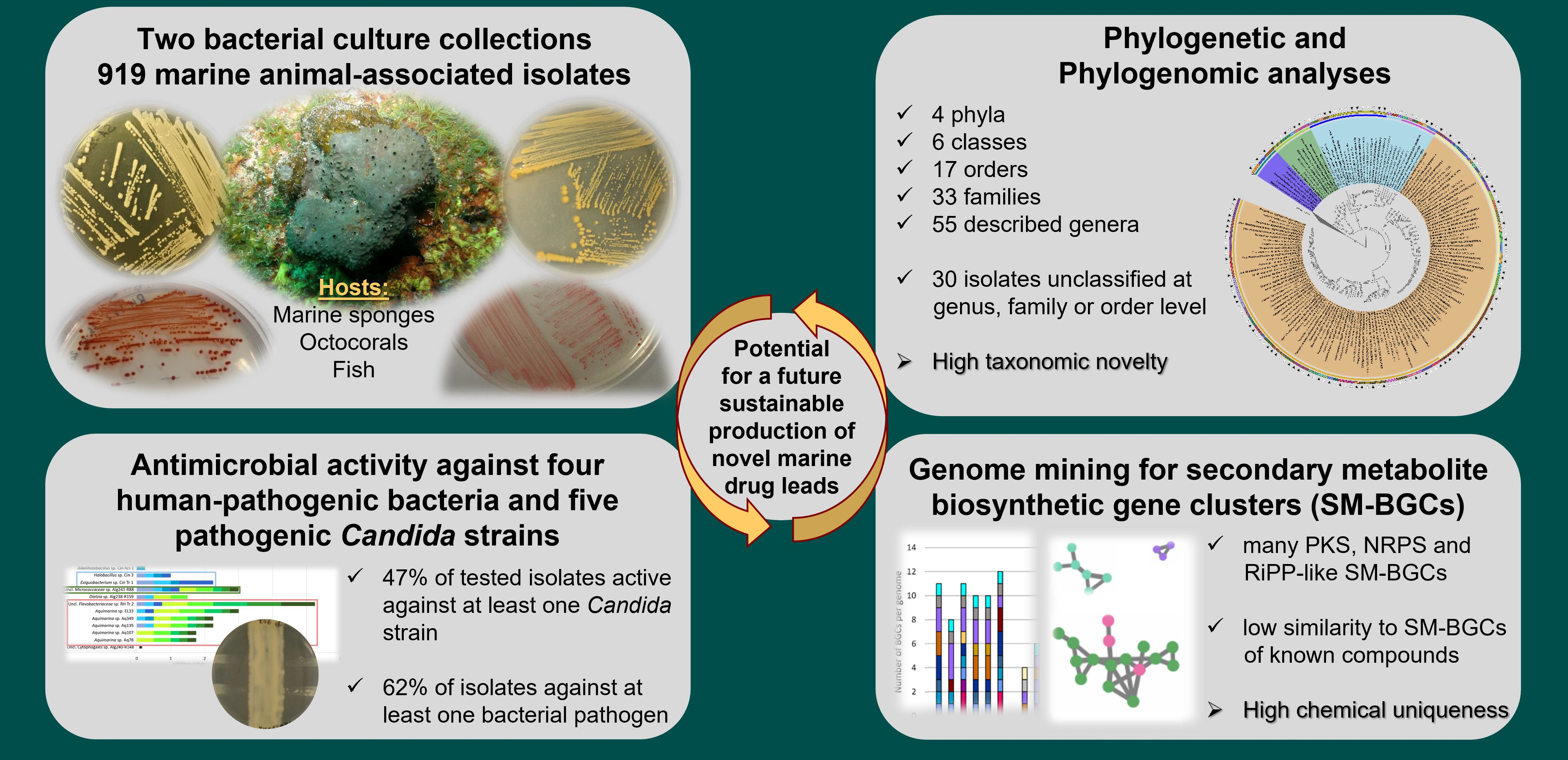
2.1. Taxonomic and Phylogenetic Diversity of the Two Marine Culture Collections

We determined the phylogenetic breadth of 919 bacterial strains, isolated from 18 marine sponge species (697 strains), two octocoral species (212 strains), and one fish species (10 strains) of two culture collections, the “MicroEcoEvo” collection (available at iBB, Instituto Superior Técnico, Lisbon Portugal) and the “EcoTech-SPONGE” collection (available at CESAM, Aveiro, Portugal). Complete lists of all isolates from both collections can be found in Tables S1 and S2 (Supplementary File S2), respectively. The “MicroEcoEvo” collection (Table S1) comprised 800 bacteria isolated from marine animals off the coast of Portugal spanning four bacterial phyla, six classes, and 45 genera (Figure 1). The most represented genera in this collection were Ruegeria (218 isolates), Vibrio (192 isolates), Pseudovibrio (140 isolates), Roseibium (62 isolates) and Shewanella (25 isolates). The collection also comprised less frequently cultured genera such as Amphritea (two isolates), Endozoicomonas (two isolates) and Parendozoicomonas (two isolates), as well as Kordia, Anderseniella and Lentilitoribacter (one isolate each) (Figure 1).
Nineteen of the isolates in this collection were not classifiable at genus level according to the Ribosomal Database Project (RDP) taxonomy [33], and represent likely at least six novel genera in the families Micrococcaceae, Rhodobacteraceae, Erythrobacteraceae and Shewanellaceae, and the orders Cytophagales and Oceanospirillales, respectively (Figure 1).
Figure 1. 16S rRNA gene-based phylogenetic inference of the 800 coral-, sponge- and fish-associated bacteria from the “MicroEcoEvo” culture collection. Black triangles indicate isolates that had their genomes sequenced. The outer ring indicates, by color, the cultivation medium used for isolation and the intermediate outer ring, the host species of each isolate. The intermediate inner ring and the inner ring indicate order and genus-level taxonomic classifications of the isolates, respectively. The legends are organized from left to right by order of appearance in the tree. Black dots on the branches indicate bootstrap support of ≥70% (500 bootstrap repetitions). Uncl.—unclassified.
Marinedrugs 21 00034 g001
The “EcoTech-SPONGE” culture collection (Table S2) comprised 119 bacteria isolated from 15 marine sponge species and spanned three bacterial phyla, five classes and 18 genera (Figure 2). All isolates in this collection were cultivated on half strength (1:2 diluted) Marine Agar (MA) at 17 °C and the number of isolates per sponge species varied from three (from Aaptos lobata) to 17 (from Cinachyrella sp.). The isolates were mostly related to Gammaproteobacteria (102 of 119), with low numbers of Bacilli (eight isolates), Alphaproteobacteria (five isolates), Cytophagia and Flavobacteriia (two isolates each). The most represented genera were Vibrio (42 isolates), Pseudoalteromonas (21 isolates) and Pseudomonas (11 isolates) (Figure 2). Less frequently cultured genera were Endozoicomonas (3 isolates), Thalassomonas (2 isolates), Flammeovirga (2 isolates), and Grimontia (1 isolate) (Figure 2). In this collection, 11 isolates could not be classified at genus, family, or order level, possibly comprising novel taxa in the family Flavobacteriaceae (n = 2), the order Oceanospirillales (n = 5) and the class Gammaproteobacteria (n = 4), respectively (Figure 2).
The “EcoTech-SPONGE” collection included taxa which were not present in the “MicroEcoEvo” collection, such as Pseudomonas (11 isolates), Alteromonas (3 isolates), Thalassomonas (2 isolates) and Grimontia (1 isolate), highlighting the taxonomic complementarity of both collections. Moreover, while Alphaproteobacteria (61% of all isolates) dominated the “MicroEcoEvo” collection, Gammaproteobacteria (86% of all isolates) dominated the “EcoTech-SPONGE” collection. The “MicroEcoEvo” collection was derived from six marine animal species and a variety of culture media and conditions, including low nutrient content media (e.g., diluted Marine Agar (MA), 50× diluted marine Gellan Gum—MG50 [25], marine R2A [34], and marine VXA [35]), and incubation temperatures of 18 °C, 19 °C or 25 °C to promote taxonomic diversity of culturable marine bacteria. Particularly MG50 and R2A provided a higher diversity of culturable genera at a low cultivation effort (≤50 isolates) compared with full strength MA. The “culturable diversity score” (i.e., the number of genera obtained over the total number of isolates gathered in a cultivation attempt) was 0.3061 for MG50 and 0.1203 for R2A but only 0.0827 for MA. In absolute numbers, however, Marine Agar (full strength and half strength (1:2 diluted)) yielded the largest number of marine host-associated bacterial genera in our collections (but also had the highest cultivation effort). While MA allowed cultivation of symbiotic taxa such as Endozoicomonas and Parendozoicomonas, the other marine media allowed the isolation of 17 genera that were not obtained on Marine Agar, such as, e.g., Anderseniella and Tateyamaria on MG50, Lentilitoribacter, and Marinicauda on R2A, and Agarivorans and Alcanivorax on VXA. The proportion of Gammaproteobacteria versus Alphaproteobacteria in the “MicroEcoEvo” collection was seemingly influenced by the nutrient content (particularly carbon and nitrogen) present in the medium. For example, in the closely related octocorals Eunicella verrucosa and E. labiata sampled off the coast of Algarve, cultivation on full strength MA at 19 °C yielded 91% Gammaproteobacteria (i.e., Vibrio, Shewanella and Pseudoalteromonas) and only 3 % Alphaproteobacteria isolates from E. verrucosa [36], while cultivation on half strength MA at a similar temperature (18 °C) yielded only 15% Gammaproteobacteria isolates and 80% Alphaproteobacteria, with Ruegeria, Roseibium, Roseovarius and Sphingorhabdus as most frequently cultured genera from E. labiata [27].
Figure 2. 16S rRNA gene-based phylogenetic inference of the 119 sponge-associated bacteria from the “EcoTech-SPONGE” culture collection. Black triangles indicate isolates that had their genomes sequenced. The outer ring indicates, by color, the host species of the bacterial isolates. The intermediate and the inner ring show order and genus-level taxonomic classifications of the isolates, respectively. The legends are organized from left to right by order of appearance in the tree. Black dots on the branches indicate bootstrap support of ≥70% (500 bootstrap repetitions).
Marinedrugs 21 00034 g002

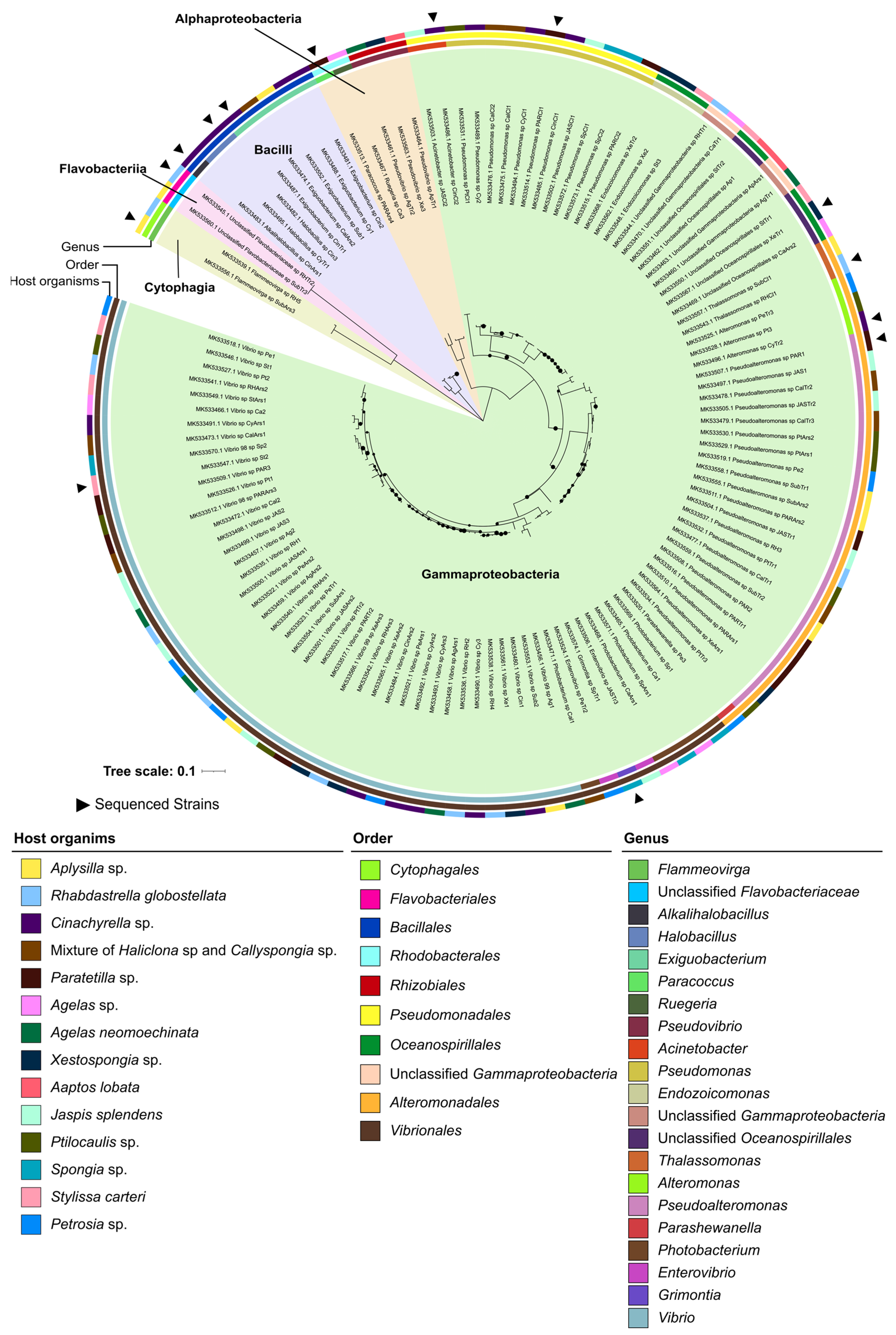
2.2. Phylogenomic Inference of Marine Host-Associated Bacteria

Seventy isolates from the four phyla and six classes of both culture collections (56 from the “MicroEcoEvo” and 14 from the “EcoTech-SPONGE” collection, to harmonize between cultivation and genome sequencing efforts) had their genomes sequenced with Illumina technology, assembled, and annotated (Table S3). Fifty of these genome assemblies (all from the “MicroEcoEvo” collection) were already publicly available, while twenty genomes are original contributions of this study and reported here for the first time (Table S3). Genomes were overall of high quality, with 66/70 showing over 97% completeness and 69/70 less than 4% contamination (Table S3). Most genomes belonged to the phylum Proteobacteria, featuring 30 alphaproteobacterial and 25 gammaproteobacterial strains (Figure 3). The remaining 15 genomes belonged to six Flavobacteriia, four Actinobacteria, three Bacilli and two Cytophagia strains (Figure 3). The genome-sequenced isolates feature multiple understudied and underrepresented species and genera. For example, our culture collection provides the only publicly available genomes for the genera Lentilitoribacter (strain Alg239-R112) and Anderseniella (strain Alg231_50), and the third genome of the genus Pelagibius (strain Alg239-R121).
To investigate the phylogenomic relatedness and taxonomic novelty of the 70 genome-sequenced bacteria, particularly of the strains not classified at genus level, a phylogenomic tree was constructed. As a reference, the genomes of 84 closely related type species (Figure 3) were included in the analysis. The closest type species of our 70 genome-sequenced strains were identified using the Microbial Genomes Atlas (MiGA) [37], based on average nucleotide identity (ANI) and average amino acid identity (AAI) scores. Most of the 70 genomes clustered with the genomes of their respective closest type strains (Figure 3). Unclassified Flavobacteriaceae sp. strain RHTr2 from the “EcoTech-SPONGE” collection branched next to the Aquimarina genus (Figure 3) and shared 66.49% AAI (close to the 65% genus-level cut-off of MiGA) with its closest type strain, Aquimarina spongiae DSM 22623 (T). Moreover, based on 16S rRNA gene sequencing, strain RHTr2 was affiliated to the Aquimarina genus with 78% confidence only (i.e., below the 80% confidence threshold of the RDP classifier tool). This, together with the phylogenomic tree topology, suggests that strain RHTr2 could represent a novel genus within the Flavobacteriaceae family, possessing close relatedness to the genus Aquimarina. Unclassified Cytophagales sp. strain Alg240-R148 from the “MicroEcoEvo” collection formed a clade with Fulvivirga lutimaris (GCA_009711525.1) on the phylogenomic tree. However, it shared only 55.33% and 55.02% AAI (much below the 65% AAI genus-level cut-off) with its closest type species, Fulvivirga kasyanovii JCM 16186T and Fulvivirga lutimaris (GCA_009711525.1), respectively. Based on 16S rRNA gene homology, the RDP classifier tool did not provide family or genus-level assignments with sufficient confidence (only 68% confidence for the family Fulvivirgaceae). This suggests that strain Alg240-R148 represents a novel genus and possibly even a novel family within the Cytophagales order, likely closely related to the Fulvivirgaceae family (Figure 3).
Unclassified Oceanospirillales sp. strain XeTr1 (“EcoTech-SPONGE” collection) clustered among genomes of the Endozoicomonadaceae family, forming a sister clade to Kistimonas asteriae (GCA_018263925), the only publicly available genome of a Kistimonas isolate (Figure 3). Yet, it shared only 58.54% AAI with its closest type strain, Endozoicomonas montiporae (GCA_001583435), as identified by MiGA, and ~58% AAI to four other Endozoicomonadaceae genomes (Table S4). Moreover, strain XeTr1 was classified into the Endozoicomonadaceae family by the RDP classifier tool with only 63% confidence (well-below the 80% cut-off). Therefore, strain XeTr1 could represent a novel genus and possibly even a novel family closely related to the Endozoicomonadaceae family, and to the Kistimonas and Endozoicomonas genera.
Figure 3. Phylogenomic inference of the 70 genome-sequenced isolates from the “MicroEcoEvo” and “EcoTechSPONGE” collections (highlighted by black triangles). In addition, 84 genomes obtained from public databases and representing type strains of species with standing in nomenclature (empty triangles) were added to the tree. Black circles on tree branches represent resampling (1000 repetitions) support greater than 75%. Five Archaea genomes of four type species (compressed clade) were used as an outgroup to root the tree, consisting of Archaeoglobus fulgidus, Methanobacterium formicicum, Nitrososphaera viennensis and Thermoplasma acidophilum. For each genome, corresponding species or strain names are shown, followed by the respective genome assembly accession number. Colored background shading represents the phylum-level classification of the genomes. The inner ring represents the class level taxonomy, and the outer ring represents the genus level affiliation of each genome. The legends are organized from left to right by order of appearance in the tree; on top of the “genus” legend, a colored bar indicates the bacterial class each genus belongs to. The evolutionary history was inferred by using a Maximum Likelihood method (FastTree2) and the Jukes-Cantor+CAT model based on alignment similarity of a set of 49 core, universal genes defined by COG (Clusters of Orthologous Groups) gene families. The evolutionary analyses were conducted using the SpeciesTreeBuilder v.01.0 app of DOE Systems Biology Knowledgebase (KBase). Uncl.—Unclassified.
Marinedrugs 21 00034 g003
Unclassified Erythrobacteraceae sp. strain Alg239-R51 (“MicroEcoEvo” collection) formed a cluster with Qipengyuania flava (GCA_004296255) (Figure 3) and shared >67% AAI with the genomes of six Qipengyuania type species (Table S4), suggesting that the strain may belong to the recently described Erythrobacteraceae genus Qipengyuania. Phylogenomics inference of the alphaproteobacterial isolates of the “MicroEcoEvo” collection further suggested re-classification of Sphingorhabdus strains EL138 and Alg231-15 into the genus Parasphingorhabdus and placement of unclassified Rhodobacteraceae strain EL27 into the genus Pseudophaeobacter. A more detailed description of the phylogenomics results for the alphaproteobacterial isolates is provided in the ‘Extended Results’ section of Supplementary File S1.
To further assess the functional and phylogenomic relatedness among the 70 genome-sequenced strains, a multivariate analysis using protein family (Pfam) annotations of the 70 genomes was conducted and is also described in the ‘Extended Results’ section of Supplementary File S1. The principal coordinates analysis easily distinguished among bacterial classes (Figure S1, Table S6), showing that, overall, functional gene profiles recapitulate phylogeny.

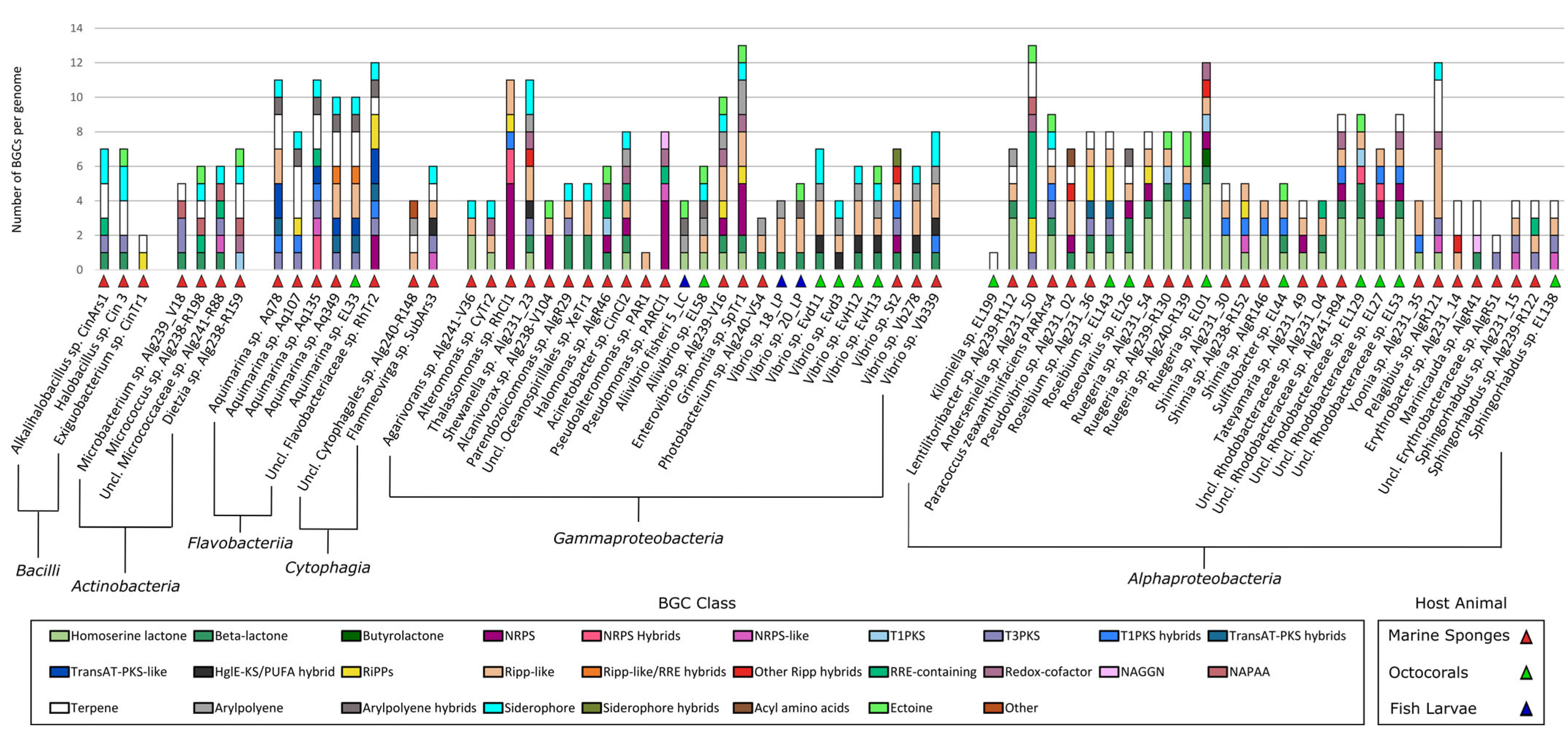
2.3. Identification of Secondary Metabolite Biosynthetic Gene Clusters (SM-BGCS) in Marine Bacterial Genomes

Using antiSMASH v6.0.0 [19], 466 SM-BGCs of 28 compound classes were identified in the 70 genomes (Figure 4). All details of the antiSMASH classification, including SM-BGC size and location in the genome are provided in Table S7. Overall, the number of SM-BGCs per genome varied from one SM-BGC in strains Kiloniella sp. EL199 and Pseudoalteromonas sp. PAR1 to 13 SM-BGCs in strains Grimontia sp. SpTr1 and Anderseniella sp. Alg231_50. Twelve isolates possessed more than 10 SM-BGCs, while the average SM-BGC count was 6.5 SM-BGCs per genome (Figure 4). Of the 466 SM-BGCs, 38 had above 70% gene cluster similarity with SM-BGCs that produce known compounds and that are present in the MIBiG database (Table S8). Of those 38 SM-BGCs, 20 coded for carotenoids, 11 for iron chelating compounds (i.e., bisucaberin B, aerobactin, acinetobactin, desferrioxamine E and amphi-enterobactins 1/2/3/4) and seven for ectoine, known to be involved in osmoregulation (Table S8).
The Bacilli isolates Alkalihalobacillus sp. CinArs1 and Halobacillus sp. Cin3 shared similar SM-BGC profiles, each harboring a type III polyketide synthase (T3PKS) and a betalactone cluster, while Exiguobacterium sp. CinTr1 harbored one ribosomally synthesized and post-translationally modified peptide (RiPP) SM-BGC (Figure 4). Likewise, Actinobacteria isolates Microbacterium sp. Alg239_V18 and unclassified Micrococcaceae sp. Alg241-R88 harbored each a T3PKS cluster, while Dietzia sp. Alg238-R159 harbored a type I polyketide synthase (T1PKS) cluster (Figure 4).
The Flavobacteriia isolates investigated in this study showed quite unique SM-BGC profiles, with Aquimarina sp. strains Aq78, Aq349 and EL33 hosting many PKS SM-BGCs, as well as RiPP-like clusters (Figure 4). Unclassified Flavobacteriaceae sp. strain RHTr2 harbored five PKS SM-BGCs (including T1PKS, T3PKS and transAT-PKS), in addition to two NRPS and two RiPP clusters (Figure 4), pointing towards a very rich and novel secondary metabolism in this potentially novel taxon.
None of the Gammaproteobacteria genomes harbored terpene SM-BGCs, which were present in all other bacterial classes investigated here (Figure 4). Overall, the Gammaproteobacteria SM-BGC content was highly diverse. The SM-BGC profile of the marine sponge derived Grimontia sp. SpTr1 genome showed similarities to that of sponge-derived Enterovibrio sp. strain Alg239-V16; both harbored beta-lactone, NRPS, RiPP and RiPP-like clusters (Figure 4). Thalassomonas sp. strain RHCl1 stood out with five NRPS clusters (Figure 4).
The Alphaproteobacteria strains also displayed a varied secondary metabolite biosynthesis potential, but most strains lacked siderophore clusters (Figure 4). The Rhodobacteraceae family was enriched in homoserine lactone SM-BGCs. Octocoral-derived Ruegeria sp. EL01 and the unclassified Rhodobacteraceae sp. strains EL27, EL53, EL129 and sponge-derived Alg241-R94 all harbored, among others, one NRPS, one RiPP-like and one T1PKS or T1PKS hybrid cluster in their genomes (Figure 4). Marine sponge-derived Pelagibius sp. strain Alg239-R121 and Anderseniella sp. strain Alg231_50 stood out for their many RiPP-like clusters (Figure 4).

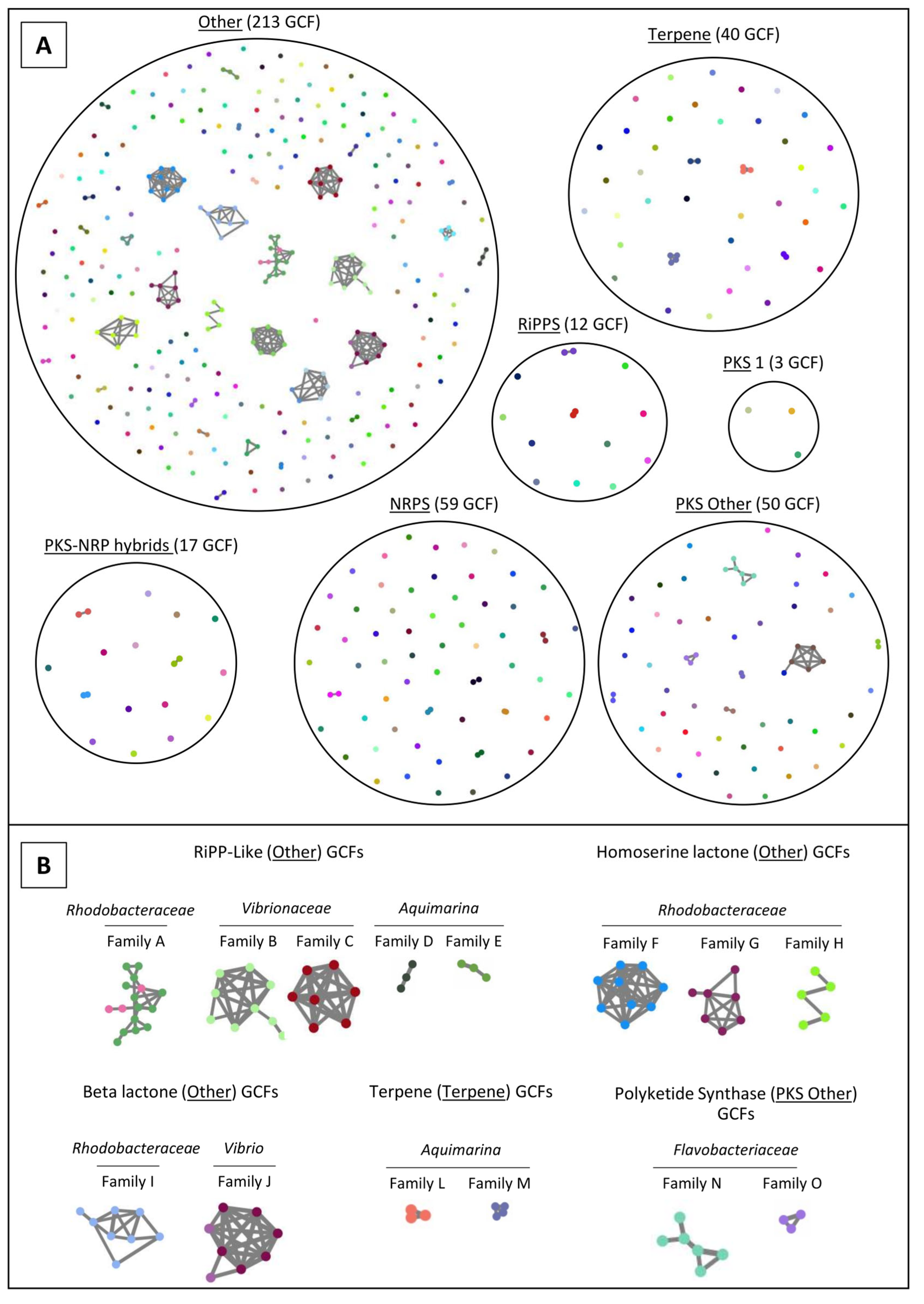
2.4. Network Analysis of SM-BGCs

To determine the similarity of the 466 SM-BGCs annotated across the 70 marine bacterial genomes, similarity networks were generated using BiG-SCAPE v1 [38]. In total, the 466 SM-BGCs grouped into 394 gene cluster families (GCFs), i.e., groups of SM-BGCs that share >70% similarity according to a BiG-SCAPE metric which integrates number of shared Pfam domains, synteny of the Pfam domains and sequence similarity. Fifty-seven GCFs consisted of two or more SM-BGCs. The remaining 337 GCFs (85%) were singletons, i.e., SM-BGCs with no relevant similarity to other SM-BGCs (Figure 5A). Many SM-BGCs annotated by antiSMASH v.6.0.0 were not classified under the BiG-SCAPE nomenclature, and instead grouped under the category “Other”.
Several large GCFs, consisting of SM-BGCs of phylogenetically related bacteria, were present in the compound categories “Other”, “Terpene” and “PKS Other” (Figure 5A). The GCFs A–E (Figure 5B) were classified as “RiPP-like” by antiSMASH, which is a broad term for SM-BGCs similar to RiPPs [19]. RiPP-like family A consisted of SM-BGCs from 16 of the 17 Rhodobacteraceae (all except Paracoccus zeaxanthinifaciens strain ParArs4) genomes of this study (Table S9). The GCFs F–H encompassed homoserine lactone SM-BGCs classified by antiSMASH. These GCFs consisted solely of SM-BGCs from isolates of the Rhodobacteraceae family (Figure 5B). Each of these GCFs (F, G, H) consisted of SM-BGCs from at least two Rhodobacteraceae genera, although SM-BGCs from one genus dominated each GCF (Table S9). Notably, many Rhodobacteraceae isolates (12 out of 16 strains) that harbored a RiPP-like SM-BGC of GCF A also had one or more homoserine lactone SM-BGCs in GCFs F, G and/or H (Figure 5B, Table S9). The genomes to which the SM-BGCs of GCFs A–O belong are detailed in Table S9.
Figure 5. Sequence similarity network analysis of the SM-BGCs from the 70 genome-sequenced marine bacterial isolates. Each colored node represents a single SM-BGC identified by antiSMASH v6. Grey lines connecting certain SM-BGCs indicate their respective grouping into Gene Cluster families (GCFs). There were 337 singleton-GFCs (i.e., SM-BGCs with no relevant similarity to other SM-BGCs) and 57 GCFs that consisted of two or more SM-BGCs in the dataset. (A) Organization of SM-BGCs and GCFs according to BiG-SCAPE’s compound class nomenclature. (B) Taxon-specific GCFs identified among the different compound classes. A list of the bacterial isolates belonging to each taxon-specific GCF is available in Table S9.
Marinedrugs 21 00034 g005

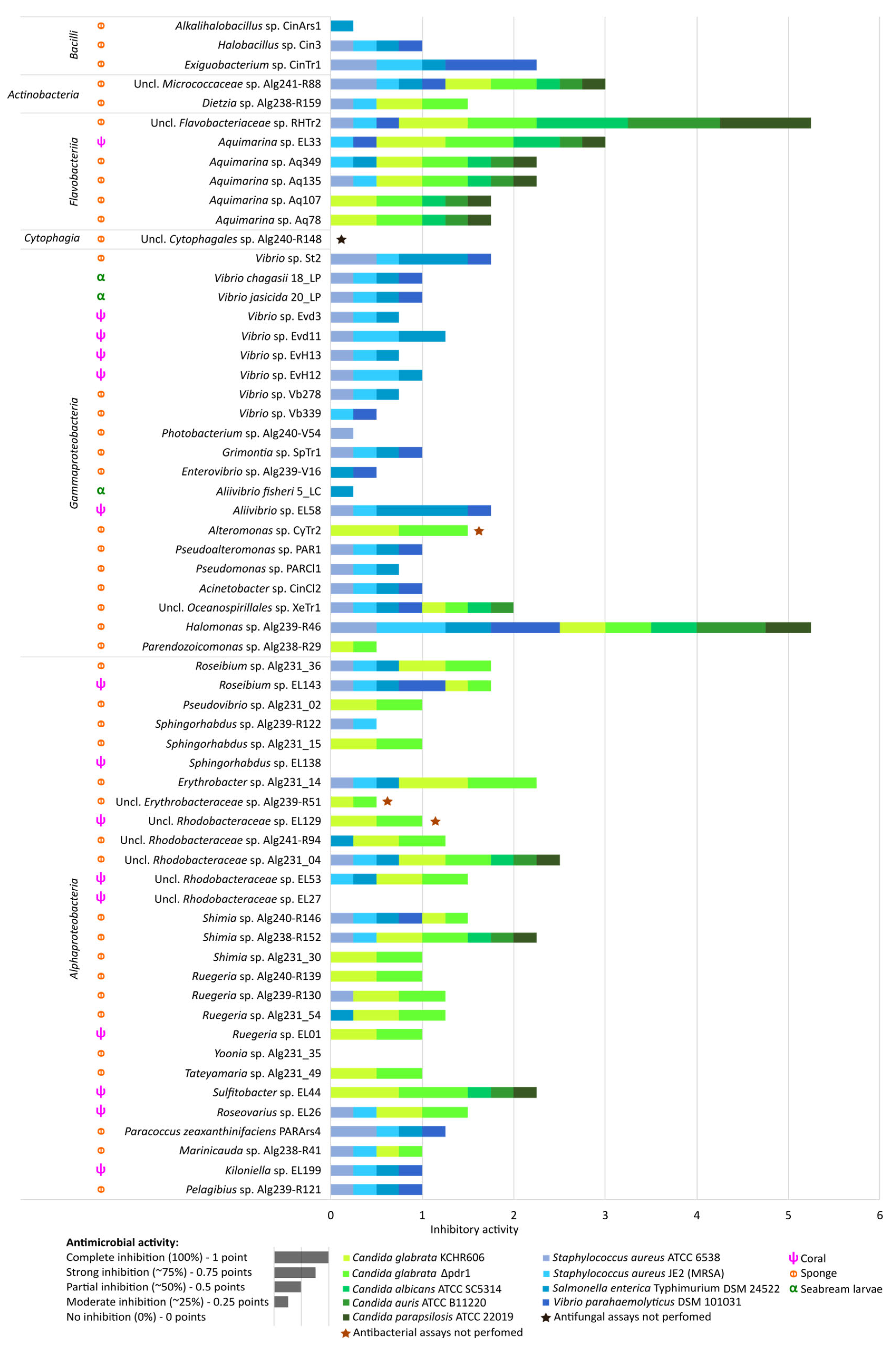
2.5. Antimicrobial Activities of Marine Bacterial Isolates

Following the detailed inspection of their secondary metabolite biosynthesis capacities, the genome-sequenced isolates were screened for antifungal and antibacterial activities against five human-pathogenic fungal strains, Candida glabrata KCHR606 [39] and Δpdr1 deletion mutant [40] (the latter with a full knockout of the PDR1 gene, encoding for the master regulator of azole drug resistance in C. glabrata), C. albicans ATCC SC5314, C. parapsilosis ATCC 22019 and C. auris ATCC B11220, and four human-pathogenic bacteria, Staphylococcus aureus ATCC 6538, methicillin-resistant Staphylococcus aureus (MRSA) strain JE2 (NR-46543), Salmonella enterica subsp. enterica serovar Typhimurium DSM 24522 and Vibrio parahaemolyticus DSM 101031 (the latter being a human, fish and shellfish pathogen).
Of the 70 genome-sequenced isolates, nine strains did not grow well enough in the cross-streak assays [41] used here for antimicrobial activity screenings. Test results are, therefore, presented for 61 genome-sequenced bacteria. Of those 61 bacteria, 57 showed some degree of activity against at least one pathogen (Figure 6). Among the Candida pathogens, C. glabrata was the most susceptible species, with 33 marine bacterial isolates causing some degree of growth inhibition in C. glabrata, irrespective of the presence or absence of the PDR1 gene. Thirteen of the 33 isolates were able to reduce growth of all Candida strains tested (Figure 6). We also observed growth inhibition of Gram-positive and Gram-negative human-pathogenic bacteria by marine isolates, with 35 isolates active against S. aureus ATCC 6538, 37 against MRSA strain JE2 (NR-46543) and 33 against S. enterica serovar Typhimurium DSM 24522 (Figure 6). Vibrio parahaemolyticus DSM 101031 was the least susceptible species in this study; only 21 marine bacterial isolates inhibited its growth.
While none of the Bacilli isolates showed antifungal activity, Halobacillus sp. CinArs1 and Exiguobacterium sp. CinTr1 showed activity against all bacterial pathogens tested here (Figure 6). Of the marine Actinobacteria, unclassified Micrococcaceae sp. Alg241-R88 showed weak activity against all tested pathogens. The six marine Flavobacteriaceae strains tested in this study showed relatively little antibacterial activity in the cross-streak assays but notable antifungal activity (Figure 6). Marine sponge derived unclassified Flavobacteriaceae sp. strain RHTr2 showed strong antifungal activity and was the only isolate which completely inhibited C. albicans, C. auris and C. parapsilosis (Figure 6).
Most (19 out of 21) marine gammaproteobacterial isolates showed some activity against pathogenic bacteria, while only four gammaproteobacterial isolates, of the orders Oceanospirillales and Alteromonadales, inhibited any of the Candida pathogens (Figure 6). Oceanospirillales isolate Halomonas sp. Alg239-R46 showed strong activity against bacterial and fungal pathogens, while unclassified Oceanospirillales sp. strain, XeTr1 was weakly active against all tested bacterial and fungal pathogens except for C. parapsilosis (Figure 6). Five strains of the Vibrionaceae family inhibited the four bacterial pathogens, whereby sponge-derived Vibrio sp. St2 and octocoral-derived Aliivibrio sp. EL58 showed strong activity against S. enterica serovar Typhimurium. However, none of the 14 tested Vibrionaceae strains, spanning five genera (i.e., Vibrio, Aliivibrio, Enterovibrio, Photobacterium and Grimontia), showed antifungal activity (Figure 6).
The marine alphaproteobacterial isolates were generally more active against fungal than bacterial pathogens (Figure 6). Most strains inhibited the growth of C. glabrata (21 out of 28), with unclassified Rhodobacteraceae sp. Alg231_04, Shimia sp. Alg238-R152 and Sulfitobacter sp. EL44 also inhibiting C. albicans, C. auris and C. parapsilosis growth (Figure 6). Five alphaproteobacterial isolates, Roseibium sp. EL143, Shimia sp. Alg240-R146, Paracoccus zeaxanthinifaciens PARArs4, Kiloniella sp. EL199 and Pelagibius sp. Alg239-R121, inhibited the four bacterial pathogens to some extent (Figure 6). Overall, the type of host animal (i.e., marine sponge, octocoral or fish) was not a determining factor for antimicrobial activity since activities were observed in isolates from the three host types. Nevertheless, two marine bacterial strains that truly stood out in this study for their strong and broad-spectrum antimicrobial activities, Halomonas sp. Alg239-R46 and Flavobacteriaceae sp. RHTr2, were both derived from marine sponges (Spongia officinalis and Rhabdastrella globostellata, respectively).
Figure 6. Antimicrobial activities of the marine bacterial isolates against human-pathogenic bacteria and Candida spp. strains. Cross-streak agar plate assay results are shown. Inhibition of pathogen growth in the presence of marine bacterial isolates was assessed qualitatively in a screening-based fashion. The stacked bar size indicates the intensity of observed growth reduction compared with negative controls (i.e., pathogens grown without the presence of marine bacteria). The antimicrobial activities were ranked on a 0—1 scale as follows: No pathogen growth inhibition—0 points; Weak inhibition—0.25 of a point; Moderate inhibition—0.50 of a point; Strong inhibition–0.75 of a point; Complete inhibition of pathogen growth–1.0 point. Different colors represent the various pathogens tested, whereby shades of green represent Candida spp. and shades of blue represent bacterial pathogens. Uncl.—unclassified.
Marinedrugs 21 00034 g006

3. Discussion

3.1. The “MicroEcoEvo” and “EcoTech-SPONGE” Culture Collections Feature a Series of Underexplored and Taxonomically Novel Marine Bacteria

This study explored the genetic diversity of 919 marine host-associated bacterial isolates from two culture collections, altogether encompassing four phyla, six classes, 17 orders, 33 families and 55 described genera, in addition to 30 isolates that were unclassifiable at either genus (19 strains), family (7 strains), or even order (4 strains) level, and most likely represent novel taxa. Unclassified Flavobacteriaceae sp. strain RHTr2 (phylum Bacteroidetes) showed strong antimicrobial activity in this study and was closely related to Aquimarina, a marine Flavobacteriaceae genus, which is receiving increased attention for its versatile secondary metabolism [42,43], antimicrobial activities [43], and as a source of novel polyketides and antibiotic peptides [44,45]. However, neither whole genome-based AAI values, nor 16S rRNA gene similarity, offered strong enough support to classify RHTr2 as Aquimarina. Interestingly, the 16S rRNA gene sequence of an uncultured Flavobacteriaceae bacterium [46] from the coast of Guam (Pacific Ocean) and associated with the marine sponge Rhabdastrella globostellata, the same sponge species from which RHTr2 was obtained, shared 99.64% similarity with isolate RHTr2. It could suggest that this novel taxon may favor associations with marine sponges.
The unclassified Micrococcaceae strain Alg241-R88 (phylum Actinobacteria) of the “MicroEcoEvo” collection, was one of only two isolates in this study that showed inhibitory activity against all tested pathogens. Phylogenomically close to the genus Pseudarthrobacter, a sibling of the Arthrobacter genus, strain Alg241-R88 possessed a NRPS-like and a T3PKS SM-BGC. Arthrobacter spp. have been reported to possess antifungal and antibacterial [47] activity. Relatively little is known about the genus Pseudarthrobacter, yet two Pseudarthrobacter oxydans strains showed activity against S. aureus and C. albicans in a recent study [48].
The “EcoTech-SPONGE” collection harbored six unclassified Oceanospirillales isolates which likely belong to a novel, not yet described family, closely related to the Endozoicomonadaceae family. The halophilic and metabolically versatile order Oceanospirillales not only contains many endosymbiotic taxa [49], but also polyester- and oil-degrading groups, as well as deep-water dwellers [50]. Our Oceanospirillales sp. strain XeTr1 from Xestospongia sp. showed weak antimicrobial activity against all tested microbial pathogens, except for C. parapsilosis, and its SM-BGC profile was characterized by beta-lactones, a compound class known for its chemical diversity and clinical potential [51].

3.2. Both Culture Collections Comprise Taxa Typically Known for Their Symbiotic Lifestyles

Seven strains in this study, one of them with a sequenced genome, belonged to the family Endozoicomonadaceae (Oceanospirillales, Gammaproteobacteria), a family typically found in association with marine animals and formally described only in 2018 [52]. Indeed, the core microbiome of corals, particularly of temperate octocorals, primarily consists of Gammaproteobacteria, with Endozoicomonadaceae phylotypes frequently dominating the prokaryotic community [18,21]. Members of this family have been suggested to be involved in sulfur and carbon cycling in their hosts [53,54] and are indicators of coral health [21]. Moreover, metagenomic analyses have suggested a role for Endozoicomonadaceae symbionts in the processing of chitin, the most abundant natural polysaccharide in marine environments and a major component of the zoo- and phytoplankton prey of suspension-feeding marine invertebrates [54,55]. In a recent study, unculturable, symbiotic Endozoicomonadaceae lineages did not possess any SM-BGCs in their genomes, suggesting that their main function is not chemical defense but rather nutrient provision and cycling [54]. Some cultured Endozoicomonadaceae representatives, however, were found by Sweet et al. to possess bacteriocin (RiPP) clusters [56]. Parendozoicomonas sp. Alg238-R29 was revealed in this study to also harbor a T3PKS cluster in addition to the RiPP cluster, although its antimicrobial activity in the cross-streak assays was negligible. The halophilic genus Halomonas (Halomonadaceae, Oceanospirillales) has been used in probiotic treatments of shrimp due to its antiviral and anti-microbial activities [57,58] and was also part of a ‘Beneficial Microbes for Corals’ (BMC) consortium that prevented corals from bleaching under pathogen and temperature stress [59]. Both the coral-associated Halomonas BMC strain [56] and our marine sponge-derived Halomonas sp. Alg239-R46 harbored a T1PKS cluster in their genomes. Indeed, Halomonas sp. Alg239-R46 was among the most bioactive isolates of this study, showing inhibitory activity against all bacterial and fungal pathogens tested and strong inhibition against MRSA, V. parahaemolyticus and C. auris. These results suggest competitive ability of Halomonas spp. in host-associated settings through the release of antimicrobial compounds, which could lead to chemical defense of their natural hosts against fungal and bacterial infections.
The marine bacterial genus Pseudoalteromonas (Pseudoalteromonadaceae, Alteromonadales) is another taxon frequently found in association with eukaryotic hosts [56,60,61] and several Pseudoalteromonas strains were among the above-mentioned BMC consortium [59]. Our two culture collections consisted of 28 Pseudoalteromonas strains, 26 isolated from marine sponges and two from the octocoral Eunicella verrucosa. Pseudoalteromonas spp. are versatile antimicrobial compound producers, with 69 antimicrobial compounds reported until 2016 [61]. More recently, a clade consisting of at least 19 Pseudoalteromonas members was reported by Chau and colleagues to possess large numbers of SM-BGCs (on average >10 SM-BGCs per genome), including SM-BGCs which produce polyketides, non-ribosomal peptides and RiPPs/bacteriocins [62]. In this study, Pseudoalteromonas sp. PARCl1 displayed moderate antibacterial activity and was also found to harbor multiple NRPS SM-BGCs. However, strain PARC11 is phylogenetically different from the SM-BGC-enriched Pseudoalteromonas clade described by Chau and colleagues [62]. In contrast, 14 other Pseudoalteromonas sp. strains of the “EcoTech-SPONGE” collection had >99% 16S rRNA gene similarity to the abovementioned SM-BGC-enriched Pseudoalteromonas [62] (Table S10). These 14 Pseudoalteromonas strains may thus be a valuable target for future bioprospection studies, possibly also displaying a complex secondary metabolism.
Finally, Roseibium (homotypic synonym Labrenzia) is a marine alphaproteobacterial genus (family Stappiaceae, order Hyphomicrobiales) that is frequently isolated from filter- or suspension-feeding, sessile invertebrates such as sponges [25], corals [27,63], and oysters [64]. Several compounds have already been isolated from this genus, including labrenzbactin, a siderophore with antibacterial and anticancer activity [65], and labrenzin, an analogue of the polyketide pederin with anticancer activity [66]. The “MicroEcoEvo” collection harbored as many as 62 Roseibium strains and, in this study, Roseibium album strains Alg231_36 and EL143 showed inhibitory activity towards human-pathogenic bacteria and C. glabrata. In line with earlier findings for the genus [67], both strains harbored transAT-PKS, T3PKS, NRPS and multiple RiPP-like SM-BGCs, underpinning their potential to produce a diverse range of secondary metabolites.

3.3. Reduction of Carbon-Content and Prolonged Incubation Periods Promote an Increased Diversity of Culturable Marine Bacteria

The cultivation of sponge-associated bacteria on a range of culture media with varying carbon sources and concentrations and additives has been used as a strategy to expand the repertoire of culturable sponge- and coral-associated bacteria [56,60]. It has been suggested that bacterial division rates within the sponge microbiome are relatively low, wherefore slow-growing bacteria may only be captured with long cultivation times [60]. Moreover, cultivation is a competitive process and high carbon-content media tend to favor fast growing genera (r-strategists) such, e.g., Vibrio and Pseudovibrio, allowing them to develop colonies quickly. Within 2–3 days of incubation, they may take over the entire surface of the cultivation plate and can prevent slow-growing taxa (k-strategists) to emerge, as observed elsewhere [24]. The Spongia officinalis-associated bacteria of the “MicroEcoEvo” collection were cultured on three low-carb media, R2A [34], MG50 [25] and VXA [35] (instead of nutrient-rich MA) with an incubation period of eight weeks, which promoted the growth of 15 bacterial families that were not obtained on full strength marine agar. These conditions likely contributed to the cultivation of slow-growing or difficult-to-cultivate marine taxa, such as Anderseniella, Lentilitoribacter, Pelagibius, Tateyamaria, Marinicauda and Parendozoicomonas, all understudied genera with limited numbers of known species and genomes available. Indeed, according to the List of Prokaryotic Names with Standing in Nomenclature (LPSN) [68] (last checked September 2022), the genera Lentilitoribacter, Anderseniella and Parendozoicomonas comprise only one formally described species each, while Pelagibius consists of two species and Tateyamaria and Marinicauda three species, respectively. Likewise, according to NCBI [69] (last checked September 2022), very few genomes are so-far available from cultured strains of these genera: seven for Tateyamaria and Marinicauda each, three for Pelagibius, two for Parendozoicomonas, and one for Anderseniella and Lentilitoribacter, respectively, including the genomes described in this study. This not only highlights the suitability of low-carb media and long incubation periods for the cultivation of understudied marine bacteria, but also emphasizes the important contribution of the here described culture collections as a resource of novel marine bacterial strains and genomes of understudied lineages.

3.4. The “MicroEcoEvo” and “EcoTech-SPONGE” Collections Are a Reservoir of Chemical Novelty with Most SM-BGCs Showing Little Homology to Those of Known Compounds

This study identified 466 SM-BGCs across 70 marine bacterial genomes, with 428 SM-BGCs (>90%) having little or no homology to SM-BGCs coding for known compounds. This suggests a vastly underexplored chemical diversity among our culture collections. RiPP-like, NRPS, PKS and terpene SM-BGCs were detected most frequently, all of which are compound classes that have been associated with antimicrobial molecule production in the past. The 38 SM-BGCs with high homology (>70%) to SM-BGCs of known compounds included those producing molecules that are pigments and/or have antioxidant functions (e.g., carotenoids, flexirubins), and those involved in osmoregulation (ectoine), and iron sequestration (e.g., bisucaberin B, aerobactin). These compounds may provide important ecological advantages to marine bacteria in their natural habitats, and are among the factors which support beneficial interactions with marine animal hosts [70].
The Flavobacteriaceae isolates examined in this study stood out for their many polyketide-synthase encoding SM-BGCs, e.g., T1PKS, T3PKS, transAT-PKS and hybrid-PKS SM-BGCs, with six of the transAT-PKS and three of the T3PKS forming GCFs N and O, respectively. TransAT-PKS GCF N was present in the genomes of Aquimarina sp. strains Aq78, Aq349 and EL33 as well as unclassified Flavobacteriaceae strain RHTr2, and corresponded to the recently identified polyketide cuniculene [44]. Silva et al. [43] demonstrated that the cuniculene SM-BGC is shared by several Aquimarina species, and the present study shows that other members of the Flavobacteriaceae family may also possess the capacity to produce cuniculene or derivatives thereof. The ecological role and bioactivity of cuniculene is yet to be described [44]. However, other natural products synthesized by transAT-PKSs exhibit antibiotic [71] and anticancer [72] activities, among others. The SM-BGCs belonging to T3PKS family O, present in the genomes of Aquimarina strains Aq78, Aq349 and EL33, were not like any SM-BGC of a known compound present in the MIBiG database, and most likely encode for a novel polyketide. These three Aquimarina strains indeed share similar metabolomes, although most of their secondary metabolites have not yet been identified [43].
Of the 25 Gammaproteobacteria genomes analyzed in this study, four isolates stood out for their large number (10–13) of SM-BGCs. One of them was Shewanella sp. strain Alg231_23, characterized by several RiPP clusters in addition to a T3PKS cluster. Two macrocyclic polyketides with antibacterial activity against several multi-resistant bacteria, including MRSA, were recently identified in Shewanella algae [73]. The “MicroEcoEvo” collection hosted as many as 25 Shewanella (Shewanellaceae, Alteromonadales) strains, obtained from the octocorals E. labiata and E. verrucosa and marine sponges S. spinosulus and I. variabilis. Shewanella spp. are common members of the microbiome of algae [74], molluscs [75] and sponges [76]. Some strains are known to have probiotic effects on Sparus aurata and Solea senegalensis, reducing larval and/or juvenile mortality [77] as they increase host resistance to pathogens [78]. The secondary metabolisms of the other three gammaproteobacterial genera with ≥10 SM-BGCs, namely Enterovibrio sp. Alg239-V16, Grimontia sp. SpTr1 and Thalassomonas sp. RhCl1, notable for their many NRPS and RiPP clusters, are still vastly underexplored and literature reports on the topic are scarce. Nevertheless, one Thalassomonas-derived RiPP cluster was recently characterized and its corresponding lanthipeptides, thalassomonasin A and B, were produced by heterologous expression in Escherichia coli [79].
RiPP BGCs from 16 of the 17 Rhodobacteraceae (Alphaproteobacteria) isolates (all except Paracoccus zeaxanthinifaciens PARArs4) analyzed in this study grouped together in one gene cluster family (A), given their high similarity. RiPP compounds are often antibacterial agents, particularly the bacteriocin subclass of RiPPs is widely known for its antibacterial activity [80]. However, peptides of this SM-BGC class can have many functions, some acting as siderophores, while others are involved in biofilm formation and maintenance [81]. All Rhodobacteraceae genomes included at least one N-acyl homoserine lactone (AHL) SM-BGCs, with all Ruegeria and four of the unclassified Rhodobacteraceae strains hosted two to five AHL BGCs per genome. AHLs are involved in chemical communication such as quorum sensing, a mechanism which, among others, frequently controls secondary metabolite production in Proteobacteria. Martens et al. (2007) found that the presence of PKS and NRPS genes in marine Rhodobacteraceae correlates to some extent with AHL production and antagonistic activity [82]. Moreover, the synthesis of the antimicrobial compound tropodithietic acid (TDA) in Phaeobacter and Ruegeria spp. is mediated by AHLs and quorum sensing [83]. Most Rhodobacteraceae-derived AHL SM-BGCs reported in this study belonged to one of three GCFs. Notably, in strains hosting multiple AHL SM-BGCs on their genomes (such as, e.g., our Ruegeria spp. strains), these SM-BGCs were not identical to one another but belonged to distinct AHL GCFs, suggesting that each AHL could have a distinct structure and possibly control the expression of a distinct gene set. It is not uncommon for marine Rhodobacteraceae genomes to possess multiple copies of luxR and/or luxI homologs, the genes which encode the proteins that produce and respond to AHLs [83]. The larger number and diversity of AHL clusters likely contributes to the structural variety of AHLs in marine Rhodobacteraceae strains, such as the high variation in the acyl side chains, with lengths ranging from C4 to C18 and differing degrees of saturation [83]. In our study, not every AHL SM-BGC carried a LuxR or LuxR-like domain, but all possessed an autoinducer synthase domain, indicating capacity to produce AHL-like molecules. The lack of LuxR domains in certain AHL SM-BGCs may suggest that other control mechanisms are present in our Rhodobacteraceae strains, as Pfam domains of unknown function were also common in these SM-BGCs.

3.5. Taxon-Specific Differences in Antimicrobial Activity of Marine Bacteria Suggest Distinct Mechanisms Involved in Antagonism against Human-Pathogenic Bacteria versus Candida

The present study showed that underexplored marine bacterial taxa have antimicrobial activities against canonical human pathogens, which warrants further investigation. Among the tested Candida pathogens, growth reduction in the presence of marine bacteria was most frequently observed in C. glabrata, with no difference in the susceptibility of the Δpdr1 deletion mutant versus the wild type. The PDR1 gene in C. glabrata encodes a transcription factor, which, in turn, induces expression of drug efflux pumps and is known to be a centerpiece in resistance to fluconazole-type antifungals [84]. Given the similar susceptibility of C. glabrata wild type and Δpdr1, the mechanisms behind the antifungal activities observed in this study are likely independent of PDR1-controlled resistance. This observation suggests that the underlying antifungal agents are likely to be effective against azole resistant clinical isolates. Noteworthily, C. glabrata appears to be more susceptible to most tested marine bacteria than the other three Candida species tested here, although C. glabrata is, of the four species, most often the one responsible for resistance to antifungal drugs, making it a particularly hard to treat pathogen [85]. Inhibition of C. albicans, C. parapsilosis and C. auris was less common in this study, but when it occurred, C. glabrata was also inhibited, indicating that the mode of action of the putative compounds involved in these cases were broad and not Candida species-specific. In contrast, cases where only C. glabrata was inhibited could suggest that the other Candida species share a common resistance mechanism, which C. glabrata lacks. Future studies should focus on the isolation and purification of the antifungal compounds to shed light on the targets for drug action.
The Gram-positive Exiguobacterium sp. strain CinTr1 (Bacillaceae, Firmicutes) inhibited the growth of all bacterial pathogens and had a particularly strong effect on the fish pathogen Vibrio parahaemolyticus. However, there was no inhibitory activity towards Candida, suggesting that activities are specific for prokaryotic cells, making this strain a good candidate for further antibiotic compound research. In line with our findings, non-marine Exiguobacterium spp. strains are also known for their potent antibacterial activities against the human-pathogenic, Gram-negative species Escherichia coli, Shigella flexneri, Klebsiella pneumonia and Salmonella enterica [86].
The 14 Vibrionaceae strains (spanning five genera) examined in the present study showed no observable antifungal activity, but all strains showed antibacterial activity, with five of the 14 isolates inhibiting all bacterial pathogens tested here. Notably, all Vibrionaceae strains but Aliivibrio fisheri 5_LC (which had only very weak antibacterial activity) harbored one or more RiPP-like SM-BGC(s), which could serve as starting point for further investigation into the antibacterial compound(s) involved. In contrast, many alphaproteobacterial isolates, particularly members of the Rhodobacteraceae family, performed well against Candida, but poorly against bacterial pathogens, hinting at different mechanisms and molecules involved in antagonism against bacterial and fungal disease agents. Sulfitobacter sp. EL44, Shimia sp. Alg238-R152 and unclassified Rhodobacteraceae Alg231_04 from the “MicroEcoEvo” collection stood out among the 17 Rhodobacteraceae strains surveyed in this study, and showed inhibitory effects against C. albicans, C. parapsilosis and C. auris, in addition to C. glabrata. However, with four to five SM-BGCs from four to five different compound classes per genome, the above-mentioned isolates were not among the Rhodobacteraceae strains with the highest number and diversity of SM-BGCs (Ruegeria sp. EL01 and Pelagibius sp. Alg239-R121 had 12 SM-BGCs and 7-8 compound classes per genome, for example). This shows that the number and diversity of SM-BGCs is not necessarily a reliable predictor of in vitro bioactivity. It is not unusual for SM-BGCs to be ‘silent’ under standard culture conditions in the laboratory, i.e., the genes are not transcribed and, therefore, the underlying metabolites are not produced. Testing different experimental conditions is one way of stimulating their expression, a strategy explored by the ‘one strain, many compounds’ (OSMAC) approach [87]. Even simple changes such as switching from solid to liquid medium can already result in the biosynthesis of different metabolites, and in different inhibitory activities. This was recently shown for several Aquimarina spp. strains, which exhibited inhibitory activity against S. aureus ATCC 6538 and MRSA in broth microdilution assays with liquid culture extracts, but not in the cross-streak assays where agar plates were used [43]. A future strategy could therefore be the application of alternative antimicrobial activity screening methods, and the OSMAC approach on those strains which revealed a rich SM-BGC profile but little antimicrobial activity in the cross-streak assays.

4. Materials and Methods

4.1. The “MicroEcoEvo” and “EcoTech-SPONGE” Culture Collections

This study was embedded in the national research project “SymbioReactor”, supported by the ‘Portuguese Ministry of the Sea’, and aimed to exploit the diversity, antimicrobial activities, and natural product biosynthesis capacities of two comprehensive culture collections, totaling 919 marine host-associated bacteria. The first “MicroEcoEvo” collection comprised 800 isolates and was established between 2010 and 2020 during multiple sampling events of the temperate marine sponges Sarcotragus spinosulus, Icrcina variabilis and Spongia officinalis [24,25,26], the octocorals Eunicella labiata [27] and Eunicella verrucosa [36] and fish larvae of Sparus aurata [28,29,88] from Portugal. Sampling and sample processing were previously described [24,25,26,27,28,29,88]. Animal tissue was washed with artificial seawater to remove external bacterial colonizers after which the soft tissue was homogenized in sterile Ca2+ and Mg2+-free artificial seawater (CMFASW: 27 g L−1 NaCl, 1 g L−1 Na2SO4, 0.8 g L−1 KCl and 0.18 g L−1 NaHCO3, 1 g of soft tissue per 9 mL CMFASW w/v) using a sterile mortar and pestle. One milliliter of each homogenate was then serially diluted with CMFASW, and various dilutions were plated for cultivation. Several marine culture media and conditions were employed, including full strength Marine Agar [24,28,29,36,88] and half strength (1:2 diluted) Marine Agar (MA) [27], 50× diluted Marine Gellan Gum (MG50) [25] as well as R2A and VXA [26] media. Incubation temperatures ranged from 18 °C [27] to 25 °C [24] and incubation time from three days [24] to up to eight weeks [25] in the different studies (see Table S1 for more details on the isolates and culture conditions of the “MicroEcoEvo” collection).
The second, “EcoTech-SPONGE” collection consisted of 119 bacterial isolates from 15 tropical marine sponge species of eight orders of the Demospongia class collected off the coast of Taiwan in 2017 [31,32]. Here, half strength (1:2 diluted) MA medium was used at 17 °C incubation temperature for cultivation of the sponge symbionts (see Table S2 for more details on the isolates and culture conditions of the “EcoTech-SPONGE” collection).
All isolates from both culture collections were picked and streaked to purity on agar plates. The purified isolates were then grown in Marine Broth (MB) and stocked in fresh MB supplemented with 20% glycerol at −80 °C. For genomic DNA extraction, 2-mL aliquots of shaken cultures (grown in MB medium until stationary phase) of the isolates were centrifuged at 10 000 g for 30 min. Genomic DNA was extracted from the resulting cell pellets using the Wizard® Genomic DNA Purification Kit (Promega, Madison, USA) according to the manufacturer’s instructions. The Purified DNA from all isolates was subjected to 16S rRNA gene amplification by PCR and Sanger sequencing for identification as described in our previous studies [24,25,26,27]. The 16S rRNA gene sequences are deposited at NCBI GenBank, and sequences and accession numbers are available in Tables S1 and S2.

4.2. 16S rRNA Gene-Based Phylogenetic Analyses

The 919 isolates from the “MicroEcoEvo” and “EcoTech-SPONGE” culture collections were taxonomically classified based on their 16S rRNA gene sequences using the “RDP Classifier” tool of the Ribosomal Database Project (RDP) v 2.11 [33]. Moreover, using the “Sequence Match” tool of RDP [33], the closest type species of each isolate (see Tables S1 and S2 for details) was recorded. Two separate 16S rRNA gene based phylogenetic trees were generated with MEGAX [89], one for the “MicroEcoEvo” culture collection, consisting of 800 16S rRNA gene sequences and one for the “EcoTech-SPONGE” collection, consisting of 119 16S rRNA gene sequences. Nucleotide sequences of each collection were aligned using the CLUSTALW algorithm. The function “find best DNA/Protein Model (ML)” was employed in MEGAX to determine the evolutionary model that fitted best each dataset. Hence, for the “MicroEcoEvo” collection, a General Time Reversible model with a discrete Gamma distribution (eight categories, +G parameter = 0.5587, [+I], 19.25%) was used while for the “EcoTech-SPONGE” collection, a Kimura 2-parameter model, also with a discrete gamma distribution (eight categories, +G parameter = 0.2898, [+I], 26.01%), fitted the data best and was employed as evolutionary model. A Maximum-Likelihood (ML) tree was then determined for each collection with bootstrap support using 500 repetitions. Trees with the highest log likelihood (–15928.12 for the “MicroEcoEvo” and –9537.38 for the “EcoTech-SPONGE” collection) were used with a total of 722 (“MicroEcoEvo” collection) and 892 (“EcoTech-SPONGE” collection) nucleotide positions in the final datasets. Both trees were drawn to scale, with branch lengths measured in the number of substitutions per site. In each tree, all positions with less than 85% site coverage were eliminated, i.e., fewer than 15% alignment gaps, missing data, and ambiguous bases were allowed at any position (partial deletion). The calculated trees were visualized and styled in iTOL (Interactive Tree Of Life) v6 [90] and Inkscape [91].

4.3. Genome Sequencing and Assembly

Seventy representative isolates (56 from the “MicroEcoEvo” and 14 from the “EcoTech-SPONGE” collection) had their genomes sequenced and deposited in public databases (See Table S3 for details and accession numbers). Fifty genomes (all from the “MicroEcoEvo” collection) were derived from previous studies and their genome sequencing and assembly procedures have been described elsewhere (e.g., [25,26,63,92,93]). Twenty genomes are original contributions of this study. To sequence these 20 genomes, genomic DNA was extracted from the microbial pellets generated by centrifugation (10 000 g for 30 min) from freshly grown liquid cultures. The Wizard genomic DNA purification kit (Promega, Madison, WI, USA) was used for DNA extraction according to the manufacturer’s instructions. DNA quality and concentration were each assessed with a NanoDropTM 2000 spectrophotometer (Thermo Fisher Scientific, Waltham, MA, USA) and a Qubit® 4.0 fluorometer (Thermo Fisher Scientific) with the dsDNA BR Assay Kit (Invitrogen, Waltham, MA, USA). DNA integrity was further evaluated by electrophoresis on a 1.2% agarose gel. Genomes were sequenced paired end (2 × 150 bp) on an Illumina NextSeq 550 platform at GENOINSEQ (Biocant, Cantanhede, Portugal). Raw reads were quality filtered, and filtered reads were assembled into contigs using SPAdes v.3.9.0 [94]. Contigs below 1000bp (800bp for Paraendozoicomonas sp. Alg238-R29) were removed.

4.4. Comparative Genomics and Phylogenomics Analysis

The 70 genome assemblies were uploaded to the Microbial Genome Atlas (MiGA) and their closest, publicly available, type species were determined using the TypeMat tool of MiGA based on the calculation of average nucleotide identity (ANI) or average amino acid identity (AAI) similarity percentage (see Table S3 for details) [37].
Moreover, to ascertain the taxonomic status of difficult-to-classify isolates (e.g., certain Sphingomonadaceae and Rhodobacteraceae strains of the “MicroEcoEvo” collection), pairwise ANI, and alignment factors (AF; i.e., the fraction of the genome that effectively aligned with the reference genome and served to calculate the ANI) with their respective closest type species were calculated using the Integrated Microbial Genomes & Microbiomes (IMG/M) system from DOE-JGI [95] (see Tables S4 and S5 for details).
For phylogenomic inference, the genome assembly files of 84 closely related type strains were downloaded from GenBank/NCBI [96]. Phylogenomics analysis of the 154 (70+84) genomes was conducted with the SpeciesTreeBuilder v.01.0 application of the DOE Systems Biology Knowledgebase (KBase) online platform [97] using the function "Insert Set of Genomes into Species Tree", after annotating all genome assemblies with Prokka within KBase. Alignments were based on a set of 49 core genes defined by Clusters of Orthologous Groups of proteins (COGs) families [97]. A maximum likelihood (ML) phylogenomic tree was then constructed using the FastTree2 algorithm [98] and the Jukes-Cantor evolutionary model with 1000 repetitions to estimate bootstrap support values. Graphical visualization and editing of the tree were performed in iTOL v6 [90] and Inkscape [91]. In addition to the phylogenomic tree, a principal coordinates analysis (PCoA) of protein family (Pfam) profiles was performed to examine clustering of the 70 genome-sequenced isolates based on their functional gene content. More information about this analysis is available in the ‘Extended Methodology’ section of Supplementary File S1.

4.5. SM-BGC Identification and Network Analysis with antiSMASH and BiG-SCAPE

To identify SM-BGCs, the genome assemblies (fasta-format) of the 70 strains were uploaded to the antiSMASH v. 6.0 [19] online server and analyzed with default settings and all available extra features “on”. Using the “Minimum Information about a Biosynthetic Gene cluster” (MIBiG) repository [19,99], the SM-BGCs identified with antiSMASH were further compared with SM-BGCs encoding the biosynthetic pathways of known compounds in the MIBiG database for the presence of homologous genes (see Tables S7 and S8 for detailed AntiSMASH and MIBiG annotation of SM-BGCs).
To construct sequence similarity networks of the SM-BGCs, the GenBank files from antiSMASH were analyzed with the BiG-SCAPE software [38] using default parameters. BiG-SCAPE defines a distance metric between gene clusters based on a comparison of their protein domain content, order, copy number and sequence identity [37], grouping SM-BGCs that share ≥70% similarity into Gene Cluster Families (GCF) [38]. The resulting SM-BGC networks were edited in Inkscape [91].

4.6. Marine Bacterial Isolates and Test Strains Used in Antimicrobial Assays

The 70 genome-sequenced isolates were revived from glycerol stocks by inoculating 50 µl of the stock solution into 10 ml of half strength MB (1:2 diluted) medium (20.05 g L−1 MB (Carl Roth GmbH + Co. KG, Karlsruhe, Germany) dissolved in a 1:1 v/v ratio of dH2O: artificial seawater (ASW)). The salt composition of ASW was as follows: 23.38 g L−1 NaCl, 2.41 g L−1 MgSO4.7H2O, 1.90 g L−1 MgCl2.6H2O, 1.11 g L−1 CaCl2.2H2O, 0.75 g L−1 KCl, 0.17 g L−1 NaHCO3. The purity of all cultures was confirmed by streaking them on half strength MA plates.
For the antifungal assays, the wild-type Candida glabrata KCHR606 [39], and Δpdr1 complete deletion mutant strains [40], kindly provided by Hiroji Chibana (Chiba University, Japan), C. albicans ATCC SC5314, C. auris ATCC B11220 and C. parapsilosis ATCC 22019 were used. For the antibacterial assays, Staphylococcus aureus ATCC 6538, methicillin-resistant Staphylococcus aureus JE2 (NR-46543, B.E.I Resources – Part of ATCC), Salmonella enterica subsp. enterica serovar Typhimurium DSM 24522 and Vibrio parahaemolyticus DSM 101031 were used. The test strains were grown and maintained in appropriate liquid media as follows: Yeast extract Peptone Dextrose (YPD) agar [20 g L−1 D(+)-glucose (anhydrous), 20 g L−1 yeast extract, 20 g L−1 agar, 10 g L−1 peptone] for Candida strains; Tryptic Soy Broth (TSB) for the human-pathogenic bacteria; full-strength MB for V. parahaemolyticus. The incubation temperatures were 37 °C for all human-pathogenic bacteria, 30 °C for the Candida strains and 24 °C for V. parahaemolyticus DSM 101031.

4.7. Cross-Streak Assays

The cross-streak assay is an antimicrobial activity screening technique that delivers qualitative or semi-quantitative results on the inhibitory activities of a certain prokaryotic isolate against a given test strain (usually also a prokaryote or a yeast) [41]. Thirty microliters of each marine bacterial isolate grown in MB were spread on 1.5% agar plates as a two-cm-wide, vertical line dividing the plate into two equal-sized halves (see scheme in Figure S2a,b). The medium content of these agar plates varied according to the test microorganisms used: full-strength MA supplemented with 0.5% (v/v) of glucose was used for Candida strains; Mueller–Hinton agar (MHA) prepared with 2% ASW was used for the pathogenic bacteria. If the marine bacterial isolates did not grow well on MHA, full strength MA was used instead. After an incubation period of 5 days at 24 °C, 5 µl of overnight-grown liquid cultures of the pathogen were placed close to the central line, ensuring the absence of contact between the different strains. For homogeneous seeding, the test strain was streaked perpendicular to the central line with an inoculation loop (Figure S2a), first toward the border of the plate and subsequently inwards. The overnight-grown liquid cultures of the pathogens were prepared as follows: pre-inoculates of the five Candida strains were prepared in YPD liquid medium and grown overnight. The next day, the optical densities (OD) of the Candida pre-cultures were determined at 600 nm, the cultures were diluted in YDP to an OD of 0.1 and incubated for five to six hours until mid-exponential phase. Before inoculation onto the cross-streak assay plates, the ODs of the Candida cultures were again adjusted to 0.1, and 5 µl of each cell suspension was then streaked onto the cross-streak plates (Figure S2a). For the antibacterial assay, overnight liquid cultures of S. aureus, MRSA, and S. enterica serovar Typhimurium were prepared in TSB and of V. parahaemolyticus in MB. Thereafter, 5 µl of these liquid cultures were spread onto the cross-streak plates (Figure S2b) as described above. After further incubation of the cross-streak assay plates (48 h for fungal pathogens and 24 h for bacterial pathogens) at 30 °C, the overall growth of test strains and the size of inhibition zones was evaluated. All experiments were performed at least in triplicate for each marine bacterium—pathogen pair. Agar plates inoculated only with the pathogens were used as negative controls (Figure S2a,b). Inhibitions were ranked as: (1.0 point) complete inhibition, no pathogen growth compared with controls, Figure S2c); (0.75 of a point) strong inhibition, ca. 75% growth reduction, Figure S2d; (0.5 of a point) moderate inhibition, ca. 50% growth reduction, Figure S2e); (0.25 of a point) weak inhibition, ca. 25% growth reduction compared with controls, Figure S2f); (0 points) no inhibition, normal growth of pathogens, similar to controls.

5. Conclusions

The present study explored two comprehensive culture collections of marine host-associated bacteria for their secondary metabolite biosynthesis capacities and antimicrobial activities against human pathogenic bacteria and Candida species known to be increasingly resistant to currently marketed drugs. It revealed the secondary metabolite coding potential and bioactivities of 70 marine host-associated bacteria, several of which belonged to understudied or novel species and genera. The here analyzed strains possessed rich and varied secondary metabolite biosynthesis potential, with many SM-BGCs having low similarity to SM-BGCs encoding known compounds, thus suggesting biosynthetic novelty. Multiple isolates showed antimicrobial activities, with Exiguobacterium sp. CinTr1, Aquimarina sp. EL33, Aq135 and Aq349, unclassified Flavobacteriaceae sp. RHTr2, Halomonas sp. Alg239-R46, unclassified Rhodobacteraceae sp. Alg231_04, Sulfitobacter sp. EL44 and Shimia sp. Alg240-R146 standing out as the most promising producers of antifungal and/or antibacterial compounds. In conclusion, the marine bacterial culture collections described here constitute a valuable resource for bioprospecting natural products and/or enzymes of pharmaceutical or biotechnological interest. Interestingly, despite extensive efforts during the last decades to isolate and screen microbial strains from marine invertebrates for pharmaceutical-biotechnological applications, our results showed that sponges and octocorals still represent an untapped resource of hitherto unknown bacterial taxa carrying genes coding for novel natural products. Beyond their potential for the blue economy sector, the microorganisms described here are also a great resource and toolbox to study the ecology and mechanisms of host-microbe interactions, and their suitability for employment as beneficial microbes for marine animals to maintain, improve and/or restore host and reef ecosystem health.

Supplementary Materials

The following supporting information can be downloaded at: https://www.mdpi.com/article/10.3390/md21010034/s1. Supplementary file S1: This file contains an extended methodology and result section on the phylogenomics assessment and the principal coordinates analysis (PCoA) of the functional (Pfam) profiles of the 70 genomes analyzed in this study, as well as supplementary figures S1 and S2: S1: Multivariate analysis of the Pfam profiles of the 70 genomes. S2: Schematic representation and examples of the antimicrobial cross-streak assays. Supplementary file S2: This file contains all Supplementary tables (S1−S10): S1 and S2: Complete list of isolates belonging to the “MicroEcoEvo” and “EcoTech-SPONGE” culture collections, respectively. S3: General genomic features, genome assembly accession numbers and quality metrices of the 70 genome sequenced isolates. S4: ANI and AAI similarity of selected, genome-sequenced isolates with the genomes of closest type species, according to MiGA. S5: ANI and AF vales of genomes of difficult-to-classify alphaproteobacterial isolates to genomes of related type species, according to DOE-JGI IMG/M system. S6: Protein family (Pfam) based functional annotation of the 70 genome-sequenced isolates. S7: antiSMASH results – SM-BGC annotation for the 70 genome sequenced isolates. S8: SM-BGCs with >70% similarity to SM-BGCs of known compounds present in the MIBiG database. S9: Overview on the gene cluster families (GCF) shown in Figure 5B and details on the origin (isolate) and compound class contributing to each GCF. S10: Pairwise comparison of the 16S rRNA gene sequences of the 14 Pseudoalteromonas isolates from the “EcoTech-SPONGE” culture collection with those of the SM-BGC enriched Pseudoalteromonas clade reported in Chau et al., 2021 [62].

Author Contributions

Conceptualization: T.K.-C., R.C.; Data curation: J.F.A., M.M., T.K.-C., R.C., C.E.; Formal analysis: J.F.A., M.M., T.K.-C., V.O.; Funding Acquisition: R.C., T.K.-C., N.C.M.G., D.F.R.C.; Investigation: J.F.A., M.M., T.K.-C., C.E., V.O., R.V., D.F.R.C., Y.M.H.; Project administration: T.K.-C., R.C.; Resources: R.C., T.K.-C., C.E., N.C.M.G., A.M.F., M.C.T., D.M.-H., D.F.R.C., Y.M.H.; Supervision: T.K.-C., R.C., N.C.M.G., M.C.T.; Validation: T.K.-C., R.C.; Visualization: J.F.A., M.M., T.K.-C.; Writing—original draft: J.F.A., T.K.-C.; Writing—review and editing. T.K.-C., R.C., M.M., V.O., C.E., D.M.-H., D.F.R.C., Y.M.H., A.M.F., M.C.T., N.C.M.G. All authors have read and agreed to the published version of the manuscript.

Funding

This work was supported by the Fundo Azul program of Direção-Geral de Política do Mar (DPGM; Ministry of the Sea, Portugal) through grant FA_05_2017_032. It also benefited from national funds from the Fundação para a Ciência e a Tecnologia (FCT), in the scope of grant “Eco-tech-Sponge” (PTDC/BIA-MIC/6473/2014–POCI-01-0145-FEDER-016531), and projects UIDB/04565/2020 and UIDP/04565/2020 of iBB, project LA/P/0140/2020 of i4HB, projects UIDP/50017/2020, UIDB/50017/2020 and LA/P/0094/2020 of CESAM and UIDB/04539/2020, UIDP/04539/2020, LA/P/0058/2020 and POCI-01-0145-FEDER-022184 of CNC. TKC is the recipient of an investigator contract (CEECIND/00788/2017) conceded by the FCT. V.O. is funded by the FCT in the scope of the framework contract foreseen in the numbers 4, 5 and 6 of article 23, of the Decree-Law 57/2016, of August 29, changed by Law 57/2017, of July 19.

Data Availability Statement

All 16S rRNA gene and whole genome sequences of this study are deposited at NCBI GenBank and European Nucleotide Archive (ENA), respectively. The 20 new genome sequences original to this study were deposited at ENA under project accession numbers PRJEB50695 and PRJEB50698. 16S rRNA gene accession numbers can be found in Table S1 (“MicroEcoEvo” collection) and Table S2 (“EcoTech-SPONGE” collection). Genome assembly accession numbers can be found in Table S3.

Acknowledgments

The authors thank Cristina Barroso for helping with genome sequencing and Ana Rita Moura Polónia from CESAM for supporting the bacterial isolation during the 2017 field sampling campaign in Taiwan. The authors are further grateful to Jorge Gonçalves from CCMAR and Nicole de Voogd from Leiden University for providing help in identifying the marine sponge and octocoral specimens used in bacterial isolation. The authors also thank Nuno Mira from iBB-IST for kindly providing the Candida parapsilosis strain ATCC 22019 used in this study.

Conflicts of Interest

The authors declare no conflict of interest. The funders had no role in the design of the study; in the collection, analyses, or interpretation of data; in the writing of the manuscript, or in the decision to publish the results.

References

  1. O’Neil, J. Review on Antimicrobial Resistance. Antimicrobial Resistance: Tackling a Crisis for the Health and Wealth of Nations; Amr-Review: London, UK, 2014. [Google Scholar]
  2. Algammal, A.M.; Hetta, H.F.; Elkelish, A.; Alkhalifah, D.H.H.; Hozzein, W.N.; Batiha, G.E.-S.; El Nahhas, N.; Mabrok, M.A. Methicillin-Resistant Staphylococcus aureus (MRSA): One Health Perspective Approach to the Bacterium Epidemiology, Virulence Factors, Antibiotic-Resistance, and Zoonotic Impact. Infect. Drug Resist. 2020, 13, 3255–3265. [Google Scholar] [CrossRef]
  3. Köck, R.; Becker, K.; Cookson, B.; van Gemert-Pijnen, J.E.; Harbarth, S.; Kluytmans, J.; Mielke, M.; Peters, G.; Skov, R.L.; Struelens, M.J.; et al. Methicillin-Resistant Staphylococcus aureus (MRSA): Burden of Disease and Control Challenges in Europe. Eurosurveillance 2010, 15, 19688. [Google Scholar] [CrossRef] [PubMed] [Green Version]
  4. Patra, S.D.; Mohakud, N.K.; Panda, R.K.; Sahu, B.R.; Suar, M. Prevalence and Multidrug Resistance in Salmonella enterica Typhimurium: An Overview in South East Asia. World J. Microbiol. Biotechnol. 2021, 37, 185. [Google Scholar] [CrossRef]
  5. Ghenem, L.; Elhadi, N.; Alzahrani, F.; Nishibuchi, M. Vibrio parahaemolyticus: A Review on Distribution, Pathogenesis, Virulence Determinants and Epidemiology. Saudi J. Med. Med. Sci. 2017, 5, 93–103. [Google Scholar] [CrossRef]
  6. Balasubramanian, R.; Im, J.; Lee, J.-S.; Jeon, H.J.; Mogeni, O.D.; Kim, J.H.; Rakotozandrindrainy, R.; Baker, S.; Marks, F. The Global Burden and Epidemiology of Invasive Non-Typhoidal Salmonella Infections. Hum. Vaccines Immunother. 2019, 15, 1421–1426. [Google Scholar] [CrossRef] [PubMed]
  7. Kumar, V.; Roy, S.; Behera, B.K.; Bossier, P.; Das, B.K. Acute Hepatopancreatic Necrosis Disease (AHPND): Virulence, Pathogenesis and Mitigation Strategies in Shrimp Aquaculture. Toxins 2021, 13, 524. [Google Scholar] [CrossRef]
  8. Nelapati, S.; Nelapati, K.; Chinnam, B.K. Vibrio parahaemolyticus—An Emerging Foodborne Pathogen—A Review. Vet. World 2011, 5, 48–62. [Google Scholar] [CrossRef]
  9. Turner, S.A.; Butler, G. The Candida Pathogenic Species Complex. Cold Spring Harb. Perspect. Med. 2014, 4, a019778. [Google Scholar] [CrossRef] [PubMed] [Green Version]
  10. Geddes-McAlister, J.; Shapiro, R.S. New Pathogens, New Tricks: Emerging, Drug-Resistant Fungal Pathogens and Future Prospects for Antifungal Therapeutics: Drug-Resistant Fungal Pathogens. Ann. N. Y. Acad. Sci. 2019, 1435, 57–78. [Google Scholar] [CrossRef]
  11. Lockhart, S.R. Candida auris and Multidrug Resistance: Defining the New Normal. Fungal Genet. Biol. 2019, 131, 103243. [Google Scholar] [CrossRef]
  12. Vivas, R.; Barbosa, A.A.T.; Dolabela, S.S.; Jain, S. Multidrug-Resistant Bacteria and Alternative Methods to Control Them: An Overview. Microb. Drug Resist. 2019, 25, 890–908. [Google Scholar] [CrossRef]
  13. Raimundo, I.; Silva, S.; Costa, R.; Keller-Costa, T. Bioactive Secondary Metabolites from Octocoral-Associated Microbes—New Chances for Blue Growth. Mar. Drugs 2018, 16, 485. [Google Scholar] [CrossRef] [PubMed] [Green Version]
  14. Anjum, K.; Abbas, S.Q.; Shah, S.A.A.; Akhter, N.; Batool, S.; Hassan, S.S. Marine Sponges as a Drug Treasure. Biomol. Ther. 2016, 24, 347–362. [Google Scholar] [CrossRef] [PubMed] [Green Version]
  15. Shetty, N.; Gupta, S. Eribulin Drug Review. South Asian J. Cancer 2014, 03, 057–059. [Google Scholar] [CrossRef] [PubMed]
  16. Cuevas, C.; Francesch, A. Development of Yondelis® (Trabectedin, ET-743). A Semisynthetic Process Solves the Supply Problem. Nat. Prod. Rep. 2009, 26, 322. [Google Scholar] [CrossRef]
  17. Pita, L.; Rix, L.; Slaby, B.M.; Franke, A.; Hentschel, U. The Sponge Holobiont in a Changing Ocean: From Microbes to Ecosystems. Microbiome 2018, 6, 46. [Google Scholar] [CrossRef]
  18. van de Water, J.A.J.M.; Allemand, D.; Ferrier-Pagès, C. Host-Microbe Interactions in Octocoral Holobionts—Recent Advances and Perspectives. Microbiome 2018, 6, 64. [Google Scholar] [CrossRef] [Green Version]
  19. Blin, K.; Shaw, S.; Kloosterman, A.M.; Charlop-Powers, Z.; van Wezel, G.P.; Medema, M.H.; Weber, T. AntiSMASH 6.0: Improving Cluster Detection and Comparison Capabilities. Nucleic Acids Res. 2021, 49, W29–W35. [Google Scholar] [CrossRef]
  20. Karimi, E.; Ramos, M.; Gonçalves, J.M.S.; Xavier, J.R.; Reis, M.P.; Costa, R. Comparative Metagenomics Reveals the Distinctive Adaptive Features of the Spongia Officinalis Endosymbiotic Consortium. Front. Microbiol. 2017, 8, 2499. [Google Scholar] [CrossRef] [Green Version]
  21. Keller-Costa, T.; Lago-Lestón, A.; Saraiva, J.P.; Toscan, R.; Silva, S.G.; Gonçalves, J.; Cox, C.J.; Kyrpides, N.; Nunes da Rocha, U.; Costa, R. Metagenomic Insights into the Taxonomy, Function, and Dysbiosis of Prokaryotic Communities in Octocorals. Microbiome 2021, 9, 72. [Google Scholar] [CrossRef]
  22. Rodriguez Jimenez, A.; Dechamps, E.; Giaux, A.; Goetghebuer, L.; Bauwens, M.; Willenz, P.; Flahaut, S.; Laport, M.S.; George, I.F. The Sponges Hymeniacidon perlevis and Halichondria panicea Are Reservoirs of Antibiotic-producing Bacteria against Multi-drug Resistant Staphylococcus aureus. J. Appl. Microbiol. 2021, 131, 706–718. [Google Scholar] [CrossRef] [PubMed]
  23. Schultz, J.; Modolon, F.; Rosado, A.S.; Voolstra, C.R.; Sweet, M.; Peixoto, R.S. Methods and Strategies to Uncover Coral-Associated Microbial Dark Matter. mSystems 2022, 7, e00367-22. [Google Scholar] [CrossRef] [PubMed]
  24. Esteves, A.I.S.; Hardoim, C.C.P.; Xavier, J.R.; Gonçalves, J.M.S.; Costa, R. Molecular Richness and Biotechnological Potential of Bacteria Cultured from Irciniidae Sponges in the North-East Atlantic. FEMS Microbiol. Ecol. 2013, 85, 519–536. [Google Scholar] [CrossRef] [PubMed] [Green Version]
  25. Karimi, E.; Keller-Costa, T.; Slaby, B.M.; Cox, C.J.; da Rocha, U.N.; Hentschel, U.; Costa, R. Genomic Blueprints of Sponge-Prokaryote Symbiosis Are Shared by Low Abundant and Cultivatable Alphaproteobacteria. Sci. Rep. 2019, 9, 1999. [Google Scholar] [CrossRef] [Green Version]
  26. Karimi, E.; Costa, R. Isolation and Genome Sequencing of 14 Spongia sp. Bacterial Associates Expands the Taxonomic and Functional Breadth of the Cultivatable Marine Sponge Microbiome. BioRxiv 2020. [Google Scholar] [CrossRef] [Green Version]
  27. Keller-Costa, T.; Eriksson, D.; Gonçalves, J.M.S.; Gomes, N.C.M.; Lago-Lestón, A.; Costa, R. The Gorgonian Coral Eunicella labiata Hosts a Distinct Prokaryotic Consortium Amenable to Cultivation. FEMS Microbiol. Ecol. 2017, 93. [Google Scholar] [CrossRef] [PubMed]
  28. Sanches-Fernandes, G.M.M.; Califano, G.; Keller-Costa, T.; Castanho, S.; Soares, F.; Ribeiro, L.; Pousão-Ferreira, P.; Mata, L.; Costa, R. Draft Genome Sequence of Vibrio chagasii 18LP, Isolated from Gilthead Seabream (Sparus aurata) Larvae Reared in Aquaculture. Microbiol. Resour. Announc. 2021, 10, e00658-21. [Google Scholar] [CrossRef]
  29. Sanches-Fernandes, G.M.M.; Califano, G.; Keller-Costa, T.; Castanho, S.; Soares, F.; Ribeiro, L.; Pousão-Ferreira, P.; Mata, L.; Costa, R. Draft Genome Sequence of Vibrio jasicida 20LP, an Opportunistic Bacterium Isolated from Fish Larvae. Microbiol. Resour. Announc. 2021, 10, e00813-21. [Google Scholar] [CrossRef]
  30. Califano, G.; Franco, T.; Gonçalves, A.C.S.; Castanho, S.; Soares, F.; Ribeiro, L.; Mata, L.; Costa, R. Draft Genome Sequence of Aliivibrio fischeri Strain 5LC, a Bacterium Retrieved from Gilthead Sea Bream (Sparus aurata) Larvae Reared in Aquaculture. Genome Announc. 2015, 3, e00593-15. [Google Scholar] [CrossRef] [Green Version]
  31. Oliveira, V.; Polónia, A.R.M.; Cleary, D.F.R.; Huang, Y.M.; Voogd, N.J.; Rocha, U.N.; Gomes, N.C.M. Characterization of Putative Circular Plasmids in Sponge-associated Bacterial Communities Using a Selective Multiply-primed Rolling Circle Amplification. Mol. Ecol. Resour. 2021, 21, 110–121. [Google Scholar] [CrossRef]
  32. Oliveira, V.; Polónia, A.R.M.; Cleary, D.F.R.; Huang, Y.M.; de Voogd, N.J.; Keller-Costa, T.; Costa, R.; Gomes, N.C.M. Assessing the Genomic Composition, Putative Ecological Relevance and Biotechnological Potential of Plasmids from Sponge Bacterial Symbionts. Microbiol. Res. 2022, 265, 127183. [Google Scholar] [CrossRef] [PubMed]
  33. Cole, J.R.; Wang, Q.; Fish, J.A.; Chai, B.; McGarrell, D.M.; Sun, Y.; Brown, C.T.; Porras-Alfaro, A.; Kuske, C.R.; Tiedje, J.M. Ribosomal Database Project: Data and Tools for High Throughput RRNA Analysis. Nucleic Acids Res. 2014, 42, D633–D642. [Google Scholar] [CrossRef] [PubMed] [Green Version]
  34. Reasoner, D.J.; Geldreich, E.E. A New Medium for the Enumeration and Subculture of Bacteria from Potable Water. Appl. Env. Microbiol. 1985, 49, 1–7. [Google Scholar] [CrossRef] [Green Version]
  35. Sait, M.; Hugenholtz, P.; Janssen, P.H. Cultivation of Globally Distributed Soil Bacteria from Phylogenetic Lineages Previously Only Detected in Cultivation-Independent Surveys: Cultivation of Soil Bacteria. Environ. Microbiol. 2002, 4, 654–666. [Google Scholar] [CrossRef] [PubMed]
  36. Cúcio, A.C. Molecular Exploration of Bacterial Communities Associated with Azooxanthellate Gorgonians in the Coast of Algarve, South Portugal. Master’s Thesis, UALG, Faro, Portugal, 2011. [Google Scholar]
  37. Rodriguez-R, L.M.; Gunturu, S.; Harvey, W.T.; Rosselló-Mora, R.; Tiedje, J.M.; Cole, J.R.; Konstantinidis, K.T. The Microbial Genomes Atlas (MiGA) Webserver: Taxonomic and Gene Diversity Analysis of Archaea and Bacteria at the Whole Genome Level. Nucleic Acids Res. 2018, 46, W282–W288. [Google Scholar] [CrossRef] [PubMed]
  38. Navarro-Muñoz, J.C.; Selem-Mojica, N.; Mullowney, M.W.; Kautsar, S.A.; Tryon, J.H.; Parkinson, E.I.; De Los Santos, E.L.C.; Yeong, M.; Cruz-Morales, P.; Abubucker, S.; et al. A Computational Framework to Explore Large-Scale Biosynthetic Diversity. Nat. Chem. Biol. 2020, 16, 60–68. [Google Scholar] [CrossRef]
  39. Ueno, K.; Uno, J.; Nakayama, H.; Sasamoto, K.; Mikami, Y.; Chibana, H. Development of a Highly Efficient Gene Targeting System Induced by Transient Repression of YKU80 Expression in Candida glabrata. Eukaryot. Cell 2007, 6, 1239–1247. [Google Scholar] [CrossRef] [Green Version]
  40. Galkina, K.V.; Okamoto, M.; Chibana, H.; Knorre, D.A.; Kajiwara, S. Deletion of CDR1 Reveals Redox Regulation of Pleiotropic Drug Resistance in Candida glabrata. Biochimie 2020, 170, 49–56. [Google Scholar] [CrossRef]
  41. Balouiri, M.; Sadiki, M.; Ibnsouda, S.K. Methods for in Vitro Evaluating Antimicrobial Activity: A Review. J. Pharm. Anal. 2016, 6, 71–79. [Google Scholar] [CrossRef] [Green Version]
  42. Silva, S.G.; Blom, J.; Keller-Costa, T.; Costa, R. Comparative Genomics Reveals Complex Natural Product Biosynthesis Capacities and Carbon Metabolism across Host-associated and Free-living Aquimarina (Bacteroidetes, Flavobacteriaceae) Species. Env. Microbiol. 2019, 21, 4002–4019. [Google Scholar] [CrossRef]
  43. Silva, S.G.; Paula, P.; da Silva, J.P.; Mil-Homens, D.; Teixeira, M.C.; Fialho, A.M.; Costa, R.; Keller-Costa, T. Insights into the Antimicrobial Activities and Metabolomes of Aquimarina (Flavobacteriaceae, Bacteroidetes) Species from the Rare Marine Biosphere. Mar. Drugs 2022, 20, 423. [Google Scholar] [CrossRef]
  44. Helfrich, E.J.N.; Ueoka, R.; Dolev, A.; Rust, M.; Meoded, R.A.; Bhushan, A.; Califano, G.; Costa, R.; Gugger, M.; Steinbeck, C.; et al. Automated Structure Prediction of Trans-Acyltransferase Polyketide Synthase Products. Nat. Chem. Biol. 2019, 15, 813–821. [Google Scholar] [CrossRef] [PubMed]
  45. Dieterich, C.L.; Probst, S.I.; Ueoka, R.; Sandu, I.; Schäfle, D.; Dal Molin, M.; Minas, H.A.; Costa, R.; Oxenius, A.; Sander, P.; et al. Aquimarins, Peptide Antibiotics with Amino-Modified C-Termini from a Sponge-Derived Aquimarina sp. Bacterium. Angew. Chem. Int. Ed. Engl. 2021, 61, e202115802. [Google Scholar] [CrossRef]
  46. Steinert, G.; Whitfield, S.; Taylor, M.W.; Thoms, C.; Schupp, P.J. Application of Diffusion Growth Chambers for the Cultivation of Marine Sponge-Associated Bacteria. Mar. Biotechnol. 2014, 16, 594–603. [Google Scholar] [CrossRef] [PubMed]
  47. Munaganti, R.K.; Muvva, V.; Konda, S.; Naragani, K.; Mangamuri, U.K.; Dorigondla, K.R.; Akkewar, D. Antimicrobial Profile of Arthrobacter kerguelensis VL-RK_09 Isolated from Mango Orchards. Braz. J. Microbiol. 2016, 47, 1030–1038. [Google Scholar] [CrossRef] [Green Version]
  48. Alvarado, P.; Huang, Y.; Wang, J.; Garrido, I.; Leiva, S. Phylogeny and Bioactivity of Epiphytic Gram-Positive Bacteria Isolated from Three Co-Occurring Antarctic Macroalgae. Antonie Van Leeuwenhoek 2018, 111, 1543–1555. [Google Scholar] [CrossRef]
  49. Hewitt, O.H.; Díez-Vives, C.; Taboada, S. Microbial Insights from Antarctic and Mediterranean Shallow-Water Bone-Eating Worms. Polar Biol. 2020, 43, 1605–1621. [Google Scholar] [CrossRef]
  50. Baelum, J.; Borglin, S.; Chakraborty, R.; Fortney, J.L.; Lamendella, R.; Mason, O.U.; Auer, M.; Zemla, M.; Bill, M.; Conrad, M.E.; et al. Deep-Sea Bacteria Enriched by Oil and Dispersant from the Deepwater Horizon Spill: Enrichment of Oil Degraders from Gulf of Mexico. Environ. Microbiol. 2012, 14, 2405–2416. [Google Scholar] [CrossRef] [PubMed]
  51. Robinson, S.L.; Christenson, J.K.; Wackett, L.P. Biosynthesis and Chemical Diversity of β-Lactone Natural Products. Nat. Prod. Rep. 2019, 36, 458–475. [Google Scholar] [CrossRef]
  52. Bartz, J.-O.; Blom, J.; Busse, H.-J.; Mvie, J.B.; Hardt, M.; Schubert, P.; Wilke, T.; Goessmann, A.; Wilharm, G.; Bender, J.; et al. Parendozoicomonas haliclonae Gen. Nov. sp. Nov. Isolated from a Marine Sponge of the Genus Haliclona and Description of the Family Endozoicomonadaceae Fam. Nov. Comprising the Genera Endozoicomonas, Parendozoicomonas, and Kistimonas. Syst. Appl. Microbiol. 2018, 41, 73–84. [Google Scholar] [CrossRef]
  53. Neave, M.J.; Michell, C.T.; Apprill, A.; Voolstra, C.R. Endozoicomonas Genomes Reveal Functional Adaptation and Plasticity in Bacterial Strains Symbiotically Associated with Diverse Marine Hosts. Sci. Rep. 2017, 7, 40579. [Google Scholar] [CrossRef] [PubMed] [Green Version]
  54. Keller-Costa, T.; Kozma, L.; Silva, S.G.; Toscan, R.; Gonçalves, J.; Lago-Lestón, A.; Kyrpides, N.C.; Nunes da Rocha, U.; Costa, R. Metagenomics-Resolved Genomics Provides Novel Insights into Chitin Turnover, Metabolic Specialization, and Niche Partitioning in the Octocoral Microbiome. Microbiome 2022, 10, 151. [Google Scholar] [CrossRef] [PubMed]
  55. Raimundo, I.; Silva, R.; Meunier, L.; Valente, S.M.; Lago-Lestón, A.; Keller-Costa, T.; Costa, R. Functional Metagenomics Reveals Differential Chitin Degradation and Utilization Features across Free-Living and Host-Associated Marine Microbiomes. Microbiome 2021, 9, 43. [Google Scholar] [CrossRef] [PubMed]
  56. Sweet, M.; Villela, H.; Keller-Costa, T.; Costa, R.; Romano, S.; Bourne, D.G.; Cárdenas, A.; Huggett, M.J.; Kerwin, A.H.; Kuek, F.; et al. Insights into the Cultured Bacterial Fraction of Corals. mSystems 2021, 6, e01249-20. [Google Scholar] [CrossRef] [PubMed]
  57. Suantika, G. The Use of Indigenous Probiotic Halomonas aquamarina and Shewanella algae for White Shrimp (Litopenaeus vannamei Boone) Hatchery Productivity in Zero Water Discharge System. J. Aquac. Res. Dev. 2013, 04, 1000194. [Google Scholar] [CrossRef] [Green Version]
  58. Velmurugan, S.; Raman, K.; Thanga Viji, V.; Donio, M.B.S.; Adlin Jenifer, J.; Babu, M.M.; Citarasu, T. Screening and Characterization of Antimicrobial Secondary Metabolites from Halomonas salifodinae MPM-TC and Its in Vivo Antiviral Influence on Indian White Shrimp Fenneropenaeus indicus against WSSV Challenge. J. King Saud Univ. Sci. 2013, 25, 181–190. [Google Scholar] [CrossRef] [Green Version]
  59. Rosado, P.M.; Leite, D.C.A.; Duarte, G.A.S.; Chaloub, R.M.; Jospin, G.; Nunes da Rocha, U.; Saraiva, J.P.; Dini-Andreote, F.; Eisen, J.A.; Bourne, D.G.; et al. Marine Probiotics: Increasing Coral Resistance to Bleaching through Microbiome Manipulation. ISME J. 2019, 13, 921–936. [Google Scholar] [CrossRef] [Green Version]
  60. Dat, T.T.H.; Steinert, G.; Cuc, N.T.K.; Smidt, H.; Sipkema, D. Bacteria Cultivated From Sponges and Bacteria Not Yet Cultivated From Sponges—A Review. Front. Microbiol. 2021, 12, 737925. [Google Scholar] [CrossRef] [PubMed]
  61. Offret, C.; Desriac, F.; Le Chevalier, P.; Mounier, J.; Jégou, C.; Fleury, Y. Spotlight on Antimicrobial Metabolites from the Marine Bacteria Pseudoalteromonas: Chemodiversity and Ecological Significance. Mar. Drugs 2016, 14, 129. [Google Scholar] [CrossRef] [PubMed] [Green Version]
  62. Chau, R.; Pearson, L.A.; Cain, J.; Kalaitzis, J.A.; Neilan, B.A. A Pseudoalteromonas Clade with Remarkable Biosynthetic Potential. Appl. Env. Microbiol. 2021, 87, e02604-20. [Google Scholar] [CrossRef]
  63. Rodrigues, G.N.; Lago-Lestón, A.; Costa, R.; Keller-Costa, T. Draft Genome Sequence of Labrenzia sp. Strain EL143, a Coral-Associated Alphaproteobacterium with Versatile Symbiotic Living Capability and Strong Halogen Degradation Potential. Genome Announc. 2018, 6, e00132-18. [Google Scholar] [CrossRef] [PubMed] [Green Version]
  64. Pujalte, M.J.; Carmen Macián, M.; Arahal, D.R.; Garay, E. Stappia alba sp. Nov., Isolated from Mediterranean Oysters. Syst. Appl. Microbiol. 2005, 28, 672–678. [Google Scholar] [CrossRef] [PubMed]
  65. Raj Sharma, A.; Zhou, T.; Harunari, E.; Oku, N.; Trianto, A.; Igarashi, Y. Labrenzbactin from a Coral-Associated Bacterium Labrenzia sp. J. Antibiot. 2019, 72, 634–639. [Google Scholar] [CrossRef] [Green Version]
  66. Kačar, D.; Schleissner, C.; Cañedo, L.M.; Rodríguez, P.; de la Calle, F.; Galán, B.; García, J.L. Genome of Labrenzia sp. PHM005 Reveals a Complete and Active Trans-AT PKS Gene Cluster for the Biosynthesis of Labrenzin. Front. Microbiol. 2019, 10, 2561. [Google Scholar] [CrossRef] [Green Version]
  67. Couceiro, J.F.; Keller-Costa, T.; Marques, M.; Kyrpides, N.C.; Woyke, T.; Whitman, W.B.; Costa, R. The Roseibium album (Labrenzia alba) Genome Possesses Multiple Symbiosis Factors Possibly Underpinning Host-Microbe Relationships in the Marine Benthos. Microbiol. Resour. Announc. 2021, 10, e00320-21. [Google Scholar] [CrossRef] [PubMed]
  68. Parte, A.C.; Sardà Carbasse, J.; Meier-Kolthoff, J.P.; Reimer, L.C.; Göker, M. List of Prokaryotic Names with Standing in Nomenclature (LPSN) Moves to the DSMZ. Int. J. Syst. Evol. Microbiol. 2020, 70, 5607–5612. [Google Scholar] [CrossRef] [PubMed]
  69. Bethesda (MD); National Library of Medicine (US); National Center for Biotechnology Information National Center for Biotechnology Information (NCBI)[Internet]. Available online: https://www.ncbi.nlm.nih.gov/ (accessed on 28 August 2022).
  70. Peixoto, R.S.; Rosado, P.M.; de Leite, D.C.A.; Rosado, A.S.; Bourne, D.G. Beneficial Microorganisms for Corals (BMC): Proposed Mechanisms for Coral Health and Resilience. Front. Microbiol. 2017, 8, 341. [Google Scholar] [CrossRef] [PubMed] [Green Version]
  71. El-Sayed, A.K.; Hothersall, J.; Cooper, S.M.; Stephens, E.; Simpson, T.J.; Thomas, C.M. Characterization of the Mupirocin Biosynthesis Gene Cluster from Pseudomonas Fluorescens NCIMB 10586. Chem. Biol. 2003, 10, 419–430. [Google Scholar] [CrossRef] [PubMed] [Green Version]
  72. Sudek, S.; Lopanik, N.B.; Waggoner, L.E.; Hildebrand, M.; Anderson, C.; Liu, H.; Patel, A.; Sherman, D.H.; Haygood, M.G. Identification of the Putative Bryostatin Polyketide Synthase Gene Cluster from “Candidatus Endobugula Sertula”, the Uncultivated Microbial Symbiont of the Marine Bryozoan Bugula neritina. J. Nat. Prod. 2007, 70, 67–74. [Google Scholar] [CrossRef]
  73. Chakraborty, K.; Kizhakkekalam, V.K.; Joy, M. Chemical Mining of Heterotrophic Shewanella algae Reveals Anti-Infective Potential of Macrocyclic Polyketides against Multidrug-Resistant Pathogens. Bioorgan. Chem. 2021, 108, 104533. [Google Scholar] [CrossRef] [PubMed]
  74. Lemaire, O.N.; Méjean, V.; Iobbi-Nivol, C. The Shewanella Genus: Ubiquitous Organisms Sustaining and Preserving Aquatic Ecosystems. FEMS Microbiol. Rev. 2020, 44, 155–170. [Google Scholar] [CrossRef] [PubMed]
  75. Mitra, S.; Gachhui, R.; Mukherjee, J. Enhanced Biofilm Formation and Melanin Synthesis by the Oyster Settlement-Promoting Shewanella colwelliana Is Related to Hydrophobic Surface and Simulated Intertidal Environment. Biofouling 2015, 31, 283–296. [Google Scholar] [CrossRef] [PubMed]
  76. Vijayan, V.; Jasmin, C.; Anas, A.; Parakkaparambil Kuttan, S.; Vinothkumar, S.; Perunninakulath Subrayan, P.; Nair, S. Sponge-Associated Bacteria Produce Non-Cytotoxic Melanin Which Protects Animal Cells from Photo-Toxicity. Appl. Biochem. Biotechnol. 2017, 183, 396–411. [Google Scholar] [CrossRef]
  77. Cámara-Ruiz, M.; Balebona, M.C.; Moriñigo, M.Á.; Esteban, M.Á. Probiotic Shewanella putrefaciens (SpPdp11) as a Fish Health Modulator: A Review. Microorganisms 2020, 8, 1990. [Google Scholar] [CrossRef]
  78. Interaminense, J.A.; Vogeley, J.L.; Gouveia, C.K.; Portela, R.W.S.; Oliveira, J.P.; Andrade, H.A.; Peixoto, S.M.; Soares, R.B.; Buarque, D.S.; Bezerra, R.S. In Vitro and in Vivo Potential Probiotic Activity of Bacillus subtilis and Shewanella algae for Use in Litopenaeus vannamei Rearing. Aquaculture 2018, 488, 114–122. [Google Scholar] [CrossRef]
  79. Thetsana, C.; Ijichi, S.; Kaweewan, I.; Nakagawa, H.; Kodani, S. Heterologous Expression of a Cryptic Gene Cluster from a Marine Proteobacterium Thalassomonas actiniarum Affords New Lanthipeptides Thalassomonasins A and B. J. Appl. Microbiol. 2022, 132, 3629–3639. [Google Scholar] [CrossRef]
  80. Joerger, R. Alternatives to Antibiotics: Bacteriocins, Antimicrobial Peptides and Bacteriophages. Poult. Sci. 2003, 82, 640–647. [Google Scholar] [CrossRef]
  81. Li, Y.; Rebuffat, S. The Manifold Roles of Microbial Ribosomal Peptide–Based Natural Products in Physiology and Ecology. J. Biol. Chem. 2020, 295, 34–54. [Google Scholar] [CrossRef] [PubMed] [Green Version]
  82. Martens, T.; Gram, L.; Grossart, H.-P.; Kessler, D.; Müller, R.; Simon, M.; Wenzel, S.C.; Brinkhoff, T. Bacteria of the Roseobacter Clade Show Potential for Secondary Metabolite Production. Microb. Ecol. 2007, 54, 31–42. [Google Scholar] [CrossRef] [PubMed]
  83. Buchan, A.; Mitchell, A.; Cude, W.N.; Campagna, S. Acyl-Homoserine Lactone-Based Quorum Sensing in Members of the Marine Bacterial Roseobacter Clade: Complex Cell-to-Cell Communication Controls Multiple Physiologies. In Stress and Environmental Regulation of Gene Expression and Adaptation in Bacteria; de Bruijn, F.J., Ed.; John Wiley & Sons, Inc.: Hoboken, NJ, USA, 2016; pp. 225–233. ISBN 978-1-119-00481-3. [Google Scholar]
  84. Pais, P.; Galocha, M.; Viana, R.; Cavalheiro, M.; Pereira, D.; Teixeira, M.C. Microevolution of the Pathogenic Yeasts Candida albicans and Candida glabrata during Antifungal Therapy and Host Infection. Microb. Cell 2019, 6, 142–159. [Google Scholar] [CrossRef] [PubMed]
  85. Pristov, K.E.; Ghannoum, M.A. Resistance of Candida to Azoles and Echinocandins Worldwide. Clin. Microbiol. Infect. 2019, 25, 792–798. [Google Scholar] [CrossRef] [PubMed]
  86. Shanthakumar, S.P.; Duraisamy, P.; Vishwanath, G.; Selvanesan, B.C.; Ramaraj, V.; Vasantharaj David, B. Broad Spectrum Antimicrobial Compounds from the Bacterium Exiguobacterium mexicanum MSSRFS9. Microbiol. Res. 2015, 178, 59–65. [Google Scholar] [CrossRef] [PubMed]
  87. Romano, S.; Jackson, S.; Patry, S.; Dobson, A. Extending the “One Strain Many Compounds” (OSMAC) Principle to Marine Microorganisms. Mar. Drugs 2018, 16, 244. [Google Scholar] [CrossRef] [PubMed] [Green Version]
  88. Califano, G.; Castanho, S.; Soares, F.; Ribeiro, L.; Cox, C.J.; Mata, L.; Costa, R. Molecular Taxonomic Profiling of Bacterial Communities in a Gilthead Seabream (Sparus aurata) Hatchery. Front. Microbiol. 2017, 8, 204. [Google Scholar] [CrossRef] [PubMed] [Green Version]
  89. Kumar, S.; Stecher, G.; Li, M.; Knyaz, C.; Tamura, K. MEGA X: Molecular Evolutionary Genetics Analysis across Computing Platforms. Mol. Biol. Evol. 2018, 35, 1547–1549. [Google Scholar] [CrossRef]
  90. Letunic, I.; Bork, P. Interactive Tree Of Life (ITOL) v5: An Online Tool for Phylogenetic Tree Display and Annotation. Nucleic Acids Res. 2021, 49, W293–W296. [Google Scholar] [CrossRef]
  91. Inkscape Project Inkscape. Available online: https://inkscape.org (accessed on 26 August 2021).
  92. Keller-Costa, T.; Silva, R.; Lago-Lestón, A.; Costa, R. Genomic Insights into Aquimarina sp. Strain EL33, a Bacterial Symbiont of the Gorgonian Coral Eunicella labiata. Genome Announc. 2016, 4, e00855-16. [Google Scholar] [CrossRef] [PubMed] [Green Version]
  93. Franco, T.; Califano, G.; Gonçalves, A.C.S.; Cúcio, C.; Costa, R. Draft Genome Sequence of Vibrio sp. Strain Evh12, a Bacterium Retrieved from the Gorgonian Coral Eunicella verrucosa. Genome Announc. 2016, 4, e01729-15. [Google Scholar] [CrossRef] [Green Version]
  94. Bankevich, A.; Nurk, S.; Antipov, D.; Gurevich, A.A.; Dvorkin, M.; Kulikov, A.S.; Lesin, V.M.; Nikolenko, S.I.; Pham, S.; Prjibelski, A.D.; et al. SPAdes: A New Genome Assembly Algorithm and Its Applications to Single-Cell Sequencing. J. Comput. Biol. 2012, 19, 455–477. [Google Scholar] [CrossRef] [Green Version]
  95. Chen, I.-M.A.; Chu, K.; Palaniappan, K.; Ratner, A.; Huang, J.; Huntemann, M.; Hajek, P.; Ritter, S.; Varghese, N.; Seshadri, R.; et al. The IMG/M Data Management and Analysis System v.6.0: New Tools and Advanced Capabilities. Nucleic Acids Res. 2021, 49, D751–D763. [Google Scholar] [CrossRef]
  96. Benson, D.A.; Cavanaugh, M.; Clark, K.; Karsch-Mizrachi, I.; Lipman, D.J.; Ostell, J.; Sayers, E.W. GenBank. Nucleic Acids Res. 2012, 41, D36–D42. [Google Scholar] [CrossRef] [PubMed] [Green Version]
  97. Arkin, A.P.; Cottingham, R.W.; Henry, C.S.; Harris, N.L.; Stevens, R.L.; Maslov, S.; Dehal, P.; Ware, D.; Perez, F.; Canon, S.; et al. KBase: The United States Department of Energy Systems Biology Knowledgebase. Nat. Biotechnol. 2018, 36, 566–569. [Google Scholar] [CrossRef] [PubMed] [Green Version]
  98. Price, M.N.; Dehal, P.S.; Arkin, A.P. FastTree 2—Approximately Maximum-Likelihood Trees for Large Alignments. PLoS ONE 2010, 5, e9490. [Google Scholar] [CrossRef]
  99. Kautsar, S.A.; Blin, K.; Shaw, S.; Navarro-Muñoz, J.C.; Terlouw, B.R.; van der Hooft, J.J.J.; van Santen, J.A.; Tracanna, V.; Suarez Duran, H.G.; Pascal Andreu, V.; et al. MIBiG 2.0: A Repository for Biosynthetic Gene Clusters of Known Function. Nucleic Acids Res. 2019, 48, D454–D458. [Google Scholar] [CrossRef] [PubMed]
Figure 4. Secondary metabolite biosynthetic gene cluster (SM-BGC) counts for each of the 70 genome-sequenced marine bacterial isolates. SM-BGCs were grouped into 28 compound classes as shown in the legend. The order (left to right) in the legend corresponds to the order of appearance in the stacked bar chart (bottom to top). Original antiSMASH classification results are available in Table S7. The class-level taxonomic affiliation of isolates is indicated below the strain names. The host origin of each isolate is represented by a colored triangle next to the strain name: red—marine sponges, green—octocorals and blue—seabream larvae. NAGGN—N-acetylglutaminylglutamine amide; NAPAA—non-alpha poly-amino acids like e-polylysin; NRPS—non-ribosomal peptide synthetase; PKS—polyketide synthase; RiPP—ribosomally synthesized and post-translationally modified peptides. Uncl.—unclassified.
Marinedrugs 21 00034 g004
Disclaimer/Publisher’s Note: The statements, opinions and data contained in all publications are solely those of the individual author(s) and contributor(s) and not of MDPI and/or the editor(s). MDPI and/or the editor(s) disclaim responsibility for any injury to people or property resulting from any ideas, methods, instructions or products referred to in the content.

