Next Issue
Volume 18, September
Previous Issue
Volume 18, July


Mar. Drugs, Volume 18, Issue 8 (August 2020) – 55 articles

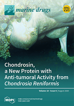
Cover Story (view full-size image): Chondrosia reniformis is a common mediterranean demosponge. In this paper, we identified a new cytotoxic protein (chondrosin) with selective activity against specific tumor cell lines. Chondrosin was extracted, purified and characterized through biochemical techniques. The procedure allowed to identify a 199-amino acid polypeptide. Cell death was determined by extracellular calcium intake, followed by cytoplasmic reactive oxygen species overproduction. The in silico modelling of chondrosin showed a high structural homology with the N-terminal region of the ryanodine receptor/channel. These results suggest a possible specific interaction of chondrosin with the Cav 1.3 ion voltage calcium channel expressed on the target cell membranes with consequent calcium-derived apoptosis. View this paper.
  • Issues are regarded as officially published after their release is announced to the table of contents alert mailing list.
  • You may sign up for e-mail alerts to receive table of contents of newly released issues.
  • PDF is the official format for papers published in both, html and pdf forms. To view the papers in pdf format, click on the "PDF Full-text" link, and use the free Adobe Reader to open them.
Order results
Result details
Section
Select all
Export citation of selected articles as:
16 pages, 3833 KiB  
Article
Collagen Peptides from Swim Bladders of Giant Croaker (Nibea japonica) and Their Protective Effects against H2O2-Induced Oxidative Damage toward Human Umbilical Vein Endothelial Cells
by Jiawen Zheng, Xiaoxiao Tian, Baogui Xu, Falei Yuan, Jianfang Gong and Zuisu Yang
Mar. Drugs 2020, 18(8), 430; https://doi.org/10.3390/md18080430 - 18 Aug 2020
Cited by 22 | Viewed by 2869
Abstract
Five different proteases were used to hydrolyze the swim bladders of Nibea japonica and the hydrolysate treated by neutrase (collagen peptide named SNNHs) showed the highest DPPH radical scavenging activity. The extraction process of SNNHs was optimized by response surface methodology, and the [...] Read more.
Five different proteases were used to hydrolyze the swim bladders of Nibea japonica and the hydrolysate treated by neutrase (collagen peptide named SNNHs) showed the highest DPPH radical scavenging activity. The extraction process of SNNHs was optimized by response surface methodology, and the optimal conditions were as follows: a temperature of 47.2 °C, a pH of 7.3 and an enzyme concentration of 1100 U/g, which resulted in the maximum DPPH clearance rate of 95.44%. Peptides with a Mw of less than 1 kDa (SNNH-1) were obtained by ultrafiltration, and exhibited good scavenging activity for hydroxyl radicals, ABTS radicals and superoxide anion radicals. Furthermore, SNNH-1 significantly promoted the proliferation of HUVECs, and the protective effect of SNNH-1 against oxidative damage of H2O2-induced HUVECs was investigated. The results indicated that all groups receiving SNNH-1 pretreatment showed an increase in GSH-Px, SOD, and CAT activities compared with the model group. In addition, SNNH-1 pretreatment reduced the levels of ROS and MDA in HUVECs with H2O2-induced oxidative damage. These results indicate that collagen peptides from swim bladders of Nibea japonica can significantly reduce the oxidative stress damage caused by H2O2 in HUVECs and provides a basis for the application of collagen peptides in the food industry, pharmaceuticals, and cosmetics. Full article
Show Figures

Graphical abstract

20 pages, 4652 KiB  
Article
Comparison of Physicochemical Characteristics and Macrophage Immunostimulatory Activities of Polysaccharides from Chlamys farreri
by Fulin Shi, Zhicong Liu, Yang Liu, Kit-Leong Cheong, Bo Teng and Bilal Muhammad Khan
Mar. Drugs 2020, 18(8), 429; https://doi.org/10.3390/md18080429 - 17 Aug 2020
Cited by 5 | Viewed by 2570
Abstract
To address the structure–activity relationship of Chlamys farreri polysaccharides on their immunostimulatory efficacy, two polysaccharides (CFP-1 and CFP-2) were extracted from Chlamys farreri by hot water extraction, and separated through column chromatography. The isolated CFPs were chemically analyzed to clarify their physicochemical characteristics [...] Read more.
To address the structure–activity relationship of Chlamys farreri polysaccharides on their immunostimulatory efficacy, two polysaccharides (CFP-1 and CFP-2) were extracted from Chlamys farreri by hot water extraction, and separated through column chromatography. The isolated CFPs were chemically analyzed to clarify their physicochemical characteristics and cultured with murine macrophage RAW264.7 cells, in order to evaluate their immunostimulatory efficacy. Despite the fact that both CFP-1 and CFP-2 were mainly comprised of glucose lacking the triple-helix structure, as revealed through preliminary physicochemical analyses, obvious differences in regard to molecular weight (Mw), glucuronic acid content (GAc) and branching degree (BD) were observed between CFP-1 and CFP-2. In in vitro immunostimulatory assays for macrophage RAW264.7 cells, it was demonstrated that CFP-2 with larger Mw, more GAc and BD could evidently promote phagocytosis and increase the production of NO, IL-6, TNF-α and IL-1β secretion, by activating the expression of iNOS, IL-6, TNF-α and IL-1β genes, respectively. Hence, CFP-2 shows great promise as a potential immunostimulatory agent in the functional foods and nutraceutical industry, while CFP-1, with lower molecular weight, less GAc and BD, displays its weaker immunostimulatory efficacy, based on the indistinctive immunostimulatory parameters of CFP-1. Full article
Show Figures

Figure 1

11 pages, 3927 KiB  
Article
Involvement of Reactive Oxygen Species in the Hepatorenal Toxicity of Actinomycin V In Vitro and In Vivo
by Fu-juan Jia, Zhuo Han, Jia-hui Ma, Shi-qing Jiang, Xing-ming Zhao, Hang Ruan, Wei-dong Xie and Xia Li
Mar. Drugs 2020, 18(8), 428; https://doi.org/10.3390/md18080428 - 15 Aug 2020
Cited by 4 | Viewed by 2285
Abstract
The high toxicity of actinomycin D (Act D) severely limits its use as a first-line chemotherapeutic agent in the clinic. Actinomycin V (Act V), an analog of Act D, exhibited strong anticancer activity in our previous studies. Here, we provide evidence that Act [...] Read more.
The high toxicity of actinomycin D (Act D) severely limits its use as a first-line chemotherapeutic agent in the clinic. Actinomycin V (Act V), an analog of Act D, exhibited strong anticancer activity in our previous studies. Here, we provide evidence that Act V has less hepatorenal toxicity than Act D in vitro and in vivo, associated with the reactive oxygen species (ROS) pathway. Compared to Act D, Act V exhibited considerably stronger sensitivity for cancer cells and less toxicity to human normal liver LO-2 and human embryonic kidney 293T cells using the MTT (3-(4,5-dimethyl-2-thiazolyl)-2,5-diphenyl-2-H-tetrazolium bromide) assay. Notably, Act V caused less damage to both the liver and kidney than Act D in vivo, indicated by organ to body weight ratios, as well as alanine aminotransferase (ALT), aspartate aminotransferase (AST), and serum creatinine (Scr) levels. Further experiments showed that the ROS pathway is involved in Act V-induced hepatorenal toxicity. Act V generates ROS and accumulates malondialdehyde (MDA), reducing levels of superoxide dismutase (SOD) and glutathione (GSH) in LO-2 and 293T cells. These findings indicate that Act V induces less hepatorenal toxicity than Act D in vitro and in vivo and merits further development as a potential therapeutic agent for the treatment of cancer. Full article
Show Figures

Figure 1

12 pages, 1858 KiB  
Article
Anti-Photoaging and Anti-Melanogenesis Effects of Fucoidan Isolated from Hizikia fusiforme and Its Underlying Mechanisms
by Lei Wang, Jae-Young Oh, Young-Sang Kim, Hyo-Geun Lee, Jung-Suck Lee and You-Jin Jeon
Mar. Drugs 2020, 18(8), 427; https://doi.org/10.3390/md18080427 - 15 Aug 2020
Cited by 34 | Viewed by 4202
Abstract
Previous studies suggested that fucoidan with a molecular weight of 102.67 kDa, isolated from Hizikia fusiforme, possesses strong antioxidant activity. To explore the cosmeceutical potential of fucoidan, its anti-photoaging and anti-melanogenesis effects were evaluated in the present study. The anti-photoaging effect was [...] Read more.
Previous studies suggested that fucoidan with a molecular weight of 102.67 kDa, isolated from Hizikia fusiforme, possesses strong antioxidant activity. To explore the cosmeceutical potential of fucoidan, its anti-photoaging and anti-melanogenesis effects were evaluated in the present study. The anti-photoaging effect was investigated in ultraviolet (UV) B-irradiated human keratinocytes (HaCaT cells), where fucoidan effectively reduced the intracellular reactive oxygen species level and improved the viability of the UVB-irradiated cells without any cytotoxic effects. Moreover, fucoidan significantly decreased UVB-induced apoptosis in HaCaT cells by regulating the protein expression of Bax, Bcl-xL, PARP, and Caspase-3 in HaCaT cells in a concentration-dependent manner. The anti-melanogenesis effect of fucoidan was evaluated in B16F10 melanoma cells that had been stimulated with alpha-melanocyte-stimulating hormone (α-MSH), and fucoidan treatment remarkably inhibited melanin synthesis in α-MSH-stimulated B16F10 cells. Further studies indicated that fucoidan significantly suppressed the expression of tyrosinase and tyrosinase-related protein-1 and -2 (TRP-1 and-2) in B16F10 cells by down-regulating microphthalmia-associated transcription factor (MITF) through regulation of the ERK–MAPK (extracellular signal regulated kinase-mitogen activated protein kinase) pathway. Taken together, these results suggest that fucoidan isolated from H. fusiforme possesses strong anti-photoaging and anti-melanogenesis activities and can be used as an ingredient in the pharmaceutical and cosmeceutical industries. Full article
(This article belongs to the Special Issue Antiphotoaging and Photoprotective Compounds from Marine Organisms)
Show Figures

Figure 1

14 pages, 2988 KiB  
Article
Secondary Metabolites with Nitric Oxide Inhibition from Marine-Derived Fungus Alternaria sp. 5102
by Senhua Chen, Yanlian Deng, Chong Yan, Zhenger Wu, Heng Guo, Lan Liu and Hongju Liu
Mar. Drugs 2020, 18(8), 426; https://doi.org/10.3390/md18080426 - 14 Aug 2020
Cited by 5 | Viewed by 2437
Abstract
Two new benzofurans, alternabenzofurans A and B (1 and 2) and two new sesquiterpenoids, alternaterpenoids A and B (3 and 4), along with 18 known polyketides (522), were isolated from the marine-derived fungus Alternaria sp. [...] Read more.
Two new benzofurans, alternabenzofurans A and B (1 and 2) and two new sesquiterpenoids, alternaterpenoids A and B (3 and 4), along with 18 known polyketides (522), were isolated from the marine-derived fungus Alternaria sp. 5102. Their structures were elucidated on the basis of extensive spectroscopic analyses (1D and 2D NMR, HR-ESIMS, and ECD) and X-ray crystallography, as well as the modified Mosher’s method. Compounds 2, 3, 5, 7, 918, and 2022 exhibited potent anti-inflammatory activity by inhibiting the production of NO in RAW264.7 cells activated by lipopolysaccharide with IC50 values in the range from 1.3 to 41.1 μM. Structure-activity relationships of the secondary metabolites were discussed. Full article
Show Figures

Figure 1

7 pages, 592 KiB  
Communication
Studies of Conorfamide-Sr3 on Human Voltage-Gated Kv1 Potassium Channel Subtypes
by Estuardo López-Vera, Luis Martínez-Hernández, Manuel B. Aguilar, Elisa Carrillo and Joanna Gajewiak
Mar. Drugs 2020, 18(8), 425; https://doi.org/10.3390/md18080425 - 13 Aug 2020
Cited by 9 | Viewed by 2303
Abstract
Recently, Conorfamide-Sr3 (CNF-Sr3) was isolated from the venom of Conus spurius and was demonstrated to have an inhibitory concentration-dependent effect on the Shaker K+ channel. The voltage-gated potassium channels play critical functions on cellular signaling, from the regeneration of action potentials in [...] Read more.
Recently, Conorfamide-Sr3 (CNF-Sr3) was isolated from the venom of Conus spurius and was demonstrated to have an inhibitory concentration-dependent effect on the Shaker K+ channel. The voltage-gated potassium channels play critical functions on cellular signaling, from the regeneration of action potentials in neurons to the regulation of insulin secretion in pancreatic cells, among others. In mammals, there are at least 40 genes encoding voltage-gated K+ channels and the process of expression of some of them may include alternative splicing. Given the enormous variety of these channels and the proven use of conotoxins as tools to distinguish different ligand- and voltage-gated ion channels, in this work, we explored the possible effect of CNF-Sr3 on four human voltage-gated K+ channel subtypes homologous to the Shaker channel. CNF-Sr3 showed a 10 times higher affinity for the Kv1.6 subtype with respect to Kv1.3 (IC50 = 2.7 and 24 μM, respectively) and no significant effect on Kv1.4 and Kv1.5 at 10 µM. Thus, CNF-Sr3 might become a novel molecular probe to study diverse aspects of human Kv1.3 and Kv1.6 channels. Full article
(This article belongs to the Special Issue Cone Snail Venom Peptides, from Treasure Hunt to Drug Leads)
Show Figures

Figure 1

21 pages, 1109 KiB  
Review
Marine Bioactive Peptides—An Overview of Generation, Structure and Application with a Focus on Food Sources
by Milica Pavlicevic, Elena Maestri and Marta Marmiroli
Mar. Drugs 2020, 18(8), 424; https://doi.org/10.3390/md18080424 - 13 Aug 2020
Cited by 38 | Viewed by 5816
Abstract
The biggest obstacles in the application of marine peptides are two-fold, as in the case of non-marine plant and animal-derived bioactive peptides: elucidating correlation between the peptide structure and its effect and demonstrating its stability in vivo. The structures of marine bioactive peptides [...] Read more.
The biggest obstacles in the application of marine peptides are two-fold, as in the case of non-marine plant and animal-derived bioactive peptides: elucidating correlation between the peptide structure and its effect and demonstrating its stability in vivo. The structures of marine bioactive peptides are highly variable and complex and dependent on the sources from which they are isolated. They can be cyclical, in the form of depsipeptides, and often contain secondary structures. Because of steric factors, marine-derived peptides can be resistant to proteolysis by gastrointestinal proteases, which presents an advantage over other peptide sources. Because of heterogeneity, amino acid sequences as well as preferred mechanisms of peptides showing specific bioactivities differ compared to their animal-derived counterparts. This review offers insights on the extreme diversity of bioactivities, effects, and structural features, analyzing 253 peptides, mainly from marine food sources. Similar to peptides in food of non-marine animal origin, a significant percentage (52.7%) of the examined sequences contain one or more proline residues, implying that proline might play a significant role in the stability of bioactive peptides. Additional problems with analyzing marine-derived bioactive peptides include their accessibility, extraction, and purification; this review considers the challenges and proposes possible solutions. Full article
Show Figures

Figure 1

32 pages, 2729 KiB  
Review
Oxidative Stress and Marine Carotenoids: Application by Using Nanoformulations
by Yasin Genç, Hilal Bardakci, Çiğdem Yücel, Gökçe Şeker Karatoprak, Esra Küpeli Akkol, Timur Hakan Barak and Eduardo Sobarzo-Sánchez
Mar. Drugs 2020, 18(8), 423; https://doi.org/10.3390/md18080423 - 13 Aug 2020
Cited by 37 | Viewed by 5452
Abstract
Carotenoids are natural fat-soluble pigments synthesized by plants, algae, fungi and microorganisms. They are responsible for the coloration of different photosynthetic organisms. Although they play a role in photosynthesis, they are also present in non-photosynthetic plant tissues, fungi, and bacteria. These metabolites have [...] Read more.
Carotenoids are natural fat-soluble pigments synthesized by plants, algae, fungi and microorganisms. They are responsible for the coloration of different photosynthetic organisms. Although they play a role in photosynthesis, they are also present in non-photosynthetic plant tissues, fungi, and bacteria. These metabolites have mainly been used in food, cosmetics, and the pharmaceutical industry. In addition to their utilization as pigmentation, they have significant therapeutically applications, such as improving immune system and preventing neurodegenerative diseases. Primarily, they have attracted attention due to their antioxidant activity. Several statistical investigations indicated an association between the use of carotenoids in diets and a decreased incidence of cancer types, suggesting the antioxidant properties of these compounds as an important factor in the scope of the studies against oxidative stress. Unusual marine environments are associated with a great chemical diversity, resulting in novel bioactive molecules. Thus, marine organisms may represent an important source of novel biologically active substances for the development of therapeutics. Marine carotenoids (astaxanthin, fucoxanthin, β-carotene, lutein but also the rare siphonaxanthin, sioxanthin, and myxol) have recently shown antioxidant properties in reducing oxidative stress markers. Numerous of bioactive compounds such as marine carotenoids have low stability, are poorly absorbed, and own very limited bioavailability. The new technique is nanoencapsulation, which can be used to preserve marine carotenoids and their original properties during processing, storage, improve their physiochemical properties and increase their health-promoting effects. This review aims to describe the role of marine carotenoids, their potential applications and different types of advanced nanoformulations preventing and treating oxidative stress related disorders. Full article
(This article belongs to the Special Issue Marine Carotenoids in Inflammation and Cancer)
Show Figures

Figure 1

14 pages, 4214 KiB  
Article
α-Conotoxin as Potential to α7-nAChR Recombinant Expressed in Escherichia coli
by Yanli Liu, Yifeng Yin, Yunyang Song, Kang Wang, Fanghui Wu and Hui Jiang
Mar. Drugs 2020, 18(8), 422; https://doi.org/10.3390/md18080422 - 12 Aug 2020
Cited by 5 | Viewed by 2656
Abstract
α7 nicotinic acetylcholine receptors (nAChR) is an important nicotinic acetylcholine receptors subtype and closely associated with cognitive disorders, such as Alzheimer’s and schizophrenia disease. The mutant ArIB (V11L, V16A) of α-conotoxin ArIB with 17-amino acid residues specifically targets α7 nAChR with no obvious [...] Read more.
α7 nicotinic acetylcholine receptors (nAChR) is an important nicotinic acetylcholine receptors subtype and closely associated with cognitive disorders, such as Alzheimer’s and schizophrenia disease. The mutant ArIB (V11L, V16A) of α-conotoxin ArIB with 17-amino acid residues specifically targets α7 nAChR with no obvious effect on other nAChR subtypes. In the study, the synthetic gene encoding mature peptide of ArIB and mutant ArIB (V11L, V16A) carried a fusion protein Trx and 6 × His-tag was separately inserted in pET-32a (+) vector and transformed into Escherichia coli strain BL21(DE3) pLysS for expression. The expressions of Trx-ArIB-His6 and Trx-ArIB (V11L, V16A)-His6 were soluble in Escherichia coli, which were purified by Ni-NTA affinity chromatography column and cleaved by enterokinase to release rArIB and rArIB (V11L, V16A). Then, rArIB and rArIB (V11L, V16A) were purified by high-performance liquid chromatography (HPLC) and identified by matrix-assisted laser desorption ionization-time of flight mass spectrometry (MALDI-TOF MS). Bioactivity of rArIB and rArIB (V11L, V16A) was assessed by two-electrode voltage-clamp electrophysiology in Xenopus laevis oocytes expressing human nAChR subtypes. The results indicated that the yield of the fusion proteins was approximately 50 mg/L and rArIB (V11L, V16A) antagonized the α7 nAChR subtype selectively with 8-nM IC50. In summary, this study provides an efficient method to biosynthesize α-conotoxin ArIB and rArIB (V11L, V16A) in Escherichia coli, which could be economical to obtain massively bioactive disulfide-rich polypeptides at fast speed. Full article
Show Figures

Figure 1

16 pages, 4520 KiB  
Article
Immunomodulatory Effects of N-Acetyl Chitooligosaccharides on RAW264.7 Macrophages
by Jun-Jin Deng, Zong-Qiu Li, Ze-Quan Mo, Shun Xu, He-Hua Mao, Dan Shi, Zhi-Wei Li, Xue-Ming Dan and Xiao-Chun Luo
Mar. Drugs 2020, 18(8), 421; https://doi.org/10.3390/md18080421 - 12 Aug 2020
Cited by 32 | Viewed by 3939
Abstract
The ongoing development of new production methods may lead to the commercialization of N-acetyl chitooligosaccharides (NACOS), such as chitosan oligosaccharides (COS). The bioactivity of NACOS, although not well detailed, differs from that of COS, as they have more acetyl groups than COS. [...] Read more.
The ongoing development of new production methods may lead to the commercialization of N-acetyl chitooligosaccharides (NACOS), such as chitosan oligosaccharides (COS). The bioactivity of NACOS, although not well detailed, differs from that of COS, as they have more acetyl groups than COS. We used two enzymatically produced NACOS with different molecular compositions and six NACOS (NACOS1–6) with a single degree of polymerization to verify their immunomodulatory effects on the RAW264.7 macrophage cell line. We aimed to identify any differences between COS and various NACOS with a single degree of polymerization. The results showed that NACOS had similar immune enhancement effects on RAW264.7 cells as COS, including the generation of reactive oxygen species (ROS), phagocytotic activity, and the production of pro-inflammation cytokines (IL-1β, IL-6, and TNF-α). However, unlike COS and lipopolysaccharide (LPS), NACOS1 and NACOS6 significantly inhibited nitric oxide (NO) production. Besides their immune enhancement effects, NACOS also significantly inhibited the LPS-induced RAW264.7 inflammatory response with some differences between various polymerization degrees. We confirmed that the NF-κB pathway is associated with the immunomodulatory effects of NACOS on RAW264.7 cells. This study could inform the application of NACOS with varying different degrees of polymerization in human health. Full article
(This article belongs to the Collection Marine Polysaccharides)
Show Figures

Figure 1

11 pages, 5357 KiB  
Article
Biocompatibility of a Marine Collagen-Based Scaffold In Vitro and In Vivo
by Dafna Benayahu, Leslie Pomeraniec, Shai Shemesh, Snir Heller, Yoav Rosenthal, Lea Rath-Wolfson and Yehuda Benayahu
Mar. Drugs 2020, 18(8), 420; https://doi.org/10.3390/md18080420 - 11 Aug 2020
Cited by 20 | Viewed by 3143
Abstract
Scaffold material is essential in providing mechanical support to tissue, allowing stem cells to improve their function in the healing and repair of trauma sites and tissue regeneration. The scaffold aids cell organization in the damaged tissue. It serves and allows bio mimicking [...] Read more.
Scaffold material is essential in providing mechanical support to tissue, allowing stem cells to improve their function in the healing and repair of trauma sites and tissue regeneration. The scaffold aids cell organization in the damaged tissue. It serves and allows bio mimicking the mechanical and biological properties of the target tissue and facilitates cell proliferation and differentiation at the regeneration site. In this study, the developed and assayed bio-composite made of unique collagen fibers and alginate hydrogel supports the function of cells around the implanted material. We used an in vivo rat model to study the scaffold effects when transplanted subcutaneously and as an augment for tendon repair. Animals’ well-being was measured by their weight and daily activity post scaffold transplantation during their recovery. At the end of the experiment, the bio-composite was histologically examined, and the surrounding tissues around the implant were evaluated for inflammation reaction and scarring tissue. In the histology, the formation of granulation tissue and fibroblasts that were part of the inclusion process of the implanted material were noted. At the transplanted sites, inflammatory cells, such as plasma cells, macrophages, and giant cells, were also observed as expected at this time point post transplantation. This study demonstrated not only the collagen-alginate device biocompatibility, with no cytotoxic effects on the analyzed rats, but also that the 3D structure enables cell migration and new blood vessel formation needed for tissue repair. Overall, the results of the current study proved for the first time that the implantable scaffold for long-term confirms the well-being of these rats and is correspondence to biocompatibility ISO standards and can be further developed for medical devices application. Full article
Show Figures

Figure 1

19 pages, 7766 KiB  
Article
Effects of Chitosan–Gentamicin Conjugate Supplement on Non-Specific Immunity, Aquaculture Water, Intestinal Histology and Microbiota of Pacific White Shrimp (Litopenaeus vannamei)
by Fengyan Liang, Chengpeng Li, Tingting Hou, Chongqing Wen, Songzhi Kong, Dong Ma, Chengbo Sun and Sidong Li
Mar. Drugs 2020, 18(8), 419; https://doi.org/10.3390/md18080419 - 10 Aug 2020
Cited by 20 | Viewed by 3502
Abstract
When the aquaculture water environment deteriorates or the temperature rises, shrimp are susceptible to viral or bacterial infections, causing a large number of deaths. This study comprehensively evaluated the effects of the oral administration of a chitosan–gentamicin conjugate (CS-GT) after Litopenaeus vannamei were [...] Read more.
When the aquaculture water environment deteriorates or the temperature rises, shrimp are susceptible to viral or bacterial infections, causing a large number of deaths. This study comprehensively evaluated the effects of the oral administration of a chitosan–gentamicin conjugate (CS-GT) after Litopenaeus vannamei were infected with Vibrio parahaemolyticus, through nonspecific immunity parameter detection, intestinal morphology observation, and the assessment of microbial flora diversification by 16S rRNA gene sequencing. The results showed that the oral administration of CS-GT significantly increased total hemocyte counts and reduced hemocyte apoptosis in shrimp (p < 0.05). The parameters (including superoxide dismutase, glutathione peroxidase, glutathione, lysozyme, acid phosphatase, alkaline phosphatase, and phenoloxidase) were significantly increased (p < 0.05). The integrity of the intestinal epithelial cells and basement membrane were enhanced, which correspondingly alleviated intestinal injury. In terms of the microbiome, the abundances of Vibrio (Gram-negative bacteria and food-borne pathogens) in the water and gut were significantly reduced. The canonical correspondence analysis (CCA) showed that the abundances of Vibrio both in the water and gut were negatively correlated with CS-GT dosage. In conclusion, the oral administration of CS-GT can improve the immunity of shrimp against pathogenic bacteria and significantly reduce the relative abundances of Vibrio in aquaculture water and the gut of Litopenaeus vannamei. Full article
(This article belongs to the Special Issue Marine-Derived Compounds Applied in Infectious Diseases)
Show Figures

Graphical abstract

14 pages, 1249 KiB  
Article
Chemistry of Renieramycins. Part 19: Semi-Syntheses of 22-O-Amino Ester and Hydroquinone 5-O-Amino Ester Derivatives of Renieramycin M and Their Cytotoxicity against Non-Small-Cell Lung Cancer Cell Lines
by Supakarn Chamni, Natchanun Sirimangkalakitti, Pithi Chanvorachote, Khanit Suwanborirux and Naoki Saito
Mar. Drugs 2020, 18(8), 418; https://doi.org/10.3390/md18080418 - 10 Aug 2020
Cited by 12 | Viewed by 2870
Abstract
Two new series of synthetic renieramycins including 22-O-amino ester and hydroquinone 5-O-amino ester derivatives of renieramycin M were semi-synthesized and evaluated for their cytotoxicity against the metastatic non-small-cell lung cancer H292 and H460 cell lines. Interestingly, the series of [...] Read more.
Two new series of synthetic renieramycins including 22-O-amino ester and hydroquinone 5-O-amino ester derivatives of renieramycin M were semi-synthesized and evaluated for their cytotoxicity against the metastatic non-small-cell lung cancer H292 and H460 cell lines. Interestingly, the series of 22-O-amino ester derivatives displayed a potent cytotoxic activity greater than the hydroquinone derivatives. The most cytotoxic derivative of the series was the 22-O-(N-Boc-l-glycine) ester of renieramycin M (5a: IC50 3.56 nM), which showed 7-fold higher potency than renieramycin M (IC50 24.56 nM) and 61-fold more than jorunnamycin A (IC50 217.43 nM) against H292 cells. In addition, 5a exhibited a significantly higher cytotoxic activity than doxorubicin (ca. 100 times). The new semi-synthetic renieramycin derivatives will be further studied and developed as potential cytotoxic agents for non-small-cell lung cancer treatment. Full article
(This article belongs to the Special Issue Chemical Modification of Marine Natural Products)
Show Figures

Graphical abstract

14 pages, 2518 KiB  
Article
Antimicrobial Peptide TP4 Targets Mitochondrial Adenine Nucleotide Translocator 2
by Bor-Chyuan Su, Yi-Chung Liu, Chen-Hung Ting, Ping-Chiang Lyu and Jyh-Yih Chen
Mar. Drugs 2020, 18(8), 417; https://doi.org/10.3390/md18080417 - 09 Aug 2020
Cited by 11 | Viewed by 3081
Abstract
Tilapia piscidin (TP) 4 is an antimicrobial peptide derived from Nile tilapia (Oreochromis niloticus), which shows broad-spectrum antibacterial activity and excellent cancer-killing ability in vitro and in vivo. Like many other antimicrobial peptides, TP4 treatment causes mitochondrial toxicity in cancer cells. [...] Read more.
Tilapia piscidin (TP) 4 is an antimicrobial peptide derived from Nile tilapia (Oreochromis niloticus), which shows broad-spectrum antibacterial activity and excellent cancer-killing ability in vitro and in vivo. Like many other antimicrobial peptides, TP4 treatment causes mitochondrial toxicity in cancer cells. However, the molecular mechanisms underlying TP4 targeting of mitochondria remain unclear. In this study, we used a pull-down assay on A549 cell lysates combined with LC-MS/MS to discover that TP4 targets adenine nucleotide translocator (ANT) 2, a protein essential for adenine nucleotide exchange across the inner membrane. We further showed that TP4 accumulates in mitochondria and colocalizes with ANT2. Moreover, molecular docking studies showed that the interaction requires Phe1, Ile2, His3, His4, Ser11, Lys14, His17, Arg21, Arg24 and Arg25 residues in TP4 and key residues within the cavity of ANT2. These findings suggest a mechanism by which TP4 may induce mitochondrial dysfunction to disrupt cellular energy metabolism. Full article
(This article belongs to the Special Issue Advances in Marine Antimicrobial Peptides)
Show Figures

Figure 1

12 pages, 3778 KiB  
Article
Characterization of a New Intracellular Alginate Lyase with Metal Ions-Tolerant and pH-Stable Properties
by Yan Ma, Jie Li, Xin-Yue Zhang, Hao-Dong Ni, Feng-Biao Wang, Hai-Ying Wang and Zhi-Peng Wang
Mar. Drugs 2020, 18(8), 416; https://doi.org/10.3390/md18080416 - 09 Aug 2020
Cited by 14 | Viewed by 2607
Abstract
Alginate lyases play an important role in alginate oligosaccharides (AOS) preparation and brown seaweed processing. Many extracellular alginate lyases have been characterized to develop efficient degradation tools needed for industrial applications. However, few studies focusing on intracellular alginate lyases have been conducted. In [...] Read more.
Alginate lyases play an important role in alginate oligosaccharides (AOS) preparation and brown seaweed processing. Many extracellular alginate lyases have been characterized to develop efficient degradation tools needed for industrial applications. However, few studies focusing on intracellular alginate lyases have been conducted. In this work, a novel intracellular alkaline alginate lyase Alyw202 from Vibrio sp. W2 was cloned, expressed and characterized. Secretory expression was performed in a food-grade host, Yarrowia lipolytica. Recombinant Alyw202 with a molecular weight of approximately 38.3 kDa exhibited the highest activity at 45 °C and more than 60% of the activity in a broad pH range of 3.0 to 10.0. Furthermore, Alyw202 showed remarkable metal ion-tolerance, NaCl independence and the capacity of degrading alginate into oligosaccharides of DP2-DP4. Due to the unique pH-stable and high salt-tolerant properties, Alyw202 has potential applications in the food and pharmaceutical industries. Full article
Show Figures

Figure 1

13 pages, 3519 KiB  
Article
Anti-Tumor Effects of Astaxanthin by Inhibition of the Expression of STAT3 in Prostate Cancer
by Shao-Qian Sun, You-Xi Zhao, Si-Yu Li, Jing-Wen Qiang and Yi-Zhi Ji
Mar. Drugs 2020, 18(8), 415; https://doi.org/10.3390/md18080415 - 07 Aug 2020
Cited by 28 | Viewed by 4913
Abstract
Astaxanthin is a natural product gaining increasing attention due to its safety and anti-cancer properties. In this study, we investigated the mechanisms of the anti-cancer effects of astaxanthin on prostate cancer (PCa) cell lines using aggressive PCa DU145 cells. Also an instantaneous silenced [...] Read more.
Astaxanthin is a natural product gaining increasing attention due to its safety and anti-cancer properties. In this study, we investigated the mechanisms of the anti-cancer effects of astaxanthin on prostate cancer (PCa) cell lines using aggressive PCa DU145 cells. Also an instantaneous silenced cell line (si-STAT3) derived from DU145 and a control cell line (si-NK) were used for the MTT and colony formation assays to determine the role of astaxanthin in proliferation and colony formation abilities. Flow cytometry assays were used to detect the apoptosis of tumor cells. Migration and invasion assays detected the weakening of the respective abilities. Western blot and RT-PCR tests detected the levels of STAT3 protein and mRNA. Astaxanthin resulted in suppression of the proliferation of DU145 cells and the level of STAT3. The treatment of DU145 cells with astaxanthin decreased the cloning ability, increased the apoptosis percentage and weakened the abilities of migration and invasion of the cells. Furthermore, astaxanthin reduced the expression of STAT3 at protein and mRNA levels. The effects were enhanced when astaxanthin and si-STAT3 were combined. The results of animal experiments were consistent with the results in cells. Thus, astaxanthin inhibits the proliferation of DU145 cells by reducing the expression of STAT3. Full article
(This article belongs to the Special Issue Marine Natural Products as Anticancer Agents)
Show Figures

Figure 1

18 pages, 6578 KiB  
Article
From Food Waste to Innovative Biomaterial: Sea Urchin-Derived Collagen for Applications in Skin Regenerative Medicine
by Cinzia Ferrario, Francesco Rusconi, Albana Pulaj, Raffaella Macchi, Paolo Landini, Moira Paroni, Graziano Colombo, Tiziana Martinello, Luca Melotti, Chiara Gomiero, M. Daniela Candia Carnevali, Francesco Bonasoro, Marco Patruno and Michela Sugni
Mar. Drugs 2020, 18(8), 414; https://doi.org/10.3390/md18080414 - 06 Aug 2020
Cited by 45 | Viewed by 6280
Abstract
Collagen-based skin-like scaffolds (CBSS) are promising alternatives to skin grafts to repair wounds and injuries. In this work, we propose that the common marine invertebrate sea urchin represents a promising and eco-friendly source of native collagen to develop innovative CBSS for skin injury [...] Read more.
Collagen-based skin-like scaffolds (CBSS) are promising alternatives to skin grafts to repair wounds and injuries. In this work, we propose that the common marine invertebrate sea urchin represents a promising and eco-friendly source of native collagen to develop innovative CBSS for skin injury treatment. Sea urchin food waste after gonad removal was here used to extract fibrillar glycosaminoglycan (GAG)-rich collagen to produce bilayer (2D + 3D) CBSS. Microstructure, mechanical stability, permeability to water and proteins, ability to exclude bacteria and act as scaffolding for fibroblasts were evaluated. Our data show that the thin and dense 2D collagen membrane strongly reduces water evaporation (less than 5% of water passes through the membrane after 7 days) and protein diffusion (less than 2% of BSA passes after 7 days), and acts as a barrier against bacterial infiltration (more than 99% of the different tested bacterial species is retained by the 2D collagen membrane up to 48 h), thus functionally mimicking the epidermal layer. The thick sponge-like 3D collagen scaffold, structurally and functionally resembling the dermal layer, is mechanically stable in wet conditions, biocompatible in vitro (seeded fibroblasts are viable and proliferate), and efficiently acts as a scaffold for fibroblast infiltration. Thus, thanks to their chemical and biological properties, CBSS derived from sea urchins might represent a promising, eco-friendly, and economically sustainable biomaterial for tissue regenerative medicine. Full article
(This article belongs to the Special Issue Regenerative Potential of Marine Natural Compounds)
Show Figures

Graphical abstract

24 pages, 3877 KiB  
Article
Transcriptomic Analysis of Four Cerianthid (Cnidaria, Ceriantharia) Venoms
by Anna M. L. Klompen, Jason Macrander, Adam M. Reitzel and Sérgio N. Stampar
Mar. Drugs 2020, 18(8), 413; https://doi.org/10.3390/md18080413 - 05 Aug 2020
Cited by 21 | Viewed by 6649
Abstract
Tube anemones, or cerianthids, are a phylogenetically informative group of cnidarians with complex life histories, including a pelagic larval stage and tube-dwelling adult stage, both known to utilize venom in stinging-cell rich tentacles. Cnidarians are an entirely venomous group that utilize their proteinaceous-dominated [...] Read more.
Tube anemones, or cerianthids, are a phylogenetically informative group of cnidarians with complex life histories, including a pelagic larval stage and tube-dwelling adult stage, both known to utilize venom in stinging-cell rich tentacles. Cnidarians are an entirely venomous group that utilize their proteinaceous-dominated toxins to capture prey and defend against predators, in addition to several other ecological functions, including intraspecific interactions. At present there are no studies describing the venom for any species within cerianthids. Given their unique development, ecology, and distinct phylogenetic-placement within Cnidaria, our objective is to evaluate the venom-like gene diversity of four species of cerianthids from newly collected transcriptomic data. We identified 525 venom-like genes between all four species. The venom-gene profile for each species was dominated by enzymatic protein and peptide families, which is consistent with previous findings in other cnidarian venoms. However, we found few toxins that are typical of sea anemones and corals, and furthermore, three of the four species express toxin-like genes closely related to potent pore-forming toxins in box jellyfish. Our study is the first to provide a survey of the putative venom composition of cerianthids and contributes to our general understanding of the diversity of cnidarian toxins. Full article
Show Figures

Graphical abstract

6 pages, 387 KiB  
Brief Report
Fucoidan Supplementation Restores Fecal Lysozyme Concentrations in High-Performance Athletes: A Pilot Study
by Amanda J. Cox, Allan W. Cripps, Phillipa A. Taylor, J. Helen Fitton and Nicholas P. West
Mar. Drugs 2020, 18(8), 412; https://doi.org/10.3390/md18080412 - 04 Aug 2020
Cited by 13 | Viewed by 3692
Abstract
Nutritional strategies to help promote immune competence are of particular interest for a range of population groups. This study aimed to assess the potential impacts of fucoidan, a seaweed-derived bioactive polysaccharide, on gut markers of immunity and inflammation. A group of professional team-sport [...] Read more.
Nutritional strategies to help promote immune competence are of particular interest for a range of population groups. This study aimed to assess the potential impacts of fucoidan, a seaweed-derived bioactive polysaccharide, on gut markers of immunity and inflammation. A group of professional team-sport athletes were selected for inclusion in the study given the recognized potential for intense physical activity to induce alterations in immune function. A retrospective analysis was performed on stored fecal samples which had been collected from professional team-sport athletes (n = 22) and healthy adults (n = 11) before and after seven days of supplementation with fucoidan (Fucus vesiculosus/Undaria pinnatifida extract, 1 g/d). Fecal concentrations of calprotectin, secretory immunoglobulin A (sIgA) and lysozyme were determined using enzyme-linked immunosorbent assays. The supplement was well tolerated by participants with no adverse events reported. At baseline, fecal lysozyme concentrations were ~73% higher in the healthy adults compared to the professional athletes (p = 0.001). For the professional athletes, a significant (~45%) increase in fecal lysozyme was observed following the supplementation period (p = 0.001). These data suggest that fucoidan supplementation may have the potential to promote the secretion of antimicrobial peptides in specific population groups and contribute to the regulation of mucosal immune health. Full article
Show Figures

Figure 1

32 pages, 5488 KiB  
Review
Progress of Bromophenols in Marine Algae from 2011 to 2020: Structure, Bioactivities, and Applications
by Hui Dong, Songtao Dong, Poul Erik Hansen, Dimitrios Stagos, Xiukun Lin and Ming Liu
Mar. Drugs 2020, 18(8), 411; https://doi.org/10.3390/md18080411 - 04 Aug 2020
Cited by 28 | Viewed by 3821
Abstract
Marine algae contain various bromophenols that have been shown to possess a variety of biological activities, including antiradical, antimicrobial, anticancer, antidiabetic, anti-inflammatory effects, and so on. Here, we briefly review the recent progress of these marine algae biomaterials and their derivatives from 2011 [...] Read more.
Marine algae contain various bromophenols that have been shown to possess a variety of biological activities, including antiradical, antimicrobial, anticancer, antidiabetic, anti-inflammatory effects, and so on. Here, we briefly review the recent progress of these marine algae biomaterials and their derivatives from 2011 to 2020, with respect to structure, bioactivities, and their potential application as pharmaceuticals. Full article
Show Figures

Figure 1

11 pages, 895 KiB  
Article
Botulinum Toxin-Chitosan Nanoparticles Prevent Arrhythmia in Experimental Rat Models
by David Sergeevichev, Vladislav Fomenko, Artem Strelnikov, Anna Dokuchaeva, Maria Vasilieva, Elena Chepeleva, Yanina Rusakova, Sergey Artemenko, Alexander Romanov, Nariman Salakhutdinov and Alexander Chernyavskiy
Mar. Drugs 2020, 18(8), 410; https://doi.org/10.3390/md18080410 - 02 Aug 2020
Cited by 7 | Viewed by 3095
Abstract
Several experimental studies have recently demonstrated that temporary autonomic block using botulinum toxin (BoNT/A1) might be a novel option for the treatment of atrial fibrillation. However, the assessment of antiarrhythmic properties of BoNT has so far been limited, relying exclusively on vagal stimulation [...] Read more.
Several experimental studies have recently demonstrated that temporary autonomic block using botulinum toxin (BoNT/A1) might be a novel option for the treatment of atrial fibrillation. However, the assessment of antiarrhythmic properties of BoNT has so far been limited, relying exclusively on vagal stimulation and rapid atrial pacing models. The present study examined the antiarrhythmic effect of specially formulated BoNT/A1-chitosan nanoparticles (BTN) in calcium chloride-, barium chloride- and electrically induced arrhythmia rat models. BTN enhanced the effect of BoNT/A1. Subepicardial injection of BTN resulted in a significant antiarrhythmic effect in investigated rat models. BTN formulation antagonizes arrhythmia induced by the activation of Ca, K and Na channels. Full article
(This article belongs to the Special Issue Ion Channels as Marine Drug Targets)
Show Figures

Figure 1

26 pages, 5569 KiB  
Article
Identification, Purification and Molecular Characterization of Chondrosin, a New Protein with Anti-tumoral Activity from the Marine Sponge Chondrosia Reniformis Nardo 1847
by Sonia Scarfì, Marina Pozzolini, Caterina Oliveri, Serena Mirata, Annalisa Salis, Gianluca Damonte, Daniela Fenoglio, Tiziana Altosole, Micha Ilan, Marco Bertolino and Marco Giovine
Mar. Drugs 2020, 18(8), 409; https://doi.org/10.3390/md18080409 - 02 Aug 2020
Cited by 9 | Viewed by 3211
Abstract
Chondrosia reniformis is a common marine demosponge showing many peculiarities, lacking silica spicules and with a body entirely formed by a dense collagenous matrix. In this paper, we have described the identification of a new cytotoxic protein (chondrosin) with selective activity against specific [...] Read more.
Chondrosia reniformis is a common marine demosponge showing many peculiarities, lacking silica spicules and with a body entirely formed by a dense collagenous matrix. In this paper, we have described the identification of a new cytotoxic protein (chondrosin) with selective activity against specific tumor cell lines, from C. reniformis, collected from the Liguria Sea. Chondrosin was extracted and purified using a salting out approach and molecular weight size exclusion chromatography. The cytotoxic fractions were then characterized by two-dimensional gel electrophoresis and mass spectrometry analysis and matched the results with C. reniformis transcriptome database. The procedure allowed for identifying a full-length cDNA encoding for a 199-amino acids (aa) polypeptide, with a signal peptide of 21 amino acids. The mature protein has a theoretical molecular weight of 19611.12 and an IP of 5.11. Cell toxicity assays showed a selective action against some tumor cell lines (RAW 264.7 murine leukemia cells in particular). Cell death was determined by extracellular calcium intake, followed by cytoplasmic reactive oxygen species overproduction. The in silico modelling of chondrosin showed a high structural homology with the N-terminal region of the ryanodine receptor/channel and a short identity with defensin. The results are discussed suggesting a possible specific interaction of chondrosin with the Cav 1.3 ion voltage calcium channel expressed on the target cell membranes. Full article
(This article belongs to the Special Issue Marine-Derived Products for Biomedicine)
Show Figures

Figure 1

10 pages, 1700 KiB  
Communication
Effects of Chitosan Oligosaccharide on Plasma and Hepatic Lipid Metabolism and Liver Histomorphology in Normal Sprague-Dawley Rats
by Shing-Hwa Liu, Rui-Yi Chen and Meng-Tsan Chiang
Mar. Drugs 2020, 18(8), 408; https://doi.org/10.3390/md18080408 - 02 Aug 2020
Cited by 17 | Viewed by 2611
Abstract
Chitosan oligosaccharide is known to ameliorate hypercholesterolemia and diabetes. However, some studies found that chitosan oligosaccharide might induce mild to moderate hepatic damage in high-fat (HF) diet-induced obese rats or diabetic rats. Chitosan oligosaccharide can be as a dietary supplement, functional food, or [...] Read more.
Chitosan oligosaccharide is known to ameliorate hypercholesterolemia and diabetes. However, some studies found that chitosan oligosaccharide might induce mild to moderate hepatic damage in high-fat (HF) diet-induced obese rats or diabetic rats. Chitosan oligosaccharide can be as a dietary supplement, functional food, or drug. Its possible toxic effects to normal subjects need to be clarified. This study is designed to investigate the effects of chitosan oligosaccharide on plasma and hepatic lipid metabolism and liver histomorphology in normal Sprague-Dawley rats. Diets supplemented with 5% chitosan oligosaccharide have been found to induce liver damage in HF diet-fed rats. We therefore selected 5% chitosan oligosaccharide as an experimental object. Rats were divided into: a normal control diet group and a normal control diet +5% chitosan oligosaccharide group. The experimental period was 12 weeks. The results showed that supplementation of 5% chitosan oligosaccharide did not significantly change the body weight, food intake, liver/adipose tissue weights, plasma lipids, hepatic lipids, plasma levels of AST, ALT, and TNF-α/IL-6, hepatic lipid peroxidation and anti-oxidative enzyme activities, fecal lipids, and liver histomorphology in normal rats. These findings suggest that supplementation of 5% chitosan oligosaccharide for 12 weeks may not induce lipid metabolism disorder and liver toxicity in normal rats. Full article
(This article belongs to the Special Issue Application of Marine Chitin and Chitosan)
Show Figures

Figure 1

18 pages, 5097 KiB  
Article
Proteo-Transcriptomic Analysis Identifies Potential Novel Toxins Secreted by the Predatory, Prey-Piercing Ribbon Worm Amphiporus lactifloreus
by Björn Marcus von Reumont, Tim Lüddecke, Thomas Timm, Günter Lochnit, Andreas Vilcinskas, Jörn von Döhren and Maria A. Nilsson
Mar. Drugs 2020, 18(8), 407; https://doi.org/10.3390/md18080407 - 01 Aug 2020
Cited by 15 | Viewed by 4755
Abstract
Nemerteans (ribbon worms) employ toxins to subdue their prey, but research thus far has focused on the small-molecule components of mucus secretions and few protein toxins have been characterized. We carried out a preliminary proteotranscriptomic analysis of putative toxins produced by the hoplonemertean [...] Read more.
Nemerteans (ribbon worms) employ toxins to subdue their prey, but research thus far has focused on the small-molecule components of mucus secretions and few protein toxins have been characterized. We carried out a preliminary proteotranscriptomic analysis of putative toxins produced by the hoplonemertean Amphiporus lactifloreus (Hoplonemertea, Amphiporidae). No variants were found of known nemertean-specific toxin proteins (neurotoxins, cytotoxins, parbolysins or nemertides) but several toxin-like transcripts were discovered, expressed strongly in the proboscis, including putative metalloproteinases and sequences resembling sea anemone actitoxins, crown-of-thorn sea star plancitoxins, and multiple classes of inhibitor cystine knot/knottin family proteins. Some of these products were also directly identified in the mucus proteome, supporting their preliminary identification as secreted toxin components. Two new nemertean-typical toxin candidates could be described and were named U-nemertotoxin-1 and U-nemertotoxin-2. Our findings provide insight into the largely overlooked venom system of nemerteans and support a hypothesis in which the nemertean proboscis evolved in several steps from a flesh-melting organ in scavenging nemerteans to a flesh-melting and toxin-secreting venom apparatus in hunting hoplonemerteans. Full article
Show Figures

Graphical abstract

25 pages, 635 KiB  
Review
Recent Advances in Astaxanthin Micro/Nanoencapsulation to Improve Its Stability and Functionality as a Food Ingredient
by Óscar Martínez-Álvarez, Marta M. Calvo and Joaquín Gómez-Estaca
Mar. Drugs 2020, 18(8), 406; https://doi.org/10.3390/md18080406 - 01 Aug 2020
Cited by 63 | Viewed by 5863
Abstract
Astaxanthin is a carotenoid produced by different organisms and microorganisms such as microalgae, bacteria, yeasts, protists, and plants, and it is also accumulated in aquatic animals such as fish and crustaceans. Astaxanthin and astaxanthin-containing lipid extracts obtained from these sources present an intense [...] Read more.
Astaxanthin is a carotenoid produced by different organisms and microorganisms such as microalgae, bacteria, yeasts, protists, and plants, and it is also accumulated in aquatic animals such as fish and crustaceans. Astaxanthin and astaxanthin-containing lipid extracts obtained from these sources present an intense red color and a remarkable antioxidant activity, providing great potential to be employed as food ingredients with both technological and bioactive functions. However, their use is hindered by: their instability in the presence of high temperatures, acidic pH, oxygen or light; their low water solubility, bioaccessibility and bioavailability; their intense odor/flavor. The present paper reviews recent advances in the micro/nanoencapsulation of astaxanthin and astaxanthin-containing lipid extracts, developed to improve their stability, bioactivity and technological functionality for use as food ingredients. The use of diverse micro/nanoencapsulation techniques using wall materials of a different nature to improve water solubility and dispersibility in foods, masking undesirable odor and flavor, is firstly discussed, followed by a discussion of the importance of the encapsulation to retard astaxanthin release, protecting it from degradation in the gastrointestinal tract. The nanoencapsulation of astaxanthin to improve its bioaccessibility, bioavailability and bioactivity is further reviewed. Finally, the main limitations and future trends on the topic are discussed. Full article
(This article belongs to the Special Issue Astaxanthin: A Potential Therapeutic Agent)
Show Figures

Figure 1

18 pages, 1724 KiB  
Article
New Cytotoxic Cerebrosides from the Red Sea Cucumber Holothuria spinifera Supported by In-Silico Studies
by Reda F. A. Abdelhameed, Enas E. Eltamany, Dina M. Hal, Amany K. Ibrahim, Asmaa M. AboulMagd, Tarfah Al-Warhi, Khayrya A. Youssif, Adel M. Abd El-kader, Hashim A. Hassanean, Shaimaa Fayez, Gerhard Bringmann, Safwat A. Ahmed and Usama Ramadan Abdelmohsen
Mar. Drugs 2020, 18(8), 405; https://doi.org/10.3390/md18080405 - 01 Aug 2020
Cited by 7 | Viewed by 3515
Abstract
Bioactivity-guided fractionation of a methanolic extract of the Red Sea cucumber Holothuria spinifera and LC-HRESIMS-assisted dereplication resulted in the isolation of four compounds, three new cerebrosides, spiniferosides A (1), B (2), and C (3), and cholesterol sulfate [...] Read more.
Bioactivity-guided fractionation of a methanolic extract of the Red Sea cucumber Holothuria spinifera and LC-HRESIMS-assisted dereplication resulted in the isolation of four compounds, three new cerebrosides, spiniferosides A (1), B (2), and C (3), and cholesterol sulfate (4). The chemical structures of the isolated compounds were established on the basis of their 1D NMR and HRMS spectral data. Metabolic profiling of the H. spinifera extract indicated the presence of diverse secondary metabolites, mostly hydroxy fatty acids, diterpenes, triterpenes, and cerebrosides. The isolated compounds were tested for their in vitro cytotoxicities against the breast adenocarcinoma MCF-7 cell line. Compounds 1, 2, 3, and 4 displayed promising cytotoxic activities against MCF-7 cells, with IC50 values of 13.83, 8.13, 8.27, and 35.56 µM, respectively, compared to that of the standard drug doxorubicin (IC50 8.64 µM). Additionally, docking studies were performed for compounds 1, 2, 3, and 4 to elucidate their binding interactions with the active site of the SET protein, an inhibitor of protein phosphatase 2A (PP2A), which could explain their cytotoxic activity. This study highlights the important role of these metabolites in the defense mechanism of the sea cucumber against fouling organisms and the potential uses of these active molecules in the design of new anticancer agents. Full article
(This article belongs to the Special Issue Nutraceuticals and Pharmaceuticals from Marine Fish and Invertebrates)
Show Figures

Graphical abstract

16 pages, 2004 KiB  
Article
Exploration of the Electrophilic Reactivity of the Cytotoxic Marine Alkaloid Discorhabdin C and Subsequent Discovery of a New Dimeric C-1/N-13-Linked Discorhabdin Natural Product
by Cary F. C. Lam, Melissa M. Cadelis and Brent R. Copp
Mar. Drugs 2020, 18(8), 404; https://doi.org/10.3390/md18080404 - 31 Jul 2020
Cited by 13 | Viewed by 2497
Abstract
The cytotoxic marine natural product discorhabdin C contains a 2,6-dibromo-cyclohexa-2,5-diene moiety, previously proposed to be a critical feature required for biological activity. We have determined that the dienone-ring of discorhabdin C is indeed electrophilic, reacting with thiol and amine nucleophiles, affording debrominated adducts. [...] Read more.
The cytotoxic marine natural product discorhabdin C contains a 2,6-dibromo-cyclohexa-2,5-diene moiety, previously proposed to be a critical feature required for biological activity. We have determined that the dienone-ring of discorhabdin C is indeed electrophilic, reacting with thiol and amine nucleophiles, affording debrominated adducts. In the case of reaction with 1-aminopentane the product contains an unusual C-2/N-18 ring closed, double-hydrate moiety. This electrophilic reactivity also extends to proteins, with lysozyme-discorhabdin C adducts being detected by ESI mass spectrometry. These results prompted further examination of an extract of discorhabdin C-producing sponge, Latrunculia (Latrunculia) trivetricillata, leading to the isolation and characterisation of a new example of a C-1/N-13 linked discorhabdin dimer that shared structural similarities with the 1-aminopentane-discorhabdin C adduct. To definitively assess the influence of the dienone moiety of discorhabdin C on cytotoxicity, a semi-synthetic hydrogenation derivative was prepared, affording a didebrominated ring-closed carbinolamine that was essentially devoid of tumour cell line cytotoxicity. Antiparasitic activity was assessed for a set of 14 discorhabdin alkaloids composed of natural products and semi-synthetic derivatives. Three compounds, (-)-discorhabdin L, a dimer of discorhabdin B and the discorhabdin C hydrogenation carbinolamine, exhibited pronounced activity towards Plasmodium falciparum K1 (IC50 30–90 nM) with acceptable to excellent selectivity (selectivity index 19–510) versus a non-malignant cell line. Full article
(This article belongs to the Special Issue Marine Natural Product of the South Pacific Area)
Show Figures

Graphical abstract

22 pages, 2411 KiB  
Article
Mass Spectrometry-Based Metabolomics Combined with Quantitative Analysis of the Microalgal Diatom (Chaetoceros calcitrans)
by Awanis Azizan, M. Maulidiani, Rudiyanto R., Khozirah Shaari, Intan Safinar Ismail, Norio Nagao and Faridah Abas
Mar. Drugs 2020, 18(8), 403; https://doi.org/10.3390/md18080403 - 30 Jul 2020
Cited by 7 | Viewed by 3553
Abstract
Although many metabolomics studies of higher land plant species have been conducted, similar studies of lower nonland plant species, which include microalgae, are still developing. The present study represents an attempt to characterize the metabolic profile of a microalgal diatom Chaetoceros calcitrans, [...] Read more.
Although many metabolomics studies of higher land plant species have been conducted, similar studies of lower nonland plant species, which include microalgae, are still developing. The present study represents an attempt to characterize the metabolic profile of a microalgal diatom Chaetoceros calcitrans, by applying high-resolution mass spectrometry detection, via Q-ExactiveTM Plus Orbitrap mass spectrometry. The results showed that 54 metabolites of various classes were tentatively identified. Experimentally, the chloroform and acetone extracts were clearly distinguished from other solvent extracts in chemometric regression analysis using PLS, showing the differences in the C. calcitrans metabolome between the groups. In addition, specific metabolites were evaluated, which supported the finding of antioxidant and anti-inflammatory activities. This study also provides data on the quantitative analysis of four carotenoids based on the identification results. Therefore, these findings could serve as a reliable tool for identifying and quantifying the metabolome that could reflect the metabolic activities of C. calcitrans. Full article
Show Figures

Figure 1

18 pages, 8509 KiB  
Article
Antiproliferative Activity of Mycalin A and Its Analogues on Human Skin Melanoma and Human Cervical Cancer Cells
by Domenica Capasso, Nicola Borbone, Monica Terracciano, Sonia Di Gaetano and Vincenzo Piccialli
Mar. Drugs 2020, 18(8), 402; https://doi.org/10.3390/md18080402 - 29 Jul 2020
Cited by 5 | Viewed by 2226
Abstract
Mycalin A, a polybrominated C15 acetogenin isolated from the encrusting sponge Mycale rotalis, displays an antiproliferative activity on human melanoma (A375) and cervical adenocarcinoma (HeLa) cells and induces cell death by an apoptotic mechanism. Various analogues and degraded derivatives of the [...] Read more.
Mycalin A, a polybrominated C15 acetogenin isolated from the encrusting sponge Mycale rotalis, displays an antiproliferative activity on human melanoma (A375) and cervical adenocarcinoma (HeLa) cells and induces cell death by an apoptotic mechanism. Various analogues and degraded derivatives of the natural substance have been prepared. A modification of the left-hand part of the molecule generates the most active substances. A structurally simplified lactone derivative of mycalin A, lacking the C1–C3 side chain, is the most active among the synthesized compounds exhibiting a strong cytotoxicity on both A375 and HeLa cells but not but not on human dermal fibroblast (HDF) used as healthy cells. Further evidence on a recently discovered chlorochromateperiodate-catalyzed process, used to oxidise mycalin A, have been collected. Full article
(This article belongs to the Collection Marine Compounds and Cancer)
Show Figures

Graphical abstract

45 pages, 5401 KiB  
Review
Marine Terpenoids from Polar Latitudes and Their Potential Applications in Biotechnology
by Laura Núñez-Pons, Andrew Shilling, Cinzia Verde, Bill J. Baker and Daniela Giordano
Mar. Drugs 2020, 18(8), 401; https://doi.org/10.3390/md18080401 - 29 Jul 2020
Cited by 23 | Viewed by 5670
Abstract
Polar marine biota have adapted to thrive under one of the ocean’s most inhospitable scenarios, where extremes of temperature, light photoperiod and ice disturbance, along with ecological interactions, have selected species with a unique suite of secondary metabolites. Organisms of Arctic and Antarctic [...] Read more.
Polar marine biota have adapted to thrive under one of the ocean’s most inhospitable scenarios, where extremes of temperature, light photoperiod and ice disturbance, along with ecological interactions, have selected species with a unique suite of secondary metabolites. Organisms of Arctic and Antarctic oceans are prolific sources of natural products, exhibiting wide structural diversity and remarkable bioactivities for human applications. Chemical skeletons belonging to terpene families are the most commonly found compounds, whereas cytotoxic antimicrobial properties, the capacity to prevent infections, are the most widely reported activities from these environments. This review firstly summarizes the regulations on access and benefit sharing requirements for research in polar environments. Then it provides an overview of the natural product arsenal from Antarctic and Arctic marine organisms that displays promising uses for fighting human disease. Microbes, such as bacteria and fungi, and macroorganisms, such as sponges, macroalgae, ascidians, corals, bryozoans, echinoderms and mollusks, are the main focus of this review. The biological origin, the structure of terpenes and terpenoids, derivatives and their biotechnological potential are described. This survey aims to highlight the chemical diversity of marine polar life and the versatility of this group of biomolecules, in an effort to encourage further research in drug discovery. Full article
(This article belongs to the Special Issue Bioactive Molecules from Extreme Environments II)
Show Figures

Figure 1

Previous Issue
Back to TopTop