Next Issue
Volume 16, March
Previous Issue
Volume 16, January


Mar. Drugs, Volume 16, Issue 2 (February 2018) – 36 articles

Cover Story (view full-size image): 
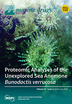
The sea anemone Bunodactis verrucosa inhabits rocky shores of the northeastern Atlantic Ocean, the North Sea and the Mediterranean Sea. Until now, the knowledge of its proteins and toxins production remained unknown. Here, B. verrucosa specimens were sampled in the coast of Portugal and the conducted proteomic analyses revealed the first proteome of this species. In addition to the identification of more than four hundred proteins and its function, this study provides new insight about toxins production in sea anemones and its putative ecological interaction. Indeed, B. verrucosa may produce a diverse repertoire of toxins to obtain its nourishment from different organism like mollusks (mussels, gastropods) and probably goby fishes. The putative toxins battery comprises proteins with enzymatic activity and neurotoxins that should act synergistically in subduing the preys. View this paper
  • Issues are regarded as officially published after their release is announced to the table of contents alert mailing list.
  • You may sign up for e-mail alerts to receive table of contents of newly released issues.
  • PDF is the official format for papers published in both, html and pdf forms. To view the papers in pdf format, click on the "PDF Full-text" link, and use the free Adobe Reader to open them.
Order results
Result details
Section
Select all
Export citation of selected articles as:
9 pages, 2088 KiB  
Article
Tetrocarcin Q, a New Spirotetronate with a Unique Glycosyl Group from a Marine-Derived Actinomycete Micromonospora carbonacea LS276
by Ting Gong, Xin Zhen, Xing-Lun Li, Jing-Jing Chen, Tian-Jiao Chen, Jin-Ling Yang and Ping Zhu
Mar. Drugs 2018, 16(2), 74; https://doi.org/10.3390/md16020074 - 24 Feb 2018
Cited by 21 | Viewed by 4688
Abstract
A new spirotetronate glycoside tetrocarcin Q (1) and six known analogues tetrocarcin A (2), AC6H (3), tetrocarcin N (4), tetrocarcin H (5), arisostatin A (6), and tetrocarcin F1 (7) [...] Read more.
A new spirotetronate glycoside tetrocarcin Q (1) and six known analogues tetrocarcin A (2), AC6H (3), tetrocarcin N (4), tetrocarcin H (5), arisostatin A (6), and tetrocarcin F1 (7) were isolated from the fermentation broth of the marine-derived actinomycete Micromonospora carbonacea LS276. Their chemical structures were established on the basis of 1D- and 2D-NMR spectroscopy, as well as HR-ESI-MS analysis. The absolute configurations of their stereogenic carbons were determined by circular dichroism (CD) analysis. Compound 1 possesses 2-deoxy-allose, which is a unique sugar type at the C-9 position. This type has not been found in the previously reported spirotetronate glycosides. Compound 1 displayed moderate antibacterial activity against Bacillus subitlis ATCC 63501 with minimum inhibitory concentration (MIC) value of 12.5 μM. Full article
(This article belongs to the Special Issue Natural Products from Coral Reef Organisms)
Show Figures

Figure 1

9 pages, 821 KiB  
Article
Preparative Separation and Purification of Trichothecene Mycotoxins from the Marine Fungus Fusarium sp. LS68 by High-Speed Countercurrent Chromatography in Stepwise Elution Mode
by Yong Liu, Xuezhen Zhou, C. Benjamin Naman, Yanbin Lu, Lijian Ding and Shan He
Mar. Drugs 2018, 16(2), 73; https://doi.org/10.3390/md16020073 - 24 Feb 2018
Cited by 8 | Viewed by 3714
Abstract
The contamination of foods and animal feeds with trichothecene mycotoxins is a growing concern for human and animal health. As such, large quantities of pure trichothecene mycotoxins are necessary for food safety monitoring and toxicological research. A new and effective method for the [...] Read more.
The contamination of foods and animal feeds with trichothecene mycotoxins is a growing concern for human and animal health. As such, large quantities of pure trichothecene mycotoxins are necessary for food safety monitoring and toxicological research. A new and effective method for the purification of trichothecene mycotoxins from a marine fungus, Fusarium sp. LS68, is described herein. Preparative high-speed countercurrent chromatography (HSCCC) was utilized for the scalable isolation and purification of four trichothecene mycotoxins for the first time in stepwise elution mode, with a biphasic solvent system composed of hexanes–EtOAc–CH3OH–H2O (6:4:5:5, v/v/v/v) and (8.5:1.5:5:5,v/v/v/v). This preparative HSCCC separation was performed on 200 mg of crude sample to yield four trichothecene mycotoxins, roridin E (1), roridin E acetate (2), verrucarin L acetate (3), and verrucarin J (4) in a single run, with each of >98% purity. These compounds were identified by MS, 1H NMR, 13C NMR, and polarimetry. The results demonstrate an efficient HSCCC method for the separation of trichothecene mycotoxins, which can be utilized to produce pure commercial and research standards. Full article
Show Figures

Graphical abstract

5 pages, 392 KiB  
Perspective
Tetrodotoxin, a Candidate Drug for Nav1.1-Induced Mechanical Pain?
by César Mattei
Mar. Drugs 2018, 16(2), 72; https://doi.org/10.3390/md16020072 - 22 Feb 2018
Cited by 10 | Viewed by 5510
Abstract
Tetrodotoxin (TTX), the mode of action of which has been known since the 1960s, is widely used in pharmacology as a specific inhibitor of voltage-gated sodium channels (Nav channels). This toxin has contributed to the characterization of the allosteric model of the Nav [...] Read more.
Tetrodotoxin (TTX), the mode of action of which has been known since the 1960s, is widely used in pharmacology as a specific inhibitor of voltage-gated sodium channels (Nav channels). This toxin has contributed to the characterization of the allosteric model of the Nav channel, and to discriminating TTX-sensitive and TTX-resistant subtypes. In addition to its role as a pharmacological tool, TTX is now considered a therapeutic molecule, and its development should lead to its use in certain pathologies involving Nav channels, particularly in the field of pain. Specifically, the blockade of Nav channels expressed in nociceptive fibres is one strategy for alleviating pain and its deleterious consequences on health. Recent work has identified, in addition to the Nav1.7, 1.8 and 1.9 channels, the Nav1.1 subtype on dorsal root ganglion (DRG) neurons as a crucial player in mechanical and non-thermal pain. The sensitivity of Nav1.1 to TTX could be exploited at the therapeutic level, especially in chronic pain conditions. Full article
(This article belongs to the Special Issue Tetrodotoxin)
Show Figures

Graphical abstract

8 pages, 1337 KiB  
Communication
Cladodionen, a Cytotoxic Hybrid Polyketide from the Marine-Derived Cladosporium sp. OUCMDZ-1635
by Guoliang Zhu, Fandong Kong, Yi Wang, Peng Fu and Weiming Zhu
Mar. Drugs 2018, 16(2), 71; https://doi.org/10.3390/md16020071 - 22 Feb 2018
Cited by 35 | Viewed by 4650
Abstract
A new hybrid polyketide, cladodionen (1), together with a new abscisic acid analogue, cladosacid (2), were isolated from the marine-derived fungus, Cladosporium sp. OUCMDZ-1635. Their structures, including the absolute configurations, were fully elucidated on the basis of spectroscopic analysis, [...] Read more.
A new hybrid polyketide, cladodionen (1), together with a new abscisic acid analogue, cladosacid (2), were isolated from the marine-derived fungus, Cladosporium sp. OUCMDZ-1635. Their structures, including the absolute configurations, were fully elucidated on the basis of spectroscopic analysis, ECD spectra, quantum chemical calculations, and chemical methods. Cladodionen (1) showed cytotoxic activities against MCF-7, HeLa, HCT-116, and HL-60 human cancer cell lines with IC50 values of 18.7, 19.1, 17.9, and 9.1 µM. Full article
Show Figures

Figure 1

9 pages, 596 KiB  
Article
Lactomycins A–C, Dephosphorylated Phoslactomycin Derivatives that Inhibit Cathepsin B, from the Marine-derived Streptomyces sp. ACT232
by Yi Sun, Rogie Royce Carandang, Yuta Harada, Shigeru Okada, Kazutoshi Yoshitake, Shuichi Asakawa, Yuichi Nogi, Shigeki Matsunaga and Kentaro Takada
Mar. Drugs 2018, 16(2), 70; https://doi.org/10.3390/md16020070 - 21 Feb 2018
Cited by 6 | Viewed by 6573
Abstract
Three new polyketides, lactomycins A (1)–C (3), were isolated from the culture broth of a marine-derived Streptomyces sp. ACT232 as cathepsin B inhibitors. Their structures were determined by a combination of NMR and MS data analyses to be the dephosphorylated derivatives of a [...] Read more.
Three new polyketides, lactomycins A (1)–C (3), were isolated from the culture broth of a marine-derived Streptomyces sp. ACT232 as cathepsin B inhibitors. Their structures were determined by a combination of NMR and MS data analyses to be the dephosphorylated derivatives of a phoslactomycin class of metabolites. Lactomycins exhibited cathepsin B inhibitory activity (IC50 0.8 to 4.5 μg/mL). Even though the biosynthetic gene clusters found in the genome of the current strain have high similarity to those of phoslactomycin, neither phoslactomycins nor leustroducsins were detected by LC-MS analyses of the crude extract. Full article
(This article belongs to the Special Issue Natural Products from Marine Actinomycetes)
Show Figures

Graphical abstract

34 pages, 3878 KiB  
Review
Exopolysaccharides from Marine and Marine Extremophilic Bacteria: Structures, Properties, Ecological Roles and Applications
by Angela Casillo, Rosa Lanzetta, Michelangelo Parrilli and Maria Michela Corsaro
Mar. Drugs 2018, 16(2), 69; https://doi.org/10.3390/md16020069 - 20 Feb 2018
Cited by 157 | Viewed by 13348
Abstract
The marine environment is the largest aquatic ecosystem on Earth and it harbours microorganisms responsible for more than 50% of total biomass of prokaryotes in the world. All these microorganisms produce extracellular polymers that constitute a substantial part of the dissolved organic carbon, [...] Read more.
The marine environment is the largest aquatic ecosystem on Earth and it harbours microorganisms responsible for more than 50% of total biomass of prokaryotes in the world. All these microorganisms produce extracellular polymers that constitute a substantial part of the dissolved organic carbon, often in the form of exopolysaccharides (EPS). In addition, the production of these polymers is often correlated to the establishment of the biofilm growth mode, during which they are important matrix components. Their functions include adhesion and colonization of surfaces, protection of the bacterial cells and support for biochemical interactions between the bacteria and the surrounding environment. The aim of this review is to present a summary of the status of the research about the structures of exopolysaccharides from marine bacteria, including capsular, medium released and biofilm embedded polysaccharides. Moreover, ecological roles of these polymers, especially for those isolated from extreme ecological niches (deep-sea hydrothermal vents, polar regions, hypersaline ponds, etc.), are reported. Finally, relationships between the structure and the function of the exopolysaccharides are discussed. Full article
(This article belongs to the Special Issue Marine Oligosaccharides and Polysaccharides)
Show Figures

Figure 1

17 pages, 4730 KiB  
Article
First Report on Chitin in a Non-Verongiid Marine Demosponge: The Mycale euplectellioides Case
by Sonia Żółtowska-Aksamitowska, Lamiaa A. Shaala, Diaa T. A. Youssef, Sameh S. Elhady, Mikhail V. Tsurkan, Iaroslav Petrenko, Marcin Wysokowski, Konstantin Tabachnick, Heike Meissner, Viatcheslav N. Ivanenko, Nicole Bechmann, Yvonne Joseph, Teofil Jesionowski and Hermann Ehrlich
Mar. Drugs 2018, 16(2), 68; https://doi.org/10.3390/md16020068 - 20 Feb 2018
Cited by 25 | Viewed by 5202
Abstract
Sponges (Porifera) are recognized as aquatic multicellular organisms which developed an effective biochemical pathway over millions of years of evolution to produce both biologically active secondary metabolites and biopolymer-based skeletal structures. Among marine demosponges, only representatives of the Verongiida order are known to [...] Read more.
Sponges (Porifera) are recognized as aquatic multicellular organisms which developed an effective biochemical pathway over millions of years of evolution to produce both biologically active secondary metabolites and biopolymer-based skeletal structures. Among marine demosponges, only representatives of the Verongiida order are known to synthetize biologically active substances as well as skeletons made of structural polysaccharide chitin. The unique three-dimensional (3D) architecture of such chitinous skeletons opens the widow for their recent applications as adsorbents, as well as scaffolds for tissue engineering and biomimetics. This study has the ambitious goal of monitoring other orders beyond Verongiida demosponges and finding alternative sources of naturally prestructured chitinous scaffolds; especially in those demosponge species which can be cultivated at large scales using marine farming conditions. Special attention has been paid to the demosponge Mycale euplectellioides (Heteroscleromorpha: Poecilosclerida: Mycalidae) collected in the Red Sea. For the first time, we present here a detailed study of the isolation of chitin from the skeleton of this sponge, as well as its identification using diverse bioanalytical tools. Calcofluor white staining, Fourier-transform Infrared Spcetcroscopy (FTIR), electrospray ionization mass spectrometry (ESI-MS), scanning electron microscopy (SEM), and fluorescence microscopy, as well as a chitinase digestion assay were applied in order to confirm with strong evidence the finding of a-chitin in the skeleton of M. euplectellioides. We suggest that the discovery of chitin within representatives of the Mycale genus is a promising step in their evaluation of these globally distributed sponges as new renewable sources for both biologically active metabolites and chitin, which are of prospective use for pharmacology and biomaterials oriented biomedicine, respectively. Full article
(This article belongs to the Special Issue Marine Chitin)
Show Figures

Figure 1

18 pages, 1503 KiB  
Article
Diverse and Abundant Secondary Metabolism Biosynthetic Gene Clusters in the Genomes of Marine Sponge Derived Streptomyces spp. Isolates
by Stephen A. Jackson, Lisa Crossman, Eduardo L. Almeida, Lekha Menon Margassery, Jonathan Kennedy and Alan D.W. Dobson
Mar. Drugs 2018, 16(2), 67; https://doi.org/10.3390/md16020067 - 20 Feb 2018
Cited by 55 | Viewed by 8184
Abstract
The genus Streptomyces produces secondary metabolic compounds that are rich in biological activity. Many of these compounds are genetically encoded by large secondary metabolism biosynthetic gene clusters (smBGCs) such as polyketide synthases (PKS) and non-ribosomal peptide synthetases (NRPS) which are modular and can [...] Read more.
The genus Streptomyces produces secondary metabolic compounds that are rich in biological activity. Many of these compounds are genetically encoded by large secondary metabolism biosynthetic gene clusters (smBGCs) such as polyketide synthases (PKS) and non-ribosomal peptide synthetases (NRPS) which are modular and can be highly repetitive. Due to the repeats, these gene clusters can be difficult to resolve using short read next generation datasets and are often quite poorly predicted using standard approaches. We have sequenced the genomes of 13 Streptomyces spp. strains isolated from shallow water and deep-sea sponges that display antimicrobial activities against a number of clinically relevant bacterial and yeast species. Draft genomes have been assembled and smBGCs have been identified using the antiSMASH (antibiotics and Secondary Metabolite Analysis Shell) web platform. We have compared the smBGCs amongst strains in the search for novel sequences conferring the potential to produce novel bioactive secondary metabolites. The strains in this study recruit to four distinct clades within the genus Streptomyces. The marine strains host abundant smBGCs which encode polyketides, NRPS, siderophores, bacteriocins and lantipeptides. The deep-sea strains appear to be enriched with gene clusters encoding NRPS. Marine adaptations are evident in the sponge-derived strains which are enriched for genes involved in the biosynthesis and transport of compatible solutes and for heat-shock proteins. Streptomyces spp. from marine environments are a promising source of novel bioactive secondary metabolites as the abundance and diversity of smBGCs show high degrees of novelty. Sponge derived Streptomyces spp. isolates appear to display genomic adaptations to marine living when compared to terrestrial strains. Full article
(This article belongs to the Special Issue Microbial Gene Clusters of Marine Origin)
Show Figures

Figure 1

17 pages, 4415 KiB  
Article
Chitin Oligosaccharide Modulates Gut Microbiota and Attenuates High-Fat-Diet-Induced Metabolic Syndrome in Mice
by Junping Zheng, Gong Cheng, Qiongyu Li, Siming Jiao, Cui Feng, Xiaoming Zhao, Heng Yin, Yuguang Du and Hongtao Liu
Mar. Drugs 2018, 16(2), 66; https://doi.org/10.3390/md16020066 - 19 Feb 2018
Cited by 74 | Viewed by 7662
Abstract
Gut microbiota has been proved to be an indispensable link between nutrient excess and metabolic syndrome, and chitin oligosaccharide (NACOS) has displayed therapeutic effects on multiple diseases such as cancer and gastritis. In this study, we aim to confirm whether NACOS can ameliorate [...] Read more.
Gut microbiota has been proved to be an indispensable link between nutrient excess and metabolic syndrome, and chitin oligosaccharide (NACOS) has displayed therapeutic effects on multiple diseases such as cancer and gastritis. In this study, we aim to confirm whether NACOS can ameliorate high-fat diet (HFD)-induced metabolic syndrome by rebuilding the structure of the gut microbiota community. Male C57BL/6J mice fed with HFD were treated with NACOS (1 mg/mL) in drinking water for five months. The results indicate that NACOS improved glucose metabolic disorder in HFD-fed mice and suppressed mRNA expression of the protein regulators related to lipogenesis, gluconeogenesis, adipocyte differentiation, and inflammation in adipose tissues. Additionally, NACOS inhibited the destruction of the gut barrier in HFD-treated mice. Furthermore, 16S ribosome RNA sequencing of fecal samples demonstrates that NACOS promoted the growth of beneficial intestinal bacteria remarkably and decreased the abundance of inflammogenic taxa. In summary, NACOS partly rebuilt the microbial community and improved the metabolic syndrome of HFD-fed mice. These data confirm the preventive effects of NACOS on nutrient excess-related metabolic diseases. Full article
(This article belongs to the Collection Marine Drugs in the Management of Metabolic Diseases)
Show Figures

Figure 1

16 pages, 906 KiB  
Review
Biosorption: An Interplay between Marine Algae and Potentially Toxic Elements—A Review
by Muhammad Bilal, Tahir Rasheed, Juan Eduardo Sosa-Hernández, Ali Raza, Faran Nabeel and Hafiz M. N. Iqbal
Mar. Drugs 2018, 16(2), 65; https://doi.org/10.3390/md16020065 - 19 Feb 2018
Cited by 307 | Viewed by 17130
Abstract
In recent decades, environmental pollution has emerged as a core issue, around the globe, rendering it of fundamental concern to eco-toxicologists, environmental biologists, eco-chemists, pathologists, and researchers from other fields. The dissolution of polluting agents is a leading cause of environmental pollution of [...] Read more.
In recent decades, environmental pollution has emerged as a core issue, around the globe, rendering it of fundamental concern to eco-toxicologists, environmental biologists, eco-chemists, pathologists, and researchers from other fields. The dissolution of polluting agents is a leading cause of environmental pollution of all key spheres including the hydrosphere, lithosphere, and biosphere, among others. The widespread occurrence of various pollutants including toxic heavy metals and other emerging hazardous contaminants is a serious concern. With increasing scientific knowledge, socioeconomic awareness, human health problems, and ecological apprehensions, people are more concerned about adverse health outcomes. Against this background, several removal methods have been proposed and implemented with the aim of addressing environmental pollution and sustainable and eco-friendly development. Among them, the biosorption of pollutants using naturally inspired sources, e.g., marine algae, has considerable advantages. In the past few years, marine algae have been extensively studied due to their natural origin, overall cost-effective ratio, and effectiveness against a broader pollutant range; thus, they are considered a potential alternative to the conventional methods used for environmental decontamination. Herein, an effort has been made to highlight the importance of marine algae as naturally inspired biosorbents and their role in biosorption. Biosorption mechanisms and factors affecting biosorption activities are also discussed in this review. The utilization of marine algae as a biosorbent for the removal of numerous potentially toxic elements has also been reviewed. Full article
(This article belongs to the Special Issue Marine Compounds Used in Biosorption)
Show Figures

Figure 1

16 pages, 434 KiB  
Review
The Anti-Cancer Effects of Frondoside A
by Thomas E. Adrian and Peter Collin
Mar. Drugs 2018, 16(2), 64; https://doi.org/10.3390/md16020064 - 19 Feb 2018
Cited by 24 | Viewed by 6514
Abstract
Frondoside A is a triterpenoid glycoside from the Atlantic Sea Cucumber, Cucumaria frondosa. Frondoside A has a broad spectrum of anti-cancer effects, including induction of cellular apoptosis, inhibition of cancer cell growth, migration, invasion, formation of metastases, and angiogenesis. In cell lines [...] Read more.
Frondoside A is a triterpenoid glycoside from the Atlantic Sea Cucumber, Cucumaria frondosa. Frondoside A has a broad spectrum of anti-cancer effects, including induction of cellular apoptosis, inhibition of cancer cell growth, migration, invasion, formation of metastases, and angiogenesis. In cell lines and animal models studied to date, the anti-cancer effects of the compound are seen in all solid cancers, lymphomas, and leukemias studied to date. These effects appear to be due to potent inhibition of p21-activated kinase 1 (PAK1), which is up-regulated in many cancers. In mouse models, frondoside A has synergistic effects with conventional chemotherapeutic agents, such as gemcitabine, paclitaxel, and cisplatin. Frondoside A administration is well-tolerated. No side effects have been reported and the compound has no significant effects on body weight, blood cells, or on hepatic and renal function tests after long-term administration. Frondoside A may be valuable in the treatment of malignancies, either as a single agent or in combination with other therapeutic modalities. Full article
(This article belongs to the Special Issue Marine Glycosides)
Show Figures

Figure 1

16 pages, 1856 KiB  
Article
Antiproliferative Activity of Glycosaminoglycan-Like Polysaccharides Derived from Marine Molluscs
by Abdullah Faisal Aldairi, Olanrewaju Dorcas Ogundipe and David Alexander Pye
Mar. Drugs 2018, 16(2), 63; https://doi.org/10.3390/md16020063 - 15 Feb 2018
Cited by 20 | Viewed by 5709
Abstract
Despite the increasing availability of new classes of cancer treatment, such as immune- and targeted therapies, there remains a need for the development of new antiproliferative/cytotoxic drugs with improved pharmacological profiles that can also overcome drug resistant forms of cancer. In this study, [...] Read more.
Despite the increasing availability of new classes of cancer treatment, such as immune- and targeted therapies, there remains a need for the development of new antiproliferative/cytotoxic drugs with improved pharmacological profiles that can also overcome drug resistant forms of cancer. In this study, we have identified, and characterised, a novel marine polysaccharide with the potential to be developed as an anticancer agent. Sulphated polysaccharides isolated from the common cockle (Cerastoderma edule) were shown to have antiproliferative activity on chronic myelogenous leukaemia and relapsed acute lymphoblastic leukaemia cell lines. Disaccharide and monosaccharide analysis of these marine polysaccharides confirmed the presence of glycosaminoglycan-like structures that were enriched in ion-exchange purified fractions containing antiproliferative activity. The antiproliferative activity of these glycosaminoglycan-like marine polysaccharides was shown to be susceptible to heparinase but not chondrotinase ABC digestion. This pattern of enzymatic and antiproliferative activity has not previously been seen, with either marine or mammalian glycosaminoglycans. As such, our findings suggest we have identified a new type of marine derived heparan sulphate/heparin-like polysaccharide with potent anticancer properties. Full article
(This article belongs to the Collection Marine Compounds and Cancer)
Show Figures

Figure 1

15 pages, 1084 KiB  
Article
A Comparative Study on Asymmetric Reduction of Ketones Using the Growing and Resting Cells of Marine-Derived Fungi
by Hui Liu, Bi-Shuang Chen, Fayene Zeferino Ribeiro De Souza and Lan Liu
Mar. Drugs 2018, 16(2), 62; https://doi.org/10.3390/md16020062 - 14 Feb 2018
Cited by 15 | Viewed by 4041
Abstract
Whole-cell biocatalysts offer a highly enantioselective, minimally polluting route to optically active alcohols. Currently, most of the whole-cell catalytic performance involves resting cells rather than growing cell biotransformation, which is one-step process that benefits from the simultaneous growth and biotransformation, eliminating the need [...] Read more.
Whole-cell biocatalysts offer a highly enantioselective, minimally polluting route to optically active alcohols. Currently, most of the whole-cell catalytic performance involves resting cells rather than growing cell biotransformation, which is one-step process that benefits from the simultaneous growth and biotransformation, eliminating the need for catalysts preparation. In this paper, asymmetric reduction of 14 aromatic ketones to the corresponding enantiomerically pure alcohols was successfully conducted using the growing and resting cells of marine-derived fungi under optimized conditions. Good yields and excellent enantioselectivities were achieved with both methods. Although substrate inhibition might be a limiting factor for growing cell biotransformation, the selected strain can still completely convert 10-mM substrates into the desired products. The resting cell biotransformation showed a capacity to be recycled nine times without a significant decrease in the activity. This is the first study to perform asymmetric reduction of ketones by one-step growing cell biotransformation. Full article
Show Figures

Figure 1

11 pages, 2383 KiB  
Article
Chlorinated Azaphilone Pigments with Antimicrobial and Cytotoxic Activities Isolated from the Deep Sea Derived Fungus Chaetomium sp. NA-S01-R1
by Weiyi Wang, Yanyan Liao, Ruixuan Chen, Yanping Hou, Wenqian Ke, Beibei Zhang, Maolin Gao, Zongze Shao, Jianming Chen and Fang Li
Mar. Drugs 2018, 16(2), 61; https://doi.org/10.3390/md16020061 - 13 Feb 2018
Cited by 58 | Viewed by 4636
Abstract
Four novel compounds, chaephilone C (1), chaetoviridides A–C (24), were obtained from the culture of a deep sea derived fungus Chaetomium sp. NA-S01-R1, together with four known compounds—chaetoviridin A (5), chaetoviridine E (6), [...] Read more.
Four novel compounds, chaephilone C (1), chaetoviridides A–C (24), were obtained from the culture of a deep sea derived fungus Chaetomium sp. NA-S01-R1, together with four known compounds—chaetoviridin A (5), chaetoviridine E (6), chaetomugilin D (7) and cochliodone A (8). Their structures, including absolute configurations, were assigned based on NMR, MS and time-dependent density functional theory (TD-DFT) ECD calculations. A plausible biogenetic pathway for compounds 13 was proposed. Compounds 2 and 3 exhibited antibacterial activities against Vibrio rotiferianus and Vibrio vulnificus. Compounds 1, 3 and 4 displayed similar anti-methicillin resistant Staphylococcus aureus (anti-MRSA) activities in comparison to chloramphenicol. Compound 2 showed the most potent cytotoxic activities towards the Hep G2 cell and compounds 1 and 3 demonstrated relatively stronger cytotoxic activities than the other compounds against the HeLa cell. Full article
(This article belongs to the Special Issue Bioactive Compounds from Marine Microbes - II)
Show Figures

Graphical abstract

18 pages, 7274 KiB  
Article
Bioactivity Assessment of Indian Origin—Mangrove Actinobacteria against Candida albicans
by J. G. S. Pavan Kumar, Ajitha Gomathi, Vitor Vasconcelos and K. M. Gothandam
Mar. Drugs 2018, 16(2), 60; https://doi.org/10.3390/md16020060 - 12 Feb 2018
Cited by 13 | Viewed by 5601
Abstract
Actinobacteria is found to have a potent metabolic activity against pathogens. The present study reveals the assessment of potent antifungal secondary metabolites from actinobacteria isolated from Indian marine mangrove sediments. The samples were collected from the coastal regions of Muthupet, Andaman and the [...] Read more.
Actinobacteria is found to have a potent metabolic activity against pathogens. The present study reveals the assessment of potent antifungal secondary metabolites from actinobacteria isolated from Indian marine mangrove sediments. The samples were collected from the coastal regions of Muthupet, Andaman and the Nicobar Islands. Identification was carried out using 16S rRNA analysis and biosynthetic genes (Polyketide synthase type I/II and Non-ribosomal peptide synthase) were screened. Actinobacteria were assayed for their antifungal activity against 16 clinical Candida albicans and the compound analysis was performed using gas chromatography-mass spectrometry GC-MS. The 31 actinobacterial strains were isolated and 16S rRNA gene sequencing revealed that this ecosystem is rich on actinobacteria, with Streptomyces as the predominant genus. The PCR based screening of biosynthetic genes revealed the presence of PKS-I in six strains, PKS-II in four strains and NRPS in 11 strains. The isolated actinobacteria VITGAP240 and VITGAP241 (two isolates) were found to have a potential antifungal activity against all the tested C. albicans. GC-MS results revealed that the actinobacterial compounds were belonging to heterocyclic, polyketides and peptides. Overall, the strains possess a wide spectrum of antifungal properties which affords the production of significant bioactive metabolites as potential antibiotics. Full article
(This article belongs to the Special Issue Screening of Bioactive Compounds from Marine Sources)
Show Figures

Figure 1

14 pages, 5871 KiB  
Article
Dual Roles of Ascidian Chondromodulin-1: Promoting Cell Proliferation Whilst Suppressing the Growth of Tumor Cells
by Xiaoju Dou, Xiang Li, Haiyan Yu and Bo Dong
Mar. Drugs 2018, 16(2), 59; https://doi.org/10.3390/md16020059 - 11 Feb 2018
Cited by 3 | Viewed by 3724
Abstract
Chondromodulin-1 (ChM-1) is an extracellular matrix protein that plays crucial roles in tumor cell growth and angiogenesis in vertebrates and humans. ChM-1 is highly expressed in the invertebrate Ciona savignyi, a marine ascidian chosen as a model. The effect of the recombinant [...] Read more.
Chondromodulin-1 (ChM-1) is an extracellular matrix protein that plays crucial roles in tumor cell growth and angiogenesis in vertebrates and humans. ChM-1 is highly expressed in the invertebrate Ciona savignyi, a marine ascidian chosen as a model. The effect of the recombinant Ciona mature ChM-1 peptide (Cs-mChM-1) on cell proliferation, migration and angiogenesis was evaluated on cultured cells. The results revealed that low concentrations of Cs-mChM-1 (12.5 nM) promoted osteoblastic cell (MC3T3-E1) growth and protected cells from H2O2-induced damage. However, a higher concentration of Cs-mChM-1 (i.e., 500 nM) not only suppressed both growth and migration of tumor cells, including human cervical cancer (HeLa) cells and human neuroblastoma (SH-SY5Y) cells, but also significantly inhibited proliferation and angiogenesis of human umbilical vein endothelial cells (HUVECs). The expression levels of cyclinD1 and mitogen-activated protein kinase 1 (MAPK1) were slightly increased in Cs-mChM-1 treated MC3T3-E1 cells, whereas these genes decreased in treated HeLa cells, SH-SY5Y cells and HUVECs. This result indicates that Cs-mChM-1 modifies cell behavior by regulating cell cycle and cell adhesion. Thus, the present results reveal that recombinant peptides of ChM-1 from invertebrates can play a dual role in cell proliferation and migration of different cell types. The inhibition effects on tumor cell growth and angiogenesis indicate potential pharmaceutical applications for recombinant Cs-mChM-1. Full article
(This article belongs to the Collection Bioactive Compounds from Marine Invertebrates)
Show Figures

Figure 1

10 pages, 763 KiB  
Article
Ecdysonelactones, Ecdysteroids from the Tropical Eastern Pacific Zoantharian Antipathozoanthus hickmani
by Paul O. Guillen, Kevin Calabro, Karla B. Jaramillo, Cristobal Dominguez, Grégory Genta-Jouve, Jenny Rodriguez and Olivier P. Thomas
Mar. Drugs 2018, 16(2), 58; https://doi.org/10.3390/md16020058 - 11 Feb 2018
Cited by 8 | Viewed by 4552
Abstract
Despite a large occurrence, especially over the Pacific Ocean, the chemical diversity of marine invertebrates belonging to the order Zoantharia is largely underexplored. For the two species of the genus Antipathozoanthus no chemical study has been reported so far. The first chemical investigation [...] Read more.
Despite a large occurrence, especially over the Pacific Ocean, the chemical diversity of marine invertebrates belonging to the order Zoantharia is largely underexplored. For the two species of the genus Antipathozoanthus no chemical study has been reported so far. The first chemical investigation of Antipathozoanthus hickmani collected at the Marine Protected Area “El Pelado”, Santa Elena, Ecuador, led to the isolation of four new ecdysteroid derivatives named ecdysonelactones. The structures of ecdysonelactones A–D (14) were determined based on their spectroscopy data, including 1D and 2D NMR and HRMS. The four compounds of this family of ecdysteroids feature an unprecedented γ-lactone fused at the C-2/C-3 position of ring A. These derivatives exhibited neither antimicrobial nor cytotoxic activities. Full article
(This article belongs to the Special Issue Isolation and Structure Elucidation of Marine Secondary Metabolites)
Show Figures

Graphical abstract

18 pages, 760 KiB  
Review
Recent Advances in Antibacterial and Antiendotoxic Peptides or Proteins from Marine Resources
by Zhenlong Wang, Xiumin Wang and Jianhua Wang
Mar. Drugs 2018, 16(2), 57; https://doi.org/10.3390/md16020057 - 10 Feb 2018
Cited by 22 | Viewed by 5377
Abstract
Infectious diseases caused by Gram-negative bacteria and sepsis induced by lipopolysaccharide (LPS) pose a major threat to humans and animals and cause millions of deaths each year. Marine organisms are a valuable resource library of bioactive products with huge medicinal potential. Among them, [...] Read more.
Infectious diseases caused by Gram-negative bacteria and sepsis induced by lipopolysaccharide (LPS) pose a major threat to humans and animals and cause millions of deaths each year. Marine organisms are a valuable resource library of bioactive products with huge medicinal potential. Among them, antibacterial and antiendotoxic peptides or proteins, which are composed of metabolically tolerable residues, are present in many marine species, including marine vertebrates, invertebrates and microorganisms. A lot of studies have reported that these marine peptides and proteins or their derivatives exhibit potent antibacterial activity and antiendotoxic activity in vitro and in vivo. However, their categories, heterologous expression in microorganisms, physicochemical factors affecting peptide or protein interactions with bacterial LPS and LPS-neutralizing mechanism are not well known. In this review, we highlight the characteristics and anti-infective activity of bifunctional peptides or proteins from marine resources as well as the challenges and strategies for further study. Full article
(This article belongs to the Special Issue Marine Antimicrobial Agents)
Show Figures

Figure 1

19 pages, 10831 KiB  
Article
Association of Alpha Tocopherol and Ag Sulfadiazine Chitosan Oleate Nanocarriers in Bioactive Dressings Supporting Platelet Lysate Application to Skin Wounds
by Maria Cristina Bonferoni, Giuseppina Sandri, Silvia Rossi, Eleonora Dellera, Alessandro Invernizzi, Cinzia Boselli, Antonia Icaro Cornaglia, Claudia Del Fante, Cesare Perotti, Barbara Vigani, Federica Riva, Carla Caramella and Franca Ferrari
Mar. Drugs 2018, 16(2), 56; https://doi.org/10.3390/md16020056 - 09 Feb 2018
Cited by 19 | Viewed by 4524
Abstract
Chitosan oleate was previously proposed to encapsulate in nanocarriers some poorly soluble molecules aimed to wound therapy, such as the anti-infective silver sulfadiazine, and the antioxidant α tocopherol. Because nanocarriers need a suitable formulation to be administered to wounds, in the present paper, [...] Read more.
Chitosan oleate was previously proposed to encapsulate in nanocarriers some poorly soluble molecules aimed to wound therapy, such as the anti-infective silver sulfadiazine, and the antioxidant α tocopherol. Because nanocarriers need a suitable formulation to be administered to wounds, in the present paper, these previously developed nanocarriers were loaded into freeze dried dressings based on chitosan glutamate. These were proposed as bioactive dressings aimed to support the application to wounds of platelet lysate, a hemoderivative rich in growth factors. The dressings were characterized for hydration capacity, morphological aspect, and rheological and mechanical behavior. Although chitosan oleate nanocarriers clearly decreased the mechanical properties of dressings, these remained compatible with handling and application to wounds. Preliminary studies in vitro on fibroblast cell cultures demonstrated good compatibility of platelet lysate with nanocarriers and bioactive dressings. An in vivo study on a murine wound model showed an accelerating wound healing effect for the bioactive dressing and its suitability as support of the platelet lysate application to wounds. Full article
(This article belongs to the Special Issue Marine Oligosaccharides and Polysaccharides)
Show Figures

Graphical abstract

22 pages, 1229 KiB  
Review
Seaweed Bioactive Compounds against Pathogens and Microalgae: Potential Uses on Pharmacology and Harmful Algae Bloom Control
by Soukaina El Amrani Zerrifi, Fatima El Khalloufi, Brahim Oudra and Vitor Vasconcelos
Mar. Drugs 2018, 16(2), 55; https://doi.org/10.3390/md16020055 - 09 Feb 2018
Cited by 60 | Viewed by 11567
Abstract
Cyanobacteria are found globally due to their adaptation to various environments. The occurrence of cyanobacterial blooms is not a new phenomenon. The bloom-forming and toxin-producing species have been a persistent nuisance all over the world over the last decades. Evidence suggests that this [...] Read more.
Cyanobacteria are found globally due to their adaptation to various environments. The occurrence of cyanobacterial blooms is not a new phenomenon. The bloom-forming and toxin-producing species have been a persistent nuisance all over the world over the last decades. Evidence suggests that this trend might be attributed to a complex interplay of direct and indirect anthropogenic influences. To control cyanobacterial blooms, various strategies, including physical, chemical, and biological methods have been proposed. Nevertheless, the use of those strategies is usually not effective. The isolation of natural compounds from many aquatic and terrestrial plants and seaweeds has become an alternative approach for controlling harmful algae in aquatic systems. Seaweeds have received attention from scientists because of their bioactive compounds with antibacterial, antifungal, anti-microalgae, and antioxidant properties. The undesirable effects of cyanobacteria proliferations and potential control methods are here reviewed, focusing on the use of potent bioactive compounds, isolated from seaweeds, against microalgae and cyanobacteria growth. Full article
(This article belongs to the Special Issue Seaweeds and Their Biological Actions)
Show Figures

Figure 1

10 pages, 1224 KiB  
Article
Dichloroisocoumarins with Potential Anti-Inflammatory Activity from the Mangrove Endophytic Fungus Ascomycota sp. CYSK-4
by Yan Chen, Zhaoming Liu, Hongju Liu, Yahong Pan, Jing Li, Lan Liu and Zhigang She
Mar. Drugs 2018, 16(2), 54; https://doi.org/10.3390/md16020054 - 09 Feb 2018
Cited by 50 | Viewed by 4804
Abstract
Three new isocoumarins—dichlorodiaportintone (1), desmethyldichlorodiaportintone (2) and desmethyldichlorodiaportinol (3)—as well as six known analogues (4–9) were isolated from the culture of the mangrove endophytic fungus Ascomycota sp. CYSK-4 from Pluchea indica. Their structures were [...] Read more.
Three new isocoumarins—dichlorodiaportintone (1), desmethyldichlorodiaportintone (2) and desmethyldichlorodiaportinol (3)—as well as six known analogues (4–9) were isolated from the culture of the mangrove endophytic fungus Ascomycota sp. CYSK-4 from Pluchea indica. Their structures were elucidated by analysis of spectroscopic data. The absolute configuration of compounds 1 and 2 were determined by the modified Mosher’s method. Compound 2 showed significant anti-inflammatory activity by inhibiting the production of NO in LPS-induced RAW 264.7 cells with IC50 value of 15.8 μM, while compounds 1, 5, and 6 exhibited weak activities with IC50 values of 41.5, 33.6, and 67.2 μM, respectively. In addition, compounds 1, 5, and 6 showed antibacterial effects against Staphylococcus aureus, Bacillus subtilis, Escherichia coli, Klebsiella pneumoniae, and Acinetobacter calcoaceticus with the MIC values in the range of 25–50 μg·mL−1. Full article
(This article belongs to the Special Issue Marine Natural Products from Symbiotic Ecosystems)
Show Figures

Graphical abstract

20 pages, 5913 KiB  
Article
Crambescidin 800, Isolated from the Marine Sponge Monanchora viridis, Induces Cell Cycle Arrest and Apoptosis in Triple-Negative Breast Cancer Cells
by Sumi Shrestha, Anabel Sorolla, Jane Fromont, Pilar Blancafort and Gavin R. Flematti
Mar. Drugs 2018, 16(2), 53; https://doi.org/10.3390/md16020053 - 08 Feb 2018
Cited by 29 | Viewed by 6031
Abstract
Triple negative breast cancer (TNBC) is currently the only group of breast cancers without an effective targeted therapy. Marine sponges have historically been a source of compounds with anticancer activity. In this study, we screened extracts from twenty marine sponges collected off the [...] Read more.
Triple negative breast cancer (TNBC) is currently the only group of breast cancers without an effective targeted therapy. Marine sponges have historically been a source of compounds with anticancer activity. In this study, we screened extracts from twenty marine sponges collected off the coast of Western Australia for cytotoxic activity against TNBC cells. One very active extract derived from the sponge Monanchora viridis was selected for bioactivity-guided fractionation. Through multiple steps of purification, we isolated a potent cytotoxic compound, which was identified as crambescidin 800 (C800). We found that C800 exhibited cytotoxic potency in a panel of breast cancer cells, of which TNBC and luminal cancer cell models were the most sensitive. In addition, C800 induced cell cycle arrest at the G2/M phase, resulting in a decline in the expression of cyclin D1, CDK4, and CDK6 in TNBC cells. This effect was associated with the inhibition of phosphorylation of Akt, NF-κB, and MAPK pathways, resulting in apoptosis in TNBC cells. Full article
(This article belongs to the Special Issue Bioactive Compounds from Marine Sponges)
Show Figures

Graphical abstract

13 pages, 2377 KiB  
Article
Identification, Characteristics and Mechanism of 1-Deoxy-N-acetylglucosamine from Deep-Sea Virgibacillus dokdonensis MCCC 1A00493
by Dian Huang, Zong-Ze Shao, Yi Yu, Min-Min Cai, Long-Yu Zheng, Guang-Yu Li, Zi-Niu Yu, Xian-Feng Yi, Ji-Bin Zhang and Fu-Hua Hao
Mar. Drugs 2018, 16(2), 52; https://doi.org/10.3390/md16020052 - 07 Feb 2018
Cited by 7 | Viewed by 5116
Abstract
Xanthomonas oryzae pv. oryzae, which causes rice bacterial blight, is one of the most destructive pathogenic bacteria. Biological control against plant pathogens has recently received increasing interest. 1-Deoxy-N-acetylglucosamine (1-DGlcNAc) was extracted from the supernatant of Virgibacillus dokdonensis MCCC 1A00493 fermentation [...] Read more.
Xanthomonas oryzae pv. oryzae, which causes rice bacterial blight, is one of the most destructive pathogenic bacteria. Biological control against plant pathogens has recently received increasing interest. 1-Deoxy-N-acetylglucosamine (1-DGlcNAc) was extracted from the supernatant of Virgibacillus dokdonensis MCCC 1A00493 fermentation through antibacterial bioassay-guided isolation. Its structure was elucidated by LC/MS, NMR, chemical synthesis and time-dependent density functional theory (TD-DFT) calculations. 1-DGlcNAc specifically suppressed X. oryzae pv. oryzae PXO99A (MIC was 23.90 μg/mL), but not other common pathogens including Xanthomonas campestris pv. campestris str.8004 and Xanthomonas oryzae pv. oryzicola RS105. However, its diastereomer (2-acetamido-1,5-anhydro-2-deoxy-d-mannitol) also has no activity to X. oryzae pv. oryzae. This result suggested that activity of 1-DGlcNAc was related to the difference in the spatial conformation of the 2-acetamido moiety, which might be attributed to their different interactions with a receptor. Eighty-four unique proteins were found in X. oryzae pv. oryzae PXO99A compared with the genome of strains8004 and RS105 by blastp. There may be unique interactions between 1-DGlcNAc and one or more of these unique proteins in X. oryzae pv. oryzae. Quantitative real-time PCR and the pharmMapper server indicated that proteins involved in cell division could be the targets in PXO99A. This research suggested that specificity of active substance was based on the active group and spatial conformation selection, and these unique proteins could help to reveal the specific mechanism of action of 1-DGlcNAc against PXO99A. Full article
(This article belongs to the Special Issue Isolation and Structure Elucidation of Marine Secondary Metabolites)
Show Figures

Figure 1

16 pages, 7623 KiB  
Article
Purification and Characterization of a Biofilm-Degradable Dextranase from a Marine Bacterium
by Wei Ren, Ruanhong Cai, Wanli Yan, Mingsheng Lyu, Yaowei Fang and Shujun Wang
Mar. Drugs 2018, 16(2), 51; https://doi.org/10.3390/md16020051 - 07 Feb 2018
Cited by 43 | Viewed by 4891
Abstract
This study evaluated the ability of a dextranase from a marine bacterium Catenovulum sp. (Cadex) to impede formation of Streptococcus mutans biofilms, a primary pathogen of dental caries, one of the most common human infectious diseases. Cadex was purified 29.6-fold and had a [...] Read more.
This study evaluated the ability of a dextranase from a marine bacterium Catenovulum sp. (Cadex) to impede formation of Streptococcus mutans biofilms, a primary pathogen of dental caries, one of the most common human infectious diseases. Cadex was purified 29.6-fold and had a specific activity of 2309 U/mg protein and molecular weight of 75 kDa. Cadex showed maximum activity at pH 8.0 and 40 °C and was stable at temperatures under 30 °C and at pH ranging from 5.0 to 11.0. A metal ion and chemical dependency study showed that Mn2+ and Sr2+ exerted positive effects on Cadex, whereas Cu2+, Fe3+, Zn2+, Cd2+, Ni2+, and Co2+ functioned as inhibitors. Several teeth rinsing product reagents, including carboxybenzene, ethanol, sodium fluoride, and xylitol were found to have no effects on Cadex activity. A substrate specificity study showed that Cadex specifically cleaved the α-1,6 glycosidic bond. Thin layer chromatogram and high-performance liquid chromatography indicated that the main hydrolysis products were isomaltoogligosaccharides. Crystal violet staining and scanning electron microscopy showed that Cadex impeded the formation of S. mutans biofilm to some extent. In conclusion, Cadex from a marine bacterium was shown to be an alkaline and cold-adapted endo-type dextranase suitable for development of a novel marine agent for the treatment of dental caries. Full article
Show Figures

Graphical abstract

11 pages, 2745 KiB  
Article
Microbial Degradation of Amino Acid-Containing Compounds Using the Microcystin-Degrading Bacterial Strain B-9
by Haiyan Jin, Yoshiko Hiraoka, Yurie Okuma, Elisabete Hiromi Hashimoto, Miki Kurita, Andrea Roxanne J. Anas, Hitoshi Uemura, Kiyomi Tsuji and Ken-Ichi Harada
Mar. Drugs 2018, 16(2), 50; https://doi.org/10.3390/md16020050 - 06 Feb 2018
Cited by 9 | Viewed by 4472
Abstract
Strain B-9, which has a 99% similarity to Sphingosinicella microcystinivorans strain Y2, is a Gram-negative bacterium with potential for use in the degradation of microcystin-related compounds and nodularin. We attempted to extend the application area of strain B-9 and applied it to mycotoxins [...] Read more.
Strain B-9, which has a 99% similarity to Sphingosinicella microcystinivorans strain Y2, is a Gram-negative bacterium with potential for use in the degradation of microcystin-related compounds and nodularin. We attempted to extend the application area of strain B-9 and applied it to mycotoxins produced by fungi. Among the tested mycotoxins, only ochratoxin A was completely hydrolyzed to provide the constituents ochratoxin α and l-phenylalanine, and levels of fumonisin B1 gradually decreased after 96 h. However, although drugs including antibiotics released into the aquatic environment were applied for microbial degradation using strain B-9, no degradation occurred. These results suggest that strain B-9 can only degrade amino acid-containing compounds. As expected, the tested compounds with amide and ester bonds, such as 3,4-dimethyl hippuric acid and 4-benzyl aspartate, were readily hydrolyzed by strain B-9, although the sulfonamides remained unchanged. The ester compounds were characteristically and rapidly hydrolyzed as soon as they came into contact with strain B-9. Furthermore, the degradation of amide and ester compounds with amino acids was not inhibited by the addition of ethylenediaminetetraacetic acid (EDTA), indicating that the responsible enzyme was not MlrC. These results suggest that strain B-9 possesses an additional hydrolytic enzyme that should be designated as MlrE, as well as an esterase. Full article
Show Figures

Figure 1

11 pages, 1830 KiB  
Article
Chloro-Furanocembranolides from Leptogorgia sp. Improve Pancreatic Beta-Cell Proliferation
by Amalia B. Gallardo, Ana R. Díaz-Marrero, José M. De la Rosa, Luis D’Croz, Germán Perdomo, Irene Cózar-Castellano, José Darias and Mercedes Cueto
Mar. Drugs 2018, 16(2), 49; https://doi.org/10.3390/md16020049 - 02 Feb 2018
Cited by 7 | Viewed by 3496
Abstract
Two new chloro-furanocembranolides (1, 2) and two new 1,4-diketo cembranolides (3, 4) were isolated from the crude extract of Leptogorgia sp. together with a new seco-furanocembranolide (5) and the known Z-deoxypukalide (6 [...] Read more.
Two new chloro-furanocembranolides (1, 2) and two new 1,4-diketo cembranolides (3, 4) were isolated from the crude extract of Leptogorgia sp. together with a new seco-furanocembranolide (5) and the known Z-deoxypukalide (6), rubifolide (7), scabrolide D (8) and epoxylophodione (9). Their structures were determined based on spectroscopic evidence. Four compounds: 1, 2, 7 and 8 were found to activate the proliferation of pancreatic insulin-producing (beta) cells. Full article
(This article belongs to the Special Issue Progress on Marine Natural Products as Lead Compounds)
Show Figures

Figure 1

13 pages, 1681 KiB  
Article
Combination Treatment of Deep Sea Water and Fucoidan Attenuates High Glucose-Induced Insulin-Resistance in HepG2 Hepatocytes
by Shan He, Wei-Bing Peng and Hong-Lei Zhou
Mar. Drugs 2018, 16(2), 48; https://doi.org/10.3390/md16020048 - 02 Feb 2018
Cited by 14 | Viewed by 5292
Abstract
Insulin resistance (IR) plays a central role in the development of several metabolic diseases, which leads to increased morbidity and mortality rates, in addition to soaring health-care costs. Deep sea water (DSW) and fucoidans (FPS) have drawn much attention in recent years because [...] Read more.
Insulin resistance (IR) plays a central role in the development of several metabolic diseases, which leads to increased morbidity and mortality rates, in addition to soaring health-care costs. Deep sea water (DSW) and fucoidans (FPS) have drawn much attention in recent years because of their potential medical and pharmaceutical applications. This study investigated the effects and mechanisms of combination treatment of DSW and FPS in improving IR in HepG2 hepatocytes induced by a high glucose concentration. The results elucidated that co-treatment with DSW and FPS could synergistically repress hepatic glucose production and increase the glycogen level in IR-HepG2 cells. In addition, they stimulated the phosphorylation levels of the components of the insulin signaling pathway, including tyrosine phosphorylation of IRS-1, and serine phosphorylation of Akt and GSK-3β. Furthermore, they increased the phosphorylation of AMPK and ACC, which in turn decreased the intracellular triglyceride level. Taken together, these results suggested that co-treatment with DSW and FPS had a greater improving effect than DSW or FPS alone on IR. They might attenuate IR by targeting Akt/GSK-3β and AMPK pathways. These results may have some implications in the treatment of metabolic diseases. Full article
(This article belongs to the Collection Marine Drugs in the Management of Metabolic Diseases)
Show Figures

Graphical abstract

10 pages, 1634 KiB  
Article
Anti-Lymphangiogenesis Components from Zoanthid Palythoa tuberculosa
by Shu-Rong Chen, Shih-Wei Wang, Chien-Jung Su, Hao-Chun Hu, Yu-Liang Yang, Chi-Ting Hsieh, Chia-Chi Peng, Fang-Rong Chang and Yuan-Bin Cheng
Mar. Drugs 2018, 16(2), 47; https://doi.org/10.3390/md16020047 - 31 Jan 2018
Cited by 11 | Viewed by 4027
Abstract
Three new compounds, tuberazines A–C (13), and eleven known compounds (414) were obtained from the ethanolic extract of Taiwanese zoanthid Palythoa tuberculosa. Compounds 14 are rare marine natural products with a pyrazine [...] Read more.
Three new compounds, tuberazines A–C (13), and eleven known compounds (414) were obtained from the ethanolic extract of Taiwanese zoanthid Palythoa tuberculosa. Compounds 14 are rare marine natural products with a pyrazine moiety, and compound 5 is a tricyclic tryptamine derivative isolated from nature for the first time. The structures of all isolated metabolites were determined by analyzing their IR, Mass, NMR, and UV spectrometric data. The absolute configuration of 1 was confirmed by comparing the trend of experimental electronic circular dichroism (ECD) with calculated ECD spectra. The anti-lymphangiogenic activities of new compounds were evaluated in human lymphatic endothelial cells (LECs). Of these, new compound 3 displayed the most potent anti-lymphangiogenesis property by suppressing cell growth and tube formation of LECs. Full article
(This article belongs to the Special Issue Natural Products from Coral Reef Organisms)
Show Figures

Graphical abstract

26 pages, 3355 KiB  
Review
Mixtures of Lipophilic Phycotoxins: Exposure Data and Toxicological Assessment
by Jimmy Alarcan, Ronel Biré, Ludovic Le Hégarat and Valérie Fessard
Mar. Drugs 2018, 16(2), 46; https://doi.org/10.3390/md16020046 - 31 Jan 2018
Cited by 23 | Viewed by 4156
Abstract
Lipophilic phycotoxins are secondary metabolites produced by phytoplanktonic species. They accumulate in filter-feeding shellfish and can cause human intoxication. Regulatory limits have been set for individual toxins, and the toxicological features are well characterized for some of them. However, phycotoxin contamination is often [...] Read more.
Lipophilic phycotoxins are secondary metabolites produced by phytoplanktonic species. They accumulate in filter-feeding shellfish and can cause human intoxication. Regulatory limits have been set for individual toxins, and the toxicological features are well characterized for some of them. However, phycotoxin contamination is often a co-exposure phenomenon, and toxicological data regarding mixtures effects are very scarce. Moreover, the type and occurrence of phycotoxins can greatly vary from one region to another. This review aims at summarizing the knowledge on (i) multi-toxin occurrence by a comprehensive literature review and (ii) the toxicological assessment of mixture effects. A total of 79 publications was selected for co-exposure evaluation, and 44 of them were suitable for toxin ratio calculations. The main toxin mixtures featured okadaic acid in combination with pectenotoxin-2 or yessotoxin. Only a few toxicity studies dealing with co-exposure were published. In vivo studies did not report particular mixture effects, whereas in vitro studies showed synergistic or antagonistic effects. Based on the combinations that are the most reported, further investigations on mixture effects must be carried out. Full article
(This article belongs to the Special Issue Algal Toxins II, 2017)
Show Figures

Figure 1

7 pages, 900 KiB  
Article
New Antibacterial Phenone Derivatives Asperphenone A–C from Mangrove-Derived Fungus Aspergillus sp. YHZ-1
by Zhi-Kai Guo, Yi-Qin Zhou, Hao Han, Wen Wang, Lang Xiang, Xin-Zhao Deng, Hui-Ming Ge and Rui-Hua Jiao
Mar. Drugs 2018, 16(2), 45; https://doi.org/10.3390/md16020045 - 30 Jan 2018
Cited by 14 | Viewed by 4604
Abstract
Marine fungi are a promising source of novel bioactive natural products with diverse structure. In our search for new bioactive natural products from marine fungi, three new phenone derivatives, asperphenone A–C (13), have been isolated from the ethyl acetate [...] Read more.
Marine fungi are a promising source of novel bioactive natural products with diverse structure. In our search for new bioactive natural products from marine fungi, three new phenone derivatives, asperphenone A–C (13), have been isolated from the ethyl acetate extract of the fermentation broth of the mangrove-derived fungus, Aspergillus sp. YHZ-1. The chemical structures of these natural products were elucidated on the basis of mass spectrometry, one- and two-dimensional NMR spectroscopic analysis and asperphenone A and B were confirmed by single-crystal X-ray crystallography. Compounds 1 and 2 exhibited weak antibacterial activity against four Gram-positive bacteria, Staphylococcus aureus CMCC(B) 26003, Streptococcus pyogenes ATCC19615, Bacillus subtilis CICC 10283 and Micrococcus luteus, with the MIC values higher than 32.0 µM. Full article
Show Figures

Figure 1

Previous Issue
Next Issue
Back to TopTop