
Impact of Plasma Rich in Growth Factors (PRGF) Eye Drops on Ocular Redness and Symptomatology in Patients with Dry Eye Disease
 , , , , ,
Abstract
:1. Introduction
2. Materials and Methods
2.1. Treatments
PRGF Preparation
2.2. Measures Analyzed
2.2.1. Ocular Surface Symptom Assessment
2.2.2. Visual Acuity
2.2.3. Frequency of Blinks
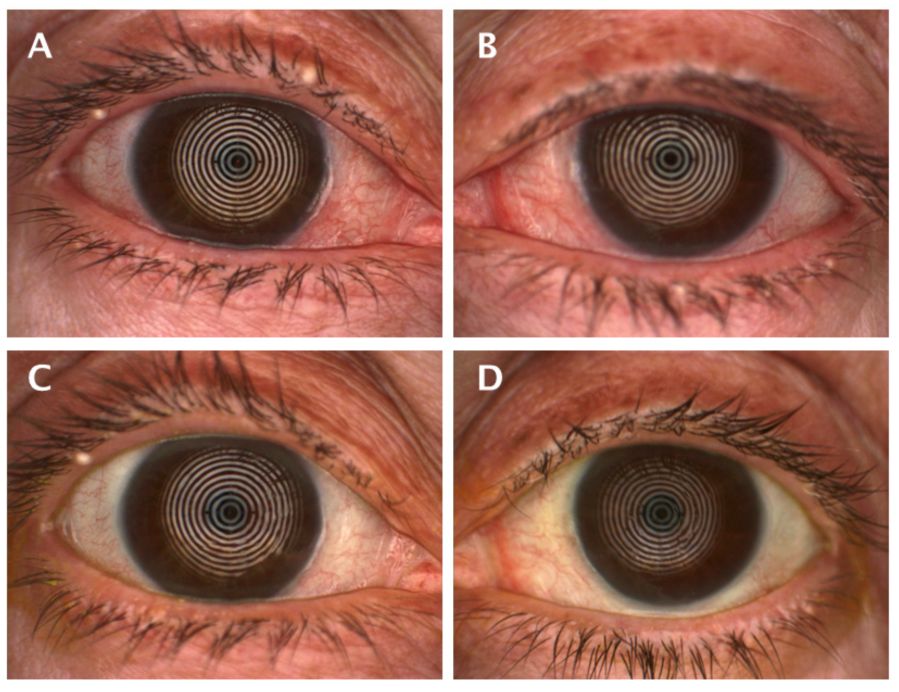
2.2.4. Ocular Redness
2.2.5. Fluorescein Staining
2.2.6. Stability of the Tear Film
2.2.7. Tear Volume
2.3. Statistical Analysis
3. Results
4. Discussion
5. Conclusions
Author Contributions
Funding
Institutional Review Board Statement
Informed Consent Statement
Data Availability Statement
Acknowledgments
Conflicts of Interest
Appendix A

| Standard Group | PRGF Group | |||
|---|---|---|---|---|
| Mean ± SEM | Mean ± SEM | p-Value | g + | |
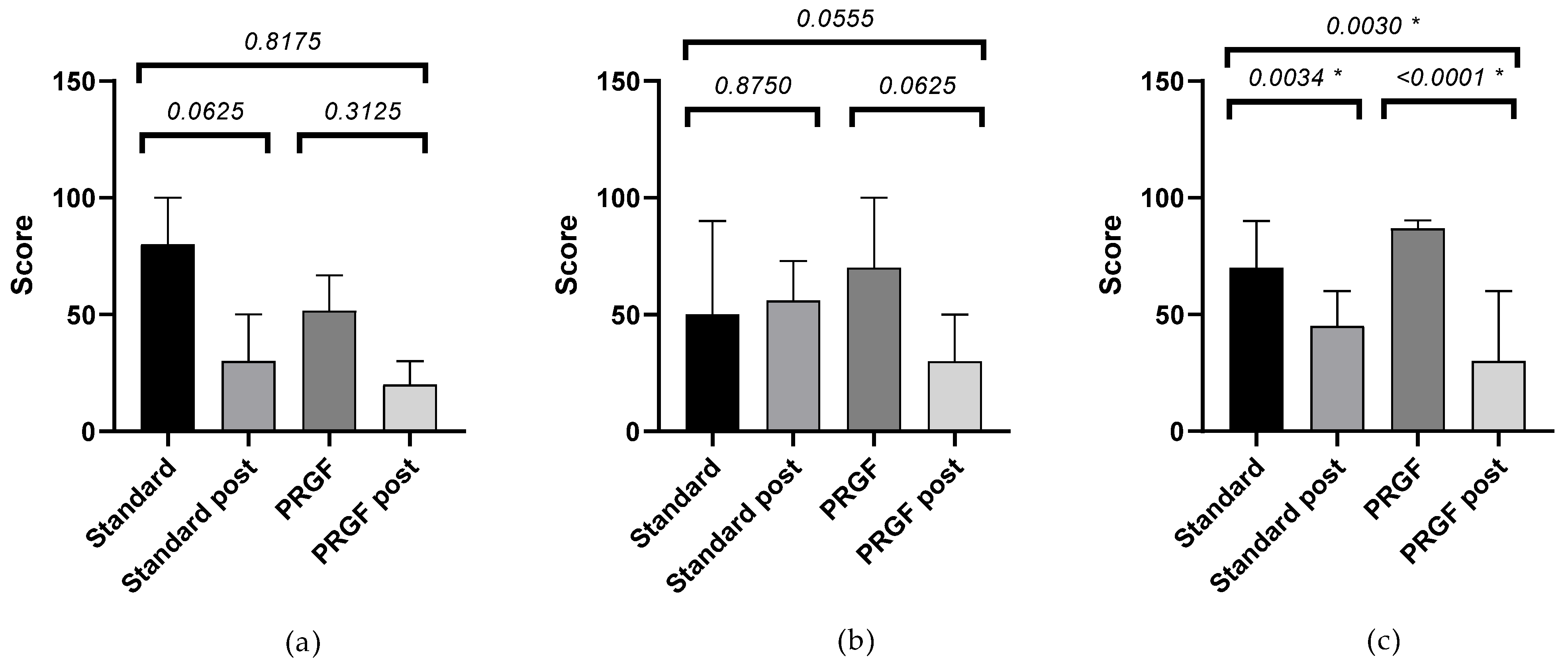
| SANDE frequency | ||||
| OSDI mild | −45.00 ± 10.25 | −30.00 ± 20.25 | 0.8175 | 0.38 |
| OSDI moderate | −6.00 ± 15.68 | −50.00 ± 11.83 | 0.0556 | 1.28 |
| OSDI severe | −21.25 ± 5.71 | −46.05 ± 4.89 | 0.0030 * | 1.16 |
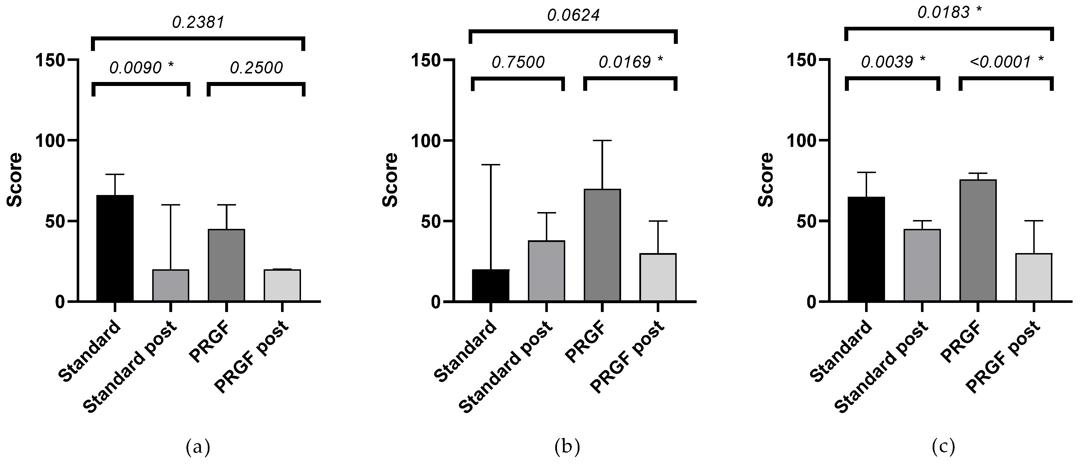
| SANDE severity | ||||
| OSDI mild | −38.00 ± 8.00 | −21.00 ± 11.45 | 0.2381 | 0.67 |
| OSDI moderate | 7.00 ± 21.54 | −46.00 ± 11.66 | 0.0624 | 0.91 |
| OSDI severe | −20.83 ± 5.18 | −41.84 ± 5.80 | 0.0183 * | 0.90 |


Appendix B
| Standard Group | PRGF Group | |||
|---|---|---|---|---|
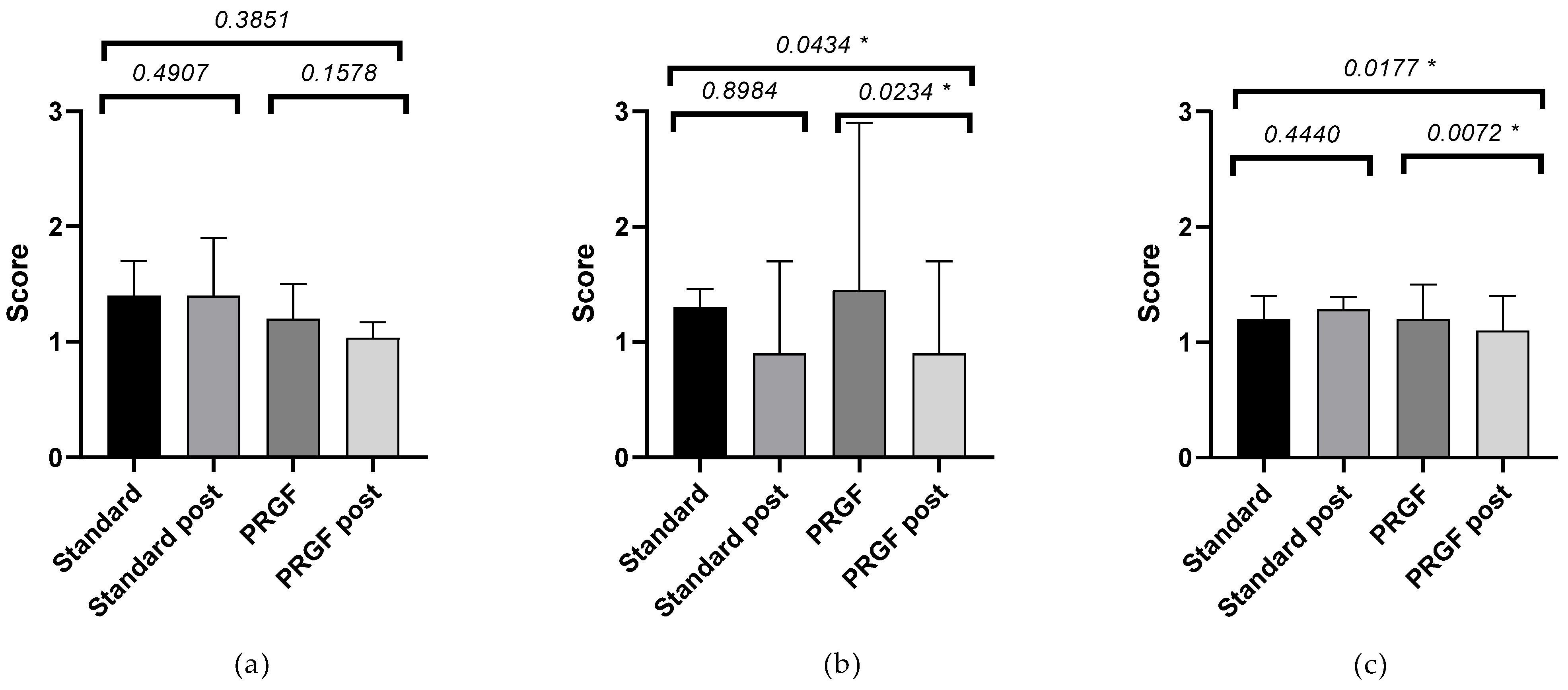
| Ocular Redness | Mean ± SEM | Mean ± SEM | p-Value | G + |
| OSDI mild | −0.08 ± 0.11 | −0.26 ± 0.17 | 0.3851 | 0.38 |
| OSDI moderate | −0.06 ± 0.12 | −0.51 ± 0.16 | 0.0434 * | 0.95 |
| OSDI severe | 0.08 ± 0.10 | −0.25 ± 0.08 | 0.0177 * | 0.34 |

References
- Tsubota, K.; Pflugfelder, S.C.; Liu, Z.; Baudouin, C.; Kim, H.M.; Messmer, E.M.; Kruse, F.; Liang, L.; Carreno-Galeano, J.T.; Rolando, M.; et al. Defining Dry Eye from a Clinical Perspective. Int. J. Mol. Sci. 2020, 21, 9271. [Google Scholar] [CrossRef] [PubMed]
- Stapleton, F.; Alves, M.; Bunya, V.Y.; Jalbert, I.; Lekhanont, K.; Malet, F.; Na, K.S.; Schaumberg, D.; Uchino, M.; Vehof, J.; et al. TFOS DEWS II Epidemiology Report. Ocul. Surf. 2017, 15, 334–365. [Google Scholar] [CrossRef] [PubMed]
- Craig, J.P.; Nichols, K.K.; Akpek, E.K.; Caffery, B.; Dua, H.S.; Joo, C.-K.; Liu, Z.; Nelson, J.D.; Nichols, J.J.; Tsubota, K.; et al. TFOS DEWS II Definition and Classification Report. Ocul. Surf. 2017, 15, 276–283. [Google Scholar] [CrossRef]
- Bron, A.J.; de Paiva, C.S.; Chauhan, S.K.; Bonini, S.; Gabison, E.E.; Jain, S.; Knop, E.; Markoulli, M.; Ogawa, Y.; Perez, V.; et al. Tfos dews II Pathophysiology Report. Ocul. Surf. 2017, 15, 438–510. [Google Scholar] [CrossRef] [PubMed]
- Maurizio Ronaldo, J.M. Management Strategies for Evaporative Dry Eye Disease and Future Perspective. Curr. Eye Res. 2022, 47, 813–823. [Google Scholar]
- Bron, A.J. The Definition and Classification of Dry Eye Disease. In Dry Eye: A Practical Approach; Springer: Berlin/Heidelberg, Germany, 2015; pp. 1–19. [Google Scholar]
- Jones, L.; Downie, L.E.; Korb, D.; Benitez-del-Castillo, J.M.; Dana, R.; Deng, S.X.; Dong, P.N.; Geerling, G.; Hida, R.Y.; Liu, Y.; et al. Tfos Dews II Management and Therapy Report. Ocul. Surf. 2017, 15, 575–628. [Google Scholar] [CrossRef] [PubMed]
- Hartwig, D.; Herminghaus, P.; Wedel, T.; Liu, L.; Schlenke, P.; Dibbelt, L.; Geerling, G. Topical Treatment of Ocular Surface Defects: Comparison of the Epitheliotrophic Capacity of Fresh Frozen Plasma and Serum on Corneal Epithelial Cells in an in Vitro Cell Culture Model. Transfus. Med. 2005, 15, 107–113. [Google Scholar] [CrossRef]
- Hartwig, D.; Harloff, S.; Liu, L.; Schlenke, P.; Wedel, T.; Geerling, G. Epitheliotrophic Capacity of a Growth Factor Preparation Produced from Platelet Concentrates on Corneal Epithelial Cells: A Potential Agent for the Treatment of Ocular Surface Defects? Transfusion 2004, 44, 1724–1731. [Google Scholar] [CrossRef]
- Liu, L.; Hartwig, D.; Harloff, S.; Herminghaus, P.; Wedel, T.; Kasper, K.; Geerling, G. Corneal Epitheliotrophic Capacity of Three Different Blood-Derived Preparations. Investig. Opthalmol. Vis. Sci. 2006, 47, 2438. [Google Scholar] [CrossRef]
- Zallio, F.; Mazzucco, L.; Monaco, F.; Astori, M.R.; Passera, R.; Drago, G.; Tamiazzo, S.; Rapetti, M.; Dolcino, D.; Guaschino, R.; et al. A Single-Center Pilot Prospective Study of Topical Application of Platelet-Derived Eye Drops for Patients with Ocular Chronic Graft-versus-Host Disease. Biol. Blood Marrow Transplant. 2016, 22, 1664–1670. [Google Scholar] [CrossRef]
- Fea, A.M.; Aragno, V.; Testa, V.; Machetta, F.; Parisi, S.; D’Antico, S.; Spinetta, R.; Fusaro, E.; Grignolo, F.M. The Effect of Autologous Platelet Lysate Eye Drops: An In Vivo Confocal Microscopy Study. BioMed Res. Int. 2016, 2016, 8406832. [Google Scholar] [CrossRef] [PubMed]
- Anitua, E.; Muruzabal, F.; Tayebba, A.; Riestra, A.; Perez, V.L.; Merayo-Lloves, J.; Orive, G. Autologous Serum and Plasma Rich in Growth Factors in Ophthalmology: Preclinical and Clinical Studies. Acta Ophthalmol. 2015, 93, e605–e614. [Google Scholar] [CrossRef]
- Ralph, R.A.; Doane, M.G.; Dohlman, C.H. Clinical Experience with a Mobile Ocular Perfusion Pump. Arch. Ophthalmol. 1975, 93, 1039–1043. [Google Scholar] [CrossRef]
- Pan, Q.; Angelina, A.; Marrone, M.; Stark, W.J.; Akpek, E.K. Autologous Serum Eye Drops for Dry Eye. Cochrane Database Syst. Rev. 2017, 2017, CD009327. [Google Scholar] [CrossRef] [PubMed]
- Tsubota, K.; Goto, E.; Shimmura, S.; Shimazaki, J. Treatment of Persistent Corneal Epithelial Defect by Autologous Serum Application. Ophthalmology 1999, 106, 1984–1989. [Google Scholar] [CrossRef] [PubMed]
- Anitua, E.; Muruzabal, F.; De la Fuente, M.; Merayo-Lloves, J.; Orive, G. Effects of Heat-Treatment on Plasma Rich in Growth Factors-Derived Autologous Eye Drop. Exp. Eye Res. 2014, 119, 27–34. [Google Scholar] [CrossRef]
- Anitua, E.; Sanchez, M.; Merayo-Lloves, J.; De la Fuente, M.; Muruzabal, F.; Orive, G. Plasma Rich in Growth Factors (PRGF-Endoret) Stimulates Proliferation and Migration of Primary Keratocytes and Conjunctival Fibroblasts and Inhibits and Reverts TGF-Β1–Induced Myodifferentiation. Investig. Opthalmol. Vis. Sci. 2011, 52, 6066. [Google Scholar] [CrossRef]
- Yoon, K.-C.; Jeong, I.-Y.; Park, Y.-G.; Yang, S.-Y. Interleukin-6 and Tumor Necrosis Factor-α Levels in Tears of Patients With Dry Eye Syndrome. Cornea 2007, 26, 431–437. [Google Scholar] [CrossRef]
- Pflugfelder, S.C.; Jones, D.; Ji, Z.; Afonso, A.; Monroy, D. Altered Cytokine Balance in the Tear Fluid and Conjunctiva of Patients with Sjogren’s Syndrome Keratoconjunctivitis Sicca. Curr. Eye Res. 1999, 19, 201–211. [Google Scholar] [CrossRef]
- Ogino, Y.; Ayukawa, Y.; Tsukiyama, Y.; Koyano, K. The Effect of Platelet-Rich Plasma on the Cellular Response of Rat Bone Marrow Cells in Vitro. Oral Surg. Oral Med. Oral Pathol. Oral Radiol. Endodontol. 2005, 100, 302–307. [Google Scholar] [CrossRef]
- Anitua, E.; Alkhraisat, M.H.; Orive, G. Perspectives and Challenges in Regenerative Medicine Using Plasma Rich in Growth Factors. J. Control. Release 2012, 157, 29–38. [Google Scholar] [CrossRef] [PubMed]
- Alio, J.L.; Colecha, J.R.; Pastor, S.; Rodriguez, A.; Artola, A. Symptomatic Dry Eye Treatment with Autologous Platelet-Rich Plasma. Ophthalmic Res. 2007, 39, 124–129. [Google Scholar] [CrossRef] [PubMed]
- López-Plandolit, S.; Morales, M.C.; Freire, V.; Grau, A.E.; Durán, J.A. Efficacy of Plasma Rich in Growth Factors for the Treatment of Dry Eye. Cornea 2011, 30, 1312–1317. [Google Scholar] [CrossRef] [PubMed]
- Bron, A.J.; Evans, V.E.; Smith, J.A. Grading of Corneal and Conjunctival Staining in the Context of Other Dry Eye Tests. Cornea 2003, 22, 640–650. [Google Scholar] [CrossRef]
- Anitua, E.; de la Fuente, M.; Muruzabal, F.; Riestra, A.; Merayo-Lloves, J.; Orive, G. Plasma Rich in Growth Factors (PRGF) Eye Drops Stimulates Scarless Regeneration Compared to Autologous Serum in the Ocular Surface Stromal Fibroblasts. Exp. Eye Res. 2015, 135, 118–126. [Google Scholar] [CrossRef]
- Merayo-Lloves, J.; Sanchez-Avila, R.M.; Riestra, A.C.; Anitua, E.; Begoña, L.; Orive, G.; Fernandez-Vega, L. Safety and Efficacy of Autologous Plasma Rich in Growth Factors Eye Drops for the Treatment of Evaporative Dry Eye. Ophthalmic Res. 2016, 56, 68–73. [Google Scholar] [CrossRef]
- Özcura, F.; Aydin, S.; Helvaci, M.R. Ocular Surface Disease Index for the Diagnosis of Dry Eye Syndrome. Ocul. Immunol. Inflamm. 2007, 15, 389–393. [Google Scholar] [CrossRef]
- Schaumberg, D.A.; Gulati, A.; Mathers, W.D.; Clinch, T.; Lemp, M.A.; Nelson, J.D.; Foulks, G.N.; Dana, R. Development and Validation of a Short Global Dry Eye Symptom Index. Ocul. Surf. 2007, 5, 50–57. [Google Scholar] [CrossRef]
- Salas Apaza, J.A.; Ariel Franco, J.V.; Meza, N.; Madrid, E.; Loézar, C.; Garegnani, L. Minimal Clinically Important Difference: The Basics. Medwave 2021, 21, e8149. [Google Scholar] [CrossRef]
- Jerchel, N.N.; Sickenberger, W.; Schulze, M.M. Objective Classification and Documentation of Bulbar Redness Using a Corneal Topographer. Contact Lens Anterior Eye 2012, 35, e18. [Google Scholar] [CrossRef]
- Fieguth, P.; Simpson, T. Automated Measurement of Bulbar Redness. Investig. Opthalmol. Vis. Sci. 2002, 43, 340–347. [Google Scholar]
- Amparo, F.; Wang, H.; Emami-Naeini, P.; Karimian, P.; Dana, R. The Ocular Redness Index: A Novel Automated Method for Measuring Ocular Injection. Investig. Opthalmol. Vis. Sci. 2013, 54, 4821–4826. [Google Scholar] [CrossRef] [PubMed]
- Wu, S.; Hong, J.; Tian, L.; Cui, X.; Sun, X.; Xu, J. Assessment of Bulbar Redness with a Newly Developed Keratograph. Optom. Vis. Sci. 2015, 92, 892–899. [Google Scholar] [CrossRef]
- Li, N.; Deng, X.G.; He, M.F. Comparison of the Schirmer I Test with and without Topical Anesthesia for Diagnosing Dry Eye. Int. J. Ophthalmol. 2012, 5, 478–481. [Google Scholar] [CrossRef]
- Tsubota, K.; Kaido, M.; Yagi, Y.; Fujihara, T.; Shimmura, S. Diseases Associated with Ocular Surface Abnormalities: The Importance of Reflex Tearing. Br. J. Ophthalmol. 1999, 83, 89–91. [Google Scholar] [CrossRef]
- Cambridge University Effect Size Calculator. Available online: https://www.cem.org/effect-size-calculator (accessed on 23 August 2022).
- Durlak, J.A. How to Select, Calculate, and Interpret Effect Sizes. J. Pediatr. Psychol. 2009, 34, 917–928. [Google Scholar] [CrossRef] [PubMed]
- Ribeiro, M.V.M.R.; de Melo, V.F.; Barbosa, M.E.F.C.; de Tozzi, M.U.F.; Ramos, M.S.B.; Gaia, N.M.S.R.S.; Santos, V.M.G.; de Neri, W.O.; Barbosa, F.T.; Ribeiro, E.A.N. The Use of Platelet Rich-Plasma in Ophthalmology: A Literature Review. Rev. Bras. Oftalmol. 2017, 76, 319–324. [Google Scholar] [CrossRef]
- García-Conca, V.; Abad-Collado, M.; Hueso-Abancens, J.R.; Mengual-Verdú, E.; Piñero, D.P.; Aguirre-Balsalobre, F.; Molina, J.C. Efficacy and Safety of Treatment of Hyposecretory Dry Eye with Platelet-Rich Plasma. Acta Ophthalmol. 2019, 97, e170–e178. [Google Scholar] [CrossRef] [PubMed]
- López-Plandolit, S.; Morales, M.C.; Freire, V.; Etxebarría, J.; Durán, J.A. Plasma Rich in Growth Factors as a Therapeutic Agent for Persistent Corneal Epithelial Defects. Cornea 2010, 29, 843–848. [Google Scholar] [CrossRef]
- Idoipe, M.; de la Sen-Corcuera, B.; Sánchez-ávila, R.M.; Sánchez-pérez, C.; Satué, M.; Sánchez-pérez, A.; Orive, G.; Muruzabal, F.; Anitua, E.; Pablo, L. Membrane of Plasma Rich in Growth Factors in Primary Pterygium Surgery Compared to Amniotic Membrane Transplantation and Conjunctival Autograft. J. Clin. Med. 2021, 10, 5711. [Google Scholar] [CrossRef]
- Merayo-Lloves, J.; Sanchez, R.M.; Riestra, A.C.; Anitua, E.; Begoña, L.; Orive, G.; Fernandez-Vega, L. Autologous Plasma Rich in Growth Factors Eyedrops in Refractory Cases of Ocular Surface Disorders. Ophthalmic Res. 2016, 55, 53–61. [Google Scholar] [CrossRef] [PubMed]
- Byun, Y.; Kim, T.; Kwon, S.M.; Seo, K.Y.; Kim, S.W.; Kim, E.K.; Park, W.C. Efficacy of Combined 0.05% Cyclosporine and 1% Methylprednisolone Treatment for Chronic Dry Eye. Cornea 2012, 31, 509–513. [Google Scholar] [CrossRef] [PubMed]
- Sheppard, J.D.; Donnenfeld, E.D.; Holland, E.J.; Slonim, C.B.; Solomon, R.; Solomon, K.D.; McDonald, M.B.; Perry, H.D.; Lane, S.S.; Pflugfelder, S.C.; et al. Effect of Loteprednol Etabonate 0.5% on Initiation of Dry Eye Treatment With Topical Cyclosporine 0.05%. Eye Contact Lens Sci. Clin. Pract. 2014, 40, 289–296. [Google Scholar] [CrossRef] [PubMed]
- Geerling, G.; Tauber, J.; Baudouin, C.; Goto, E.; Matsumoto, Y.; O’Brien, T.; Rolando, M.; Tsubota, K.; Nichols, K.K. The International Workshop on Meibomian Gland Dysfunction: Report of the Subcommittee on Management and Treatment of Meibomian Gland Dysfunction. Investig. Opthalmol. Vis. Sci. 2011, 52, 2050. [Google Scholar] [CrossRef]
- Pinto-Fraga, J.; López-de la Rosa, A.; Blázquez Arauzo, F.; Urbano Rodríguez, R.; González-García, M.J. Efficacy and Safety of 0.2% Hyaluronic Acid in the Management of Dry Eye Disease. Eye Contact Lens Sci. Clin. Pract. 2017, 43, 57–63. [Google Scholar] [CrossRef]
- Takamura, E.; Tsubota, K.; Watanabe, H.; Ohashi, Y. A Randomised, Double-Masked Comparison Study of Diquafosol versus Sodium Hyaluronate Ophthalmic Solutions in Dry Eye Patients. Br. J. Ophthalmol. 2012, 96, 1310–1315. [Google Scholar] [CrossRef]
- Kinoshita, S.; Oshiden, K.; Awamura, S.; Suzuki, H.; Nakamichi, N.; Yokoi, N. A Randomized, Multicenter Phase 3 Study Comparing 2% Rebamipide (OPC-12759) with 0.1% Sodium Hyaluronate in the Treatment of Dry Eye. Ophthalmology 2013, 120, 1158–1165. [Google Scholar] [CrossRef]
- Lekhanont, K.; Chuckpaiwong, V.; Vongthongsri, A.; Sangiampornpanit, T. Effects of Sodium Hyaluronate on Wavefront Aberrations in Dry Eye Patients. Optom. Vis. Sci. 2014, 91, 39–46. [Google Scholar] [CrossRef]
- Stevenson, W. Dry Eye Disease. Arch. Ophthalmol. 2012, 130, 90. [Google Scholar] [CrossRef]
- Barabino, S.; Chen, Y.; Chauhan, S.; Dana, R. Ocular Surface Immunity: Homeostatic Mechanisms and Their Disruption in Dry Eye Disease. Prog. Retin. Eye Res. 2012, 31, 271–285. [Google Scholar] [CrossRef]
- Rolando, M.; Zierhut, M.; Barabino, S. Should We Reconsider the Classification of Patients with Dry Eye Disease? Ocul. Immunol. Inflamm. 2021, 29, 521–523. [Google Scholar] [CrossRef]
- Marsh, P.; Pflugfelder, S.C. Topical Nonpreserved Methylprednisolone Therapy for Keratoconjunctivitis Sicca in Sjögren Syndrome. Ophthalmology 1999, 106, 811–816. [Google Scholar] [CrossRef]
- Papas, E.B. Key Factors in the Subjective and Objective Assessment of Conjunctival Erythema. Investig. Ophthalmol. Vis. Sci. 2000, 41, 687–691. [Google Scholar]
- McMonnies, C.W.; Chapman-Davies, A. Assessment of Conjunctival Hyperemia in Contact Lens Wearers. Part I. Optom. Vis. Sci. 1987, 64, 246–250. [Google Scholar] [CrossRef] [PubMed]
- Efron, N.; Morgan, P.B.; Katsara, S.S. Validation of Grading Scales for Contact Lens Complications. Ophthalmic Physiol. Opt. 2001, 21, 17–29. [Google Scholar] [CrossRef]
- Sorbara, L.; Simpson, T.; Duench, S.; Schulze, M.; Fonn, D. Comparison of an Objective Method of Measuring Bulbar Redness to the Use of Traditional Grading Scales. Contact Lens Anterior Eye 2007, 30, 53–59. [Google Scholar] [CrossRef]
- Peterson, R.C.; Wolffsohn, J.S. Sensitivity and Reliability of Objective Image Analysis Compared to Subjective Grading of Bulbar Hyperaemia. Br. J. Ophthalmol. 2007, 91, 1464–1466. [Google Scholar] [CrossRef] [PubMed]
- Barros, A.; Lozano-Sanroma, J.; Queiruga-Piñeiro, J.; Fernández-Vega Cueto, L.; Anitua, E.; Alcalde, I.; Merayo-Lloves, J. Recovery of Corneal Innervation after Treatment in Dry Eye Disease: A Confocal Microscopy Study. J. Clin. Med. 2023, 12, 1841. [Google Scholar] [CrossRef]
- Anitua, E.; de la Fuente, M.; Merayo-Lloves, J.; Muruzabal, F. Optimization of a Plasma Rich in Growth Factors Membrane for the Treatment of Inflammatory Ocular Diseases. Bioengineering 2022, 9, 508. [Google Scholar] [CrossRef]
- Tsubota, K.; Hata, S.; Okusawa, Y.; Egami, F.; Ohtsuki, T.; Nakamori, K. Quantitative Videographic Analysis of Blinking in Normal Subjects and Patients with Dry Eye. Arch. Ophthalmol. 1996, 114, 715–720. [Google Scholar] [CrossRef] [PubMed]
- Descalzi, F.; Ulivi, V.; Cancedda, R.; Piscitelli, F.; Luongo, L.; Guida, F.; Gatta, L.; Maione, S.; Di Marzo, V. Platelet-Rich Plasma Exerts Antinociceptive Activity by a Peripheral Endocannabinoid-Related Mechanism. Tissue Eng. Part A 2013, 19, 2120–2129. [Google Scholar] [CrossRef] [PubMed]
- Bendinelli, P.; Matteucci, E.; Dogliotti, G.; Corsi, M.M.; Banfi, G.; Maroni, P.; Desiderio, M.A. Molecular Basis of Anti-Inflammatory Action of Platelet-Rich Plasma on Human Chondrocytes: Mechanisms of NF-ΚB Inhibition via HGF. J. Cell. Physiol. 2010, 225, 757–766. [Google Scholar] [CrossRef]
- van Buul, G.M.; Koevoet, W.L.M.; Kops, N.; Bos, P.K.; Verhaar, J.A.N.; Weinans, H.; Bernsen, M.R.; van Osch, G.J.V.M. Platelet-Rich Plasma Releasate Inhibits Inflammatory Processes in Osteoarthritic Chondrocytes. Am. J. Sports Med. 2011, 39, 2362–2370. [Google Scholar] [CrossRef] [PubMed]
- Sanchez-Avila, R.M.; Merayo-Lloves, J.; Riestra, A.C.; Anitua, E.; Muruzabal, F.; Orive, G.; Fernández-Vega, L. The Effect of Immunologically Safe Plasma Rich in Growth Factor Eye Drops in Patients with Sjögren Syndrome. J. Ocul. Pharmacol. Ther. 2017, 33, 391–399. [Google Scholar] [CrossRef] [PubMed]
- Sanchez-Avila, R.M.; Merayo-Lloves, J.; Riestra, A.C.; Fernandez-Vega Cueto, L.; Anitua, E.; Begoña, L.; Muruzabal, F.; Orive, G. Treatment of Patients with Neurotrophic Keratitis Stages 2 and 3 with Plasma Rich in Growth Factors (PRGF-Endoret) Eye-Drops. Int. Ophthalmol. 2018, 38, 1193–1204. [Google Scholar] [CrossRef]
- Sullivan, B.D.; Crews, L.A.; Messmer, E.M.; Foulks, G.N.; Nichols, K.K.; Baenninger, P.; Geerling, G.; Figueiredo, F.; Lemp, M.A. Correlations between Commonly Used Objective Signs and Symptoms for the Diagnosis of Dry Eye Disease: Clinical Implications. Acta Ophthalmol. 2014, 92, 161–166. [Google Scholar] [CrossRef]
- Wolffsohn, J.S.; Arita, R.; Chalmers, R.; Djalilian, A.; Dogru, M.; Dumbleton, K.; Gupta, P.K.; Karpecki, P.; Lazreg, S.; Pult, H.; et al. Tfos Dews II Diagnostic Methodology Report. Ocul. Surf. 2017, 15, 539–574. [Google Scholar] [CrossRef]
- Viso, E.; Rodriguez-Ares, M.T.; Gude, F. Prevalence of and Associated Factors for Dry Eye in a Spanish Adult Population (The Salnes Eye Study). Ophthalmic Epidemiol. 2009, 16, 15–21. [Google Scholar] [CrossRef]
- Moss, S.E.; Klein, R.; Klein, B.E.K. Long-Term Incidence of Dry Eye in an Older Population. Optom. Vis. Sci. 2008, 85, 668–674. [Google Scholar] [CrossRef]


| Standard | PRGF | ||
|---|---|---|---|
| Mean ± SEM | Mean ± SEM | p-Value | |
| n (right eye/left eye) | 43 (22/21) | 57 (29/28) | 0.894 |
| Gender (male/female) | 9/13 | 3/27 | 0.009 * |
| Age | 60.73 ± 3.11 | 54.47 ± 3.10 | 0.169 |
| OSDI | 36.36 ± 3.66 | 47.70 ± 4.38 | 0.151 |
| FBUT | 4.05 ± 0.48 | 5.44 ± 0.55 | 0.106 |
| Local corneal staining | 1.23 ± 0.07 | 1.24 ± 0.09 | 0.449 |
| AT | TC | PHP | PP | C | |
|---|---|---|---|---|---|
| Standard | 100% (n = 22) | 0% (n = 0) | 18.22% (n = 4) | 0% (n = 0) | 0% (n = 0) |
| PRGF | 100% (n = 30) | 20% (n = 6) | 20% (n = 6) | 3.3% (n = 1) | 3.3% (n = 1) |
| Standard Group | PRGF Group | |||||||
|---|---|---|---|---|---|---|---|---|
| Baseline | Follow-Up | Baseline | Follow-Up | |||||
| Mean (±SEM) | Mean (±SEM) | p-Value | Effect Size + | Mean (±SEM) | Mean (±SEM) | p-Value | Effect Size + | |
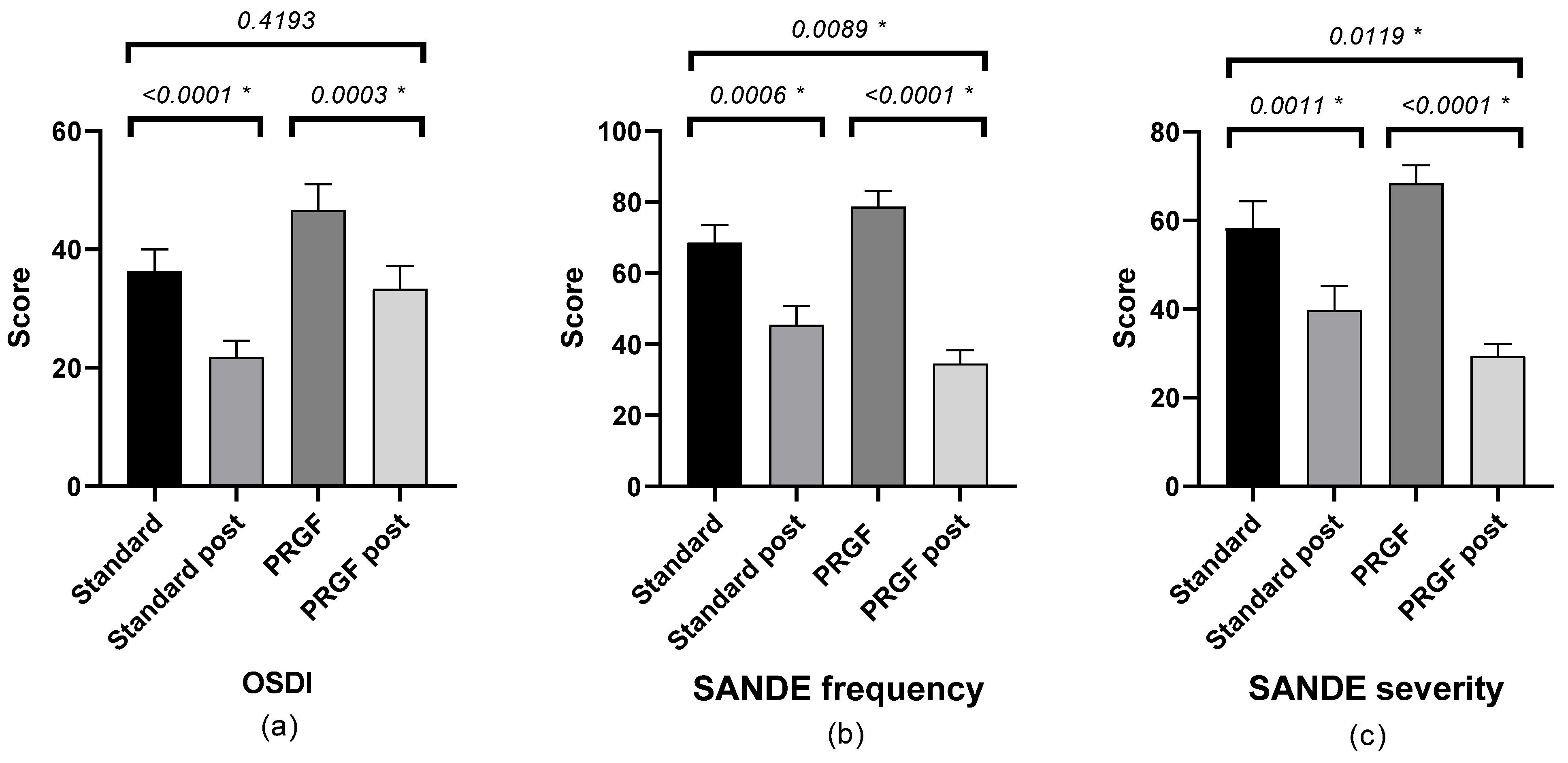
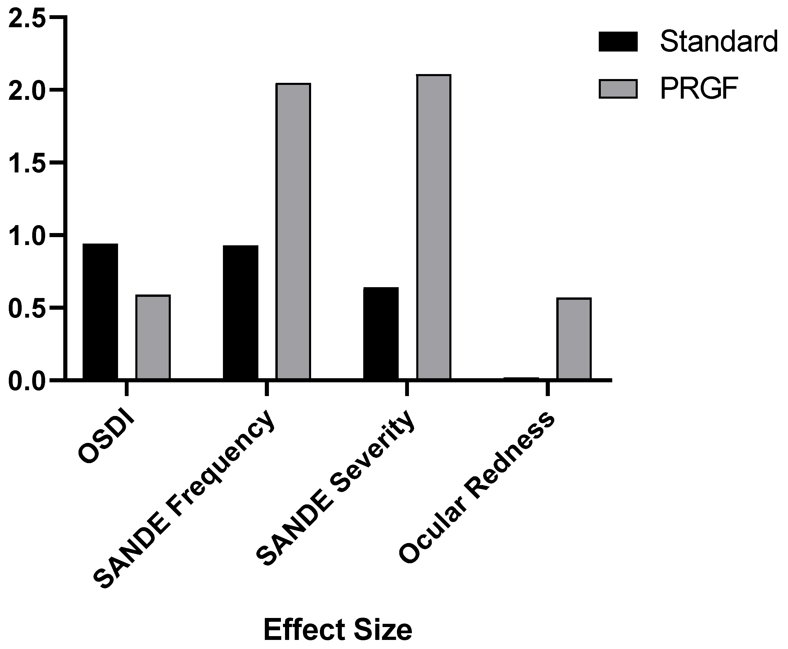
| OSDI Score | 36.36 ± 3.66 | 21.81 ± 2.78 | <0.0001 * | 0.94 | 47.70 ± 4.38 | 34.32 ± 3.90 | 0.0003 * | 0.59 |
| SANDE Frequency | 68.64 ± 4.95 | 45.45 ± 5.38 | 0.0006 * | 0.93 | 78.97 ± 4.62 | 35.00 ± 3.90 | <0.0001 * | 2.05 |
| SANDE Severity | 58.18 ± 6.17 | 39.77 ± 5.49 | 0.0011 * | 0.64 | 68.62 ± 4.25 | 29.66 ± 2.95 | <0.0001 * | 2.11 |
| BCVA (LogMAR) | 0.02 ± 0.008 | 0.01 ± 0.008 | 0.1316 | 0.20 | 0.08 ± 0.03 | 0.05 ± 0.02 | 0.0165 * | 0.10 |
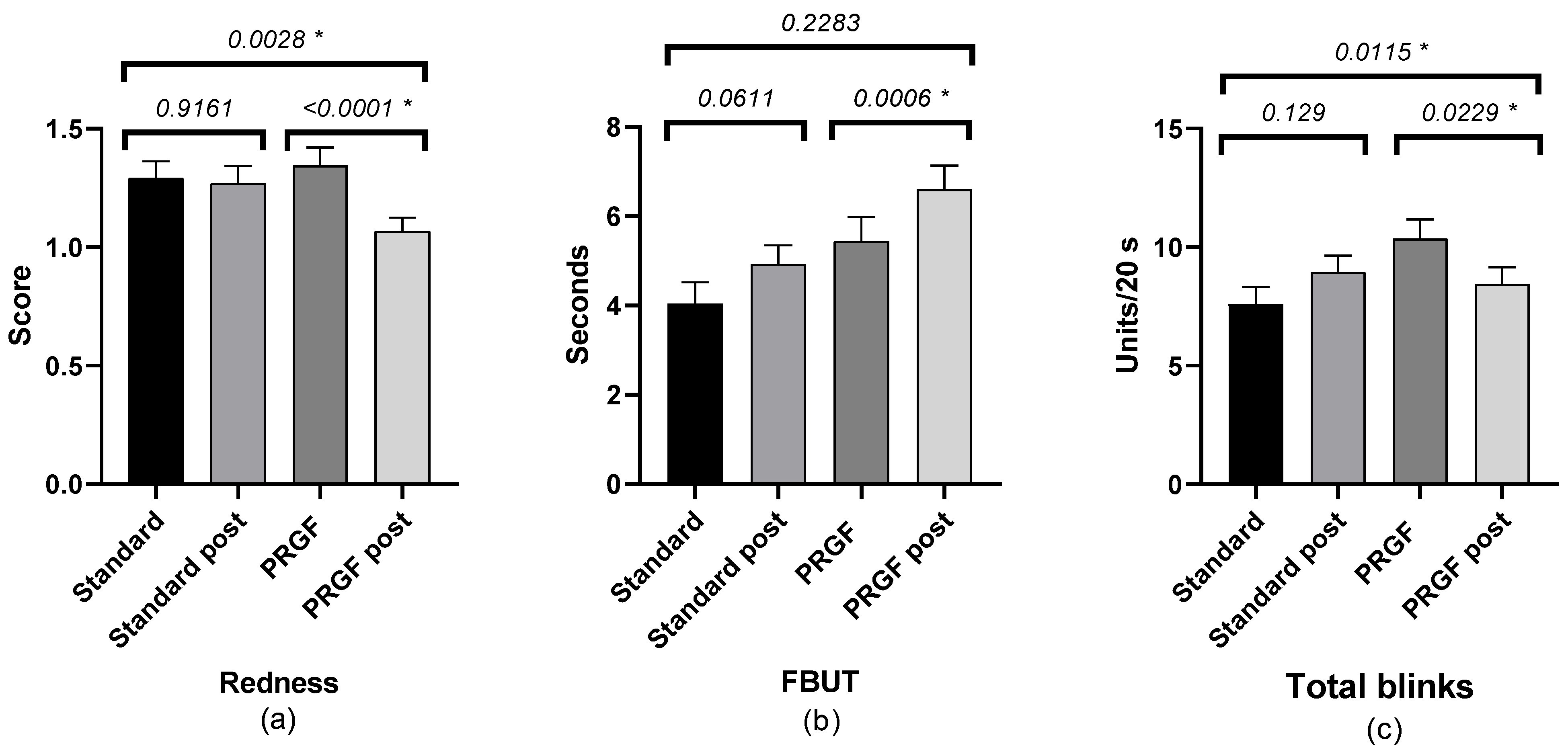
| Total blinks (n) | 7.61 ± 0.73 | 8.95 ± 0.69 | 0.1290 | 0.29 | 10.35 ± 0.81 | 8.46 ± 0.70 | 0.229 * | 0.33 |
| Ocular Redness (Index) | 1.26 ± 0.07 | 1.27 ± 0.08 | 0.9161 | 0.02 | 1.35 ± 0.08 | 1.05 ± 0.06 | <0.0001 * | 0.57 |
| Corneal Staining (Score) | 1.23 ± 0.07 | 1.28 ± 0.07 | 0.7539 | 0.11 | 1.24 ± 0.09 | 1.18 ± 0.07 | 0.2876 | 0.13 |
| FBUT (s) | 4.05 ± 0.48 | 4.93 ± 0.42 | 0.0611 | 0.30 | 5.44 ± 0.55 | 6.81 ± 0.57 | 0.0006 * | 0.34 |
| Schirmer (mm) | 6.74 ± 0.84 | 5.21 ± 0.68 | 0.0298 * | 0.30 | 5.88 ± 0.64 | 5.93 ± 0.70 | 0.9402 | 0.01 |
Disclaimer/Publisher’s Note: The statements, opinions and data contained in all publications are solely those of the individual author(s) and contributor(s) and not of MDPI and/or the editor(s). MDPI and/or the editor(s) disclaim responsibility for any injury to people or property resulting from any ideas, methods, instructions or products referred to in the content. |
© 2023 by the authors. Licensee MDPI, Basel, Switzerland. This article is an open access article distributed under the terms and conditions of the Creative Commons Attribution (CC BY) license (https://creativecommons.org/licenses/by/4.0/).
Share and Cite
Lozano-Sanroma, J.; Barros, A.; Alcalde, I.; Sánchez-Ávila, R.M.; Queiruga-Piñeiro, J.; Fernández-Vega Cueto, L.; Merayo-Lloves, J. Impact of Plasma Rich in Growth Factors (PRGF) Eye Drops on Ocular Redness and Symptomatology in Patients with Dry Eye Disease. Medicina 2023, 59, 928. https://doi.org/10.3390/medicina59050928
Lozano-Sanroma J, Barros A, Alcalde I, Sánchez-Ávila RM, Queiruga-Piñeiro J, Fernández-Vega Cueto L, Merayo-Lloves J. Impact of Plasma Rich in Growth Factors (PRGF) Eye Drops on Ocular Redness and Symptomatology in Patients with Dry Eye Disease. Medicina. 2023; 59(5):928. https://doi.org/10.3390/medicina59050928
Chicago/Turabian StyleLozano-Sanroma, Javier, Alberto Barros, Ignacio Alcalde, Ronald M. Sánchez-Ávila, Juan Queiruga-Piñeiro, Luis Fernández-Vega Cueto, and Jesús Merayo-Lloves. 2023. "Impact of Plasma Rich in Growth Factors (PRGF) Eye Drops on Ocular Redness and Symptomatology in Patients with Dry Eye Disease" Medicina 59, no. 5: 928. https://doi.org/10.3390/medicina59050928