
Histologic Evidence of Oral and Periodontal Regeneration Using Recombinant Human Platelet-Derived Growth Factor

Abstract
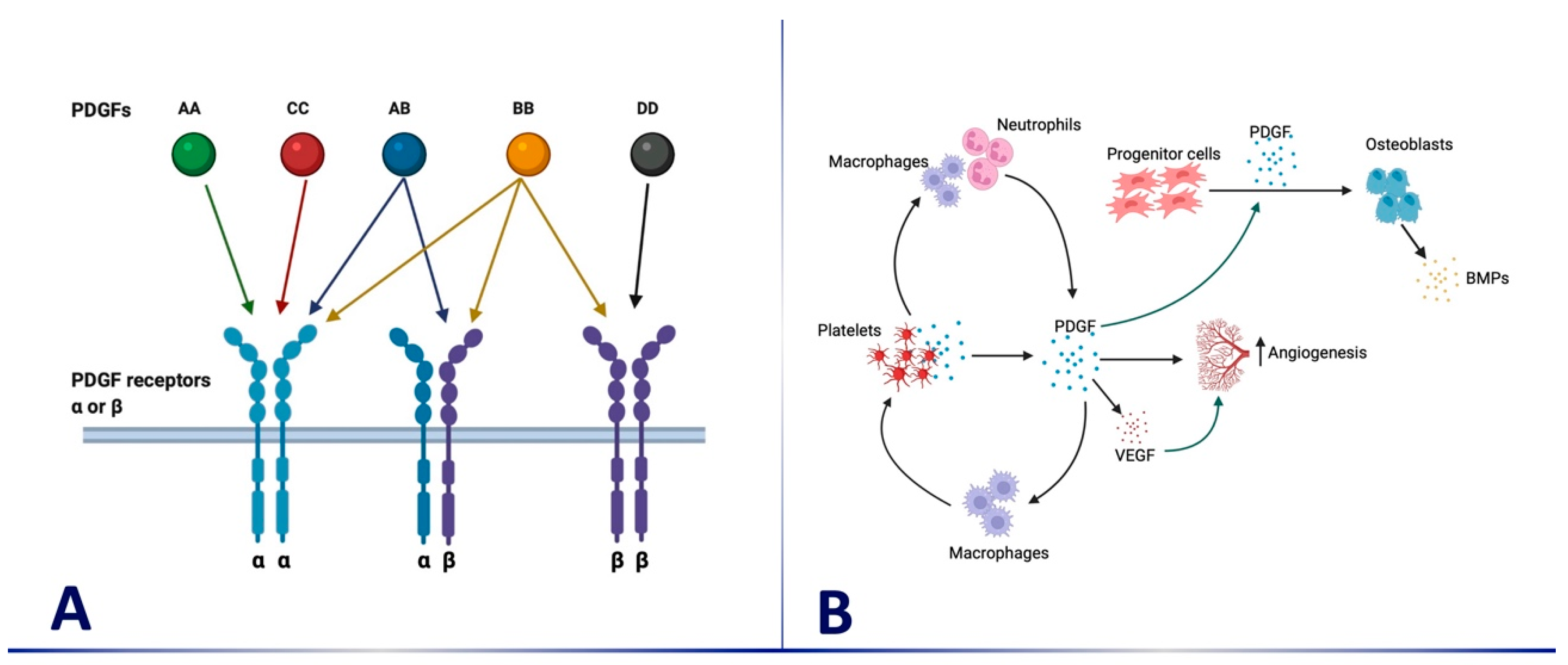
:1. Introduction
2. The Effects of PDGF-BB on Periodontal Regenerative Procedures
2.1. Histological Outcomes in Intrabony Defects
2.2. Histological Outcomes in Furcation Defects
2.3. Histological Outcomes in Root Coverage Procedures and Soft Tissue Augmentation
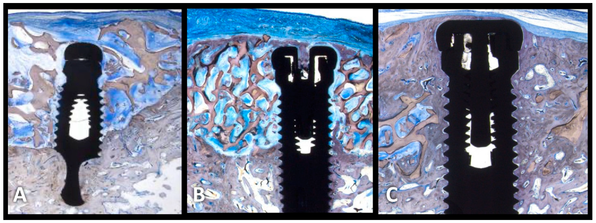
2.4. Histological Outcomes in Peri-Implant Regenerative Therapy
2.5. Histological Outcomes in Alveolar Ridge Preservation
2.6. Histological Outcomes in Guided Bone Regeneration
2.7. Histological Outcomes in Sinus Augmentation
3. Conclusions
- No abnormal or unfavorable effects, such as increased inflammation, ankylosis, or tissue overgrowth, have been reported following the use of rhPDGF-BB, even at the tissue and cellular levels under careful microscopic evaluation following nearly 30 years of histologic evaluation in animal models and many different human clinical indications.
- The osteopromotive effects of rhPDGF are present histologically as early as two weeks following treatment in animal models.
- The use of rhPDGF has been most thoroughly evaluated in humans histologically in periodontal infrabony and furcation defects. These studies have consistently found that true periodontal regeneration of cementum, PDL, and bone can be achieved using rhPDGF with allograft, xenograft, and, to a lesser extent, beta-TCP. In contrast, open-flap surgical debridement results in a long junctional epithelium.
- The histologic effects of rhPDGF have also been evaluated in numerous other clinical indications including peri-implant defects, GBR, extraction sockets, and, to a lesser extent, sinus grafts. In all cases, there was either a statistical improvement in bone regeneration (mostly) or a strong trend towards more bone in rhPDGF-treated sites. Such consistency of observations, despite the differences that might be expected across investigators, indications, models, and surgical techniques, etc., is striking.
- The use of collagen membranes is contra-indicated when using rhPDGF in intrabony defects and other sites where graft containment and stabilization can be achieved without the use of membranes. If a membrane is indicated for graft containment or stabilization, one should consider a membrane that is not cell occlusive (such as perforated PTFE) when using rhPDGF.
Author Contributions
Funding
Institutional Review Board Statement
Informed Consent Statement
Data Availability Statement
Conflicts of Interest
References
- Lynch, S.E.; Genco, R.; Marx, R.E. Tissue Engineering Applications in Oral and Maxillofacial Surgery and Periodontics; Quintessence Publishing Co., Inc.: Batavia, IL, USA, 2008. [Google Scholar]
- Mooney, D.J.; Silva, E.A. Tissue engineering: A glue for biomaterials. Nat. Mater. 2007, 6, 327–328. [Google Scholar] [CrossRef] [PubMed]
- Kohler, N.; Lipton, A. Platelets as a source of fibroblast growth-promoting activity. Exp. Cell Res. 1974, 87, 297–301. [Google Scholar] [CrossRef] [PubMed]
- Ross, R.; Glomset, J.; Kariya, B.; Harker, L. A platelet-dependent serum factor that stimulates the proliferation of arterial smooth muscle cells in vitro. Proc. Natl. Acad. Sci. USA 1974, 71, 1207–1210. [Google Scholar] [CrossRef] [PubMed] [Green Version]
- Westermark, B.; Wasteson, A. A platelet factor stimulating human normal glial cells. Exp. Cell Res. 1976, 98, 170–174. [Google Scholar] [CrossRef] [PubMed]
- Alvarez, R.H.; Kantarjian, H.M.; Cortes, J.E. Biology of platelet-derived growth factor and its involvement in disease. Mayo Clin. Proc. 2006, 81, 1241–1257. [Google Scholar] [CrossRef]
- Deuel, T.F.; Senior, R.M.; Huang, J.S.; Griffin, G.L. Chemotaxis of monocytes and neutrophils to platelet-derived growth factor. J. Clin. Investig. 1982, 69, 1046–1049. [Google Scholar] [CrossRef] [Green Version]
- Shimokado, K.; Raines, E.W.; Madtes, D.K.; Barrett, T.B.; Benditt, E.P.; Ross, R. A significant part of macrophage-derived growth factor consists of at least two forms of PDGF. Cell 1985, 43, 277–286. [Google Scholar] [CrossRef]
- Graham, S.; Leonidou, A.; Lester, M.; Heliotis, M.; Mantalaris, A.; Tsiridis, E. Investigating the role of PDGF as a potential drug therapy in bone formation and fracture healing. Expert Opin. Investig. Drugs 2009, 18, 1633–1654. [Google Scholar] [CrossRef]
- Li, A.; Xia, X.; Yeh, J.; Kua, H.; Liu, H.; Mishina, Y.; Hao, A.; Li, B. PDGF-AA promotes osteogenic differentiation and migration of mesenchymal stem cell by down-regulating PDGFRα and derepressing BMP-Smad1/5/8 signaling. PLoS ONE 2014, 9, e113785. [Google Scholar] [CrossRef] [Green Version]
- Bouletreau, P.J.; Warren, S.M.; Spector, J.A.; Steinbrech, D.S.; Mehrara, B.J.; Longaker, M.T. Factors in the fracture microenvironment induce primary osteoblast angiogenic cytokine production. Plast. Reconstr. Surg. 2002, 110, 139–148. [Google Scholar] [CrossRef]
- Carano, R.A.; Filvaroff, E.H. Angiogenesis and bone repair. Drug Discov. Today 2003, 8, 980–989. [Google Scholar] [CrossRef]
- Lynch, S.E.; De Castilla, G.R.; Williams, R.C.; Kiritsy, C.P.; Howell, T.H.; Reddy, M.S.; Antoniades, H.N. The effects of short-term application of a combination of platelet-derived and insulin-like growth factors on periodontal wound healing. J. Periodontol. 1991, 62, 458–467. [Google Scholar] [CrossRef]
- Wang, H.; Pappert, T.D.; Castelli, W.A.; Chiego, D.J.; Shyr, Y.; Smith, B.A. The effect of platelet-derived growth factor on the cellular response of the periodontium: An autoradiographic study on dogs. J. Periodontol. 1994, 65, 429–436. [Google Scholar] [CrossRef] [PubMed]
- Sculean, A.; Karring, T.; Theilade, J.; Lioubavina, N. The regenerative potential of oxytalan fibers. An experimental study in the monkey. J. Clin. Periodontol. 1997, 24, 932–936. [Google Scholar] [CrossRef] [PubMed]
- Nevins, M.; Camelo, M.; Nevins, M.L.; Schenk, R.K.; Lynch, S.E. Periodontal regeneration in humans using recombinant human platelet-derived growth factor-BB (rhPDGF-BB) and allogenic bone. J. Periodontol. 2003, 74, 1282–1292. [Google Scholar] [CrossRef] [PubMed] [Green Version]
- Ridgway, H.K.; Mellonig, J.T.; Cochran, D.L. Human histologic and clinical evaluation of recombinant human platelet-derived growth factor and beta-tricalcium phosphate for the treatment of periodontal intraosseous defects. Int. J. Periodontics Restor. Dent. 2008, 28, 171–179. [Google Scholar]
- Shirakata, Y.; Taniyama, K.; Yoshimoto, T.; Miyamoto, M.; Takeuchi, N.; Matsuyama, T.; Noguchi, K. Regenerative effect of basic fibroblast growth factor on periodontal healing in two-wall intrabony defects in dogs. J. Clin. Periodontol. 2010, 37, 374–381. [Google Scholar] [CrossRef]
- Camelo, M.; Nevins, M.L.; Schenk, R.K.; Lynch, S.E.; Nevins, M. Periodontal regeneration in human Class II furcations using purified recombinant human platelet-derived growth factor-BB (rhPDGF-BB) with bone allograft. Int. J. Periodontics Restor. Dent. 2003, 23, 213–225. [Google Scholar]
- Mellonig, J.T.; del Pilar Valderrama, M.; Cochran, D.L. Histological and clinical evaluation of recombinant human platelet-derived growth factor combined with beta tricalcium phosphate for the treatment of human Class III furcation defects. Int. J. Periodontics Restor. Dent. 2009, 29, 169–177. [Google Scholar]
- Nevins, M.; Garber, D.; Hanratty, J.J.; McAllister, B.S.; Nevins, M.L.; Salama, M.; Schupbach, P.; Wallace, S.; Bernstein, S.M.; Kim, D.M. Human histologic evaluation of anorganic bovine bone mineral combined with recombinant human platelet-derived growth factor BB in maxillary sinus augmentation: Case series study. Int. J. Periodontics Restor. Dent. 2009, 29, 583–591. [Google Scholar]
- McAllister, B.S.; Haghighat, K.; Prasad, H.S.; Rohrer, M.D. Histologic evaluation of recombinant human platelet-derived growth factor-BB after use in extraction socket defects: A case series. Int. J. Periodontics Restor. Dent. 2010, 30, 365–373. [Google Scholar]
- Wallace, S.C.; Snyder, M.B.; Prasad, H. Postextraction ridge preservation and augmentation with mineralized allograft with or without recombinant human platelet-derived growth factor BB (rhPDGF-BB): A consecutive case series. Int. J. Periodontics Restor. Dent. 2013, 33, 599–609. [Google Scholar] [CrossRef]
- Geurs, N.; Ntounis, A.; Vassilopoulos, P.; Van Der Velden, U.; Loos, B.G.; Reddy, M. Using growth factors in human extraction sockets: A histologic and histomorphometric evaluation of short-term healing. Int. J. Oral Maxillofac. Implant. 2014, 29, 485–496. [Google Scholar] [CrossRef] [PubMed] [Green Version]
- Mendoza-Azpur, G.; Cornejo, H.; Olaechea, A.; Padial-Molina, M.; O’Valle, F.; Galindo-Moreno, P. Anorganic Bovine Bone Plus Recombinant Human Platelet-Derived Growth Factor-BB in Ridge Preservation: A Pilot Study. Int. J. Oral Maxillofac. Implant. 2022, 37, 356–364. [Google Scholar] [CrossRef]
- Simion, M.; Rocchietta, I.; Dellavia, C. Three-dimensional ridge augmentation with xenograft and recombinant human platelet-derived growth factor-BB in humans: Report of two cases. Int. J. Periodontics Restor. Dent. 2007, 27, 109–115. [Google Scholar]
- De Angelis, N.; Scivetti, M. Lateral ridge augmentation using an equine flex bone block infused with recombinant human platelet-derived growth factor BB: A clinical and histologic study. Int. J. Periodontics Restor. Dent. 2011, 31, 383–388. [Google Scholar]
- Nevins, M.; Al Hezaimi, K.; Schupbach, P.; Karimbux, N.; Kim, D.M. Vertical ridge augmentation using an equine bone and collagen block infused with recombinant human platelet-derived growth factor-BB: A randomized single-masked histologic study in non-human primates. J. Periodontol. 2012, 83, 878–884. [Google Scholar] [CrossRef]
- Nevins, M.L.; Reynolds, M.A.; Camelo, M.; Schupbach, P.; Kim, D.M.; Nevins, M. Recombinant human platelet-derived growth factor BB for reconstruction of human large extraction site defects. Int. J. Periodontics Restor. Dent. 2014, 34, 157–163. [Google Scholar] [CrossRef] [PubMed] [Green Version]
- McGuire, M.K.; Scheyer, E.T.; Schupbach, P. Growth factor-mediated treatment of recession defects: A randomized controlled trial and histologic and microcomputed tomography examination. J. Periodontol. 2009, 80, 550–564. [Google Scholar] [CrossRef]
- Simion, M.; Rocchietta, I.; Fontana, F.; Dellavia, C. Evaluation of a resorbable collagen matrix infused with rhPDGF-BB in peri-implant soft tissue augmentation: A preliminary report with 3.5 years of observation. Int. J. Periodontics Restor. Dent. 2012, 32, 273–282. [Google Scholar]
- Simion, M.; Rocchietta, I.; Kim, D.; Nevins, M.; Fiorellini, J. Vertical ridge augmentation by means of deproteinized bovine bone block and recombinant human platelet-derived growth factor-BB: A histologic study in a dog model. Int. J. Periodontics Restor. Dent. 2006, 26, 415–423. [Google Scholar]
- Simion, M.; Nevins, M.; Rocchietta, I.; Fontana, F.; Maschera, E.; Schupbach, P.; Kim, D.M. Vertical ridge augmentation using an equine block infused with recombinant human platelet-derived growth factor-BB: A histologic study in a canine model. Int. J. Periodontics Restor. Dent. 2009, 29, 245–255. [Google Scholar]
- Lynch, S.E.; Williams, R.C.; Poison, A.M.; Howell, T.H.; Reddy, M.S.; Zappa, U.E.; Antoniades, H.N. A combination of platelet-derived and insulin-like growth factors enhances periodontal regeneration. J. Clin. Periodontol. 1989, 16, 545–548. [Google Scholar] [CrossRef] [PubMed]
- Giannobile, W.V.; Hernandez, R.A.; Finkelman, R.D.; Ryarr, S.; Kiritsy, C.P.; D’Andrea, M.; Lynch, S.E. Comparative effects of platelet-derived growth factor-BB and insulin-like growth factor-I, individually and in combination, on periodontal regeneration in Macaca fascicularis. J. Periodontal. Res. 1996, 31, 301–312. [Google Scholar] [CrossRef] [PubMed]
- Zhang, Y.; Miron, R.J.; Li, S.; Shi, B.; Sculean, A.; Cheng, X. Novel MesoPorous BioGlass/silk scaffold containing adPDGF-B and adBMP7 for the repair of periodontal defects in beagle dogs. J. Clin. Periodontol. 2015, 42, 262–271. [Google Scholar] [CrossRef] [PubMed]
- Al-Hazmi, B.A.; Al-Hamdan, K.S.; Al-Rasheed, A.; Babay, N.; Wang, H.-L.; Al-Hezaimi, K. Efficacy of using PDGF and xenograft with or without collagen membrane for bone regeneration around immediate implants with induced dehiscence-type defects: A microcomputed tomographic study in dogs. J. Periodontol. 2013, 84, 371–378. [Google Scholar] [CrossRef] [Green Version]
- Nevins, M.L.; Camelo, M.; Schupbach, P.; Kim, D.M.; Camelo, J.M.B.; Nevins, M. Human histologic evaluation of mineralized collagen bone substitute and recombinant platelet-derived growth factor-BB to create bone for implant placement in extraction socket defects at 4 and 6 months: A case series. Int. J. Periodontics Restor. Dent. 2009, 29, 129–139. [Google Scholar]


| Intrabony Defects | |||||||
| Reference | Species/Model | Bone Graft Type/Carrier | Amt of rhPDGF: Saturated | Sample Size | Defect Type | Healing Type Histologically | Clinical Gains (Mean mm) |
| Lynch et al., 1991 [13] PDGF in combination with IGF | Animal (Beagle dogs) | Methylcellulose gel | 10 ng of I-PDGF-B in combination with IGF | 13 | Not reported | New Bone, new cementum, highly organized CT | Not reported |
| Wang et al., 1994 [14] | Animal (Mongrel dogs) | PDGF alone or in combination with ePTFE membrane | Not reported | 6 | Fenestration defects created into dentin | Increased fibroblasts, cemetoblasts, osteoblasts, perivacular cells, and endothelial cells proliferation | Not reported |
| Sculean et al., 1997 [15] | Animal (Monkey) | PDGF in gel | 0.1 µg/mL | 1 | Surgically- created intrabony defects on either the mesial or distal aspect of teeth | New PDL, new cementum, new bone | Not reported |
| Nevins et al., 2003 [16] | Human | DFDBA | 0.3 mg/mL, 1.0 mg/mL, and 5.0 mg/mL | 9 | Interproximal intrabony defects | New PDL, new cementum, new bone | PD reduction 6.42 ± 1.69 CAL gain 6.17 ± 1.94 Bone fill 2.14 ± 0.85 |
| Ridgway et al., 2008 [17] | Human | β-TCP | 0.3 mg/mL, and 1.0 mg/mL | 8 | Interproximal intrabony | New PDL, new cementum, new bone | PD reduction 4.6 ± 1.5 (0.3 mg/mL) 4.3 ± 1.5 (1 mg/mL) CAL gain 3.1 ± 1.8 (0.3 mg/mL) 3.2 ± 1.9 (1 mg/mL) |
| Shirakata et al., 2010 [18] | Animal (Beagle dogs) | β-TCP | 0.3 mg/mL | 4 | Surgically created interproximal defects | New cementum, new bone | Bone formation 4.66 ± 0.7 |
| Furcation defects | |||||||
| Reference | Species/Model | Bone Graft type/Carrier | Amt of rhPDGF: Saturated | Sample size | Defect type | Healing type Histologically | Clinical gains |
| Nevins et al., 2003 [16] | Human | DFDBA | 0.3 mg/mL, 1.0 mg/mL, and 5.0 mg/mL | 9 | Class III furcation defects | New PDL, new cementum, new bone | PD reduction 6.42 ± 1.69 CAL gain 6.17 ± 1.94 Bone fill 2.14 ± 0.85 |
| Camelo et al., 2003 [19] | Human | DFDBA | 0.5 mg/mL and 1.0 mg/mL | 4 | Class II furcation defects | New PDL, new cementum, new bone | PD reduction (Vertical 4.25) (Horizontal 3.5) CAL 3.75 |
| Mellonig et al., 2009 [20] | Human | β-TCP | 0.3 mg/mL | 4 | Class III furcation defects | New PDL, new cementum, new bone | PD reduction 4 CAL gain 2.86 |
| Alveolar ridge preservation | |||||||
| Reference | Species/Model | Bone Graft type /Carrier | Amt of rhPDGF: Saturated | Sample size | Defect type | Healing type Histologically | Clinical gains |
| Nevins et al., 2009 [21] | Human | MCBS | 0.3 mg/mL | 8 | Buccal wall extraction defects | New bone formation | Not reported |
| McAllister et al., 2010 [22] | Human | Either ABB-C or β-TCP | 0.3 mg/mL | 11 | Extraction sockets | Vital bone formation | Not reported |
| Wallace et al., 2013 [23] | Human | Mineralized allograft | 0.3 mg/mL | 30 | Buccal wall extraction defects | Vital bone formation | Not reported |
| Geurs et al., 2014 [24] | Human | FDBA + β-TCP + Collagen plug | NR | 41 | Extraction sockets | New woven bone formation, less organic matrix | Not reported |
| Mendoza-Azpur et al., 2022 [25] | Human | Anorganic bovine bone and collagen | 0.3 mg/mL | 5 | Extraction sockets | New mineralized tissue formation | Significant differences in the bucco-lingual width between the groups, favoring the PDGF group |
| Guided bone regeneration | |||||||
| Reference | Species/Model | Bone Graft type/Carrier | Amt of rhPDGF: Saturated | Sample size | Defect type | Healing type Histologically | Clinical gains |
| Simion et al., 2007 [26] | Human | Deproteinized bovine bone block | NR | 2 | Vertical and horizontal ridge defects | New bone formation | Not reported |
| De Angelis et al., 2011 [27] | Human | Equine block | 0.3 mg/mL | 1 | Horizontal ridge defect | New bone formation | Not reported |
| Nevins et al., 2012 [28] | Monkey | eHAC block | NR | 6 | Vertical bone defects | New bone formation | Not reported |
| Nevins et al., 2014 [29] | Human | Either with bovine or equine matrix | 0.3 mg/mL | 8 | Large alveolar extraction defects | New bone formation | Not reported |
| Sinus augmentation | |||||||
| Reference | Species/Model | Bone Graft type/Carrier | Amt of rhPDGF: Saturated | Sample size | Defect type | Healing type Histologically | Clinical gains |
| Nevins et al., 2009 [21] | Human | ABBM | 0.3 mg/mL | 10 | Lamellar bone and woven bone formation | Not reported | |
| Soft tissue augmentation and recession coverage | |||||||
| Reference | Species/Model | Bone Graft type/Carrier | Amt of rhPDGF: Saturated | Sample size | Defect type | Healing type Histologically | Clinical gains |
| McGuire et al., 2009 [30] | Human | β-TCP | 0.3 mg/mL | 30 | Miller Class II buccal gingival recession | New PDL New Cementum New Bone | Root coverage (90.8%) Recession depth reduction (−2.9 + 0.5 mm) |
| Simion et al., 2012 [31] | Human | Resorbable collagen matrix | Not reported | 6 | Insufficient soft tissue volume | Well organized epithelium and CT | Increased soft tissue volume |
| Dental Implant-related Regeneration | |||||||
| Reference | Species/Model | Bone Graft type/Carrier | Amt of rhPDGF: Saturated | Sample size | Defect type | Healing type Histologically | Clinical gains |
| Simion et al., 2006 [32] | Canine | Deproteinized bovine bone block | Not reported | 6 | Vertical ridge defects | New bone formation | Not reported |
| Simion et al., 2009 [33] | Canine | eHAC block | Not reported | 12 | Vertical ridge defects | New bone formation | Not reported |
Disclaimer/Publisher’s Note: The statements, opinions and data contained in all publications are solely those of the individual author(s) and contributor(s) and not of MDPI and/or the editor(s). MDPI and/or the editor(s) disclaim responsibility for any injury to people or property resulting from any ideas, methods, instructions or products referred to in the content. |
© 2023 by the authors. Licensee MDPI, Basel, Switzerland. This article is an open access article distributed under the terms and conditions of the Creative Commons Attribution (CC BY) license (https://creativecommons.org/licenses/by/4.0/).
Share and Cite
Meghil, M.M.; Mandil, O.; Nevins, M.; Saleh, M.H.A.; Wang, H.-L. Histologic Evidence of Oral and Periodontal Regeneration Using Recombinant Human Platelet-Derived Growth Factor. Medicina 2023, 59, 676. https://doi.org/10.3390/medicina59040676
Meghil MM, Mandil O, Nevins M, Saleh MHA, Wang H-L. Histologic Evidence of Oral and Periodontal Regeneration Using Recombinant Human Platelet-Derived Growth Factor. Medicina. 2023; 59(4):676. https://doi.org/10.3390/medicina59040676
Chicago/Turabian StyleMeghil, Mohamed M., Obada Mandil, Myron Nevins, Muhammad H. A. Saleh, and Hom-Lay Wang. 2023. "Histologic Evidence of Oral and Periodontal Regeneration Using Recombinant Human Platelet-Derived Growth Factor" Medicina 59, no. 4: 676. https://doi.org/10.3390/medicina59040676