
McKittrick–Wheelock Syndrome: A Case Report

Abstract
:1. Introduction
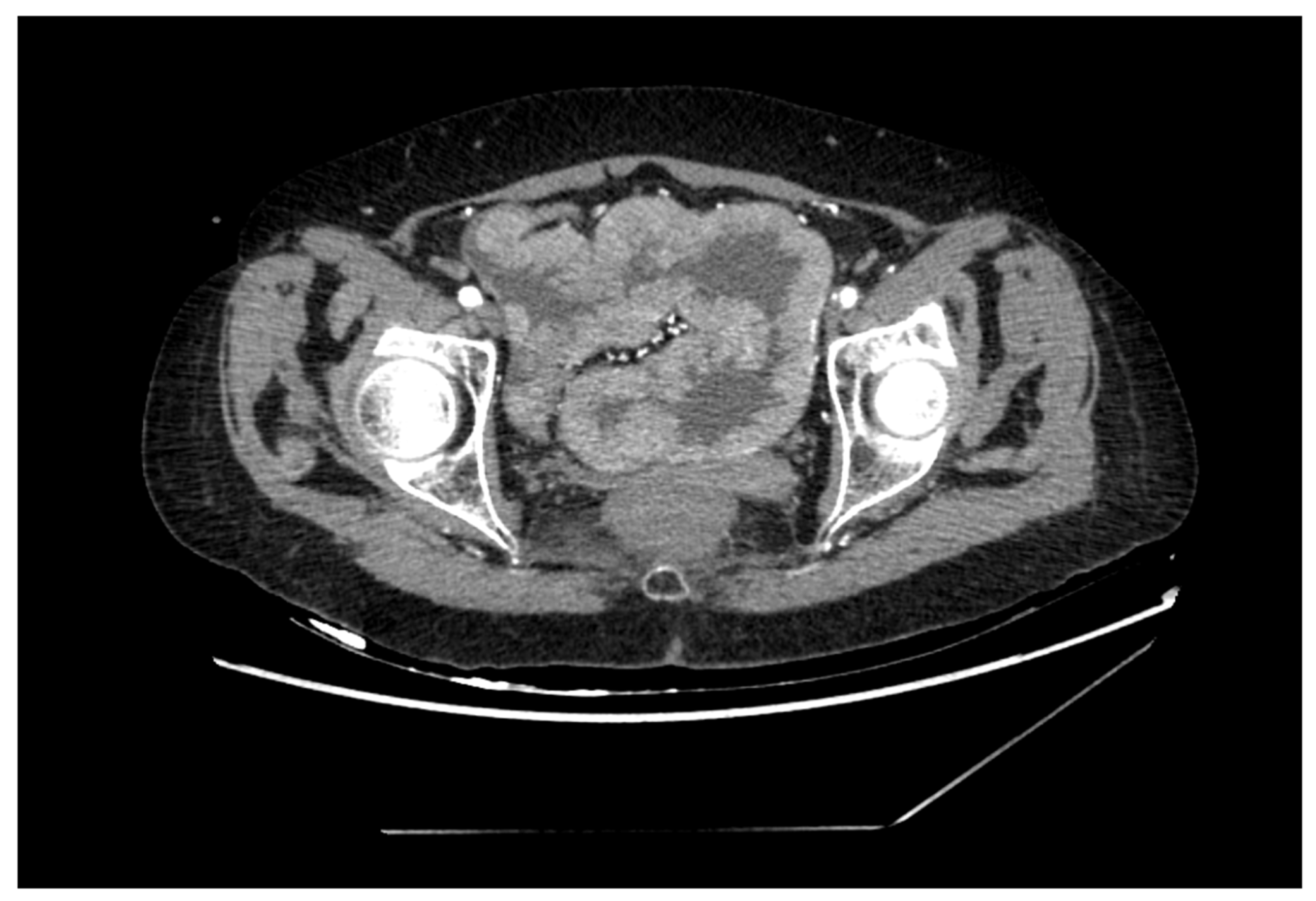
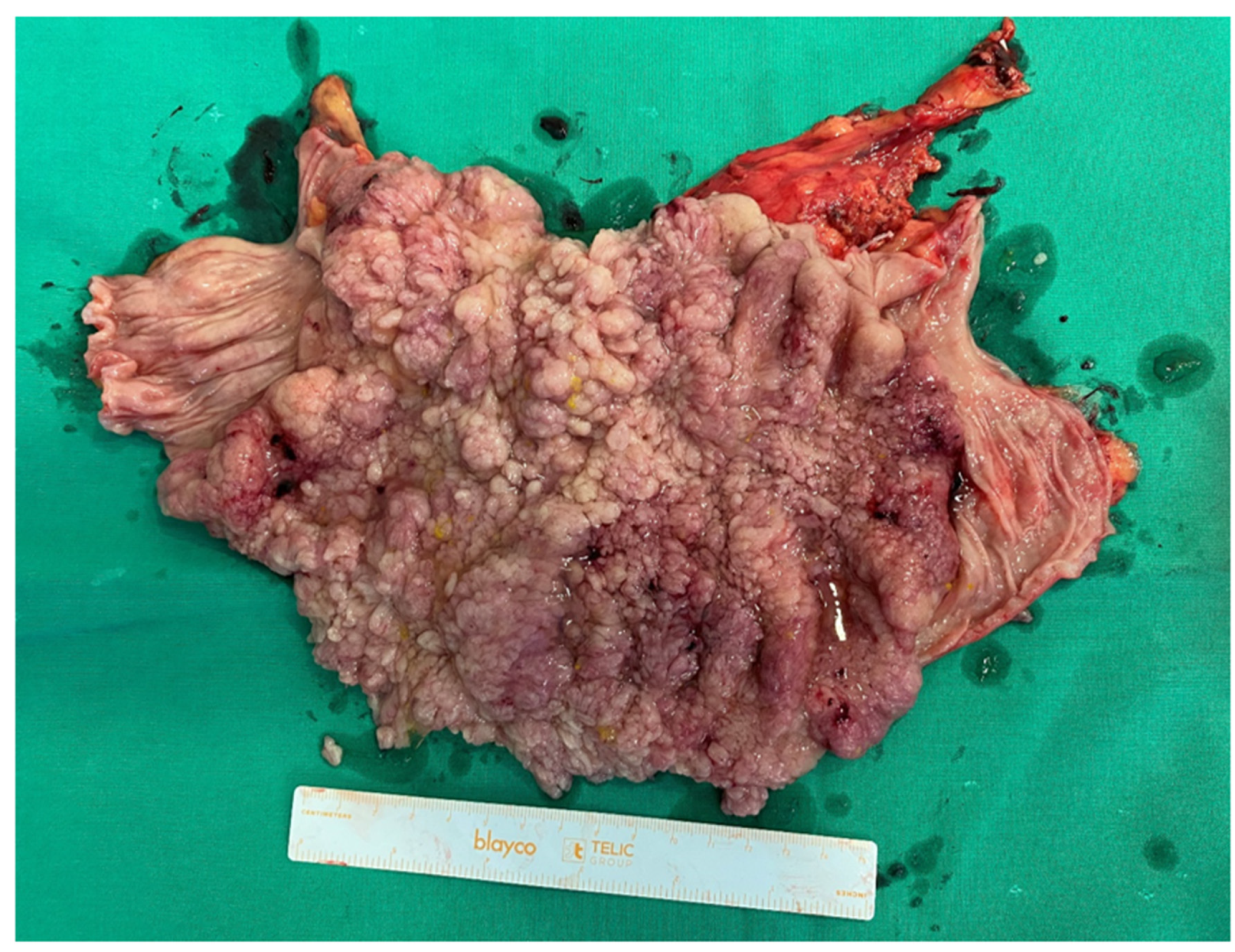
2. Case Report
3. Discussion
4. Conclusions
Author Contributions
Funding
Institutional Review Board Statement
Informed Consent Statement
Data Availability Statement
Conflicts of Interest
Abbreviations
| MWS | McKittrick–Wheelock syndrome |
| ED | Emergency department |
| CT | Computed tomography |
| ARF | Acute renal failure |
| MRI | Magnetic resonance imaging |
References
- Cernevskis, H.; Staka, A.; Pukitis, A.; Silda, A.; Kuzema, V.; Mihailova, I. Mckittrick-Wheelock Syndrome: A Case Report. Arch. Case Rep. Clin. Med. 2016, 2, 115. [Google Scholar] [CrossRef] [Green Version]
- Raphael, M.J.; McDonald, C.M.; Detsk, Y.A.S. McKittrick–Wheelock syndrome. CMAJ 2015, 187, 676–678. [Google Scholar] [CrossRef] [PubMed] [Green Version]
- Dagan, A.; Reissman, P. Giant secretory villous adenoma of the rectum and sigmoid presenting as McKittrick–Wheelock syndrome. Int. J. Color. Dis. 2010, 25, 909–910. [Google Scholar] [CrossRef] [PubMed]
- Popescu, A.; Orban-Schiopu, A.M.; Becheanu, G.; Diculescu, M. McKittrick-Wheelock syndrome—A rare cause of acute renal failure. J. Gastrointest. Liver Dis. 2005, 14, 63–66. [Google Scholar]
- Pheils, M.T. Villous tumors of the rectum. Dis. Colon. Rectum. 1979, 22, 406–407. [Google Scholar] [CrossRef] [PubMed]
- Miles, L.F.; Wakeman, C.J.; Farmer, K.C. Giant villous adenoma presenting as McKittrick-Wheelock syndrome and pseudo-obstruction. Med. J. Aust. 2010, 192, 225–227. [Google Scholar] [CrossRef]
- McKittrick, L.S.; Wheelock, F.C., Jr. Carcinoma of the colon. Dis. Colon. Rectum. 1997, 40, 1494–1496. [Google Scholar] [CrossRef] [PubMed]
- Orchard, M.; Hooper, J.; Wright, J.; McCarthy, K. A systematic review of McKittrick–Wheelock syndrome. Ann. R Coll. Surg. Engl. 2018, 100, 591–597. [Google Scholar] [CrossRef]
- Jyala, A.; Mehershahi, S.; Shah, N.; Shaikh, D.H.; Patel, H. McKittrick-Wheelock Syndrome: A Rare Cause of Chronic Diarrhea. Cureus 2021, 13, e13308. [Google Scholar] [CrossRef]
- Kryzauskas, M.; Jakubauskas, M.; Gendvilaite, N.; Rudaitis, V.; Poskus, T. Bowel Rest with Total Parenteral Nutrition as an Alternative to Diverting Ileostomy in High-Risk Colorectal Anastomosis: A Pilot Study. Medicina 2022, 58, 510. [Google Scholar] [CrossRef]
- Challis, B.G.; Lim, C.T.; Cluroe, A.; Cameron, E.; O’Rahilly, S. The McKittrick–Wheelock syndrome: A rare cause of curable diabetes. Endocrinol Diabetes Metab. Case Rep. 2016, 2016, 160013. [Google Scholar] [CrossRef] [PubMed]
- Kapoor, S. Mckittrick–Wheelock syndrome: An often, overlooked complication of rectal adenomas. Surg. Endosc. 2014, 28, 2247. [Google Scholar] [CrossRef] [PubMed] [Green Version]
- Mois, E.; Graur, F.; Sechel, R.; Al-Hajjar, N. McKittrick-Wheelock syndrome: A rare case report of acute renal failure. Clujul. Med. 2016, 89, 301–303. [Google Scholar] [CrossRef] [PubMed] [Green Version]
- Choi, W.H.; Ryuk, J.; Kim, H.J.; Park, S.Y.; Park, J.S.; Kim, J.G.; Choi, G.S. A case of giant rectal villous tumor with severe fluid-electrolyte imbalance treated by laparoscopic low anterior resection. J. Korean Surg. Soc. 2012, 82, 325–329. [Google Scholar] [CrossRef] [Green Version]
- Targarona, E.M.; Hernandez, P.M.; Balague, C.; Martinez, C.; Hernández, J.; Pulido, D.; Berindoague, R.; Trias, M. McKittrick-Wheelock Syndrome Treated by Laparoscopy: Report of 3 Cases. Surg. Laparosc. Endosc. Percutaneous Tech. 2008, 18, 536–538. [Google Scholar] [CrossRef]
- Podestà, M.A.; Cucchiari, D.; Merizzoli, E.; Elmore, U.; Angelini, C.; Badalamenti, S. McKittrick–Wheelock syndrome: A rare cause of acute renal failure and hypokalemia not to be overlooked. Ren. Fail. 2014, 36, 811–813. [Google Scholar] [CrossRef]
- Khalife, M.; Eloubeidi, M.A.; Hosn, M.A. McKittrick–Wheelock syndrome presenting with dermatomyositis and rectal prolapsed. Clin. Exp. Gastroenterol. 2013, 6, 85–89. [Google Scholar]
- Villanueva, M.E.P.; Onglao, M.A.S.; Tampo, M.M.T.; Lopez, M.P.J. McKittrick-Wheelock Syndrome: A Case Series. Ann. Coloproctol. 2022, 38, 266–270. [Google Scholar] [CrossRef]
- Malik, S.; Mallick, B.; Makkar, K.; Kumar, V.; Sharma, V.; Rana, S.S. Malignant McKittrick-Wheelock syndrome as a cause of acute kidney injury and hypokalemia: Report of a case and review of literature. Intractable Rare Dis. Res. 2016, 5, 218–221. [Google Scholar] [CrossRef] [Green Version]
- Hashash, J.G.; Holder-Murray, J.; Aoun, E.; Yadav, D. The McKittrick-Wheelock syndrome: A rare cause of chronic diarrhoea. BMJ Case Rep. 2013, 2013, bcr2013009208. [Google Scholar] [CrossRef] [Green Version]
- Caron, M.; Dubrûle, C.E.; Letarte, F.; Lemelin, V.; Lafleur, A. McKittrick-Wheelock Syndrome Presenting with Acute Kidney Injury and Metabolic Alkalosis: Case Report and Narrative Review. Case Rep. Gastrointest. Med. 2019, 2019, 3104187. [Google Scholar] [CrossRef] [PubMed] [Green Version]
- Nakhla, S.G.; Murakami, T.T.; Sundararajan, S. Poorly Differentiated Neuroendocrine Tumor of the Rectum Coexistent with Giant Rectal Villous Adenoma Presenting as McKittrick-Wheelock Syndrome. Case Rep. Oncol. Med. 2015, 2015, 242760. [Google Scholar] [CrossRef] [PubMed] [Green Version]
- Emrich, J.; Niemeyer, C. The secreting villous adenoma as a rare cause of acute renal failure. Med. Klin. (Munich) 2002, 97, 619–623. [Google Scholar] [CrossRef] [PubMed]


Disclaimer/Publisher’s Note: The statements, opinions and data contained in all publications are solely those of the individual author(s) and contributor(s) and not of MDPI and/or the editor(s). MDPI and/or the editor(s) disclaim responsibility for any injury to people or property resulting from any ideas, methods, instructions or products referred to in the content. |
© 2023 by the authors. Licensee MDPI, Basel, Switzerland. This article is an open access article distributed under the terms and conditions of the Creative Commons Attribution (CC BY) license (https://creativecommons.org/licenses/by/4.0/).
Share and Cite
Marcinkevičiūtė, K.; Kryžauskas, M.; Poškus, T. McKittrick–Wheelock Syndrome: A Case Report. Medicina 2023, 59, 633. https://doi.org/10.3390/medicina59030633
Marcinkevičiūtė K, Kryžauskas M, Poškus T. McKittrick–Wheelock Syndrome: A Case Report. Medicina. 2023; 59(3):633. https://doi.org/10.3390/medicina59030633
Chicago/Turabian StyleMarcinkevičiūtė, Kristina, Marius Kryžauskas, and Tomas Poškus. 2023. "McKittrick–Wheelock Syndrome: A Case Report" Medicina 59, no. 3: 633. https://doi.org/10.3390/medicina59030633