

Periodontal Phenotype Modification Using Subepithelial Connective Tissue Graft and Bone Graft in the Mandibular Anterior Teeth with Mucogingival Problems Following Orthodontic Treatment

Abstract
:1. Introduction
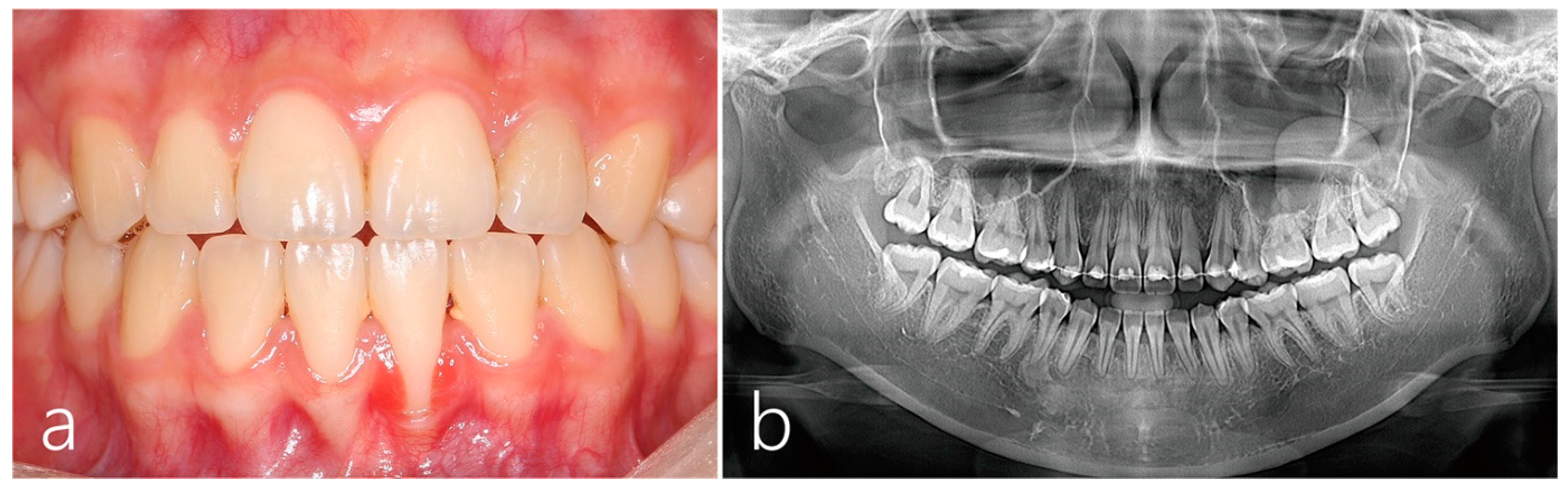
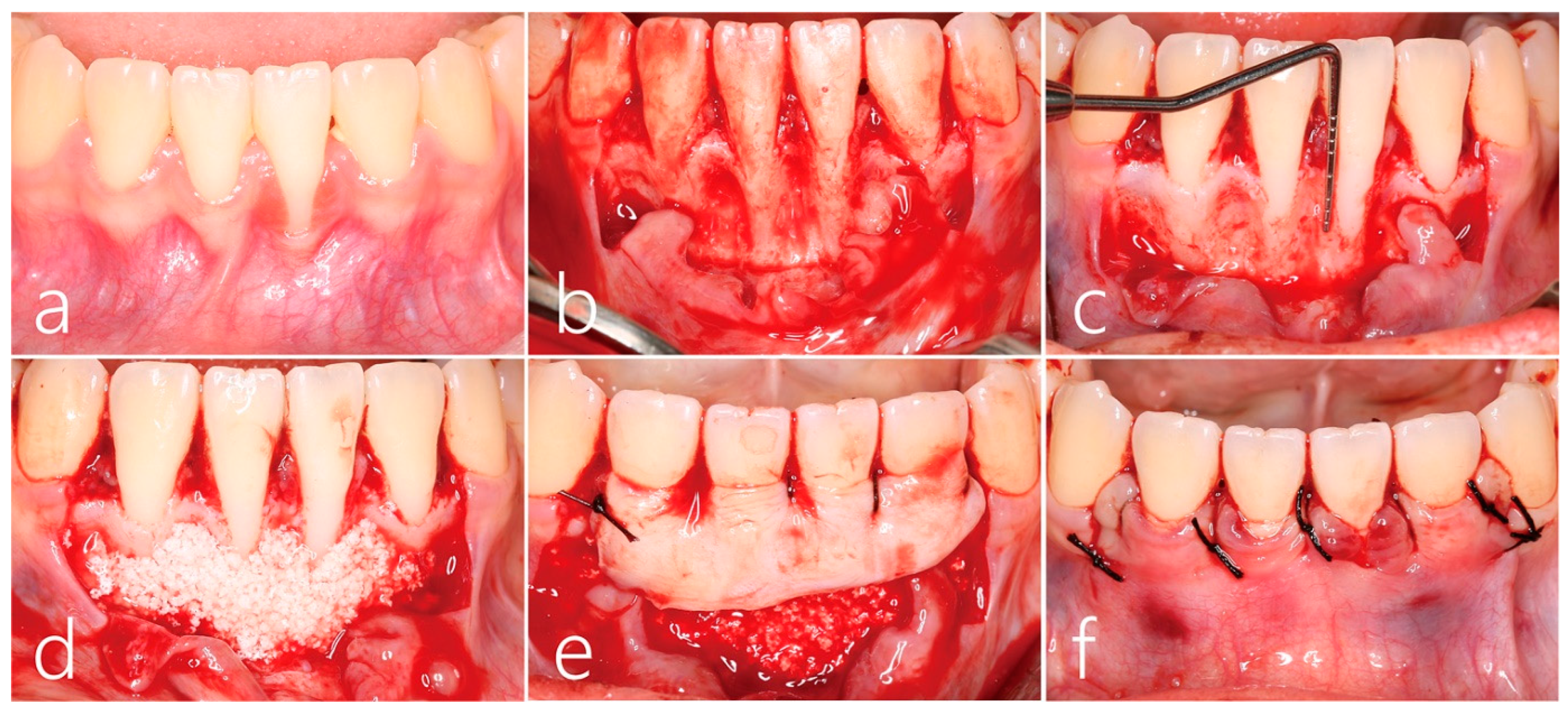
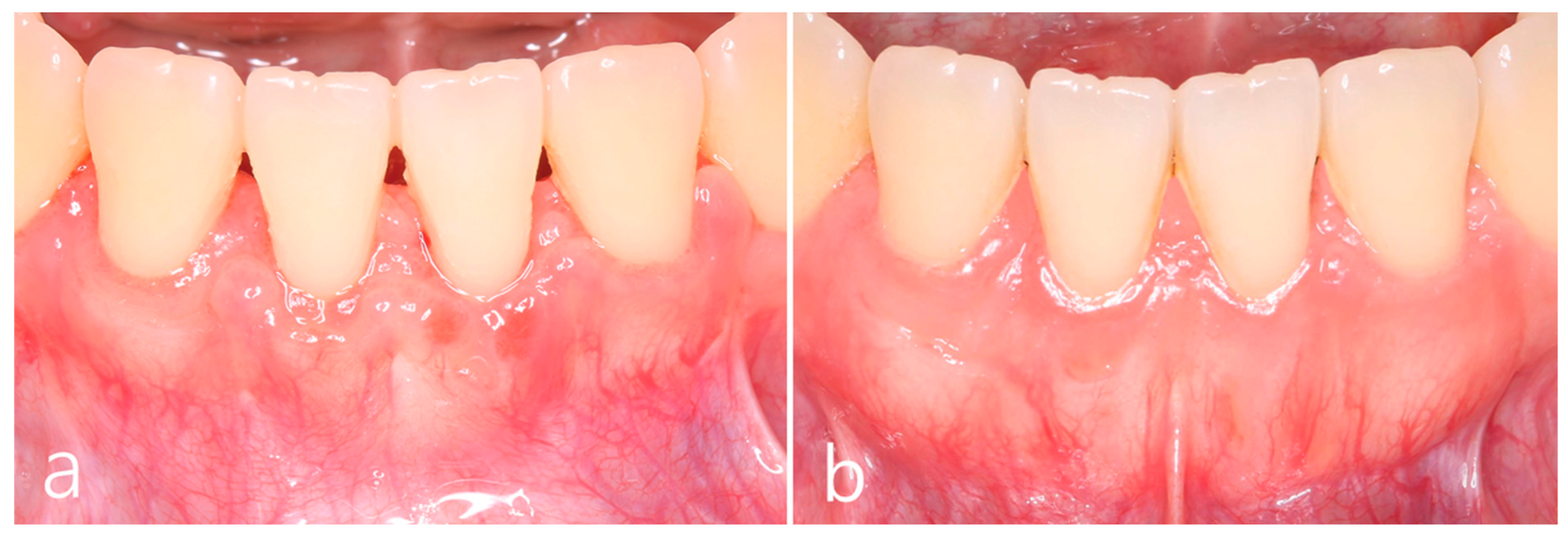
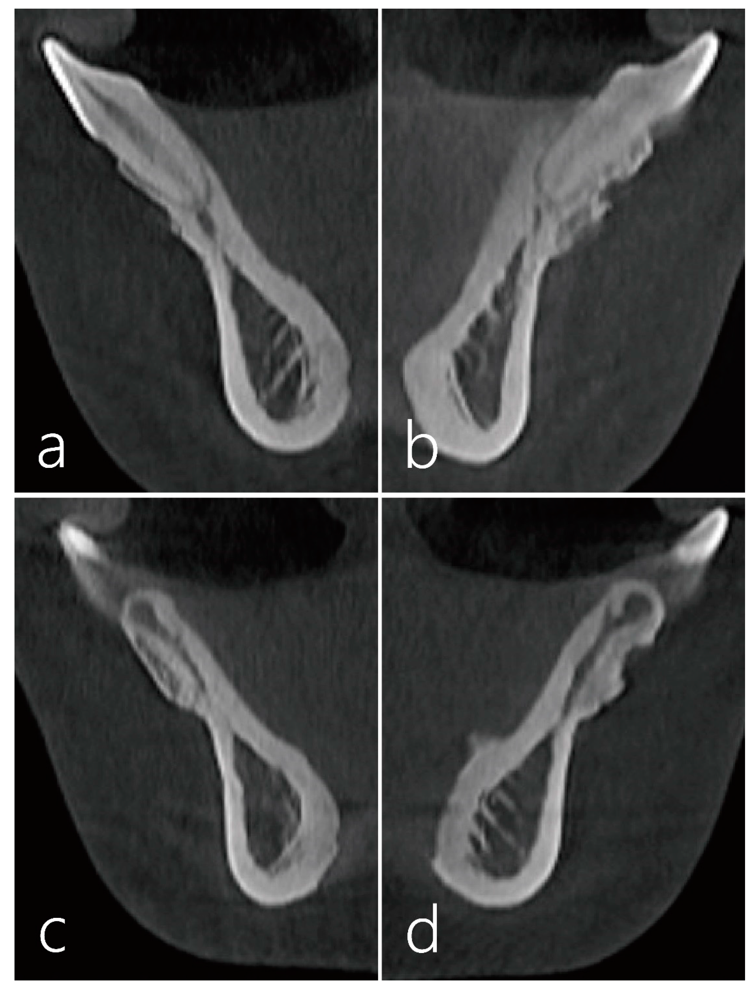
2. Case Report
3. Discussion
4. Conclusions
Author Contributions
Funding
Institutional Review Board Statement
Informed Consent Statement
Data Availability Statement
Conflicts of Interest
References
- Cortellini, P.; Bissada, N.F. Mucogingival conditions in the natural dentition: Narrative review, case definitions, and diagnostic considerations. J. Clin. Periodontol. 2018, 45 (Suppl. S20), S190–S198. [Google Scholar] [CrossRef] [Green Version]
- Imber, J.C.; Kasaj, A. Treatment of Gingival Recession: When and How? Int. Dent. J. 2021, 71, 178–187. [Google Scholar] [CrossRef]
- Joss-Vassalli, I.; Grebenstein, C.; Topouzelis, N.; Sculean, A.; Katsaros, C. Orthodontic therapy and gingival recession: A systematic review. Orthod. Craniofac. Res. 2010, 13, 127–141. [Google Scholar] [CrossRef]
- Lee, J.B.; Baek, S.J.; Kim, M.; Pang, E.K. Correlation analysis of gingival recession after orthodontic treatment in the anterior region: An evaluation of soft and hard tissues. J. Periodontal Implant Sci. 2020, 50, 146–158. [Google Scholar] [CrossRef]
- Wennstrom, J.L. Mucogingival considerations in orthodontic treatment. Semin. Orthod. 1996, 2, 46–54. [Google Scholar] [CrossRef]
- Bollen, A.M.; Cunha-Cruz, J.; Bakko, D.W.; Huang, G.J.; Hujoel, P.P. The effects of orthodontic therapy on periodontal health: A systematic review of controlled evidence. J. Am. Dent. Assoc. 2008, 139, 413–422. [Google Scholar] [CrossRef]
- Allen, E.; Irwin, C.; Ziada, H.; Mullally, B.; Byrne, P.J. Periodontics: 6. The management of gingival recession. Dent. Update 2007, 34, 534–536. [Google Scholar] [CrossRef] [PubMed]
- Ji, J.J.; Li, X.D.; Fan, Q.; Liu, X.J.; Yao, S.; Zhou, Z.; Yang, S.; Shen, Y. Prevalence of gingival recession after orthodontic treatment of infraversion and open bite. J. Orofac. Orthop. 2019, 80, 1–8. [Google Scholar] [CrossRef] [PubMed] [Green Version]
- Morris, J.W.; Campbell, P.M.; Tadlock, L.P.; Boley, J.; Buschang, P.H. Prevalence of gingival recession after orthodontic tooth movements. Am. J. Orthod. Dentofac. Orthop. 2017, 151, 851–859. [Google Scholar] [CrossRef]
- Gorbunkova, A.; Pagni, G.; Brizhak, A.; Farronato, G.; Rasperini, G. Impact of Orthodontic Treatment on Periodontal Tissues: A Narrative Review of Multidisciplinary Literature. Int. J. Dent. 2016, 2016, 4723589. [Google Scholar] [CrossRef] [PubMed] [Green Version]
- Bertl, K.; Spineli, L.M.; Mohandis, K.; Stavropoulos, A. Root coverage stability: A systematic overview of controlled clinical trials with at least 5 years of follow-up. Clin. Exp. Dent. Res. 2021, 7, 692–710. [Google Scholar] [CrossRef] [PubMed]
- Barootchi, S.; Tavelli, L.; Zucchelli, G.; Giannobile, W.V.; Wang, H.L. Gingival phenotype modification therapies on natural teeth: A network meta-analysis. J. Periodontol. 2020, 91, 1386–1399. [Google Scholar] [CrossRef]
- Cairo, F. Periodontal plastic surgery of gingival recessions at single and multiple teeth. Periodontol. 2000 2017, 75, 296–316. [Google Scholar] [CrossRef] [PubMed]
- Banihashemrad, A.; Aghassizadeh, E.; Radvar, M. Treatment of gingival recessions by guided tissue regeneration and coronally advanced flap. N. Y. State Dent. J. 2009, 75, 54–58. [Google Scholar]
- Tozum, T.F.; Keceli, H.G.; Guncu, G.N.; Hatipoglu, H.; Sengun, D. Treatment of gingival recession: Comparison of two techniques of subepithelial connective tissue graft. J. Periodontol. 2005, 76, 1842–1848. [Google Scholar] [CrossRef] [PubMed]
- Cieslik-Wegemund, M.; Wierucka-Mlynarczyk, B.; Tanasiewicz, M.; Gilowski, L. Tunnel Technique With Collagen Matrix Compared With Connective Tissue Graft for Treatment of Periodontal Recession: A Randomized Clinical Trial. J. Periodontol. 2016, 87, 1436–1443. [Google Scholar] [CrossRef]
- Cortellini, P.; Tonetti, M.; Prato, G.P. The partly epithelialized free gingival graft (pe-fgg) at lower incisors. A pilot study with implications for alignment of the mucogingival junction. J. Clin. Periodontol. 2012, 39, 674–680. [Google Scholar] [CrossRef] [PubMed]
- Bakhishov, H.; Isler, S.C.; Bozyel, B.; Yildirim, B.; Tekindal, M.A.; Ozdemir, B. De-epithelialized gingival graft versus subepithelial connective tissue graft in the treatment of multiple adjacent gingival recessions using the tunnel technique: 1-year results of a randomized clinical trial. J. Clin. Periodontol. 2021, 48, 970–983. [Google Scholar] [CrossRef]
- Vincent-Bugnas, S.; Laurent, J.; Naman, E.; Charbit, M.; Borie, G. Treatment of multiple gingival recessions with xenogeneic acellular dermal matrix compared to connective tissue graft: A randomized split-mouth clinical trial. J. Periodontal Implant Sci. 2021, 51, 77–87. [Google Scholar] [CrossRef]
- Chambrone, L.; Garcia-Valenzuela, F.S. Periodontal phenotype modification of complexes periodontal-orthodontic case scenarios: A clinical review on the applications of allogenous dermal matrix as an alternative to subepithelial connective tissue graft. J. Esthet. Restor. Dent. 2023, 35, 158–167. [Google Scholar] [CrossRef]
- Renkema, A.M.; Fudalej, P.S.; Renkema, A.; Bronkhorst, E.; Katsaros, C. Gingival recessions and the change of inclination of mandibular incisors during orthodontic treatment. Eur. J. Orthod. 2013, 35, 249–255. [Google Scholar] [CrossRef] [PubMed] [Green Version]
- Steiner, G.G.; Pearson, J.K.; Ainamo, J. Changes of the marginal periodontium as a result of labial tooth movement in monkeys. J. Periodontol. 1981, 52, 314–320. [Google Scholar] [CrossRef] [PubMed]
- Batenhorst, K.F.; Bowers, G.M.; Williams, J.E., Jr. Tissue changes resulting from facial tipping and extrusion of incisors in monkeys. J. Periodontol. 1974, 45, 660–668. [Google Scholar] [CrossRef] [PubMed]
- Laursen, M.G.; Rylev, M.; Melsen, B. The role of orthodontics in the repair of gingival recessions. Am. J. Orthod. Dentofacial Orthop. 2020, 157, 29–34. [Google Scholar] [CrossRef] [PubMed]
- Kim, D.M.; Neiva, R. Periodontal soft tissue non-root coverage procedures: A systematic review from the AAP Regeneration Workshop. J. Periodontol. 2015, 86, S56–S72. [Google Scholar] [CrossRef] [PubMed]
- Coatoam, G.W.; Behrents, R.G.; Bissada, N.F. The width of keratinized gingiva during orthodontic treatment: Its significance and impact on periodontal status. J. Periodontol. 1981, 52, 307–313. [Google Scholar] [CrossRef]
- Cardaropoli, D.; Tamagnone, L.; Roffredo, A.; Gaveglio, L. Coronally advanced flap with and without a xenogenic collagen matrix in the treatment of multiple recessions: A randomized controlled clinical study. Int. J. Periodontics Restor. Dent. 2014, 34 (Suppl. S3), s97–s102. [Google Scholar]
- Thoma, D.S.; Zeltner, M.; Hilbe, M.; Hammerle, C.H.; Husler, J.; Jung, R.E. Randomized controlled clinical study evaluating effectiveness and safety of a volume-stable collagen matrix compared to autogenous connective tissue grafts for soft tissue augmentation at implant sites. J. Clin. Periodontol. 2016, 43, 874–885. [Google Scholar] [CrossRef]
- Miguel, M.M.V.; Ferraz, L.F.F.; Rossato, A.; Cintra, T.M.F.; Mathias-Santamaria, I.F.; Santamaria, M.P. Comparison between connective tissue graft and xenogeneic acellular dermal matrix to treat single gingival recession: A data reanalysis of randomized clinical trials. J. Esthet. Restor. Dent. 2022, 34, 1156–1165. [Google Scholar] [CrossRef]
- Meza-Mauricio, J.; Cortez-Gianezzi, J.; Duarte, P.M.; Tavelli, L.; Rasperini, G.; de Faveri, M. Comparison between a xenogeneic dermal matrix and connective tissue graft for the treatment of multiple adjacent gingival recessions: A randomized controlled clinical trial. Clin. Oral Investig. 2021, 25, 6919–6929. [Google Scholar] [CrossRef]
- Karam, P.S.; Sant’Ana, A.C.; de Rezende, M.L.; Greghi, S.L.; Damante, C.A.; Zangrando, M.S. Root surface modifiers and subepithelial connective tissue graft for treatment of gingival recessions: A systematic review. J. Periodontal Res. 2016, 51, 175–185. [Google Scholar] [CrossRef] [PubMed]
- de Sanctis, M.; Clementini, M. Flap approaches in plastic periodontal and implant surgery: Critical elements in design and execution. J. Clin. Periodontol. 2014, 41 (Suppl. S15), S108–S122. [Google Scholar] [CrossRef]
- Barootchi, S.; Tavelli, L.; Ravidà, A.; Wang, C.W.; Wang, H.L. Effect of EDTA root conditioning on the outcome of coronally advanced flap with connective tissue graft: A systematic review and meta-analysis. Clin. Oral Investig. 2018, 22, 2727–2741. [Google Scholar] [CrossRef]
- Górski, B.; Szerszeń, M.; Kaczyński, T. Effect of 24% EDTA root conditioning on the outcome of modified coronally advanced tunnel technique with subepithelial connective tissue graft for the treatment of multiple gingival recessions: A randomized clinical trial. Clin. Oral Investig. 2022, 26, 1761–1772. [Google Scholar] [CrossRef] [PubMed]
- Schwarz, F.; Herten, M.; Ferrari, D.; Wieland, M.; Schmitz, L.; Engelhardt, E.; Becker, J. Guided bone regeneration at dehiscence-type defects using biphasic hydroxyapatite + beta tricalcium phosphate (Bone Ceramic) or a collagen-coated natural bone mineral (BioOss Collagen): An immunohistochemical study in dogs. Int. J. Oral Maxillofac. Surg. 2007, 36, 1198–1206. [Google Scholar] [CrossRef] [PubMed]
- Sanz, M.; Vignoletti, F. Key aspects on the use of bone substitutes for bone regeneration of edentulous ridges. Dent. Mater. 2015, 31, 640–647. [Google Scholar] [CrossRef]
- Francisco, I.; Basílio, Â.; Ribeiro, M.P.; Nunes, C.; Travassos, R.; Marques, F.; Pereira, F.; Paula, A.B.; Carrilho, E.; Marto, C.M. Three-Dimensional Impression of Biomaterials for Alveolar Graft: Scoping Review. Funct. Biomater. 2023, 29, 76. [Google Scholar] [CrossRef] [PubMed]
- Deo, V.; Gupta, S.; Ansari, S.; Kumar, P.; Yadav, R. Evaluation of effectiveness of connective tissue graft as a barrier with bioresorbable collagen membrane in the treatment of mandibular Class II furcation defects in humans: 4-year clinical results. Quintessence Int. 2014, 45, 15–22. [Google Scholar]
- Santagata, M.; Guariniello, L.; Prisco, R.V.; Tartaro, G.; D’Amato, S. Use of subepithelial connective tissue graft as a biological barrier: A human clinical and histologic case report. J. Oral Implantol. 2014, 40, 465–468. [Google Scholar] [CrossRef]
- Ribeiro, F.S.; Pontes, A.E.; Zuza, E.P.; da Silva, V.C.; Lia, R.C.; Junior, E.M. Connective tissue graft as a biological barrier for guided tissue regeneration in intrabony defects: A histological study in dogs. Clin. Oral Investig. 2015, 19, 997–1004. [Google Scholar] [CrossRef]


Disclaimer/Publisher’s Note: The statements, opinions and data contained in all publications are solely those of the individual author(s) and contributor(s) and not of MDPI and/or the editor(s). MDPI and/or the editor(s) disclaim responsibility for any injury to people or property resulting from any ideas, methods, instructions or products referred to in the content. |
© 2023 by the authors. Licensee MDPI, Basel, Switzerland. This article is an open access article distributed under the terms and conditions of the Creative Commons Attribution (CC BY) license (https://creativecommons.org/licenses/by/4.0/).
Share and Cite
Park, W.-B.; Park, W.; Lim, S.-W.; Han, J.-Y. Periodontal Phenotype Modification Using Subepithelial Connective Tissue Graft and Bone Graft in the Mandibular Anterior Teeth with Mucogingival Problems Following Orthodontic Treatment. Medicina 2023, 59, 584. https://doi.org/10.3390/medicina59030584
Park W-B, Park W, Lim S-W, Han J-Y. Periodontal Phenotype Modification Using Subepithelial Connective Tissue Graft and Bone Graft in the Mandibular Anterior Teeth with Mucogingival Problems Following Orthodontic Treatment. Medicina. 2023; 59(3):584. https://doi.org/10.3390/medicina59030584
Chicago/Turabian StylePark, Won-Bae, Wonhee Park, Seung-Weon Lim, and Ji-Young Han. 2023. "Periodontal Phenotype Modification Using Subepithelial Connective Tissue Graft and Bone Graft in the Mandibular Anterior Teeth with Mucogingival Problems Following Orthodontic Treatment" Medicina 59, no. 3: 584. https://doi.org/10.3390/medicina59030584