Article

Comparative Genotypic Analysis of RAPD and RFLP Markers for Molecular Variation Detection of Methicillin-Resistant Staphylococcus aureus Clinical Isolates

by
Fathy M. Elkady
1,*,
Abdulaziz A. Al-Askar
2,
Ahmed Abdel Tawab
3,
Mohammad M. Alkherkhisy
3,
Amr A. Arishi
2,4 and
Amr H. Hashem
5,*
1
Microbiology and Immunology Department, Faculty of Pharmacy (Boys), Al-Azhar University, Cairo 11884, Egypt
2
Department of Botany and Microbiology, Faculty of Science, King Saud University, Riyadh 11451, Saudi Arabia
3
Department of Microbiology and Immunology, Faculty of Medicine, Al-Azhar University, Cairo 11884, Egypt
4
School of Molecular Sciences, The University of Western Australia, Perth, WA 6009, Australia
5
Botany and Microbiology Department, Faculty of Science, Al-Azhar University, Cairo 11884, Egypt
*
Authors to whom correspondence should be addressed.
Medicina 2022, 58(9), 1245; https://doi.org/10.3390/medicina58091245
Submission received: 11 August 2022 / Revised: 1 September 2022 / Accepted: 5 September 2022 / Published: 8 September 2022
(This article belongs to the Special Issue Pathogenesis, Diagnostics, and Therapeutics of Infectious Diseases)

Abstract

:
Background and Objectives: Methicillin-resistant Staphylococcus aureus (MRSA) isolates are associated with various diseases ranged from mild superficial impairments to invasive infections. This study aimed to evaluate the ability of polymerase chain reaction (PCR) based methods namely, restriction fragment length polymorphism (RFLP) of the coa gene and random amplified polymorphic DNA (RAPD), to determine the genetic diversity of MRSA isolates. Materials and Methods: A total of 37 MRSA isolates were conventionally identified depending on their biochemical and microbiological culture characteristics. Genotypic confirmation was based on detection of the associated mecA gene. The genetic variation amongst MRSA isolates was evaluated following the coa gene-based RFLP and RAPD fingerprints. Results: Results illustrated that, the species specific coa gene was detected in all MRSA isolates. The irregular bands intensity, number, and molecular sizes of the PCR amplicons demonstrated the coa gene polymorphism. The incompatible AluI digestion patterns of these amplicons classified the tested MRSA isolates into 20 RFLP patterns which confirm the coa gene polymorphism. Additionally, the PCR-based RAPD analysis showed variable bands number with size range of approximately 130 bp to 4 kbp, which indicated the genetic variation of the tested MRSA isolates as it created 36 variable RAPD banding profiles. Conclusions: coa gene AluI enzymatic restriction sites, amongst the tested MRSA isolates, certify their genetic variation on the basis of the accurate but complicated and relatively expensive coa gene-based RFLP. Conversely, the results verified the excellent ability of the simple and cost-effective PCR-based RAPD analysis to discriminate between MRSA isolates without any preface data about the genome.

1. Introduction

Staphylococcus aureus (S. aureus), a considerable Gram-positive opportunistic bacterium, may affect human skin, heart, bone, ear, and respiratory tract. This pathogen is associated with various diseases ranging from mild superficial impairment to serious, invasive, and life-threatening infections [1]. Some S. aureus strains have acquired the mecA gene, coding for altered penicillin-binding protein, with the consequent worldwide emergence of methicillin-resistant S. aureus (MRSA) strains. This altered protein is characterized by a low affinity for binding to all β-lactams which diminishes its clinical usefulness in controlling hospital and community acquired MRSA infections [2]. The widespread occurrence of S. aureus infections is due to multiple virulence factors of the pathogen besides their abundance in the environment and in normal microbiota [3]. All S. aureus strains have a unique capability to manipulate the coagulation process. Induction of human blood clotting formation represents one of the essential virulent activities associated with their pathogenesis. This plasma clotting activity is directly caused by the production of coagulase enzymes that catalyse the fibrin generation via prothrombin activation. Moreover, the formation of fibrin may provide protection against phagocytosis [4]. Accordingly, coagulase enzymes created by all S. aureus strains are considered one of the fundamental phenotypic measures for the detection of their clinical infections [5]. These enzymes are found either extracellularly or membrane-bounded and encoded by the coa gene, one of the foremost species-specific virulent genes that are used for molecular confirmation of the diseases caused by S. aureus [6].
The Pulsed-Field Gel Electrophoresis (PFGE) is the gold standard technique for epidemiological investigation of microbial infections. Nevertheless, the comparatively high expense of performance and the requirement of specialized apparatus obstruct their use [7]. The inconsistent nature of the coa gene 3′ coding region is responsible for their highly polymorphic nature and subsequent variable amino acid sequence in coagulase protein. This region contains a chain of 81 base pair (bp) tandem repeats or short repetitive sequences coding for 27 amino acid repeated sequences. The number of these tandem repeats as well as the positions of restriction enzyme cutting sites varied amongst S. aureus isolates [8]. Consequently, this variation permits the discrimination and DNA-based genotyping of S. aureus isolates. The coa gene amplification, using DNA primer complementary to a definite sequence in its conserved region, produced dissimilar amplicon sizes in the tested isolates [9]. Additionally, analysis of the polymerase chain reaction-based restriction fragment length polymorphism (PCR-based RFLP) of coa gene amplicons can be used for S. aureus isolates genotyping. This technique depends on the dissimilarity in the sequence of the coding region at the 3′ end with subsequent variation in the sites of AluI and HaeIII restriction enzymes effect [10]. Moreover, the PCR-based random amplified polymorphic DNA (PCR-based RAPD) is a rapid, simple, and effortless technique at a low annealing temperature. It utilizes primers with arbitrary short oligonucleotides that able to randomly amplify various DNA targets. The created fragments vary in their numbers and sizes depending on the divergence in the length between the primer annealing sites that is unique for each bacterial isolate [11]. This method permits strain typing on the basis of a large part of their genomic DNA, independently on well-known data, and provides different banding profiles with subsequent better strain fingerprinting [12].
Herein, this search seeks to assess the capability of two PCR depending methods, the costly and laborious RFLP based on the coa gene and the inexpensive RAPD technique with little stringent conditions based on the whole genome, to detect the genomic variation and subsequent diversity of MRSA clinical isolates.

2. Materials and Methods

2.1. Isolation and Phenotypic Identification of S. aureus

Clinical specimens from hospitalized patients were instantly cultured on nutrient agar (Oxoid, Basingstoke, Hampshire, UK) and blood agar followed by incubation at 37 °C for 24 h. Staphylococcus aureus was phenotypically identified depending on their distinctive ability to grow on mannitol salt agar (Oxoid, Basingstoke, Hampshire, UK) with sugar fermentation. As well, their Gram stain appearance and biochemical behaviours, following standard microbiological procedures, including catalase, coagulase, and DNase production, were examined [13].

2.2. Phenotypic Recognition of MRSA

The MRSA clinical isolates were identified on the bases of the cefoxitin disc diffusion test following the Kirby–Bauer disk diffusion method using 0.5 McFarland standard bacterial suspension on Muller Hinton agar medium (Oxoid, UK). The inhibition zone diameter around the cefoxitin (30 µg) disc (Oxoid, UK) was determined after 18 h incubation at 35 °C [14].

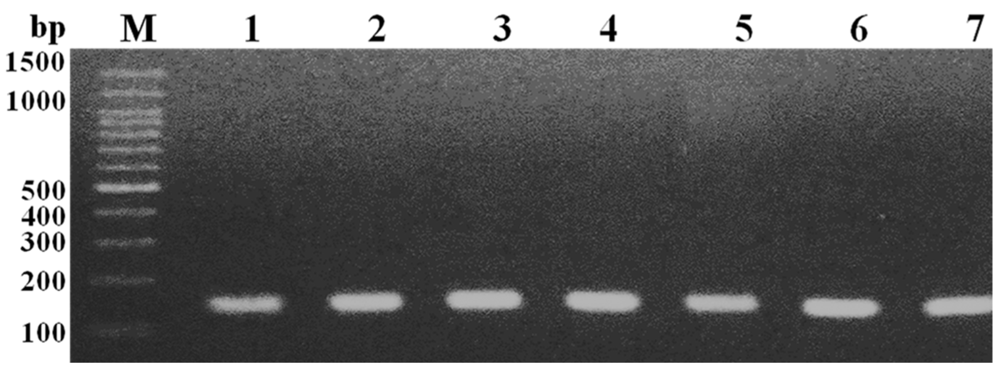
2.3. Genotypic Confirmation of MRSA

Definite identification of MRSA isolates was dependent on the detection of their characteristic mecA gene [15]. Briefly, genomic DNA was extracted using GeneJET Genomic DNA Purification Kit according to the manufacturer’s instructions (Thermo Fisher Scientific Inc., Vilnius, Lithuania). The mecA gene was amplified, by PCR technique, using a Thermal cycler (Biometra UNO–Thermoblock, Germany) in presence of 1 µL from each particular 10 pmol primer (Table 1), 12.5 µL Cosmo PCR Red Master Mix (Willowfort, Birmingham, UK), 1 µL of 50 ng genomic DNA, and nuclease-free double distilled water to final reaction volume of 25 µL. The mecA gene amplification cycling program includes 1 cycle of initial denaturation at 94 °C for 4 min (min) followed by 35 cycles of denaturation at 94 °C for 1 min, annealing at 55 °C for 1 min, and elongation at 72 °C for 1 min, and lastly 1 cycle of final elongation at 72 °C for 10 min. The obtained PCR products were loaded into 1.5% agarose gel containing 5 µL ethidium bromide (0.05 mg/mL) followed by electrophoresis at 100 V in Tris-acetate EDTA buffer (Sigma Aldrich, Hamburg, Germany). The UV transilluminator (HERMLE Labortechnik GmbH, Wehingen, Germany) was used to observe the mecA gene-specific bands and determine their molecular size with the aid of a 100 bp DNA ladder (Geneaid Biotech Lt., New Taipei City, Taiwan) as a marker [16].

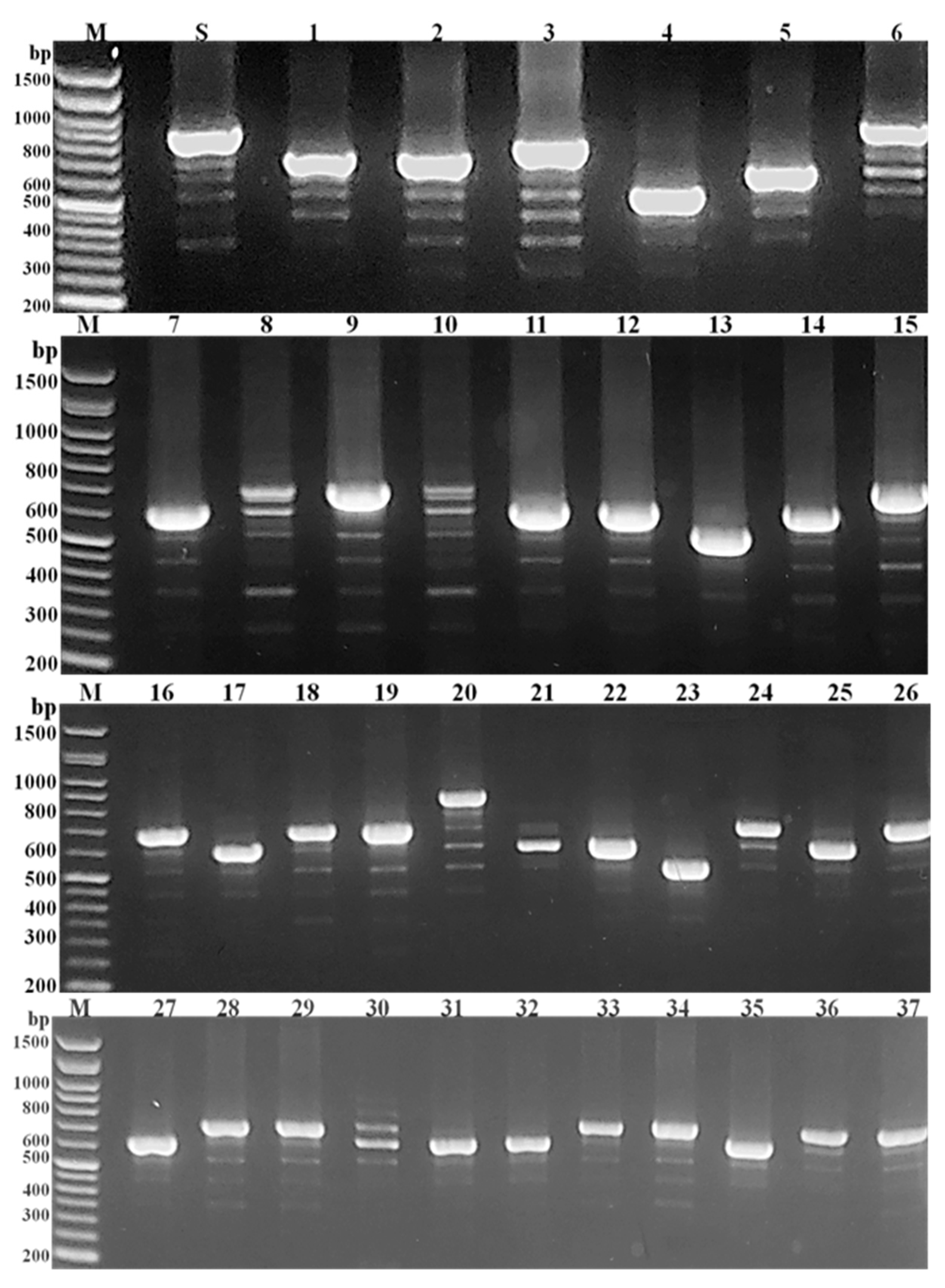
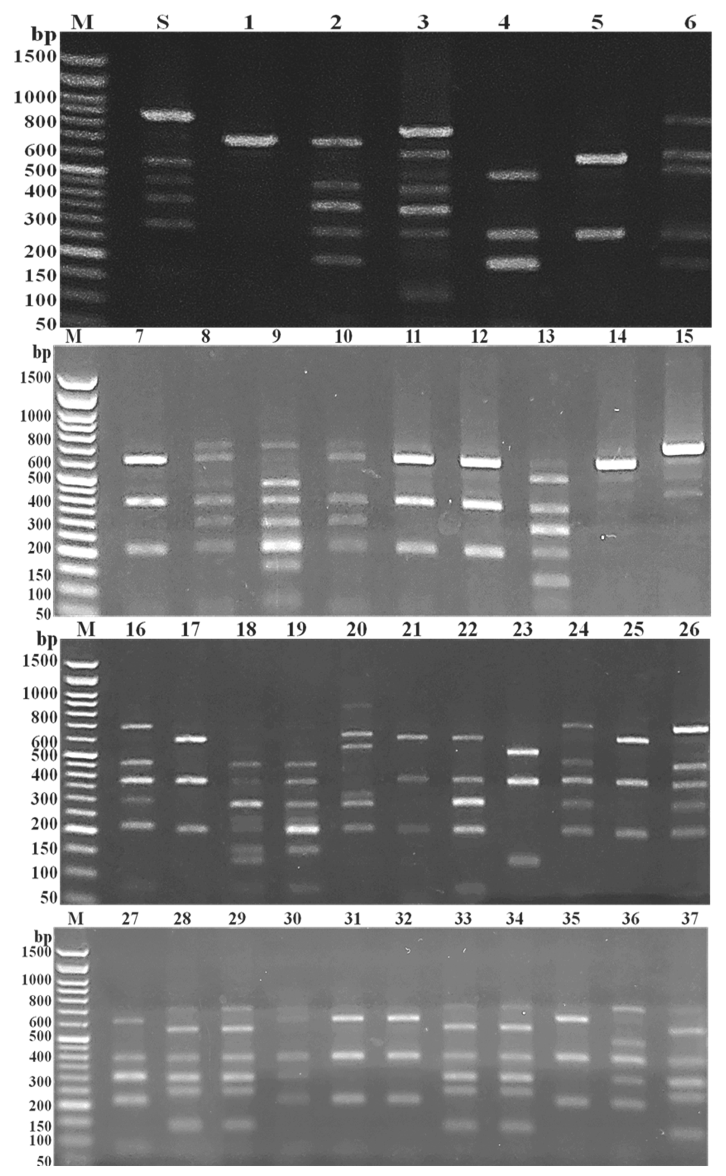
2.4. Coagulase (Coa) Gene Molecular Typing of MRSA Isolates

The coding region of the coa gene 3′ end was amplified by PCR using 1 µL from each 10 pmol primer (Table 1), 10 µL Cosmo PCR Red Master Mix (Willowfort, UK), 1 µL of 50 ng genomic DNA, and nuclease-free double distilled water to final reaction volume of 20 µL. The PCR cycling profile includes 1 cycle of initial denaturation at 95 °C for 2 min, 35 cycles of denaturation at 95 °C for 30 s (s), annealing at 55.5 °C for 30 s, and extension at 72 °C for 50 s, followed by 1 cycle of final extension at 72 °C for 5 min. Staphylococcus aureus ATCC 6538 was used as a positive control and PCR amplicons were detected with the aid of a UV transilluminator after 1.5% agarose gel electrophoresis in presence of a 50 bp DNA marker [6]. The PCR amplification products of coa gene were analysed after treatment with AluI restriction enzyme (BioLabs, Inc., New England, MA, USA) following the manufacturer’s information. Briefly, 50 µL reaction mixture consists of 10 µL from each PCR product, 34 µL nuclease-free distilled water, and 6 µL of 10 U AluI (1 µL of the enzyme in 5 µL rCutSmart buffer) was prepared and incubated for 15 min at 37 °C followed by enzyme inactivation by heating at 80 °C for 20 min. The RFLP pattern for each isolate was separated by electrophoresis using agarose gel 1.5% and matched with a 50 bp DNA marker [18].

2.5. PCR-Based RAPD Fingerprinting of MRSA Isolates

The random DNA fragments amplification reactions were achieved in a total reaction volume of 20 µL containing 10 µL Cosmo PCR Red Master Mix (Willowfort, UK), 1 µL of 50 ng genomic DNA, 2 µL of 25 pmol primer that targets the whole genome (Table 1), and 7 µL of nuclease-free double distilled water. The PCR amplification program involves the application of 1 cycle of initial denaturation at 94 °C for 5 min with subsequent 40 cycles of denaturation at 94 °C for 1 min, annealing at 40 °C for 1 min, and extension at 72 °C for 3 min, and to finish 1 cycle of final extension at 72 °C for 7 min. The RAPD pattern for each isolate was separated by electrophoresis using 1% agarose gel and visualized by a transilluminator and compared with 1 kbp plus DNA ladder (Enzynomics, Daejeon, South Korea) as a marker [17].

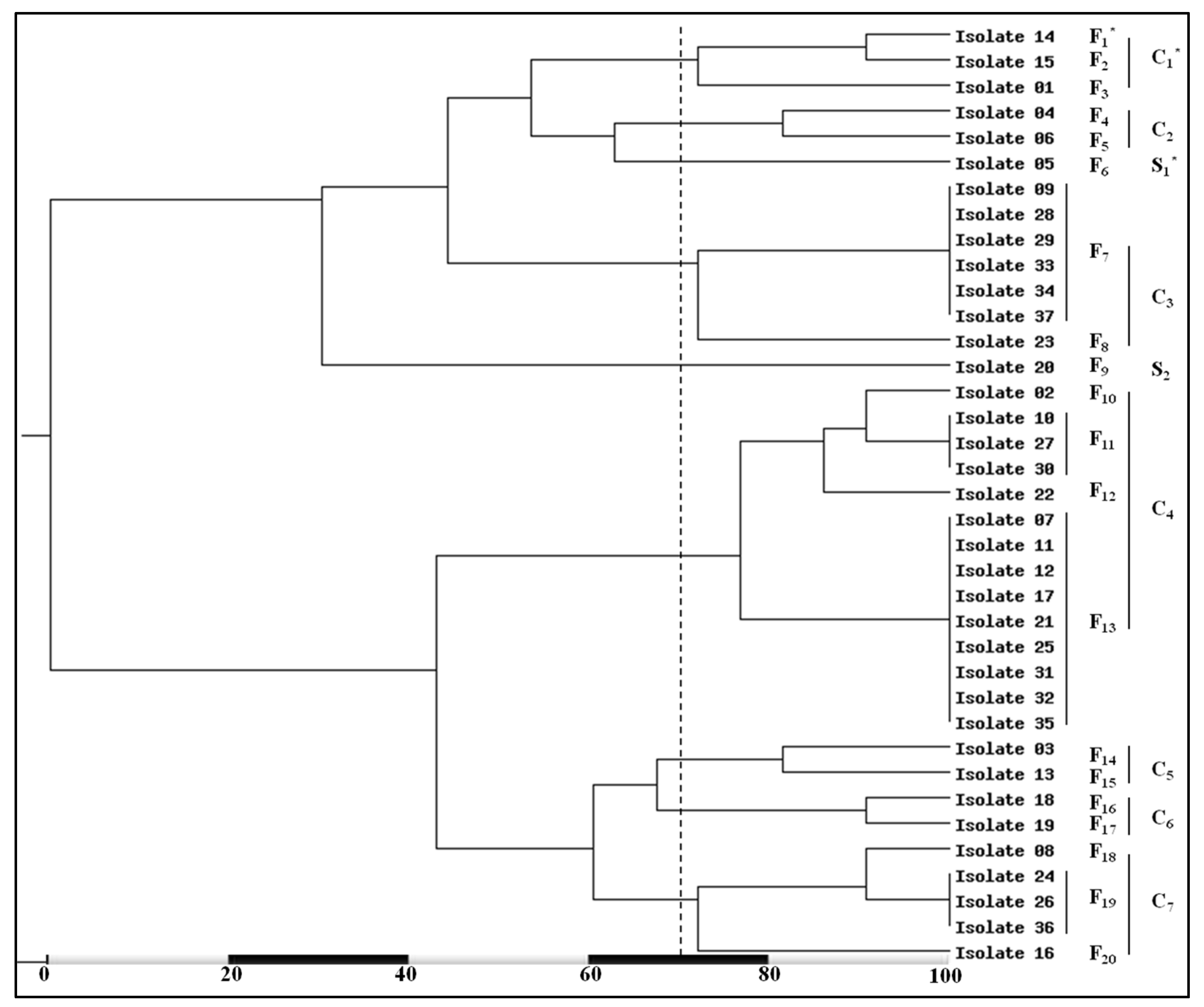
2.6. Phylogenetic Analysis

The variable bands number and size of the coa gene PCR-based RFLP and PCR-RAPD patterns of the tested MRSA isolates were separately analysed. Dice Similarity Coefficient and Unweighted Pair Group Method with Arithmetic Average (UPGMA) were used to construct the phylogenetic trees (dendrograms). The presence or absence of a specific band was coded by binary numbers of 1 (for presence) or 0 (for absence). Isolates with about 70% similarity in the dendrogram were collected in groups identified as clusters.

2.7. Statistical Analysis

The discriminatory power of each typing method was evaluated by calculating the Simpson’s index of diversity (discriminatory index (D)) according to the following equation’:
D = 1 1 N ( N 1 ) . j = 1 s n j   ( n j 1 )  
where N is the total number of isolates in the sample population, S is the total number of the obtained types, and nj is the isolates number belonging to the most frequent type [19].

3. Results

3.1. Identification of MRSA

A total of 37 Gram-positive cocci with a typical bunch of grapes-like appearance, a β-haemolytic character on blood agar, golden-yellow colonies on nutrient agar, and mannitol fermenting activity were assumed to be S. aureus. Additionally, the positive behaviours for catalase, coagulase, and DNase tests were biochemically confirmed their identification. Furthermore, these isolates were phenotypically emphasized as MRSA depending on their growth inhibition around cefoxitin disc (30 µg) at a diameter ≤ 21 mm. Genotypically, detection of the expected mecA gene-specific band, of approximately 147 bp (Figure 1), in all the 37 tested S. aureus isolates indicated their confirmed identification as MRSA.

3.2. PCR-Based RFLP of Coa Gene

All the tested MRSA isolates were coa gene-positive with uneven PCR amplicon sizes. The coa gene polymorphism was demonstrated by the detection of their inconsistent band’s number (1 to 6 bands). Moreover, these bands showed variable intensity and molecular sizes of approximately 81 bp multiplied in the range of 243 to 972 bp (Figure 2). As well, the agarose gel electrophoresis of AluI enzymatic digestion of the coa gene amplicons showed 3 to 7 DNA fragments. The size of each fragment was approximately equal to one of the 81 bp multiplied and in the range of 81 to 891 bp (Figure 3).
The incompatible digestion patterns confirm the coa gene polymorphism among the tested MRSA isolates and resulted in 20 diverse fragmentation classes nominated as genotype F1–F20. Amongst these genotypes, the majority of the tested isolates (9/37; 24.3%) were mostly related to genotype F13 (isolates number 7, 11, 12, 17, 21, 25, 31, 32, and 35) with three DNA fragments size of 648, 405, and 243 bp. Genotype F7 was contained 6/37 isolates (16.2%), isolates number 9, 28, 29, 33, 34, and 37, with six fragmentation sizes of 729, 567, 405, 324, 243, and 162 bp. Additionally, 3/37 isolates (8.1%) were included in genotype F11 (isolates number 10, 27, and 30) with four DNA fragments sizes of 648, 405, 324, and 243 bp. As well, genotype F19 included isolates number 24, 26, and 36 (3/37 isolates: 8.1%) with five fragmentation sizes of 648, 405, 324, 243, and 162 bp. On the other hand, the remaining 16 isolates revealed high degree of heterogeneity as each isolate represented a separate genotype with variable restriction patterns. Additionally, isolate number 1 showed an indigestible coa gene amplicon (genotype F3).
The generated dendrogram, depending on 100% similarity, and its clusters showed variable similarity percentages (Figure 4). Based on approximately 70% similarity, the 20 RFLP genotypes were grouped into 7 RFLP clusters (C1–C7) and 2 singletons (S1 and S2). The highest percentage (37.8%; 14/37) of isolates were included in C4 (the most prevalent cluster) while, C2 (the lowest prevalent cluster) comprised only two isolate (5.4%).

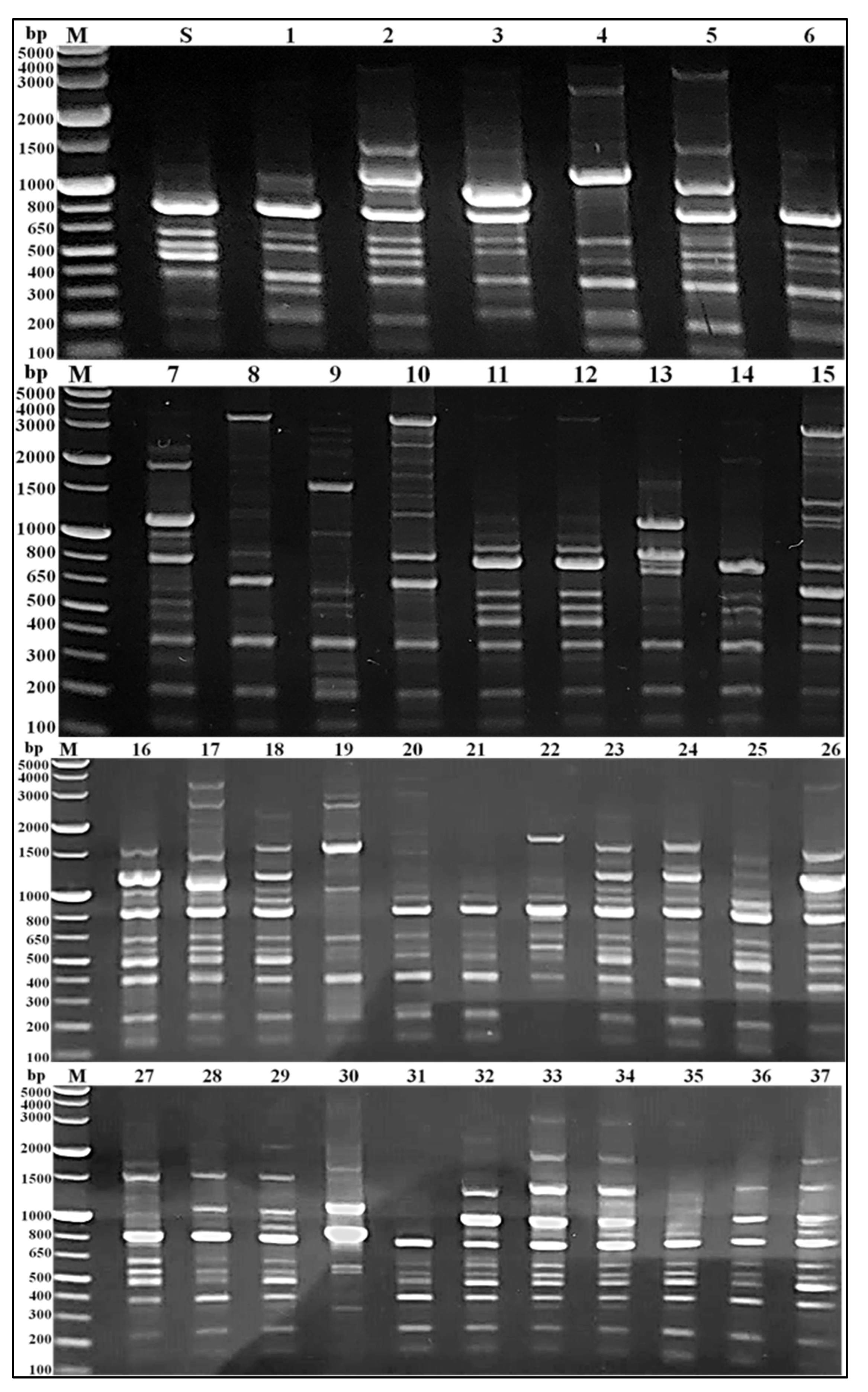
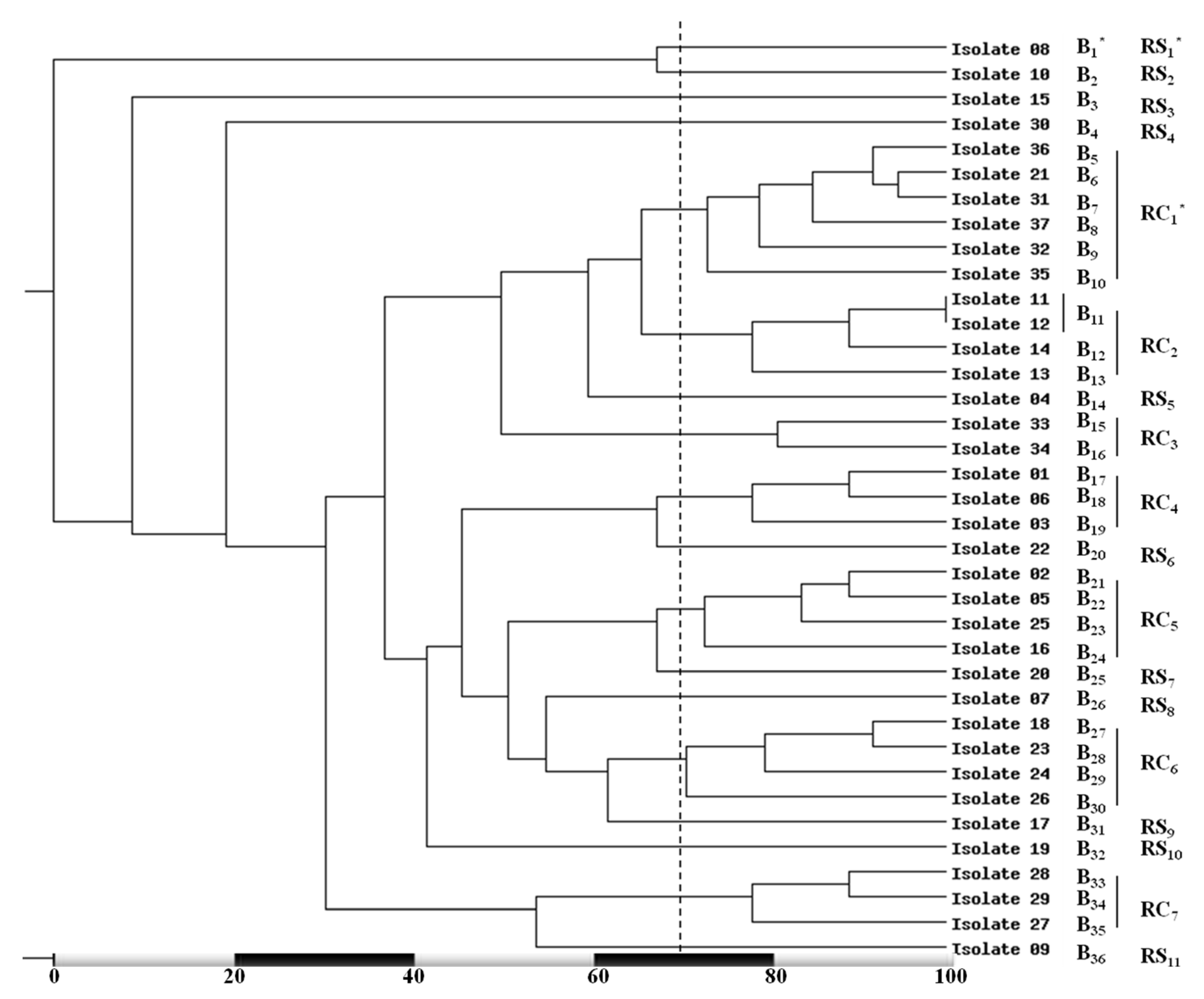
3.3. RAPD Fingerprinting of MRSA Isolates

The genetic variation of the tested MRSA isolates was decided by PCR-based RAPD (Figure 5). The results showed several bands (6–18), with a molecular size range of approximately 130 bp to 4 kbp. As well, some conserved bands e.g., 220 bp and 400 bp were mostly detected in the tested isolates. The 220 bp bands were detected in all isolates except isolates number 22 and 30. While, bands with the molecular size of 400 bp were detected in all isolates except isolate number 30.
Importantly, nine unique bands of approximately 4000, 3500, 3100, 2700, 2600, 2100, 1700, 750, and 580 bp molecular size were detected isolates number 20, 17, 34, 19, 9, 14, 10, 13, and 30, respectively. Similarly, each of 10 distinctive bands with the molecular size of approximately 3600, 2800, 2300, 1900, 1850, 1750, 920, 600, 450, and 230 bp were observed in only two of the tested isolates; numbers (5 and 25), (15 and 16), (7 and 9), (7 and 33), (22 and 34), (15 and 27), (33 and 34), (10 and 30), (8 and 10), and (9 and 20), respectively.
The unpredictable appearance of the conserved and variable bands as well as, the changeable band’s numbers and sizes creates 36 variable RAPD banding profiles, voted genotypes B1–B36. The MRSA isolates number 11 and 12 represented the same RAPD banding profiles (B11) while, other tested MRSA isolates revealed a high level of variability as each isolate was represented only one RAPD profiles.
Based on about 70% similarity, the phylogenetic analysis of the obtained 36 RAPD-PCR fingerprints revealed seven variable RAPD clusters (RC1–RC7) and 11 RAPD singletons (RS1–RS11). The results recorded RC1 as the most ubiquitous cluster which included six (16.2%) isolates while RC3 included only two isolates (5.4%) and represent the less common cluster (Figure 6).

3.4. Discrimination Index

The discriminatory power for each genotyping method was evaluated by calculation of their numerical index of discrimination. The Simpsons index of diversity showed the highest capability of RAPD-PCR, with a D value of more than 0.97, for isolates discrimination. On the other hand, the coa gene-based RFLP revealed lower discrimination ability, with a D value of 0.35.

4. Discussion

Methicillin-resistant S. aureus isolates represent one of the most considerable infectious agents with an increased tendency to resist a wide range of currently available antimicrobial agents [20]. Therefore, this study was performed to detect the MRSA isolates and to assess the capability of the coa gene-based RFLP and whole genome-based RAPD analysis for detection of their genetic variation. In the current study, thirty-seven S. aureus isolates were identified following conventional phenotypic microscopical, cultural, and biochemical characteristics. In a related study, S. aureus was identified based on their capability on mannitol fermentation, golden yellow endopigment production, β-haemolytic activity, and coagulase positive test [21]. The overspread and increased incidence of MRSA infections necessitate the urgent need for their rapid identification. In the present study, phenotypic cefoxitin resistance with subsequent mecA gene molecular finding was considered as a marker for MRSA isolates confirmation. A similar study carried out by Bokharaei, et al. [22] described the cefoxitin disc test and PCR amplification of the mecA gene as an effective tool for MRSA strains recognition.
Molecular detection of the coa gene, which presents in all phenotypically coagulase-positive S. aureus isolates, is an important genotypic measure for their identification. Additionally, recognition of this gene is an easy, precise, and discriminatory method for S. aureus isolates genotyping [23]. In this study, the coa gene was detected in all the tested MRSA isolates and its polymorphic nature was initially predicted. The PCR amplicons of the coa gene were inconsistent with variable bands number (1 to 6 bands), size (243 to 972 bp), and intensity. In a related study carried out in Egypt, up to eight bands of coa gene amplicons were recorded among the tested MRSA isolates with a band size range of 80 to 810 bp [24]. These results were in accordance with Al-Ruwaili [25] study who differentiate 11 coa gene band classes in the range of 500 to 1000 bp. The tested MRSA isolates showed unequal coa gene bands number (1 to 4 bands) with incompatible intensity. Insertion or deletion mutations, at the 3′ end, might be responsible for these coa gene polymorphism with subsequent change in the PCR amplification products size. Additionally, the coexistence of numerous coa gene allelic form allows a single MRSA isolate to produce several gene variants which explained the feasible formation of a number of bands by the same isolate [10].
Molecular typing methods depending on specific gene amplification with subsequent enzymatic digestion are used for MRSA isolates characterization. These genotyping methods exhibit valuable discrimination ability and are supportive in the determination of the variability amongst MRSA isolates on genetic bases [26]. In the current study, coa gene-based RFLP, using AluI restriction enzyme, of the 37 tested MRSA isolates, yielded 20 banding patterns. These patterns exhibited 3 to 7 DNA fragments of approximately one of the 81 bp multiplied in the size range of 81 to 891 bp. Additionally, one isolate showed coa gene amplicon resistant to AulI cutting. These results were relatively following a comparative study carried out by Al-Ajealy, et al. [27], they recorded seven AulI restriction patterns of coa gene amplicons with 1 to 3 DNA fragments in a size range of 80 to 700 bp. Additionally, their results also recorded AulI enzyme undigested coa amplicons in 20% of their tested isolates. Similarly, in the study carried out by Chadi Dendani, et al. [28], the PCR-based RFLP of coa gene showed nine different AluI restriction patterns with approximately band sizes of 90 to 550 bp. The S. aureus isolates with faint coa amplification product yielded no detectable bands after AluI enzymatic digestion. The obtained results disagreed with Ibrahim, et al. [29] who observed just three PCR-based RFLP patterns showed 3 to 4 DNA fragments with a size range of 80 to 500 bp. The existence of the AluI target site in the coa gene sequence may reflect the ability of this enzyme to digest the coa gene amplicons in the susceptible isolates. Additionally, the altered coa gene sequence amongst diverse isolates provide dissimilar restriction locations [30]. Conversely, a point mutation at the 3′ region of coa gene and subsequent lack of AluI enzyme-specific restriction sites may reflect the incapability of the AluI enzyme to digest the coa gene PCR amplicon of the digestion resistant isolates [31].
Recently, epidemiological investigations of bacterial outbreaks on the basis of the whole genome may provide an incomparable resolution [26]. In the present study, 36 inconsistent RAPD banding patterns were observed amongst the 37 tested MRSA isolates with elevated variability levels. As well, these banding patterns were divided into seven RAPD clusters and 11 RAPD singletons. The study conducted by Mobasherizadeh, et al. [32] described the RAPD-PCR as a perfect, fast, simple, and low-cost genotyping technique depending on a small amount of total DNA. Their study classified 25 MRSA isolates into five genotypes based on RAPD-PCR. The tested isolates were divided into two clusters (groups each with more than three members), a small group containing three members, and single isolates each with distinctive RAPD-PCR patterns. In a related study, all the tested MRSA isolates were generating an abundance band, in the range of 400 to 1000 bp, by RAPD-PCR which subsequently divided the tested isolates into five different clusters [33]. The different frequencies and locations of the target sequences within the chromosomal DNA reflect the variation in DNA fragments number and size in different RAPD-PCR patterns with subsequent genetic variation depending on the sequence of the arbitrarily short primer used [34].
In the present study, the RAPD-PCR numerical index of discrimination of more than 0.97 illustrates their highest ability for isolates differentiation. Similarly, the study carried out by Ye, et al. [35] demonstrated the high capacity of RAPD-PCR for differentiation between S. aureus isolates with discriminatory index of about 0.95. The random reliance of PCR-based RAPD method on the whole bacterial genome might explain the excellent ability of such technique for isolates characterization as well as its usefulness as a molecular genotyping tool in the epidemiological investigation.

5. Conclusions

Bacterial genotyping represents an essential part of infection control measures. These methods help in outbreaks investigation and clarification of the infection source which might be another patient, staff members, apparatus, food, or the environment. The incompatible coa gene digestion patterns, among the tested MRSA isolates, validate their genetic inconsistency and illustrate the confirmatory discrimination ability based on the precise but relatively expensive and multi-step coa gene-RFLP typing method. In contrast, the present study verified the ability of the simple and inexpensive RAPD-PCR analysis to discriminate between MRSA isolates based on the whole bacterial genome without any preface information. As well, this technique provides an improved insight on MRSA isolates typing with consequent useful epidemiological investigations and superior patient concern.

Author Contributions

Conceptualization, F.M.E.; methodology, F.M.E., A.A.T., M.M.A., A.A.A.-A., A.A.A. and A.H.H.; software, F.M.E. and A.H.H.; validation, F.M.E.; formal analysis, F.M.E.; investigation, F.M.E.; resources, F.M.E.; data curation, F.M.E. and A.H.H.; writing—original draft preparation, F.M.E. and A.H.H.; writing—review and editing, F.M.E., A.A.T., M.M.A., A.A.A.-A., A.A.A. and A.H.H.; funding acquisition, A.A.A.-A. All authors have read and agreed to the published version of the manuscript.

Funding

Open access funding provided by King Saud University, Riyadh, Saudi Arabia with project number (RSP2022R505).

Institutional Review Board Statement

Not applicable.

Informed Consent Statement

Not applicable.

Data Availability Statement

Not applicable.

Acknowledgments

The authors express their sincere thanks to Faculty of pharmacy, Faculty of Science and Faculty of Medicine (Boys), Al-Azhar University, Cairo, Egypt for providing the necessary research facilities. Also, thanks to King Saud University, Riyadh, Saudi Arabia with project number (RSP2022R505).

Conflicts of Interest

The authors declare no conflict of interest.

References

  1. Michalik, M.; Samet, A.; Podbielska-Kubera, A.; Savini, V.; Międzobrodzki, J.; Kosecka-Strojek, M. Coagulase-negative staphylococci (CoNS) as a significant etiological factor of laryngological infections: A review. Ann. Clin. Microbiol. Antimicrob. 2020, 19, 26. [Google Scholar] [CrossRef] [PubMed]
  2. Salah, R.; Karmy, M.; Abdelraouf, A.; Kotb, S. Evaluation of the bactericidal effect of silver nanoparticles against Methicillin Resistant Staphylococcus aureus (MRSA) and Methicillin Sensitive Staphylococcus aureus (MSSA) strains isolated from mastitic milk of small ruminants and their surrounding environment in Aswan. J. Veter.-Med. Res. 2021, 27, 143–151. [Google Scholar] [CrossRef]
  3. Gajdács, M. The Continuing Threat of Methicillin-Resistant Staphylococcus aureus. Antibiotics 2019, 8, 52. [Google Scholar] [CrossRef] [PubMed]
  4. Liesenborghs, L.; Verhamme, P.; Vanassche, T. Staphylococcus aureus, master manipulator of the human hemostatic system. J. Thromb. Haemost. 2018, 16, 441–454. [Google Scholar] [CrossRef] [PubMed]
  5. Abdeen, E.; Mousa, W.; Abdelsalam, S.; Heikal, H.; Shawish, R.; Nooruzzaman, M.; Soliman, M.; Batiha, G.; Hamad, A.; Abdeen, A. Prevalence and Characterization of Coagulase Positive Staphylococci from Food Products and Human Specimens in Egypt. Antibiotics 2021, 10, 75. [Google Scholar] [CrossRef] [PubMed]
  6. Tahmasbi, F.; Sheikhi, R.; Ashraf, A.; Mojtahedi, A. Biofilm formation and molecular characterization of methicillin-resistant Staphylococcus aureus strains isolated from the patients, personnel, air and environment of ICUs. Gene Rep. 2020, 20, 100736. [Google Scholar] [CrossRef]
  7. Stefańska, I.; Kwiecień, E.; Górzyńska, M.; Sałamaszyńska-Guz, A.; Rzewuska, M. RAPD-PCR-Based Fingerprinting Method as a Tool for Epidemiological Analysis of Trueperella pyogenes Infections. Pathogens 2022, 11, 562. [Google Scholar] [CrossRef]
  8. Dallal, M.M.S.; Khoramizadeh, M.R.; Amiri, S.A.; Yaraghi, A.A.S.; Fard, R.M.N. Coagulase gene polymorphism of Staphylococcus aureus isolates: A study on dairy food products and other foods in Tehran, Iran. Food Sci. Hum. Wellness 2016, 5, 186–190. [Google Scholar] [CrossRef]
  9. Javid, F.; Taku, A.; Bhat, M.A.; Badroo, G.A.; Mudasir, M.; Sofi, T.A. Molecular typing of Staphylococcus aureus based on coagulase gene. Veter.-World 2018, 11, 423–430. [Google Scholar] [CrossRef]
  10. Omar, N.Y.; Ali, H.A.S.; Harfoush, R.A.H.; El Khayat, E.H. Molecular Typing of Methicillin Resistant Staphylococcus aureus Clinical Isolates on the Basis of Protein A and Coagulase Gene Polymorphisms. Int. J. Microbiol. 2014, 2014, 650328. [Google Scholar] [CrossRef] [Green Version]
  11. Rahimi-Moghaddam, A.; Salmanzadeh-Ahrabi, S.; Tahereh, T.; Seifali, M.; Pourramezan, Z. Genotyping of Streptococcus Pyogenes Isolates using Optimized RAPD-PCR Protocol. Biol. J. Microorg. 2019, 8, 131–138. [Google Scholar]
  12. Abed, S.; Assie, A.; Abu-Elteen, K.; Dheeb, B.; Abu-Qatouseh, L. Molecular Characterization of Methicillin-Resistant and Methicillin- Susceptible Staphylococcus aureus Isolates Obtained from Human-Skin Samples in Iraq. Biomed. Pharmacol. J. 2020, 13, 737–746. [Google Scholar] [CrossRef]
  13. Fernandes Queiroga Moraes, G.; Cordeiro, L.V.; de Andrade Júnior, F.P. Main laboratory methods used for the isolation and identification of Staphylococcus spp. Rev. Colomb. Cienc. Químico-Farm. 2021, 50, 5–28. [Google Scholar]
  14. Madhavan, A.; Sachu, A.; Balakrishnan, A.; Vasudevan, A.; Balakrishnan, S.; Vasudevapanicker, J. Comparison of PCR and phenotypic methods for the detection of methicillin resistant Staphylococcus aureus. Iran. J. Microbiol. 2021, 13, 31–36. [Google Scholar] [CrossRef] [PubMed]
  15. Fahim, K.M.; Khalf, M.A.M.; Nader, S.M.; Ismael, E. Impacts of Housing and Storage Environments on Physical Quality and the Potential Public Health Risks of Chicken Table Eggs. Adv. Anim. Veter.-Sci. 2021, 9, 1176–1189. [Google Scholar] [CrossRef]
  16. Mkhize, S.; Amoako, D.G.; Shobo, C.O.; Zishiri, O.T.; Bester, L.A. Genotypic and Phenotypic Characterizations of Methicillin-Resistant Staphylococcus aureus (MRSA) on Frequently Touched Sites from Public Hospitals in South Africa. Int. J. Microbiol. 2021, 2021, 6011045. [Google Scholar] [CrossRef]
  17. Sogut, M.U.; Bas, B.; Bilgin, M.; Sezener, M.G.; Findik, A. Molecular analysis and genotyping of methicillin-resistant Staphylococcus aureus strains isolated from different clinical sources. Niger. J. Clin. Pract. 2020, 23, 912. [Google Scholar] [CrossRef]
  18. Mahmoudi, H.; Arabestani, M.; Mousavi, S.F.; Alikhani, M.Y. Molecular analysis of the coagulase gene in clinical and nasal carrier isolates of methicillin-resistant Staphylococcus aureus by restriction fragment length polymorphism. J. Glob. Antimicrob. Resist. 2017, 8, 41–45. [Google Scholar] [CrossRef]
  19. Pacha, P.A.; Munoz, M.A.; González-Rocha, G.; Martín, I.S.; Quezada-Aguiluz, M.; Aguayo-Reyes, A.; Bello-Toledo, H.; Latorre, A.A. Molecular diversity of Staphylococcus aureus and the role of milking equipment adherences or biofilm as a source for bulk tank milk contamination. J. Dairy Sci. 2021, 104, 3522–3531. [Google Scholar] [CrossRef]
  20. Dhungel, S.; Rijal, K.R. Methicillin-Resistant Staphylococcus aureus (MRSA): Prevalence, Antimicrobial Susceptibility Pattern, and Detection of mecA Gene among Cardiac Patients from a Tertiary Care Heart Center in Kathmandu, Nepal. Infect. Dis. Res. Treat. 2021, 14, 11786337211037355. [Google Scholar] [CrossRef]
  21. Gharib, A.; Attia, A.; Bendary, M. Detection of the Coa Gene In Staphylococcus aureus From Different Sources By Polymerase Chain Reaction. Suez. Canal. Veter.-Med. J. SCVMJ 2013, 18, 167–177. [Google Scholar] [CrossRef]
  22. Bokharaei, N.M.; Dallal, M.S.; Pourmand, M.; Rajabi, Z. Antibiotic Resistance Pattern and Detection of mecA Gene in Staphylococcus aureus Isolated from Iranian Hamburger Samples. J. Food Qual. Hazards Control 2020, 7, 188–195. [Google Scholar] [CrossRef]
  23. Mohajeri, P.; Azizkhani, S.; Farahani, A.; Norozi, B. Genotyping of coa and aroA Genes of Methicillin-Resistant Staphylococcus aureus Strains Isolated From Nasal Samples in Western Iran. Jundishapur J. Microbiol. 2016, 9, e26460. [Google Scholar] [CrossRef] [PubMed]
  24. Abdulghany, H.M.; Khairy, R.M. The Frequency of Methicillin-Resistant Staphylococcus aureus and Coagulase Gene Polymorphism in Egypt. Int. J. Bacteriol. 2014, 2014, 680983. [Google Scholar] [CrossRef]
  25. A Al-Ruwaili, M. The coa, mec, and spa genes diversity among methicillin-resistant Staphylococcus aureus Strains from health-care workers and patients. Niger. J. Clin. Pract. 2018, 21, 1065–1074. [Google Scholar] [CrossRef]
  26. Naorem, R.S.; Urban, P.; Goswami, G.; Fekete, C. Characterization of methicillin-resistant Staphylococcus aureus through genomics approach. 3 Biotech 2020, 10, 401. [Google Scholar] [CrossRef]
  27. Al-Ajealy, B.A.; Al-Shukri, M.S.; Al-Jumaily, H.S. Detection of newly defined superantigenic toxin genes and coagulase gene polymorphism in Staphylococcus aureus isolates. Rev. Med. Microbiol. 2017, 28, 158–163. [Google Scholar] [CrossRef]
  28. Dendani, Z.C.; Bezille, P.; Arcangioli, M.-A. PCR and PCR-RFLP genotyping of Staphylococcus aureus coagulase gene: Convenience compared to pulse-field gel electrophoresis. Comp. Clin. Pathol. 2016, 25, 1061–1064. [Google Scholar] [CrossRef]
  29. Ibrahim, O.M.A.; Bilal, N.E.; Azoz, M.E.H.; Eltahir, H.B. Coagulase gene polymorphisms of Staphylococcus aureus isolates from patients at Kosti Teaching Hospital, Sudan. Access Microbiol. 2019, 1, e000026. [Google Scholar] [CrossRef]
  30. IL-Zengena, A.A.; Al-Taai, H.R.; Al-Dulaimi, A.A. Molecular and genetics analysis of coagulase (COA) gene polymorphism in clinical isolates of staphylococcus aureus by PCR-RFLP in patient of Baquba city, Iraq. Biochem. Cell. Arch. 2020, 20, 1513–1517. [Google Scholar]
  31. Khazaie, F.; Ahmadi, E. Bovine subclinical mastitis-associated methicillin-resistant Staphylococcus aureus, selective genotyping and antimicrobial susceptibility profile of the isolates in Kurdistan province of Iran. Iran. J. Microbiol. 2021, 13, 65–73. [Google Scholar] [CrossRef] [PubMed]
  32. Mobasherizadeh, S.; Shojaei, H.; Havaei, S.A.; Mostafavizadeh, K.; Davoodabadi, F.; Khorvash, F.; Ataei, B.; Daei-Naser, A. Application of the Random Amplified Polymorphic DNA (RAPD) Fingerprinting to Analyze Genetic Variation in Community Associated-Methicillin Resistant Staphylococcus Aureus (CA-MRSA) Isolates in Iran. Glob. J. Health Sci. 2015, 8, 185–191. [Google Scholar] [CrossRef] [PubMed]
  33. Halil, B.; Altanlar, N. Determination of Genetic Diversity and Similarity among Methicillin Resistant Staphylococcus aureus strains by RAPD-PCR. Turk. J. Sci. Health 2012, 3, 62–69. [Google Scholar]
  34. Hado, H.A.; Assafi, M.S. Molecular fingerprinting of methicillin resistant Staphylococcus aureus strains isolated from human and poultry in Duhok, Iraq. Iraqi J. Veter.-Sci. 2021, 35, 99–103. [Google Scholar] [CrossRef]
  35. Ye, Y.; Jiang, Q.; Wu, Q.; Zhang, J.; Lu, J.; Lin, L. The characterization and comparison of Staphylococcus aureus by antibiotic susceptibility testing, enterobacterial repetitive intergenic consensus–polymerase chain reaction, and random amplified polymorphic DNA–polymerase chain reaction. Foodborne Pathog. Dis. 2012, 9, 168–171. [Google Scholar] [CrossRef]
Figure 1. Agarose gel electrophoresis of mecA gene PCR amplicons at about 147 bp. Lanes, M; 100 bp DNA ladder and 1–7; showed the representative some positive results of mecA gene specific bands in phenotypic MRSA isolates 1–7, respectively.
Medicina 58 01245 g001
Figure 2. Agarose gel (1.5%) electrophoresis of coa gene PCR products. Lanes, M; 50 bp DNA ladder, S; S. aureus ATCC 6538, and 1 to 37; showed the coa gene variable bands size, number, and intensity in the tested MRSA isolates number 1–37, respectively.
Medicina 58 01245 g002
Figure 3. Agarose gel (1.5%) electrophoresis profiles of coa gene based AluI enzymatic digestion. Lanes, M; 50 bp DNA ladder, S; S. aureus ATCC 6538, and 1 to 37; showed the miscellaneous RFLP patterns of coa gene amongst the tested MRSA isolates number 1–37, respectively.
Medicina 58 01245 g003
Figure 4. Dendrogram clarifying the variation amongst the tested MRSA isolates on the bases of AluI enzymatic digestion of coa gene PCR amplicons. * F1–F20; the RFLP genotypes, C1–C7; the obtained clustered, and S1 and S2; the established singletons, based on approximately 70% similarity (dotted line).
Medicina 58 01245 g004
Figure 5. Agarose gel (1%) electrophoresis patterns of PCR-based RAPD. Lanes, M; 1 kbp plus DNA ladder, S; S. aureus ATCC 6538, and 1 to 37; showed the diverse RAPD patterns of the tested MRSA isolates number 1–37, respectively.
Medicina 58 01245 g005
Figure 6. Dendrogram demonstrates the high level of genetic variation between the tested MRSA isolates on the bases of PCR-based RAPD banding patterns analysis. * B1–B36; the RAPD genotypes, RC1–RC7; the created clustered, and S1 and S11; the recognized singletons, based on approximately 70% similarity (dotted line).
Medicina 58 01245 g006
Table 1. Nucleotide sequences of the primers used in this study.
Target SequencePrimer Sequence (3′ Medicina 58 01245 i001 5′)Amplicon Size (bp)Reference
mecA geneGTGAAGATATACCAAGTGATT
ATGCGCTATAGATTGAAAGGAT
147[15]
coa geneATAGAGATGCTGGTACAGG
GCTTCCGATTGTTCGATGC
Variable[6]
Whole-genomeGAGGGTGGCGGTTCTVariable[17]
Publisher’s Note: MDPI stays neutral with regard to jurisdictional claims in published maps and institutional affiliations.

