
The Use of Acellular Fish Skin Grafts in Burn Wound Management—A Systematic Review

 , , ,
, , ,
Abstract
:1. Introduction
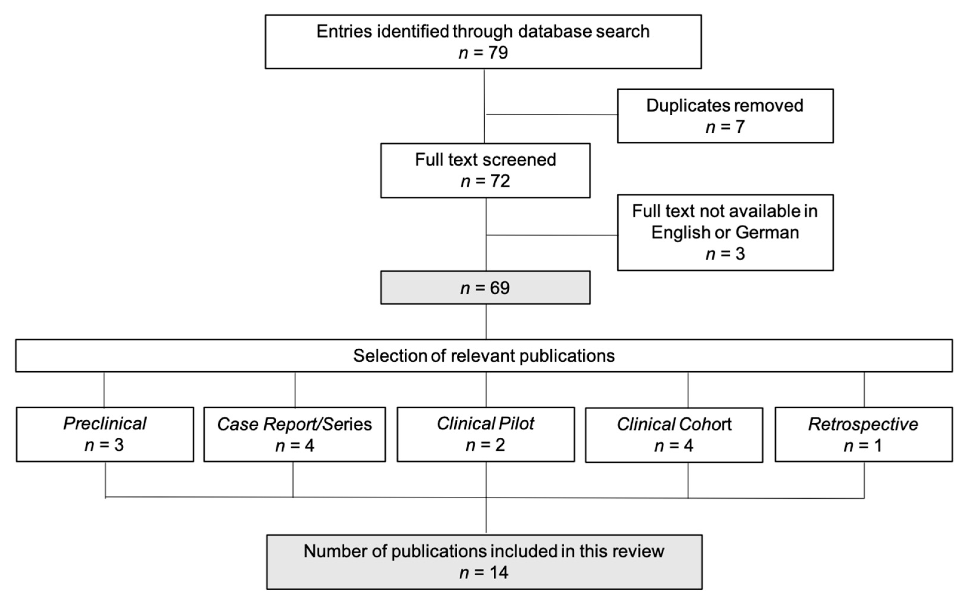
2. Methods
2.1. Aim Construct
2.2. Literature Search
2.3. Outcome Data
- -
- Reepithelialization time.
- -
- Change of the wound surface area over time.
- -
- Secondary outcomes:
- -
- Number of dressing changes,
- -
- Cost of the dressings,
- -
- Level of pain associated with the application, change or removal of the dressing,
- -
- Analgesic intake,
- -
- Hospital length of stay,
- -
- Need for further intervention (surgery),
- -
- Scar quality.
2.4. Reporting of the Systematic Review and Evidence-Based Process
3. Results and Discussion
3.1. Reepithelialization Time
3.2. Pain Intensity
3.3. Dressing Changes
3.4. Treatment-Related Costs
3.5. Application in Split-Thickness Donor Sites
3.6. Long-Term Outcomes
3.7. Limitations
4. Conclusions
Author Contributions
Funding
Institutional Review Board Statement
Informed Consent Statement
Data Availability Statement
Conflicts of Interest
References
- Oryan, A.; Alemzadeh, E.; Moshiri, A. Burn wound healing: Present concepts, treatment strategies and future directions. J. Wound Care 2017, 26, 5–19. [Google Scholar] [CrossRef] [PubMed]
- Markiewicz-Gospodarek, A.; Kozioł, M.; Tobiasz, M.; Baj, J.; Radzikowska-Büchner, E.; Przekora, A. Burn Wound Healing: Clinical Complications, Medical Care, Treatment, and Dressing Types: The Current State of Knowledge for Clinical Practice. Int. J. Environ. Res. Public Health 2022, 19, 1338. [Google Scholar] [CrossRef]
- Alam, K.; Jeffery, S.L.A. Acellular Fish Skin Grafts for Management of Split Thickness Donor Sites and Partial Thickness Burns: A Case Series. Mil. Med. 2019, 184, 16–20. [Google Scholar] [CrossRef] [PubMed] [Green Version]
- Ii, R.S.; Saathoff, E.C.; Larson, D.A.; Wall, J.T.; Wienandt, N.A.; Magnusson, S.; Kjartansson, H.; Natesan, S.; Christy, R.J. Accelerated Wound Closure of Deep Partial Thickness Burns with Acellular Fish Skin Graft. Int. J. Mol. Sci. 2021, 22, 1590. [Google Scholar] [CrossRef]
- Júnior, E.M.L.; Filho, M.O.D.M.; Costa, B.A.; Fechine, F.V.; Vale, M.L.; Diógenes, A.K.D.L.; Neves, K.R.T.; Uchôa, A.M.D.N.; Soares, M.F.A.D.N.; de Moraes, M.E.A. Nile Tilapia Fish Skin–Based Wound Dressing Improves Pain and Treatment-Related Costs of Superficial Partial-Thickness Burns: A Phase III Randomized Controlled Trial. Plast. Reconstr. Surg. 2021, 147, 1189–1198. [Google Scholar] [CrossRef] [PubMed]
- Nischwitz, S.P.; Luze, H.; Popp, D.; Winter, R.; Draschl, A.; Schellnegger, M.; Kargl, L.; Rappl, T.; Giretzlehner, M.; Kamolz, L.P. Global burn care and the ideal burn dressing reloaded—A survey of global experts. Burns 2021, 47, 1665–1674. [Google Scholar] [CrossRef] [PubMed]
- Michael, S.; Winters, C.; Khan, M. Acellular Fish Skin Graft Use for Diabetic Lower Extremity Wound Healing: A Retrospective Study of 58 Ulcerations and a Literature Review. Wounds A Compend. Clin. Res. Pract. 2019, 31, 262–268. Available online: http://www.ncbi.nlm.nih.gov/pubmed/31730505 (accessed on 24 April 2022).
- Verde, M.E.Q.L.; Ferreira-Júnior, A.E.C.; de Barros-Silva, P.G.; Miguel, E.D.C.; Mathor, M.B.; Lima-Júnior, E.M.; de Moraes-Filho, M.O.; Alves, A.P.N.N. Nile tilapia skin (Oreochromis niloticus) for burn treatment: Ultrastructural analysis and quantitative assessment of collagen. Acta Histochem. 2021, 123, 151762. [Google Scholar] [CrossRef]
- Yang, C.K.; Polanco, T.O.; Ii, J.C.L. A Prospective, Postmarket, Compassionate Clinical Evaluation of a Novel Acellular Fish-skin Graft Which Contains Omega-3 Fatty Acids for the Closure of Hard-to-heal Lower Extremity Chronic Ulcers. Wounds 2016, 28, 112–118. Available online: http://www.ncbi.nlm.nih.gov/pubmed/27071138 (accessed on 24 April 2022).
- Le Guellec, D.; Morvan-Dubois, G.; Sire, J.Y. Skin development in bony fish with particular emphasis on collagen deposition in the dermis of the zebrafish (Danio rerio). Int. J. Dev. Biol. 2004, 48, 217–231. [Google Scholar] [CrossRef] [Green Version]
- Brown, P. Bovine Spongiform Encephalopathy and Variant Creutzfeldt-Jakob Disease: Background, Evolution, and Current Concerns. Emerg. Infect. Dis. 2001, 7, 6–16. [Google Scholar] [CrossRef] [PubMed] [Green Version]
- Kjartansson, H.; Olafsson, I.H.; Karason, S.; Thorisson, H.; Baldursson, B.T.; Gunnarsson, E.; Jorundsson, E.; Sigurjonsson, G.F. Use of Acellular Fish Skin for Dura Repair in an Ovine Model: A Pilot Study. Open J. Mod. Neurosurg. 2015, 5, 124–136. [Google Scholar] [CrossRef] [Green Version]
- Lullove, E.; Liden, B.; Winters, C.; McEneaney, P.; Raphael, A.; Ii, J.L. A Multicenter, Blinded, Randomized Controlled Clinical Trial Evaluating the Effect of Omega-3–Rich Fish Skin in the Treatment of Chronic, Nonresponsive Diabetic Foot Ulcers. Wounds A Compend. Clin. Res. Pract. 2021, 33, 169–177. [Google Scholar] [CrossRef] [PubMed]
- Tan, S.W.; Wong, J.; Kee, T.; Chai, Z.T.; Ho, Q.Y.; Chan, M.; Chew, K.Y.; Oh, C.C. Successful treatment of calciphylaxis in a renal transplant recipient with combination of intralesional sodium thiosulphate, intravenous sodium thiosulphate and fish skin graft. Australas. J. Dermatol. 2021, 62, e358–e359. [Google Scholar] [CrossRef]
- Dardari, D.; Lequint, C.; Jugnet, A.C.; Bénard, T.; Bouly, M.; Penfornis, A. Curing Necrotic Angiodermatitis with an Intact Fish Skin Graft in a Patient Living with Diabetes. Medicina 2022, 58, 292. [Google Scholar] [CrossRef]
- Ahn, K.H.; Park, E.S. A rare case report of neonatal calcinosis cutis induced by distant and delayed extravasation of intravenous calcium gluconate. Arch. Plast. Surg. 2021, 48, 641–645. [Google Scholar] [CrossRef]
- Dias, M.T.P.M.; Bilhar, A.P.M.; Rios, L.C.; Costa, B.A.; Júnior, E.M.L.; Alves, A.P.N.N.; Bruno, Z.V.; Filho, M.O.D.M.; Bezerra, L.R.P.S. Neovaginoplasty Using Nile Tilapia Fish Skin as a New Biologic Graft in Patients with Mayer-Rokitansky-Küster-Hauser Syndrome. J. Minim. Invasive Gynecol. 2019, 27, 966–972. [Google Scholar] [CrossRef]
- Shi, Y.; Zhang, H.; Zhang, X.; Chen, Z.; Zhao, D.; Ma, J. A comparative study of two porous sponge scaffolds prepared by collagen derived from porcine skin and fish scales as burn wound dressings in a rabbit model. Regen. Biomater. 2019, 7, 63–70. [Google Scholar] [CrossRef]
- Costa, B.A.; Júnior, E.M.L.; Filho, M.O.D.M.; Fechine, F.V.; De Moraes, M.E.A.; Júnior, F.R.S.; Soares, M.F.A.D.N.; Rocha, M.B.S. Use of Tilapia Skin as a Xenograft for Pediatric Burn Treatment: A Case Report. J. Burn. Care Res. 2019, 40, 714–717. [Google Scholar] [CrossRef]
- Wallner, C.; Holtermann, J.; Drysch, M.; Schmidt, S.; Reinkemeier, F.; Wagner, J.M.; Dadras, M.; Sogorski, A.; Houschyar, K.S.; Becerikli, M.; et al. The Use of Intact Fish Skin as a Novel Treatment Method for Deep Dermal Burns Following Enzymatic Debridement: A Retrospective Case-Control Study. Eur. Burn J. 2022, 3, 43–55. [Google Scholar] [CrossRef]
- Lima-Junior, E.M.; Filho, M.O.D.M.; Costa, B.A.; Fechine, F.V.; De Moraes, M.E.A.; Silva-Junior, F.R.; Soares, M.F.A.D.N.; Rocha, M.B.S.; Leontsinis, C.M.P. Innovative treatment using tilapia skin as a xenograft for partial thickness burns after a gunpowder explosion. J. Surg. Case Rep. 2019, 2019, rjz181. [Google Scholar] [CrossRef] [PubMed] [Green Version]
- Pan, S.C. Burn blister fluids in the neovascularization stage of burn wound healing: A comparison between superficial and deep partial-thickness burn wounds. Burn. Trauma 2013, 1, 27–31. [Google Scholar] [CrossRef] [PubMed] [Green Version]
- Hu, Z.; Yang, P.; Zhou, C.; Li, S.; Hong, P. Marine Collagen Peptides from the Skin of Nile Tilapia (Oreochromis niloticus): Characterization and Wound Healing Evaluation. Mar. Drugs 2017, 15, 102. [Google Scholar] [CrossRef]
- Júnior, E.M.L.; Filho, M.O.D.M.; Forte, A.J.; Costa, B.A.; Fechine, F.V.; Alves, A.P.N.N.; De Moraes, M.E.A.; Rocha, M.B.S.; Júnior, F.R.S.; Soares, M.F.A.D.N.; et al. Pediatric Burn Treatment Using Tilapia Skin as a Xenograft for Superficial-Partial Thickness Wounds: A Pilot Study. J. Burn. Care Res. 2019, 41, 241–247. [Google Scholar] [CrossRef]
- Júnior, E.M.L.; Filho, M.O.D.M.; Costa, B.A.; Rohleder, A.V.P.; Rocha, M.B.S.; Fechine, F.V.; Forte, A.J.; Alves, A.P.N.N.; Júnior, F.R.S.; Martins, C.B.; et al. Innovative Burn Treatment Using Tilapia Skin as a Xenograft: A Phase II Randomized Controlled Trial. J. Burn. Care Res. 2020, 41, 585–592. [Google Scholar] [CrossRef]
- Reese, A.D.; Keyloun, J.W.; Garg, G.; McLawhorn, M.M.; Moffatt, L.T.; Travis, T.E.; Johnson, L.S.; Shupp, J.W. Compounded Cerium Nitrate–Silver Sulfadiazine Cream is Safe and Effective for the Treatment of Burn Wounds: A Burn Center’s 4-Year Experience. J. Burn. Care Res. 2021, 43, 716–721. [Google Scholar] [CrossRef] [PubMed]
- Moi, A.L.; Haugsmyr, E.; Heisterkamp, H. Long-Term Study Of Health And Quality of Life after Burn Injury. Ann. Burn. Fire Disasters 2016, 29, 295–299. Available online: http://www.ncbi.nlm.nih.gov/pubmed/28289366 (accessed on 27 April 2022).
- Ahuja, R.B.; Goswami, P. Cost of providing inpatient burn care in a tertiary, teaching, hospital of North India. Burns 2013, 39, 558–564. [Google Scholar] [CrossRef]
- Júnior, E.M.L.; Filho, M.O.D.M.; Costa, B.A.; Alves, A.P.N.N.; De Moraes, M.E.A.; Uchôa, A.M.D.N.; Martins, C.B.; Bandeira, T.D.J.P.G.; Rodrigues, F.A.R.; Paier, C.R.K.; et al. Lyophilised tilapia skin as a xenograft for superficial partial thickness burns: A novel preparation and storage technique. J. Wound Care 2020, 29, 598–602. [Google Scholar] [CrossRef]
- Badois, N.; Bauër, P.; Cheron, M.; Hoffmann, C.; Nicodeme, M.; Choussy, O.; Lesnik, M.; Poitrine, F.C.; Fromantin, I. Acellular fish skin matrix on thin-skin graft donor sites: A preliminary study. J. Wound Care 2019, 28, 624–628. [Google Scholar] [CrossRef]
- Yoon, J.; Yoon, D.; Lee, H.; Lee, J.; Jo, S.; Kym, D.; Yim, H.; Hur, J.; Chun, W.; Kim, G.; et al. Wound healing ability of acellular fish skin and bovine collagen grafts for split-thickness donor sites in burn patients: Characterization of acellular grafts and clinical application. Int. J. Biol. Macromol. 2022, 205, 452–461. [Google Scholar] [CrossRef] [PubMed]
- Wallace, H.J.; Fear, M.W.; Crowe, M.M.; Martin, L.J.; Wood, F.M. Identification of factors predicting scar outcome after burn injury in children: A prospective case-control study. Burn. Trauma 2017, 5, 19. [Google Scholar] [CrossRef] [PubMed]


| Study | Acellular Fish Skin | Comparison Product | Animal Model | Scalding Conditions | Treatment Period | Endpoints | Main Findings |
|---|---|---|---|---|---|---|---|
| Accelerated Wound Closure of Deep Partial Thickness Burns with Acellular Fish Skin Graft. (Stone II et al., 2021) [2] | North Atlantic cod (Kerecis® Omega3) | Fetal bovine dermis (Primatrix TM) | 6 female Yorkshire pigs of weights 51.8 ± 3.3 kg at the time of the burn wound creation | Creation of ten 5 cm × 5 cm wounds (4 DPT and 6 FT) with a thermocouple brass burn device by Alam et al. [3] heated to 100 °C.
| 60 days | Reepithelialization time, skin function (skin moisture properties, microcirculation) | AFS: faster reepithelialization time in DPT and FT wounds. |
| A comparative study of two porous sponge scaffolds prepared by collagen derived from porcine skin and fish scales as burn wound dressings in a rabbit model. (Shi et al., 2020) [4] | Grass carp | Porcine skin-derived Collagen, dry gauze, Vaseline gauze | 2 New Zealand white rabbits | Creation of ten 2 cm × 2 cm wounds with 35 layers of boiled gauze which was applied with gravitational pressure.
| 28 days | Wound size, dressing properties | AFS: faster wound healing after 12 days, higher moisture permeability |
| Marine Collagen Peptides from the skin of Nile Tilapia (Oreochromis niloticus): Characterization and Wound Healing Evaluation. (Hu et al., 2017) [5] | Nile Tilapia | - | 48 New Zealand white rabbits | Creation of one DPT 4 × 4 cm burn wound with a scalding device (YLS-5Q, Bejing, China) heated to 100 °C.
| 18 days | Reepithelialization time, histological analysis of the skin structure integrity, cell types and granulation tissue | AFS: faster reepithelialization time in comparison to the control group. Reduction of inflammation and promotion of granulation tissue formation |
| Study | Study Type | Fish Skin | Comparison to | Study Cohort | Treatment Period | Endpoints | Main Findings |
|---|---|---|---|---|---|---|---|
| The Use of Intact Fish Skin as a Novel Treatment Method for Deep Dermal Burns following Enzymatic Debridement: A retrospective Case-Control Study. (Wallner et al., 2022) [6] | Retrospective case–control study | North Atlantic cod (Kerecis® Omega3) | absorbable, synthetic skin substitute (Suprathel®) in SPTB split-thickness skin graft in DPTB | 12 patients (age range 18–60 years) with SPTB or DPTB mean TBSA of 12.5 ± 9.4% after enzymatic debridement | 28 days | Wound quality assessment and size, reepithelialization time, scar quality | AFS: accelerated wound healing (total reepithelialization time of 22 ± 6.3 days), higher water-storage capacity, improved aesthetic and functional outcomes, decreased pain and itching |
| Wound healing ability of acellular fish skin and bovine collagen grafts for split-thickness donor sites in burn patients: Characterization of acellular grafts and clinical application. (Yoon et al., 2022) [7] | In vitro and clinical comparison study | North Atlantic cod (Kerecis® Omega3) | Bovine collagen graft (ProHeal®) | 52 patients with acute burns who underwent split-thickness skin grafting | Up to 17 days | In vitro: cellular responses to the grafts In vivo: reepithelialization time, wound complications | AFS: accelerated reepithelialization time by 2 days |
| Nile Tilapia Fish Skin-Based Wound Dressing Improves Pain and Treatment-Related Costs of Superficial Partial-Thickness Burns: A Phase III Randomized Controlled Trial. (Lima Júnior et al., 2021) [8] | Open-label, monocentric, Phase III Randomized Controlled Trial | Nile Tilapia | Silver Sulfadiazine Cream 1% | 115 patients (age range: 18–70 years) with SPTB < 15% TBSA | Up to 11 days | Reepithelialization time, number of dressing changes, treatment related costs, pain intensity | AFS: Fewer days of reepithelialization and dressing changes. Lower analgesic needs and scores in BSPAS and mechanical pain threshold measurements. Reduction of average treatment related costs per patient by 42.1% |
| A Randomized Comparison Study of Lyophilized Nile Tilapia Skin and Silver-Impregnated Sodium Carboxymethylcellulose for the Treatment of Superficial Partial-Thickness Burns. (Lima Júnior et al., 2021) [9] | Open-label, randomized, prospective, controlled pilot study | Nile Tilapia | silver-impregnated sodium carboxymethylcellulose dressing (Aquacel Ag®) | 24 patients (age range 18–70 years) with SPTB ≤ 10% TBSA | Up to 11 days | Number of dressing changes, pain intensity, pain-related anxiety, analgesic intake | AFS: Reduced number of dressing changes, lower overall pain intensity measured via VAS score. Comparable analgesic intake and pain-related anxiety. |
| Lyophilised tilapia skin as a xenograft for superficial partial thickness burns: a novel preparation and storage technique. (Lima Júnior et al., 2020) [10] | Case Report | Nile Tilapia | - | 33-year-old female patient with SPTB of 10% TBSA | 10 days | Reepithelialization time | Good adherence to the wound bed, total reepithelialization time of 10 days |
| Innovative Burn Treatment Using Tilapia Skin as a Xenograft: A Phase II Randomized Controlled Trial. (Lima Júnior et al., 2020) [11] | Open-label, monocentric, Phase II Randomized Controlled Trial | Nile Tilapia | Silver Sulfadiazine Cream 1% | 62 patients (age range: 18–50 years) with SPTB ≤ 20% TBSA or DPTB between 5 and 15% TBSA | Up to 23 days | Reepithelialization time, number of dressing changes, burn improvement, anesthetic/analgesic intake, pain intensity | AFS: Fewer days of reepithelialization and dressing changes. Lower pain intensity and amount of anesthetics/analgesics |
| Innovative treatment using tilapia skin as a xenograft for partial thickness burns after a gunpowder explosion. (Lima Júnior et al., 2019) [12] | Case Report | Nile Tilapia | 23-year-old male patient with 16% TBSA SPTB and DPTB | Up to 17 days | Reepithelialization time | Good adherence of the biomaterial to the wound bed, reepithelialization of SPTB in 12 days and 17 days in DPTB | |
| Pediatric Burn Treatment Using Tilapia Skin as a Xenograft for Superficial Partial Thickness Wounds: A Pilot Study. (Lima Júnior et al., 2019) [13] | Open-label, monocentric, randomized phase II pilot study | Nile Tilapia | Silver Sulfadiazine Cream 1% | 30 children (age range: 2–12 years) with SPTB | Up to 11 days | Reepithelialization time, number of dressing changes | AFS: Reduced total number of dressings (3.00 ± 0.76) in comparison to the Silver Sulfadiazine cream 1% group (9.27 ± 1.39) Comparable reepithelialization time and rate, anesthetic and analgesics intake |
| Use of Tilapia Skin as a Xenograft for Pediatric Burn Treatment: A Case Report. (Costa et al., 2019) [14] | Case Report | Nile Tilapia | 3-year-old boy with SPTB of 18% TBSA | 10 days | Reepithelialization time | Total reepithelialization time of 10 days | |
| Acellular Fish Skin Grafts for Management of Split Thickness Donor Sites and Partial Thickness Burns: A Case Series. (Alam et al., 2019) [2] | Case series | North Atlantic cod (Kerecis® Omega3) | 10 patients (age range 18–90 years) undergoing split-thickness skin grafting for burn injuries | Up to 16 days | Reepithelialization time, pain | Total reepithelialization time of 11.5 days (range: 10–16), Average pain score of 2.3 (range 1–4) of 10 at dressing changes | |
| Acellular fish skin matrix on thin-skin graft donor sites: a preliminary study. (Badois et al., 2019) [15] | Prospective, comparative, before-after cohort study | North Atlantic cod (Kerecis® Omega3) | Paraffin gauze (Jelonet) | 21 patients (age range: 33–84 years) with split-thickness skin graft donor sites of 30–45 cm2 | Up to 134 days | Reepithelialization time, wound evaluation, pain | AFS: Average total reepithelialization time was 31.5 days (±24.7) in comparison to 67.9 days (±66.2) in the Jelonet group. AFS: reduced pain levels and infection |
Publisher’s Note: MDPI stays neutral with regard to jurisdictional claims in published maps and institutional affiliations. |
© 2022 by the authors. Licensee MDPI, Basel, Switzerland. This article is an open access article distributed under the terms and conditions of the Creative Commons Attribution (CC BY) license (https://creativecommons.org/licenses/by/4.0/).
Share and Cite
Luze, H.; Nischwitz, S.P.; Smolle, C.; Zrim, R.; Kamolz, L.-P. The Use of Acellular Fish Skin Grafts in Burn Wound Management—A Systematic Review. Medicina 2022, 58, 912. https://doi.org/10.3390/medicina58070912
Luze H, Nischwitz SP, Smolle C, Zrim R, Kamolz L-P. The Use of Acellular Fish Skin Grafts in Burn Wound Management—A Systematic Review. Medicina. 2022; 58(7):912. https://doi.org/10.3390/medicina58070912
Chicago/Turabian StyleLuze, Hanna, Sebastian Philipp Nischwitz, Christian Smolle, Robert Zrim, and Lars-Peter Kamolz. 2022. "The Use of Acellular Fish Skin Grafts in Burn Wound Management—A Systematic Review" Medicina 58, no. 7: 912. https://doi.org/10.3390/medicina58070912