Article

Oral Microbiome Using Colocasia antiquorum var. esculenta Extract Varnish in a Mouse Model with Oral Gavage of P. gingivalis ATCC 53978

1
Department of Dental Biomaterials, College of Dentistry, Wonkwang University, 460 Iksan-daero, Iksan 54538, Jeonbuk, Korea
2
Institute of Biomaterials and Implant, College of Dentistry, Wonkwang University, 460 Iksan-daero, Iksan 54538, Jeonbuk, Korea
3
Department of Oral Anatomy, College of Dentistry, Wonkwang University, 460 Iksan-daero, Iksan 54538, Jeonbuk, Korea
*
Author to whom correspondence should be addressed.
These authors contributed equally to this work.
Medicina 2022, 58(4), 506; https://doi.org/10.3390/medicina58040506
Submission received: 1 March 2022 / Revised: 30 March 2022 / Accepted: 30 March 2022 / Published: 1 April 2022
(This article belongs to the Topic State-of-the-Art Dentistry and Oral Health)

Abstract

:
Background and Objective: There is increasing interest in preventing periodontitis using natural products. The purpose of this study was to investigate the effect of Colocasia antiquorum var. esculenta (CA) varnish on the oral microbiome and alveolar bone loss in a mouse periodontitis model. Materials and Methods: Antibacterial activity against Porphyromonas gingivalis (P. gingivalis) ATCC 53978 and cell cytotoxicity using CCK-8 on L929 cells were measured. Balb/c mice were assigned into five groups (negative control, positive control, CA in drinking water, varnish, and CA varnish). P. gingivalis was administered to the mice by oral gavage three times. After sacrifice, the oral microbiome and the levels of the inflammatory cytokine IL-1β and matrix metalloproteinase-9 were analyzed. Alveolar bone loss was measured using micro-computed tomography. Results: CA extract showed an antibacterial effect against P. gingivalis (p < 0.05) and showed no cytotoxicity at that concentration (p > 0.05). Although alpha diversity of the oral microbiome did not statistically differ between the groups (p > 0.05), the relative abundance of dominant bacteria tended to be different between the groups. The inflammatory cytokine IL-1β was reduced in the CA varnish group (p < 0.05), and no difference was observed in MMP-9 expression and alveolar bone loss (p > 0.05). Conclusions: CA varnish did not affect the overall microflora and exhibited an anti-inflammatory effect, suggesting that it is possibility a suitable candidate for improving periodontitis.

1. Introduction

Periodontal disease is an inflammatory disease caused by bacteria and changes in the oral bacterial composition are known to cause periodontal disease [1]. Oral bacteria attached to calculus on tooth surfaces produce toxins such as lipopolysaccharides and endotoxins [2]. The toxins cause an inflammatory response in the gingival tissue, creating a countering immune response [3]. In this inflammatory immune response, inflammatory cytokines such as IL-1β and TNF-α are expressed [4] and matrix metalloproteases (MMPs) cause collagen degradation [5]. Inflammatory cytokines and MMPs can cause severe damage to the surrounding tissues [6].
The first approach to treating periodontal disease is the removal of the calculus attached to the teeth through scaling and root planning [7]. However, when the calculus is located in the deep gingival sulcus, it is difficult to remove it entirely [8]. Antibiotics and chlorhexidine can assist in the non-surgical treatment of periodontal disease but have side effects such as hepatotoxicity, renal toxicity, opportunistic infection, and discoloration [9,10]. In order to reduce these side effects, natural herb medicine must be increased [11]. Local drug delivery systems have also been developed to localize drugs in periodontal pockets [12].
Colocasia antiquorum var. esculenta (CA) is a plant of the family Tauraceae, called taro, eddoe, and is used for food and medicinal purposes [13]. CA extract is known to have antioxidant, antifungal, and antimicrobial effects [14,15]. The methanol extract of CA has exhibited antimicrobial effects against Porphyromonas gingivalis (P. gingivalis) with anti-inflammatory and anti-osteoclastogenic activities [16]. However, the effects of the ethanol extract of CA are not well known.
Several mouse models have been used to simulate periodontitis to mimic the human condition, including the ligature model, the lipopolysaccharide (LPS) injection model, and the P. gingivalis inoculation model [17,18,19]. The ligature model can induce alveolar bone loss quickly; however, it can cause damage to the gingival tissue due to ligation pressure [20]. The LPS injection model promotes aggressive lesions and rapid alveolar bone destruction but exhibits a different pattern than chronic periodontitis, where the alveolar bone is destroyed over an extended period [18]. This study adopted the P. gingivalis inoculation model, in which P. gingivalis was administered by oral gavage to initiate dysbiosis of oral bacteria and induce periodontitis with chronic behavior [21]. This model simulates human chronic periodontitis caused by P. gingivalis [22].
More than 700 microorganisms closely related with periodontitis have been detected in the oral cavity [23]. With the development of next-generation sequencing technology, it has become possible to analyze the oral microbiome, a collection of all oral microorganisms [24]. The oral microflora, oral microbiota, and more recently, the oral microbiome have all been used to describe the bacteria found in the human oral cavity [25]. Changes in the oral microbiome related to periodontitis, dental caries, and oral cancer have been investigated [26,27]; however, few papers have examined the changes in the oral microbiome by applying dental materials. If an effective agent, such as an antibiotic, alters the oral microbiome, it can cause other diseases such as candidiasis [2]. Since the oral microbiome examines the interaction of hundreds of species, in vivo tests should be done. It is crucial to analyze the oral microbiome using an animal model before clinical trials. Recently, the murine oral microbiome has been studied [28,29]. Mouse microbiome models have been the primary choice for preclinical tests for studying relationships between the microbiomes and host physiological, metabolic, immune, and neurologic phenotypes [30].
The purpose of this study was to investigate whether CA varnish effectively inhibits factors related to periodontal disease. Antibacterial effects against P. gingivalis and the cytotoxicity of the ethanol extract of CA were also evaluated in vitro. Using a mouse model with oral gavage of P. gingivalis the American Type Culture Collection (ATCC) 53978, the changes in the oral microbiome, inflammatory cytokines, and alveolar bone levels were analyzed. The null hypothesis was that the oral microbiome, alveolar bone loss, and inflammatory factor would not differ among the groups.

2. Materials and Methods

2.1. Ethanol Extract of CA

The ethanol extract of Colocasia antiquorum var. esculenta (CA) in this study was obtained from the Korea Plant Extract Bank (Korea Research Institute of Bioscience and Biotechnology, Daejeon, Korea). A stock solution (50 mg/mL) was prepared in dimethyl sulfoxide (DMSO) and stored at −20 °C before use.

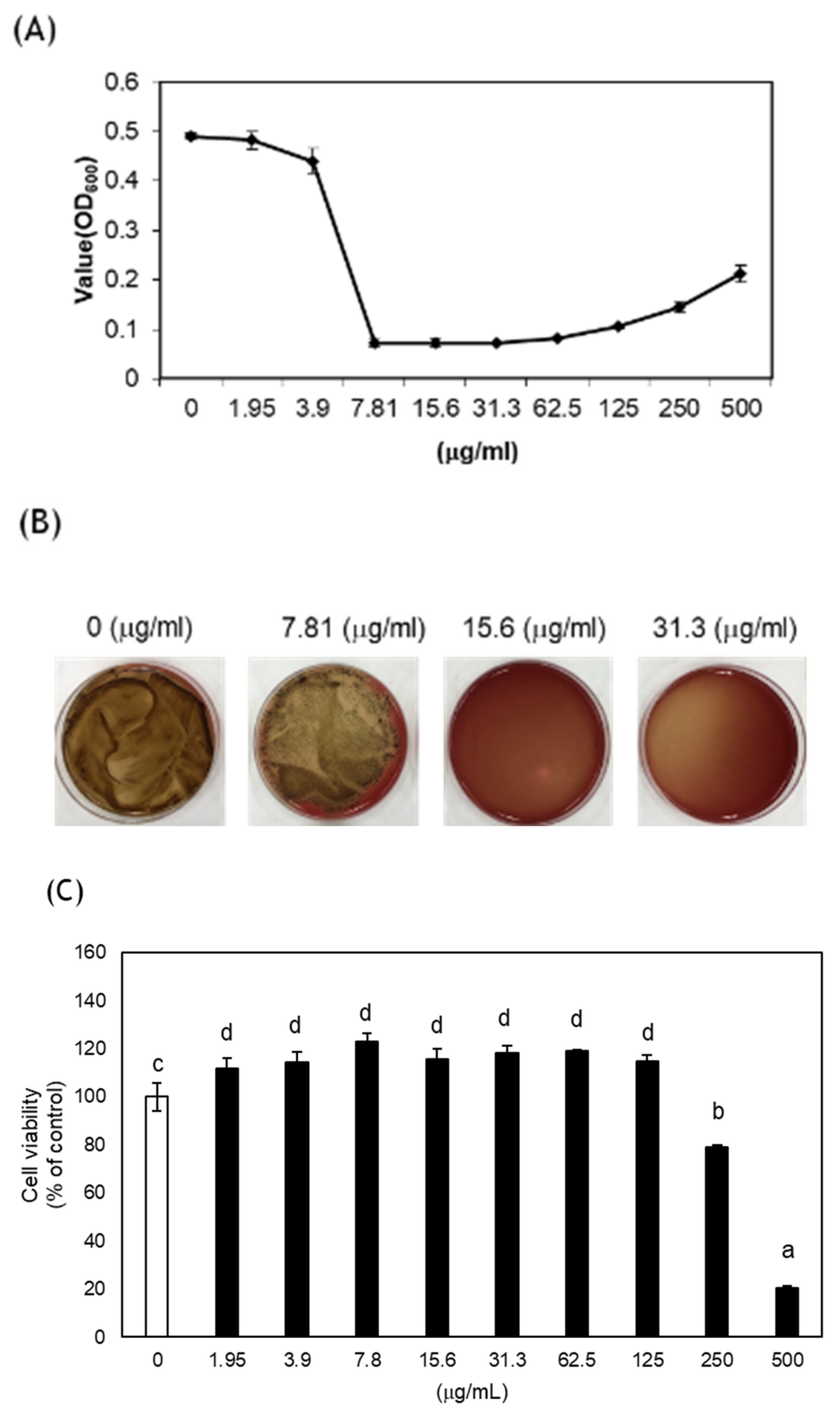
2.2. Antibacterial Assay

To investigate the antibacterial effects of CA, P. gingivalis (ATCC 53978) was purchased from the American Type Culture Collection (ATCC, Manassas, VA, USA) and cultured at 37 °C in an anaerobic chamber in brain heart infusion (BHI) broth with 5 g/mL hemin and 0.5 g/mL menadione. Serial dilutions of CA were added to the 96-well plate, and P. gingivalis suspensions with a final concentration of 5 × 10⁷ colony-forming units (CFU) were added. The minimum inhibitory concentration (MIC) was determined as the lowest concentration of CA that inhibited the visible growth of P. gingivalis. For the minimal bactericidal concentration (MBC), concentrations equal to and higher than the MIC were streaked on blood agar plates and incubated for seven days. MBC was determined as the lowest concentration of CA that had no visible bacterial colonies on the blood agar plate after incubation.

2.3. Cytotoxicity

L929, a mouse fibroblast cell line, was obtained from the America Type Culture Collection. RPMI 1640 (HyClone, Logan, UT, USA) was used to culture the cells and was supplemented with 10% FBS and 1% penicillin and streptomycin. CA was diluted with medium for the assay, and the same amount of DMSO (0.1%) was added to the negative control. According to the manufacturer’s protocol, a Cell Counting Kit-8 (CCK-8, Dojindo Molecular Technologies, Kumamoto, Japan) was used to assess cytotoxicity.

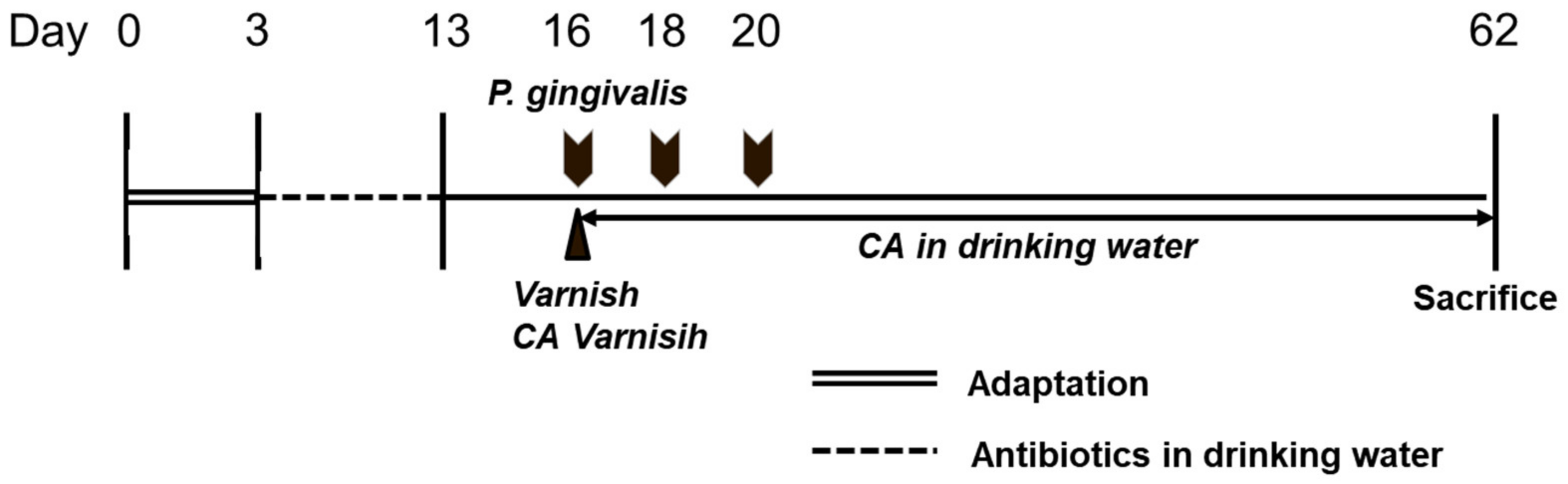
2.4. Oral Gavage of the P. gingivalis Mouse Model

Eleven-week-old female BALB/c mice were purchased from Samtako Bio Korea (Osan, Korea). This experiment was approved by the Wonkwang University Animal Ethics Committee (WKU20-97). The mice were randomly divided into five groups. Eight animals were assigned per group, and four mice were housed per cage. The experimental groups were divided, and the code, explanation, and treatment were specified in the parenthesis as follows:
  • Group 1: Negative control (NC; no bacterial inoculation, no treatment)
  • Group 2: Positive control (PC; bacterial inoculation, no treatment)
  • Group 3: CA in drinking water (WCA; bacterial inoculation, 0.5% CA extract in drinking water)
  • Group 4: Varnish (V; bacterial inoculation, varnish)
  • Group 5: CA varnish (VCA; bacterial inoculation, varnish mixed with CA)
The experimental schedule is shown in Figure 1. All mice were subjected to three days of acclimatization and received deionized water and food ad libitum. After the adaptation period, the mice received sulfamethoxazole (870 μg/mL) and trimethoprim (170 μg/mL) in deionized water for 10 days. Three days after antibiotic treatment, log-phase broth P. gingivalis was centrifuged and suspended to make 1 × 10¹⁰ CFU/mL in 2% carboxymethyl cellulose in PBS. P. gingivalis (100 μL/mouse) was inoculated by oral gavage three times every two days in Groups 2, 3, 4, and 5. Three-quarters of P. gingivalis was injected into the esophagus using an oral zonde (20G, JEUNGDO BIO PLANT Co., Ltd., Seoul, Korea), while the rest was injected into the oral cavity.
For the WCA group, distilled water mixed with 0.5 w/v% CA dissolved in 1% ethanol solution was provided as drinking water. Varnish was fabricated with rosin and ethanol as described in the previous study [16]. For the VCA group, 15 wt% CA was mixed into the varnish. For the V and VCA groups, the manufactured varnish and the varnish combined with CA were applied around the upper molar with a micro-brush for 15 s under anesthesia with intraperitoneal injections of 400 mg/kg chloral hydrate (Sigma, St. Louis, MO, USA). All mice were euthanized by CO2 gas 42 days after the final bacteria inoculation.

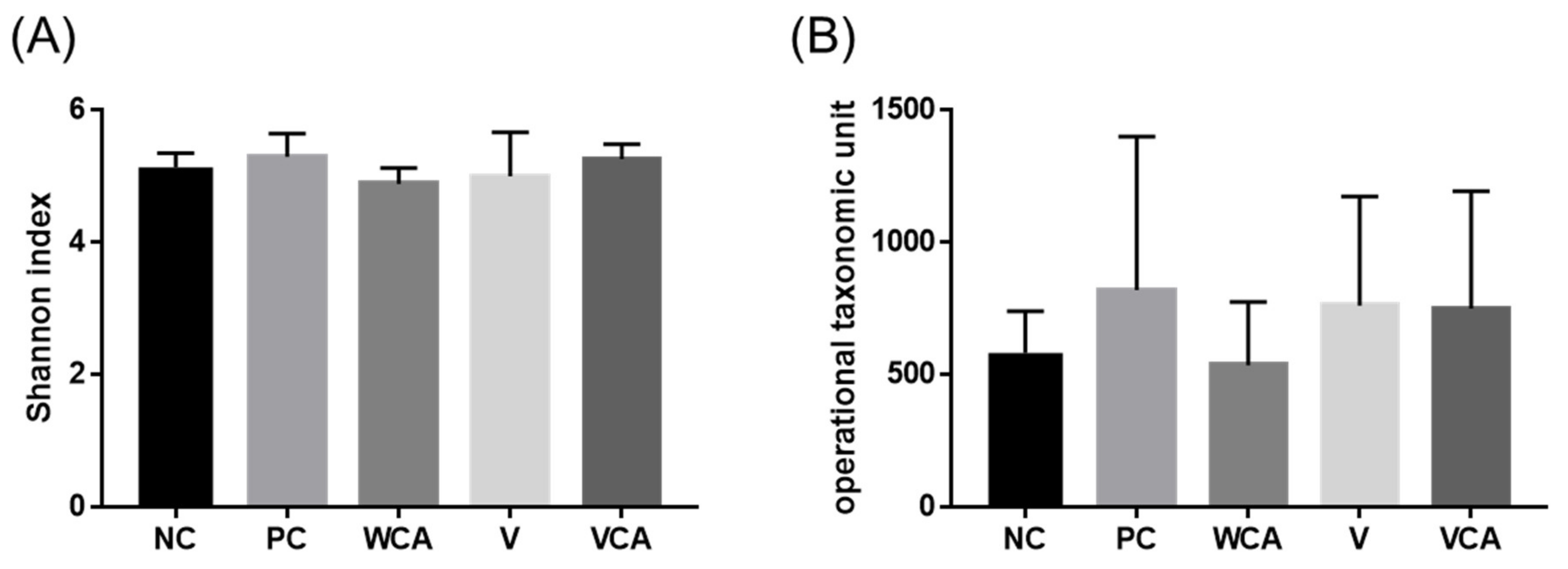
2.5. Oral Microbiome

The experimental animals assigned eight per group were divided into two subgroups. Half of the mice (n = 4) were utilized for oral microbiome analysis, while the other four mice were used for inflammatory cytokine analysis. Mouse gingival tissue was collected from the palatal gingiva around the upper molars [30] (Figure 2A). The sample was placed in 500 μL of buffer (Nucleic acid preservation & transport medium, Noblebio, Korea). The collected mouse gingival tissue was transferred into the QIAamp DNA Stool Mini Kit (QIAGEN, Hilden, Germany) solution and the bacterial genomic DNA was extracted according to the manufacturer’s instructions. The library was prepared by referring to Illumina’s 16S metagenomic library prep guide sequencing (Illumina, San Diego, CA, USA). The library was quantified through Qubit4.0 (Thermo Fisher Scientific, Waltham, MA, USA), and the size was measured using Qsep1 (Bioptic Inc., New Taipei, Taiwan). After adjusting the concentration of the prepared library to 20 pmol and pooling to create a mixture, 20 µL of the sample was placed into the cartridge, and analysis was performed using Iseq100 (Illumina) equipment. The FASTQ file created by the Iseq100 equipment was analyzed for its metagenome through the EZBioCloud (ChunLab, Seoul, Korea) platform and the BaseSpace (Illumina, San Diego, CA, USA) platform. After analyzing the samples, the Shannon index, operational taxonomic units (OTUs) counts, and the relative abundances of species, genus, class, and phylum were analyzed. The Shannon index is a well-known diversity index, and the higher the Shannon index value, the higher the community diversity [31]. OTUs are a cluster of similar sequence variants of the 16s rDNA marker gene sequence [32].

2.6. mRNA Expression by Quantitative RT-PCR

Real-time polymerase chain reaction (RT-PCR) was performed to analyze the inflammatory factors IL-1β and MMP-9. The mouse gingival tissue collected around the second molar (Figure 2B) was homogenized (BioMasher II; Nippi, Tokyo, Japan) for several minutes in a TRIzol solution (Invitrogen, Carlsbad, CA, USA). cDNA was synthesized from 100 ng of RNA isolated using the Omniscript Reverse Transcriptase Kit (Qiagen, Germantown, MD, USA). Real-time quantitative PCR was analyzed using StepOne Plus Real-Time OCR Systems (Applied Biosystem, Foster City, CA, USA) by adding cDNA synthesized, each with 10 pmol of prime and PowerUp SYBR Green Master Mix (AppliedBiosystem, Waltham, MA, USA). The expression levels of inflammatory cytokine (IL-1β) and MMP-9, as quantified by GAPDH, were compared. Information on the primers used in the experiment is as in Table 1.

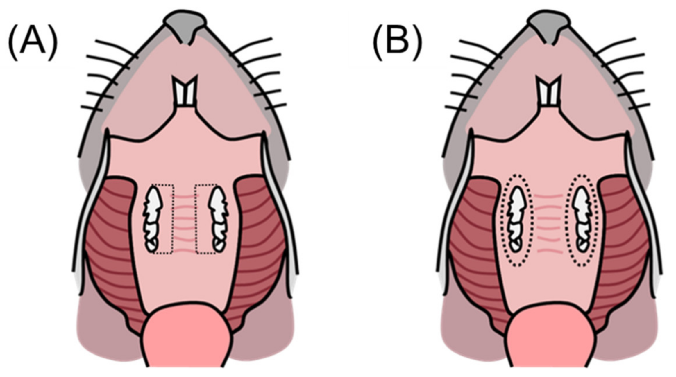
2.7. Alveolar Bone Loss

For oral microbiome analysis, the skulls were defleshed and stored in 4% paraformaldehyde overnight. Before microCT analysis, paraformaldehyde was replaced with phosphate-buffered saline. The maxilla was scanned with a micro-CT (SkyScan 1076, Bruker, Kontich, Belgium) under 88 kV and 112 μA of X-ray tube voltage and current, respectively; pixel size of 9 μm; and exposure time of 2000 ms at 0.42° intervals. DataViewer software (Bruker, Kontich, Belgium) was used to reconstruct the two-dimensional images, while CTVox (Bruker) was utilized to create a three-dimensional (3D) image. Using Image J software (NIH, Bethesda, MD, USA), alveolar bone loss was measured as the distance from the cementoenamel junction (CEJ) to the alveolar bone crest (ABC) on the 3D images at a total of seven buccal sites [33].

2.8. Statistical Analysis

Statistical analysis was performed using IBM SPSS Statistics (Version 26, Armonk, NY, USA). When the data has its normality and homogeneity of variance with the Shapiro–Wilk test and the Levene test, it was analyzed with a one-way analysis of variance test. Cytotoxicity was analyzed using a one-way analysis of variance with Tukey’s multiple range test. Alveolar bone loss data were analyzed using one-way analysis of variance with the post hoc Duncan’s multiple range test. Real-time PCR data were tested with the Kruskal–Wallis test, with the post hoc Mann–Whitney test and the Bonferroni correction. Oral microbiome data were tested by the Kruskal—Wallis test. A p-value < 0.05 was considered statistically significant.

3. Results

3.1. Antibacterial Effect and Cytotoxicity of CA

CA significantly inhibited the growth of P. gingivalis ATCC 53978 with a minimal inhibitory concentration of 7.81 μg/mL (Figure 3A) and a minimal bactericidal concentration of 15.6 μg/mL (Figure 3B). CA exhibited no cytotoxicity up to the concentration of 125 μg/mL. The cytotoxicity significantly increased at 250 μg/mL or higher concentrations than NC (Figure 3C).

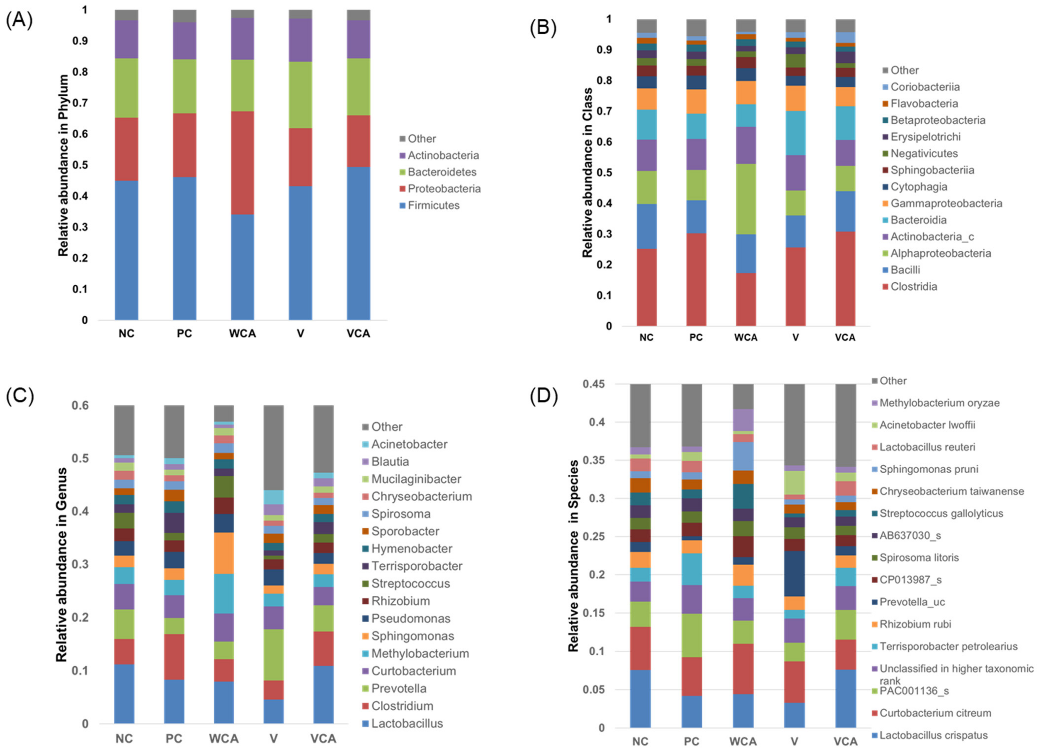
3.2. Oral Microbiome

In the microbiome analysis, a total of 3073 species were analyzed. Alpha diversity was assessed by the Shannon index and operational taxonomic unit (OTU) analysis (Figure 4). There were no statistically significant differences among the groups in Shannon index or OTUs (p > 0.05).
The relative abundance at the phylum, class, genus, and species levels are demonstrated in Figure 5. Bacteria that comprised less than 0.5% of the total bacteria were classified as other. At the phylum level, Firmicutes were the most dominant in all groups. At the class level, Clostridia dominated, followed by Bacilli. At the genus level, Lactobacillus was the most dominant. At the species level, Lactobacillus crispatus was the most dominant in the NC and VCA groups.
Bacteria related to periodontitis are summarized in Table 2. The relative abundance of Spirochaetes was smaller in NC than PC, and those of the experimental groups tended to be smaller. The relative abundance of Bacilli, Lactobacillus, and Lactobacillus crispatus were larger in NC than PC, and the VCA group exhibited a similar tendency to NC.

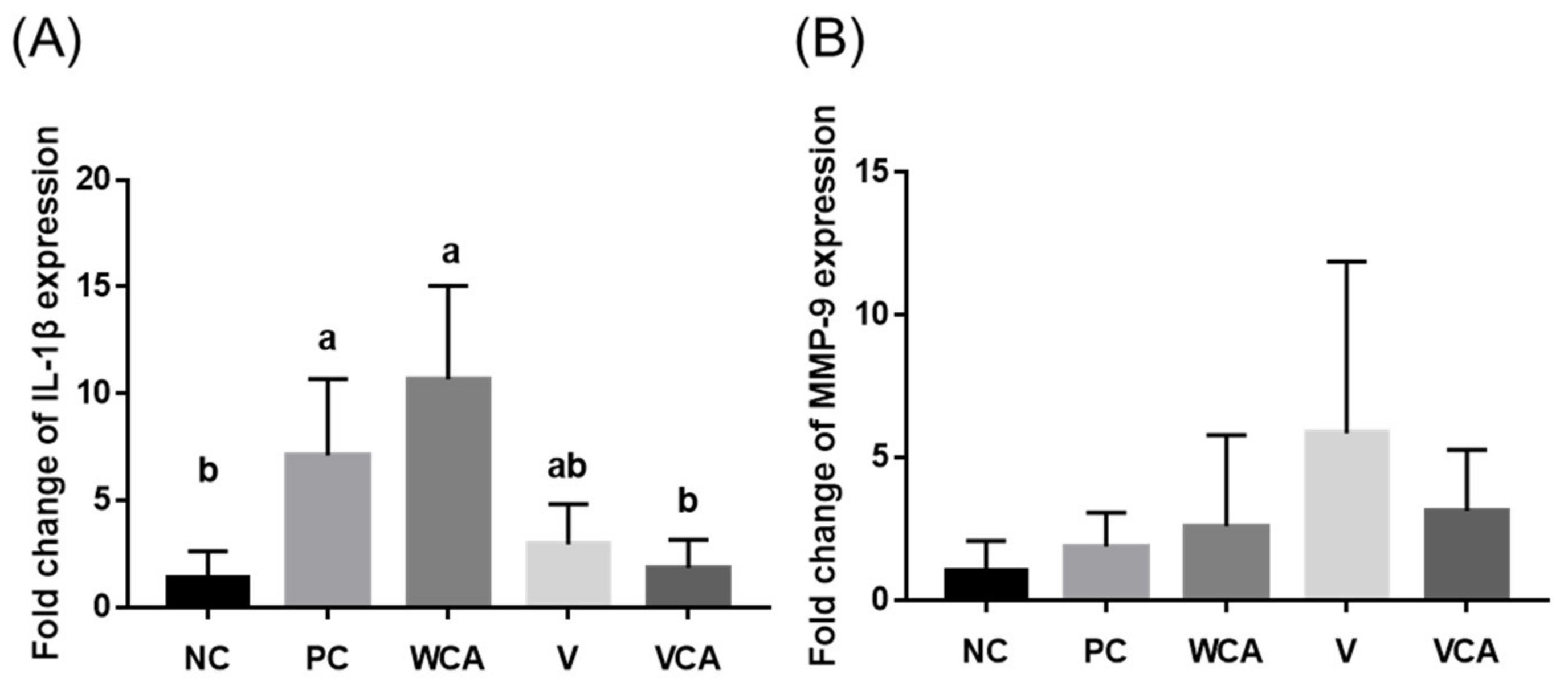
3.3. Anti-Inflammatory Effect

The VCA group showed significantly lower IL-1β expression level than PC and WCA groups (p < 0.05), with no significant differences from the NC and V groups (p > 0.05) (Figure 6A). There were no statistically significant differences in MMP-9 expression between the groups (Figure 6B).

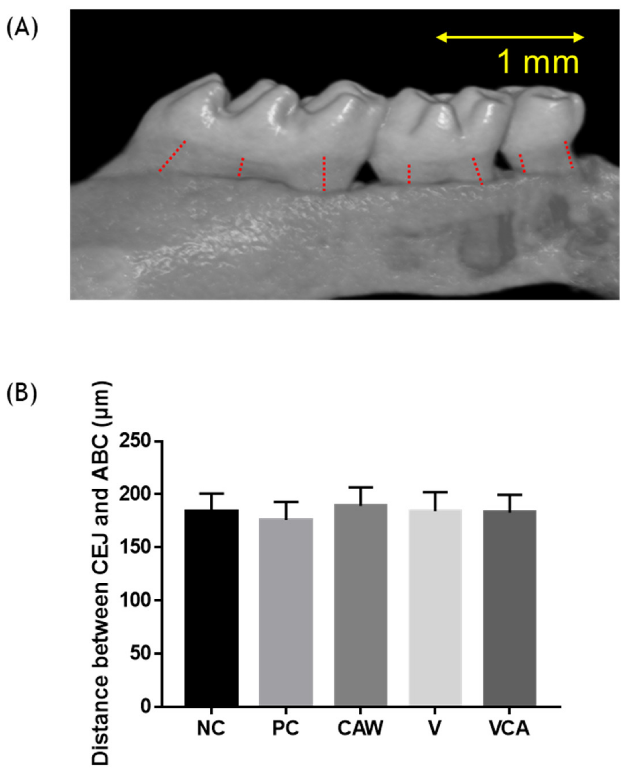
3.4. Alveolar Bone Loss

There were no significant differences in alveolar bone loss, defined as the distance from CEJ to the ABC, between the groups (p > 0.05) (Figure 7).

4. Discussion

The purpose of this study was to evaluate the effect of CA extract in vitro and CA varnish in vivo in terms of improving periodontitis. The antibacterial effects against P. gingivalis and cell cytotoxicity of CA extract were measured in vitro, while the oral microbiome, mRNA expression (IL-1β and MMP-9), and alveolar bone loss were evaluated by oral gavage of the P. gingivalis mouse model treated with CA varnish.
For the antibacterial effect of CA extract against P. gingivalis ATCC 53978, MIC and MBC were 7.81 and 15.6 μg/mL, respectively, with no cytotoxicity. In a previous study, MIC and MBC against P. gingivalis ATCC 33277 were 31.3 and 62.5 μg/mL, respectively [34]. P. gingivalis is a Gram-negative anaerobic bacterium and one of the main causes of periodontal disease [35]. Since fimbriae vary according to the substrain of P. gingivalis, the response according to the pathogenicity or agent may be different [36]. There are multiple substrains in P. gingivalis, and the virulence varies between them [37]. It was confirmed that MIC and MBC were altered according to the substrain of P. gingivalis, and higher antibacterial activities of CA extract were demonstrated against P. gingivalis ATCC 53978 than P. gingivalis ATCC33277.
In another previous study, a methanol extract of CA exhibited antibacterial activity of 125 μg/mL against P. gingivalis ATCC 33277 [16]. Although the solvent was different, the antibacterial activity was maintained. Since methanol is neurotoxic, the ethanol extraction is expected to be useful in the future.
The cytotoxicity of CA was not observed up to 125 μg/mL but significantly increased at 250 μg/mL; however, considering that ISO 10993-5 specifies cell viability less than 70% of the NC as cytotoxic [38], a concentration of up to 250 μg/mL is not cytotoxic. It was confirmed that there was no cytotoxicity at the concentration exhibiting effective antibacterial activity. It was shown that CA extract is a potential substance for preventing periodontitis in vitro.
For the oral microbiome analysis, oral swabs or gingival tissues were used [39]. The dominant species were clearly visible in the oral swab method, while the results were more varied in the tissue analysis [29]. Since the microbiome in the oral cavity varies by region, oral swabs might be advantageous for obtaining overall microbiome data; however, in order to evaluate periodontitis-related bacteria, evaluation of the gingival tissue around the gingival sulcus is more critical than the overall bacteria of the oral cavity [26]. Therefore, sectioned gingival tissue was used to analyze the microbiome in this study. As a result of the animal experiments, overall microbiome dysbiosis was not observed. There were no significant differences in the Shannon index and OTU, indicating diversity within the group. Antibiotics and chlorhexidine mouthwash used to control the periodontal disease can alter the oral microbiome to cause side effects [40,41]. In this respect, CA varnish, which exhibited little effect on the diversity of the microbiome, is considered advantageous.
At the phylum level, Firmicutes, Proteobacteria, Bacteroidetes, and Actinobacteria were found in more than 95% of all groups. Between them, Firmicutes appeared to be dominant and were mostly Gram-positive. In addition, Spirochaete, which is known to increase in periodontitis [42], tended to increase in PC compared with the NC group and decreased in the experimental groups (Table 2). Bacilli are known to decrease in proportion to the presence of periodontitis [43]. Bacilli at the class level decreased in PC and increased in the VCA group as in the NC group (Table 2).
At the genus level, Lactobacillus was dominant. Lactobacillus plays an important role in maintaining healthy conditions by stimulating natural immunity and contributing to the balance of microflora [44]. In VCA group, Lactobacillus was as dominant, as in NC group. However, Lactobacilli, which can appear in the oral cavity of human beings such as L. acidophilus and L. casei, were hardly found in mice gingival tissue [45]. At the species level, Lactobacillus crispatus represents the majority of Lactobacilli and has not been well studied in the human oral cavity. Lactobacillus crispatus tended to increase in VCA similar to NC. Lactobacillus crispatus inoculation was reported to improve periodontitis [46]. According to the study of the mouse microbiome with oral swabs, Streprococcus danieliae was the dominant species [33]. Although statistical differences were not noted among the bacterial taxonomic distributions due to the limited number of specimens, a tendency to change in the microbiome composition was observed.
Alveolar bone loss analyzed using micro-CT showed no statistically significant differences between the groups. Possible reasons why are as follows. Firstly, the number and amount (CFU) of inoculations were not enough to cause periodontitis. Although there have been several inoculation doses in several inoculation models [47,48], three inoculations were performed in this study, following the protocol of PJ Baker [49,50]. Secondly, several studies have adopted periodontitis models of mixed infection with P. gingivalis and other bacteria such as Fusobacterium nucleatum or Streptococcus gordonii [51,52]. The previous study was performed using P. gingivalis ATCC 33277 [34]. According to Baker’s research, P. gingivalis ATCC 53978 exhibited more potent virulence and bone loss than ATCC 33277 [53]. Therefore, P. gingivalis ATCC 53978 was used to induce alveolar bone loss in this study. In this study, P. gingivalis single infection did not cause alveolar bone loss, despite using P. gingivalis ATCC 53978, which is known to have higher virulence [52]. Thirdly, instead of using bacteria grown on plates, the bacteria were grown in broth. Therefore, there is a high possibility of inducing bone loss, because bacteria with black pigmentation on the plate are more toxic than those grown in broth [54]. Further research is needed to evoke alveolar bone loss by adjusting for these factors.
The expression of IL-1β of the gingival tissue was higher in PC than NC and decreased in the VCA group. IL-1β is one of the pro-inflammatory cytokines observed in the early stages of inflammation [55]. MMP-9 is induced by various inflammatory stimuli and associated with bone destruction [56]; however, MMP-9 did not exhibit a statistically significant difference between groups, which is consistent with those of alveolar bone loss. Although bone destruction was not induced in this study, significant changes in the pro-inflammatory cytokine had occurred. Therefore, it is considered that slight inflammation such as gingivitis had occurred but had not progressed to periodontitis. It is necessary to increase the concentration of the CA extract for further study.
The WCA group was included to evaluate the systemic effect of CA by adding it to drinking water; however, a higher level of IL-1β was observed in the WCA group, where CA was metabolized and circulated throughout the body. The concentration used in this study seemed not to be strong enough to exhibit an anti-inflammatory effect. If CA is administered through oral gavage directly at higher and consistent concentrations, it is expected to demonstrate more reliable systemic anti-inflammatory effects.
In this study, it was not possible to verify which compounds in CA extracts were effective for antibacterial and anti-inflammatory activities. In addition, when mixed with varnish, the release profile of the extracts was not evaluated. It is necessary to analyze the active substances with HPCL or LC-MC. For inflammatory factors, future studies on other cytokines that cause inflammation and bone loss are needed [57,58]. For microbiome analysis, increasing the number of animals in each group or analysis using oral swabs seems to be necessary for further studies.
Our research suggests that CA extracts have an antibacterial effect without cytotoxicity at active concentrations in vitro. Using oral gavage of the P. gingivalis mouse model, alveolar bone loss was not observed. However, pro-inflammatory cytokine levels were decreased in the CA varnish group. Although there were no significant changes in the overall oral microbiome, there were tendencies to increase the levels of bacteria known to inhibit periodontitis and decrease the levels of bacteria-associated periodontitis in the CA varnish group.

5. Conclusions

Within the limitations of this study, CA exhibited antibacterial activity against P. gingivalis ATCC 53978 and revealed no cytotoxicity at a concentration that showed antibacterial activity. CA varnish did not change the oral microbiome and alveolar bone loss but showed an anti-inflammatory effect in the oral gavage of the P. gingivalis mouse model. CA varnish has the potential to be a suitable candidate for improving periodontitis.

Author Contributions

Conceptualization, J.-M.B.; methodology, S.-J.S. and S.-H.M.; software, S.-J.S. and H.-J.K.; validation, S.-H.M. and S.-H.O.; formal analysis, S.-J.S.; investigation, S.-H.O.; resources, S.-J.S.; data curation, S.-H.M. and S.-J.S.; writing—original draft preparation, S.-J.S.; writing—review and editing, S.-H.M. and J.-M.B.; visualization, S.-J.S.; supervision, J.-M.B. project administration, J.-M.B.; funding acquisition, J.-M.B. All authors have read and agreed to the published version of the manuscript.

Funding

This work was supported by the National Research Foundation of Korea (NRF) grant, funded by the Korea government (MSIT) (No. 2021R1A2C1009204).

Institutional Review Board Statement

This study was approved by the Institutional Animal Care and Use Committee of Wonkwang University (WKU20-97, 25 September 2020).

Informed Consent Statement

Not applicable.

Data Availability Statement

The data presented in this study are available on request from the corresponding author.

Acknowledgments

The plant extract used in this study was obtained from the Korea Plant Extract Bank (KPEB) at the Korea Research Institute of Bioscience and Biotechnology (Daejeon, Korea).

Conflicts of Interest

The authors declare no conflict of interest.

References

  1. Kilian, M.; Chapple, I.L.C.; Hannig, M.; Marsh, P.D.; Meuric, V.; Pedersen, A.M.L.; Tonneti, M.S.; Wade, W.G.; Zaura, E. The oral microbiome—An update for oral healthcare professionals. Br. Dent. J. 2016, 221, 657–666. [Google Scholar] [CrossRef] [PubMed]
  2. Daly, C.G.; Seymour, G.J.; Kieser, J.B. Bacterial endotoxin: A role in chronic inflammatory periodontal disease? J. Oral Pathol. Med. 1980, 9, 1–15. [Google Scholar] [CrossRef] [PubMed]
  3. Ide, M.; Jagdev, D.; Coward, P.Y.; Crook, M.; Barclay, G.R.; Wilson, R.F. The Short-Term Effects of Treatment of Chronic Periodontitis on Circulating Levels of Endotoxin, C-Reactive Protein, Tumor Necrosis Factor-α, and Interleukin-6. J. Periodontol. 2004, 75, 420–428. [Google Scholar] [CrossRef] [PubMed]
  4. Casarin, R.C.V.; Del Peloso, R.É.; Mariano, F.S.; Nociti, F.H., Jr.; Casati, M.Z.; Gonçalves, R.B. Levels of Aggregatibacter actinomycetemcomitans, Porphyromonas gingivalis, Inflammatory Cytokines and Species-Specific Immunoglobulin G in Generalized Aggressive and Chronic Periodontitis. J. Periodontal. Res. 2010, 45, 635–642. [Google Scholar] [CrossRef]
  5. Rai, B.; Kharb, S.; Jain, R.; Anand, S.C. Biomarkers of periodontitis in oral fluids. J. Oral Sci. 2008, 50, 53–56. [Google Scholar] [CrossRef] [Green Version]
  6. De Araújo, R.F., Jr.; de Souza, T.O.; de Medeiros, C.A.X.; de Souza, L.B.; de Freitas, M.L.; de Lucena, H.F.; do Socorro Costa Feitosa Alves, M.; de Araújo, A.A. Carvedilol Decrease IL-1β and TNF-α, Inhibits MMP-2, MMP-9, COX-2, and RANKL Expression, and Up-Regulates OPG in a Rat Model of Periodontitis. PLoS ONE 2013, 8, e66391. [Google Scholar] [CrossRef]
  7. Cobb, C.M.; Sottosanti, J.S. A re-evaluation of scaling and root planing. J. Periodontol. 2021, 92, 1370–1378. [Google Scholar] [CrossRef]
  8. Buchanan, S.A.; Robertson, P.B. Calculus Removal by Scaling/Root Planing with and without Surgical Access. J. Periodontol. 1987, 58, 159–163. [Google Scholar] [CrossRef]
  9. Machtei, E.E.; Hirsh, I.; Falah, M.; Shoshani, E.; Avramoff, A.; Penhasi, A. Multiple applications of flurbiprofen and chlorhexidine chips in patients with chronic periodontitis: A randomized, double blind, parallel, 2-arms clinical trial. J. Clin. Periodontol. 2011, 38, 1037–1043. [Google Scholar] [CrossRef] [Green Version]
  10. Rainsford, K. Profile and mechanisms of gastrointestinal and other side effects of nonsteroidal anti-inflammatory drugs (NSAIDs). Am. J. Med. 1999, 107, 27–35. [Google Scholar] [CrossRef]
  11. Laleman, I.; Teughels, W. Novel natural product-based oral topical rinses and toothpastes to prevent periodontal diseases. Periodontology 2000, 84, 102–123. [Google Scholar] [CrossRef]
  12. Rajeshwari, H.R.; Dhamecha, D.; Jagwani, S.; Rao, M.; Jadhav, K.; Shaikh, S.; Puzhankara, L.; Jalalpure, S. Local drug delivery systems in the management of periodontitis: A scientific review. J. Control Release 2019, 307, 393–409. [Google Scholar] [CrossRef]
  13. Kumar, M.; Bhardwaj, R. Wetting characteristics of Colocasia esculenta (Taro) leaf and a bioinspired surface thereof. Sci. Rep. 2020, 10, 935. [Google Scholar] [CrossRef]
  14. Simsek, S.; El Nehir, S. In vitro starch digestibility, estimated glycemic index and antioxidant potential of taro (Colocasia esculenta L. Schott) corm. Food Chem. 2015, 168, 257–261. [Google Scholar] [CrossRef]
  15. Yang, A.H.; Yeh, K.W. Molecular cloning, recombinant gene expression, and antifungal activity of cystatin from taro (Colocasia esculenta cv. Kaosiung no. 1). Planta 2005, 221, 493–501. [Google Scholar] [CrossRef]
  16. Moon, S.-H.; Ji, S.H.; Son, J.-L.; Shin, S.-J.; Oh, S.; Kim, S.H.; Bae, J.-M. Antibacterial, anti-inflammatory, and anti-osteoclastogenic activities of Colocasia antiquorum var. esculenta: Potential applications in preventing and treating periodontal diseases. Dent. Mater. J. 2020, 39, 1096–1102. [Google Scholar] [CrossRef]
  17. Abe, T.; Hajishengallis, G. Optimization of the ligature-induced periodontitis model in mice. J. Immunol. Methods 2013, 394, 49–54. [Google Scholar] [CrossRef] [Green Version]
  18. Li, Y.; Lu, Z.; Zhang, X.; Yu, H.; Kirkwood, K.L.; Lopes-Virella, M.F.; Huang, Y. Metabolic Syndrome Exacerbates Inflammation and Bone Loss in Periodontitis. J. Dent. Res. 2015, 94, 362–370. [Google Scholar] [CrossRef] [Green Version]
  19. Cai, Y.; Chen, Z.; Liu, H.; Xuan, Y.; Wang, X.; Luan, Q. Green tea epigallocatechin-3-gallate alleviates Porphyromonas gingivalis-induced periodontitis in mice. Int. Immunopharmacol. 2015, 29, 839–845. [Google Scholar] [CrossRef]
  20. De Molon, R.S.; de Avila, E.D.; Cirelli, J.A. Host responses induced by different animal models of periodontal disease: A literature review. J. Investig. Clin. Dent. 2013, 4, 211–218. [Google Scholar] [CrossRef]
  21. Boyer, E.; Leroyer, P.; Malherbe, L.; Fong, S.B.; Loréal, O.; Bonnaure Mallet, M.; Meuric, V. Oral dysbiosis induced by Porphyromonas gingivalis is strain-dependent in mice. J. Oral Microbiol. 2020, 12, 1832837. [Google Scholar] [CrossRef]
  22. Oz, H.S.; Puleo, D.A. Animal Models for Periodontal Disease. J. Biomed. Biotechnol. 2011, 2011, 754857. [Google Scholar] [CrossRef] [Green Version]
  23. Berezow, A.B.; Darveau, R.P. Microbial shift and periodontitis. Periodontology 2000, 55, 36–47. [Google Scholar] [CrossRef]
  24. Mougeot, J.-L.C.; Stevens, C.B.; Cotton, S.L.; Morton, D.S.; Krishnan, K.; Brennan, M.T.; Paster, B.C.; Bahrani Mougeot, F.K. Concordance of HOMIM and HOMI NGS technologies in the microbiome analysis of clinical samples. J. Oral Microbiol. 2016, 8, 30379. [Google Scholar] [CrossRef] [Green Version]
  25. Dewhirst, F.E.; Chen, T.; Izard, J.; Paster, B.J.; Tanner, A.C.R.; Yu, W.-H.; Lakshmanan, A.; Wade, W.G. The Human Oral Microbiome. J. Bacteriol. 2010, 192, 5002–5017. [Google Scholar] [CrossRef] [Green Version]
  26. Costalonga, M.; Herzberg, M.C. The oral microbiome and the immunobiology of periodontal disease and caries. Immunol. Lett. 2014, 162, 22–38. [Google Scholar] [CrossRef] [PubMed] [Green Version]
  27. Healy, C.M.; Moran, G.P. The microbiome and oral cancer: More questions than answers. Oral Oncol. 2019, 89, 30–33. [Google Scholar] [CrossRef]
  28. Abusleme, L.; O’Gorman, H.; Dutzan, N.; Greenwell-Wild, T.; Moutsopoulos, N.M. Establishment and Stability of the Murine Oral Microbiome. J. Dent. Res. 2020, 99, 721–729. [Google Scholar] [CrossRef]
  29. Abusleme, L.; Hong, B.-Y.; Hoare, A.; Konkel, J.; Diaz, P.; Moutsopoulos, N. Oral Microbiome Characterization in Murine Models. Bio-Protocol 2017, 7, 139–148. [Google Scholar] [CrossRef] [Green Version]
  30. Vikrant Berde, C.; Salvi Sagar, P.; Kajarekar Kunal, V.; Joshi Suyoj, A.; Berde Vikrant, B. Insight into the Animal Models for Microbiome Studies. In Microbiome in Human Health Disease; Springer: Singapore, 2021; pp. 259–273. [Google Scholar] [CrossRef]
  31. Yin, L.; Wan, Y.-D.; Pan, X.-T.; Zhou, C.-Y.; Lin, N.; Ma, C.-T.; Yao, J.; Su, Z.; Wan, C.; Yu, W.-Y.; et al. Association Between Gut Bacterial Diversity and Mortality in Septic Shock Patients: A Cohort Study. Med. Sci. Monit. 2019, 25, 7376–7382. [Google Scholar] [CrossRef]
  32. Nguyen, N.-P.; Warnow, T.; Pop, M.; White, B. A perspective on 16S rRNA operational taxonomic unit clustering using sequence similarity. NPJ Biofilms Microbiomes 2016, 2, 16004. [Google Scholar] [CrossRef] [PubMed] [Green Version]
  33. Baker, P.J. Genetic control of the immune response in pathogenesis. J. Periodontol. 2005, 76, 2042–2046. [Google Scholar] [CrossRef] [PubMed]
  34. Moon, S.-H.; Shin, S.-J.; Tae, H.-J.; Oh, S.-H.; Bae, J.-M. Effects of Colocasia antiquorum var. esculenta Extract In Vitro and In Vivo against Periodontal Disease. Medicina 2021, 57, 1054. [Google Scholar] [CrossRef]
  35. Jain, S.; Darveau, R.P. Contribution of Porphyromonas gingivalis lipopolysachharide to periodontitis. Periodontology 2000, 54, 53. [Google Scholar] [CrossRef] [Green Version]
  36. Dahlen, G.; Manji, F.; Baelum, V.; Fejerskov, O. Black pigmented Bacteroides species and Actinobacillus actinomycetemcomitans in subgingival plaque of adult Kenyans. J. Clin. Periodontol. 1989, 16, 305–310. [Google Scholar] [CrossRef]
  37. Lee, J.-Y.; Sojar, H.T.; Bedi, G.S.; Genco, R.J. Porphyromonas (Bacteroides) gingivalis fimbrillin: Size, amino-terminal sequence, and antigenic heterogeneity. Infect. Immun. 1991, 59, 383–389. [Google Scholar] [CrossRef] [Green Version]
  38. ISO10993-5; Biological Evaluation of Medical Devices—Part 5: Tests for In Vitro Cytotoxicity. International Organisation for Standardization: Geneva, Switzerland, 2009.
  39. Welch, M.J.L.; Ramírez-Puebla, S.T.; Borisy, G.G. Oral Microbiome Geography: Micron-Scale Habitat and Niche. Cell Host Microbe 2020, 28, 160–168. [Google Scholar] [CrossRef]
  40. Li, H.; Xiao, B.; Zhang, Y.; Xiao, S.; Luo, J.; Huang, W. Impact of maternal intrapartum antibiotics on the initial oral microbiome of neonates. Pediatr. Neonatol. 2019, 60, 654–661. [Google Scholar] [CrossRef] [Green Version]
  41. Bescos, R.; Ashworth, A.; Cutler, C.; Brookes, Z.L.; Belfield, L.; Rodiles, A.; Casas-Agustench, P.; Farnham, G.; Liddle, L.; Burleigh, M.; et al. Effects of Chlorhexidine mouthwash on the oral microbiome. Sci. Rep. 2020, 10, 5254. [Google Scholar] [CrossRef] [Green Version]
  42. Willis, J.R.; Gabaldon, T. The human oral microbiome in health and disease: From sequences to ecosystems. Microorganisms 2020, 8, 308. [Google Scholar] [CrossRef] [Green Version]
  43. Booth, V.; Downes, J.; Van den Berg, J.; Wade, W.G. Gram-positive anaerobic bacilli in human periodontal disease. J. Periodontal Res. 2004, 39, 213–220. [Google Scholar] [CrossRef]
  44. Koll-Klais, P.; Mandar, R.; Leibur, E.; Marcotte, H.; Hammarstrom, L.; Mikelsaar, M. Oral lactobacilli in chronic periodontitis and periodontal health: Species composition and antimicrobial activity. Oral Microbiol. Immunol. 2005, 20, 354–361. [Google Scholar] [CrossRef]
  45. Liu, T.-H.; Tsai, T.-Y.; Pan, T.-M. The Anti-Periodontitis Effects of Ethanol Extract Prepared Using Lactobacillus paracasei subsp. paracasei NTU 101. Nutrients 2018, 10, 472. [Google Scholar] [CrossRef] [Green Version]
  46. Taguchi, C.; Arikawa, K.; Saitou, M.; Uchiyama, T.; Watanabe, I.; Tobita, K.; Kobayashi, R.; Ochiai, T.; Nasu, I. Orally Ingested Lactobacillus crispatus KT-11 Inhibits Porphyromonas gingivalis infected Alveolar Bone Resorption. Int. J. Oral-Med. Sci. 2015, 13, 102–109. [Google Scholar] [CrossRef] [Green Version]
  47. Zhang, W.; Ju, J.; Rigney, T.; Tribble, G. Porphyromonas gingivalis infection increases osteoclastic bone resorption and osteoblastic bone formation in a periodontitis mouse model. BMC Oral Health 2014, 14, 89. [Google Scholar] [CrossRef] [Green Version]
  48. Toyama, T.; Todoki, K.; Takahashi, Y.; Watanabe, K.; Takahashi, S.S.; Sugiyama, S.; Lee, M.-C.-I.; Hamada, N. Inhibitory effects of Jixueteng on P. gingivalis-induced bone loss and osteoclast differentiation. Arch. Oral Biol. 2012, 57, 1529–1536. [Google Scholar] [CrossRef]
  49. Baker, P.J.; Evans, R.T.; Roopenian, D.C. Oral infection with Porphyromonas gingivalis and induced alveolar bone loss in immunocompetent and severe combined immunodeficient mice. Arch. Oral Biol. 1994, 39, 1035–1040. [Google Scholar] [CrossRef]
  50. Baker, P.J.; Dixon, M.; Roopenian, D.C. Genetic control of susceptibility to Porphyromonas gingivalis-induced alveolar bone loss in mice. Infect. Immun. 2000, 68, 5864–5868. [Google Scholar] [CrossRef] [Green Version]
  51. Polak, D.; Wilensky, A.; Shapira, L.; Halabi, A.; Goldstein, D.; Weiss, E.I.; Houri-Haddad, Y. Mouse model of experimental periodontitis induced by Porphyromonas gingivalis/Fusobacterium nucleatum infection: Bone loss and host response. J. Clin. Periodontol. 2009, 36, 406–410. [Google Scholar] [CrossRef]
  52. Daep, C.A.; Novak, E.A.; Lamont, R.J.; Demuth, D.R. Structural Dissection and In Vivo Effectiveness of a Peptide Inhibitor of Porphyromonas gingivalis Adherence to Streptococcus gordonii. Infect. Immun. 2011, 79, 67–74. [Google Scholar] [CrossRef] [Green Version]
  53. Baker, P.J.; Dixon, M.; Evans, R.T.; Roopenian, D.C. Heterogeneity of Porphyromonas gingivalis strains in the induction of alveolar bone loss in mice. Oral Microbiol. Immunol. 2000, 15, 27–32. [Google Scholar] [CrossRef] [PubMed]
  54. Bostanci, N.; Belibasakis, G.N. Porphyromonas gingivalis: An invasive and evasive opportunistic oral pathogen. FEMS Microbiol. Lett. 2012, 333, 1–9. [Google Scholar] [CrossRef] [PubMed] [Green Version]
  55. D’aiuto, F.; Parkar, M.; Brett, P.M.; Ready, D.; Tonetti, M.S. Gene polymorphisms in pro-inflammatory cytokines are associated with systemic inflammation in patients with severe periodontal infections. Cytokine 2004, 28, 29–34. [Google Scholar] [CrossRef] [PubMed]
  56. Hernandez Rios, M.; Sorsa, T.; Obregon, F.; Tervahartiala, T.; Valenzuela, M.A.; Pozo, P.; Dutzan, N.; Lesaffre, E.; Molas, M.; Gamonal, J. Proteolytic roles of matrix metalloproteinase (MMP) 13 during progression of chronic periodontitis: Initial evidence for MMP 13/MMP 9 activation cascade. J. Clin. Periodontol. 2009, 36, 1011–1017. [Google Scholar] [CrossRef]
  57. Graves, D. Cytokines That Promote Periodontal Tissue Destruction. J. Periodontol. 2008, 79, 1585–1591. [Google Scholar] [CrossRef] [Green Version]
  58. Lo Giudice, R.; Militi, A.; Nicita, F.; Bruno, G.; Tamà, C.; Lo Giudice, F.; Puleio, F.; Calapai, F.; Mannucci, C. Correlation between Oral Hygiene and IL-6 in Children. Dent. J. 2020, 8, 91. [Google Scholar] [CrossRef]
Figure 1. Experimental design. An adaptation period of 3 days was provided, followed by antibiotic drinking water for 10 days. After 3 antibiotic-free days, Varnish and Colocasia antiquorum var. esculenta (CA) varnish were applied to the teeth, and P. gingivalis was inoculated. CA in the drinking water group was provided as distilled water with 0.5 w/v% CA extract. After 62 days, the animals were sacrificed.
Medicina 58 00506 g001
Figure 2. Schematic view of the gingival tissue collection area in the upper jaw of the mouse as expressed with a dotted line. (A) Gingival tissue for oral microbiome analysis, (B) gingival tissue around the maxillary molar for quantitative real-time PCR analysis.
Medicina 58 00506 g002
Figure 3. Antibacterial effects and cytotoxicity of Colocasia antiquorum var. esculenta (CA). (A) The minimal inhibitory concentration of CA against P. gingivalis ATCC 53978, (B) the minimal bactericidal concentration of CA against P. gingivalis, (C) the cytotoxicity of CA on L929 cells as determined by the CCK-8 assay. Different letters indicate significant differences between the groups by one-way ANOVA with Tukey’s multiple comparisons (p < 0.05).
Medicina 58 00506 g003
Figure 4. Alpha diversity of the oral microbiome, (A) Shannon index of the mouse gingival tissue microbiome. (B) Operational taxonomic unit (OTU) of the microbiome. There were no significant differences in alpha diversity. NC, negative control; PC, positive control; WCA, water in Colocasia antiquorum var. esculenta (CA) group; V, varnish group; VCA, Varnish with CA group.
Medicina 58 00506 g004
Figure 5. Bacterial taxonomic distributions. (A) Phylum level, (B) class level, (C) genus level, and (D) species level. NC, negative control; PC, positive control; WCA, water in Colocasia antiquorum var. esculenta (CA) group; V, varnish group; VCA, varnish in CA group.
Medicina 58 00506 g005
Figure 6. (A) Interleukin 1-β and (B) matrix metallopeptidase-9 (MMP-9) levels in mouse gingival tissue. Different lowercase letters indicate significant differences by the Kruskal–Wallis and Mann–Whitney U tests with the Bonferroni correction at α = 0.05. NC, negative control; PC, positive control; WCA, water in CA group; V, varnish group; VCA, varnish with CA group.
Medicina 58 00506 g006
Figure 7. Micro X-ray computed tomography (CT) analysis of alveolar bone loss. (A) The micro-CT image shows the distance between the cemento-enamel junction (CEJ) and the alveolar bone crest (ABC) represented by the dotted line. (B) Alveolar bone loss is expressed as the measured distance of the CEJ to the ABC of the groups. NC, negative control; PC, positive control; WCA, water in Colocasia antiquorum var. esculenta (CA) group; V, varnish group; VCA, varnish in CA group.
Medicina 58 00506 g007
Table 1. Sequence of the primers used for quantitative real-time PCR in the study.
PrimersForwardReverse
GAPDHAGGTTGTCTCCTGCGACTTCACTGTTGCTGTAGCCGTATTCATTG
IL-βCTATACCTGTCCTGTGTAATGAAAGATCTGCTTGTGAGGTGCTGATGTA
MMP-9CCCTGGAACTCACACGACATCGTCCACCTGGTTCACCTCATG
Table 2. The relative abundance of bacteria associated with periodontitis expressed as mean and standard deviation in parentheses.
NCPCWCAVVCA
PhylaSpirochaetes0.724
(0.488)
1.438
(1.550)
0.628
(0.428)
0.555
(0.478)
0.930
(1.031)
ClassBacilli14.554
(8.278)
10.784
(2.647)
12.632
(7.476)
10.409
(6.532)
13.187
(6.558)
GenusLactobacillus11.206
(8.017)
8.333
(2.527)
7.986
(3.252)
4.592
(2.774)
10.912
(6.546)
SpeciesLactobacillus crispatus6.529
(4.981)
3.585
(1.580)
3.539
(2.593)
2.648
(1.689)
6.632
(4.537)
NC, negative control; PC, positive control; WCA, water in Colocasia antiquorum var. esculenta (CA) group; V, varnish group; VCA, Varnish with CA group.
Publisher’s Note: MDPI stays neutral with regard to jurisdictional claims in published maps and institutional affiliations.

