Review

Molecular Pathways Related to Sulforaphane as Adjuvant Treatment: A Nanomedicine Perspective in Breast Cancer

by
María Zenaida Saavedra-Leos
1,
Euclides Jordan-Alejandre
2,
Jonathan Puente-Rivera
3 and
Macrina Beatriz Silva-Cázares
1,*
1
Academic Coordination of the Altiplano Region, University Autonomous of San Luis Potosi, San Luis Potosi 78700, Mexico
2
Genomics Sciences Prograde, Autonomous University of Mexico City, Mexico City 03100, Mexico
3
Division of Health, Biological and Environmental Sciences, Open and Distance University of Mexico, Mexico City 03330, Mexico
*
Author to whom correspondence should be addressed.
Medicina 2022, 58(10), 1377; https://doi.org/10.3390/medicina58101377
Submission received: 19 August 2022 / Revised: 23 September 2022 / Accepted: 28 September 2022 / Published: 1 October 2022
(This article belongs to the Collection Frontiers in Breast Cancer Diagnosis and Treatment)

Abstract

:
Because cancer is a multifactorial disease, it is difficult to identify the specific agents responsible for the disease’s progression and development, but lifestyle and diet have been shown to play a significant role. Diverse natural compounds are demonstrating efficacy in the development of novel cancer therapies, including sulforaphane (1-isothiocyanate-4-(methylsulfinyl)butane), a compound found in broccoli and other cruciferous vegetables that promotes key biological processes such as apoptosis, cell cycle arrest, autophagy, and suppression of key signalling pathways such as the PI3K/AKT/mTOR pathway in breast cancer cells. However, one of the primary challenges with sulforaphane treatment is its low solubility in water and oral bioavailability. As a consequence, several investigations were conducted using this component complexed in nanoparticles, which resulted in superior outcomes when combined with chemotherapy drugs. In this study, we discuss the properties and benefits of sulforaphane in cancer therapy, as well as its ability to form complexes with nanomolecules and chemotherapeutic agents that synergize the antitumour response in breast cancer cells.

1. Introduction

Breast cancer is the most frequent malignancy in women and the second most common cancer worldwide, characterized by the aberrant proliferation of cells and deregulation of cell cycle control [1,2,3]. Breast cancer is thought to be caused by a mix of genetic and lifestyle variables, but the extent to which an overall healthy lifestyle can reduce the influence of many genetic variations on the risk of invasive breast cancer is unclear. Advances in epidemiological and clinical breast cancer research have allowed researchers to identify a number of risk factors linked to women’s reproductive history and lifestyle such as food, body weight, and physical exercise. Obesity and being overweight in post-menopausal women have been associated with an increased risk of breast cancer. At the same time, protective factors such as physical exercise, breastfeeding, and a well-balanced diet can significantly reduce the risk of breast cancer [4]. In this perspective, it is clear that food plays a dual function as a protective or risk factor. In fact, nutritional oxidative stress can be caused by an imbalance between antioxidant response and pro-oxidant load as a result of insufficient or excessive food availability [5,6,7]. The analysis results of the various nutrients or bioactive chemicals found in vegetables for cancer prevention is becoming increasingly important, since these studies have revealed a favourable influence on tumour growth and development by inducing apoptosis and decreasing cell proliferation [8]. In general, certain bioactive and other compounds can suppress or promote the expression of certain key genes in breast cancer by modulating their transcription, blocking or activating signalling pathways involved in proliferation, differentiation, and apoptosis, while counteracting the toxic effects of chemotherapy drugs. For example, Bober et al. pointed to an assumed role of doxorubicin (DOX) in dysregulation of actin cytoskeleton and cell death [1,9,10,11,12,13].

2. Effects of Glucosinolates Derived from Cruciferous Vegetables

In 2013, an epidemiological study with a cohort of 69,120 participants described one of the first approaches to the relationship between dietary patterns and cancer development. The dietary patterns considered different food ingestion, and the results suggested a protective effect of a vegetarian-like diet showing a protection for overall cancer incidence, and interestingly, the lacto-ovo-vegetarian regiment appears to be a gastro-intestinal prevention factor [14]. Various small phytochemicals, especially dietary phytochemicals such as sulforaphane, mahanine, resveratrol, linolenic acid, diallyl sulfide, benzyl/phenethyl isothiocyanate, etc., have been considered in line with the recent advancement in the acceptance of these types of potential dietary phytochemicals as chemo-preventive agents against the development of breast cancer [15]. These relations between vegetarian-like diets could explain the major ingestion of vegetal antioxidants molecules; Razis et al. described the role of cruciferous vegetables as cancer protection factors specifically the glycosylates—they described the relation between these molecules and the reduction of different cancer types like colorectum, lung, prostate, and breast [16]. The prevention factor related to vegetarian diets is constituted in part by the consumption of cruciferous vegetables such as Brussel sprouts, cabbage, cauliflower, collard greens, kale, kohlrabi, mustard, rutabaga, turnips, bok choy, and Chinese cabbage; results of different investigations suggested that the anticancer effect is related to the high concentrations of glucosinolates [17]. Broccoli is a vegetable related to the Brassica oleracea group (var. Italica) [18]. These vegetables have multiples benefits for the healthy. In different studies, the authors related a broccoli-enriched diet as a preventive factor for diseases such as cancer and neurodegenerative diseases, while diabetes studies revealed the prevention of complications and inclusive randomized studies revealed the reduction of autism symptoms [1,19,20,21,22]. Broccoli is an economical vegetable whose growth occurs at 18 °C to 25 °C [23]. Studies suggest that broccoli can prevent and improve the treatment of diseases, especially diseases related to the oxidative and inflammatory process because it produces antioxidative molecules.
The glucosinolates are a class of sulphur containing glycosides, common glucosilates are glucoraphanin, glucoerucin, glucoraphasatin, and gluconasturtiin among others—these glucosinolates are found in broccoli, recked salad, radish and watercress respectively [16]. Such molecules play an important role in cancer prevention, although they could be activated by hydrolysis—myrosinases are enzymes capable of ‘activating’ glucosinolates. Furthermore, these enzymes activate molecules when vegetable tissue is cut or chewed, generating a substance known as isothiocyanate [24]. Sulforaphane (1-isothiocyanato-4-(methylsulfinyl)butane) is an isothiocyanate generated by hydrolysis of isothiocyanates by myrosinases enzymes; the process occurs when mastication disintegrates the myrosinase compartments. The interaction between the enzyme and the glucosinolates generates hydrolysis of the thioglucosidic bond, digestion generates two different products, (I) glucose and (II) unstable aglycone while the next step generates different products (Table 1) depending on the physicochemical conditions generated in the first step [25].
There is a stable correlation of isothiocyanate formation with a pH range of 6–7, despite the fact that glucosinolates possess a β-hydroxy-isothiocyanate that being unstable spontaneously cyclises to oxazolidine-2-thiones (e.g., goitrin) and indole isothiocyanates after lysis generates the corresponding alcohol as indole-3-carbinol. In contrast to a pH between 4 and 7 the presence of the nitrile generates principally ascorbigen and thiocyanate, nevertheless the mechanism of ‘nitrile-forming factor’ is not clear [35].

3. Apoptosis, Cell Cycle, Autophagy, and Suppression of the PI3K-AKT-mTOR Pathway Are the Key Biological Processes Influenced by Sulforaphane

3.1. Sulforaphane Promotes Mitochondria-Mediated Apoptosis

The main method of treatment related to cancer involves increasing the susceptibility of apoptosis in cancer cells. Sulforaphane treatment studies provide a consistent mechanism of action that is related to a reduced mitochondrial membrane potential that promotes apoptosis in cancer cells and tumour xenografts. Cancer research shows that sulforaphane treatment increases caspase-9 and poly-ADP ribose polymerase (PARP-1) activity, as well as cyclooxygenase IV (COX IV) activity, sensitizing cancer cells to mitochondria-mediated apoptosis [36]. Interestingly, the apoptotic effect of sulforaphane is also mediated by the receptor type 1 inositol 1,4,5-trisphosphate (IP3R1) known to promote mitochondrial Ca2+ overload leading to cell apoptosis 24 h after treatment. Additionally, sustained treatment for 7 days led to a significant reduction in tumour size observed in mice tumours [37]. Remarkably, the combination of Lactobacillus and sulforaphane could enhance apoptotic effects and reduce the cell growth of colon cancer cells HCT116 and SW480 [38]. The study shows that the up-regulation of pro-apoptotic factors such as TNFR1, cIAP-1, cIAP-2, Bax, and mitochondrial membrane potential decreased after co-treatment, suggesting apoptosis activation [38]. On the other hand, Myzak et al. showed that after 48 h of sulforaphane treatment, an increase in acetylation in the promoter region of p21 and Bax is produced by HDAC, triggered cell cycle arrest and activation of multicaspase activity [39]. Caspase activity was also detected in H727 and H720 cells treated with sulforaphane, showing in treated cells a cleaved caspase-3 in 70%, caspase-7 in 89%, and cleaved PARP in 113% which triggered the induction of apoptosis in 53% of treated cells after 2 weeks of sulforaphane treatment [40]. Other experimental investigations demonstrated that sulforaphane therapy promotes the expression of a lysosome-associated membrane protein 2 (LAMP2), which decreases sulforaphane’s capacity to trigger apoptotic cell death, at least in prostate cancer. LAMP2 knockdown enhanced apoptosis in PC-3 and 22Rv1 cells treated with 20 μm/L sulforaphane. Finally, another mechanism of apoptosis related to sulforaphane is the activation of p34cdc2 kinase by dephosphorylation—the activation induced apoptosis in 25% and 35% of treated cells after 24 and 48 h. In addition the authors suggested that the induction of the proteasome by ubiquitination is essential to induce the mechanism of apoptosis in treatment with sulforaphane [41].

3.2. Effects of Sulforaphane Treatment on Cell Cycle

Understanding oncogenesis and apoptosis reveals the critical function of cell cycle control in malignant transformation. When normal cells acquire DNA damage or mutations, checkpoints are activated to stop cell cycle progression and promote DNA repair or induce cell apoptosis. For example, Sundaram et al. showed apoptotic changes such as nuclear condensation, fragmentation, and formation of apoptotic bodies probably due to the induction of NOS2 and NOS3 expression, triggering increased nitric oxide. Furthermore, the study reveals the upregulation of HSP-90AB1, PRKAR1B, ALOX12, PRG3, and NCF2. On the other hand, sulforaphane treatment led to downregulation of genes such as CCNA1, SOD3, and GPX4 among other genes related to maintenance of redox balance [42]. However, cancer cells often lack these regulatory systems, resulting in uncontrolled cell cycle progression and proliferation. Interestingly, multiple reports have shown that sulforaphane has an influence on the cell cycle in cancer cells. For example, Myzak, and Dashwood, and Hao et al. described cell cycle arrest of at least 18.5% (in control contrast) in phase G0/G1, triggered by sulforaphane treatment for 24 h. Furthermore, the percentage of cells inducing apoptosis was 43% higher compared to the control and the maximum rate observed was 106% higher in the treated group compared to the control [43]. In addition, sulforaphane treatment decreased migration and inhibited the metastasis process [44]. Another similar study showed that treating HCT116 cells with 20 µm of sulforaphane promote cell cycle arrest and increased apoptosis. These effects were associated with blocking of cyclin B1, Cdc25B, and Cdc25C 24 h after treatment. Although the results do not show an impact of sulforaphane on Cdk1, it was observed that increased Cdk1 on Tyr15 residue phosphorylation, inhibiting in this way the activation of the Cdk1/Cyclin B1 complex; the treatment induces the phosphorylation of the Ser216 residue Cdc25C and the subsequent bind of 14-3-3β favours translocation of Cdc25C to the cytoplasm. Furthermore, the results suggested the activation of Chk2 by phosphorylation of Thr-68 1 h post-treatment, notwithstanding the effect on Chk1 was not significant—this effect was maintained even 24 h after withdrawal of the sulforaphane treatment [45].
Other research has shown sulforaphane might decrease the expression of cell cycle proteins such as Cyclin D1, Cyclin A, and C-Myc 4 h after treatment with 50 μM of sulforaphane in HT-29 cells. In contrast, higher expression of cell cycle inhibitory proteins such as p21 was reported. Interestingly, the MAPK pathway, ERK, JNK, and p38 activation were linked with sulforaphane therapy. However, the molecular mechanism that permits inhibition of cell growth by these pathways and proteins is not well known since they are contradictorily connected with cell growth [46]. P21 expression seems to be one of the factors most related to sulforaphane therapy. Transcriptomic research has shown that 50 μM of sulforaphane enhances the restoration of p21 levels in Caco-2 cells. Transcriptomic analysis also found 169 genes with differential expression, with 106 genes increasing and 63 decreasing, suggesting that sulforaphane is a potential molecule with numerous molecular targets which might be studied for a better understanding of cancer therapy with sulforaphane [47]. Parnaud et al. also showed p21 over-expression and G2/M arrest after sulforaphane treatment in HT29 cells. Additionally, the treatment promoted the phosphorylation of retinoblastoma protein tumour suppressor after 24 h having the maximal expression after 48 h of treatment favouring apoptosis induction [48].

3.3. Sulforaphane Treatment Promotes Autophagy

Cell autophagy is a tightly controlled catabolic process by which cells recycle their internal components by sending them to lysosomes. Autophagy has been shown in several studies to perform a broad range of physiological and pathological functions in cells. Autophagy acts as a tumour suppressor in cancer, enabling cells to eliminate damaged cellular contents, consequently reducing ROS and DNA damage [49]. In terms of autophagy, a rising number of studies demonstrate that sulforaphane enhances its activation in cancer cells. In squamous cell carcinoma, an autophagosomal protein known as LC3B-II was shown to be increased until it peaked at 24 h after sulforaphane therapy, promoting the development of autophagosomes and auto-lysosomes. Surprisingly, the action of autophagy could be connected to the decrease in tumour weight of in vivo models after therapy [50]. Reports in prostate cancer show similar results, in relation to the overexpression of LC3-BII in treated cells with 20 μM/L of sulforaphane. In addition, microarray analysis showed 25 genes related to the autophagy regulation process in sulforaphane treatment after 6 to 9 h including only 13 genes identified over the entire period, namely HSP90AA1, MAP1LC3B, MAP1LC3A, EIF2AK3, HSPA8, and UVRAG which are upregulated and ATG4C, FAS, PTEN, ATG10, PRKAA1, TP53, and NF-κB 1 as downregulated genes [41].

3.4. Sulforaphane Treatment Has Negative Effects on the PI3K-AKT-mTOR Oncogenic Signalling Pathway Preventing Carcinogenesis and Angiogenesis Process

The PI3K/AKT/mTOR signalling pathway is constitutively active in several cancer processes and plays an important role in carcinogenesis and development. Second, the PI3K/AKT/mTOR pathway regulates cancer cell survival, proliferation, migration, and therapy response [51]. A number of studies have shown that sulforaphane suppresses the mTOR signalling pathway and induces autophagy [40]. Particularly, phosphorylation of AKT, mTOR, ribosomal protein S6 kinase, and eukaryotic translation initiation factor 4E binding protein 1 were inhibited by sulforaphane and linked to a substantial decrease in tumour growth in mice after 24 days of therapy [36]. Sulforaphane co-treatment with chemotherapeutic drugs such as acetazolamide inhibits the PI3K/AKT/mTOR signalling pathway, which interacts synergistically to induce apoptosis. In both in vitro and in vivo xenograft tissues, this combination decreased tumour cell survival relative to each drug alone. Both H727 and H720 cell treatments were associated with the induction of apoptosis, elevation of the p21 cell cycle inhibitor, and downregulation of the PI3K/Akt/mTOR pathway, indicating that the PI3K/Akt/mTOR pathway is a primary target of the acetazolamide + sulforaphane combination treatment [40]. On the other hand, angiogenesis is considered to play a key role in the development and progression of cancer, generating vascularized solid tumours with a high micro-vessel density. Recent research reveals that tumour angiogenesis includes signalling cascades between tumour cells and the stromal microenvironment, resulting in the creation of aberrant vasculature, which contributes to tumour growth and metastasis. Liu et al. analysed in liver cancer the effect of 20 μM of sulforaphane, demonstrating an essential role in inhibiting STAT3/HIF-1/VEGF signalling and promoting anti-angiogenesis effects [52]. Another mechanism described for sulforaphane anti-angiogenic activity is described by Davis et al. Their data suggest FOXO activation triggers cell migration and capillary tube formation inhibition [53]. Further, angiogenesis is involved in the exacerbation of hypoxia by activating the expression of hypoxia-inducible factor 1 (HIF-1), which is connected with cancer and angiogenesis. Many genes, including vascular endothelial growth factor (VEGF), inducible nitric oxide synthase, and lactate dehydrogenase A, are regulated by it. Surprisingly, sulforaphane was reported to decrease hypoxia-induced HIF-1 expression by activating the JNK and ERK signalling pathways. The inhibition of HIF-1 by sulforaphane resulted in a decrease in VEGF expression. These findings indicate that sulforaphane is a potent chemo preventive agent against angiogenesis in vitro in cancer cells, suggesting that the HIF-1 target throws light on the processes behind sulforaphane’s suppression of tumour cell growth [54]. Different molecular pathways are summarized in Figure 1.

4. Tumour-Suppressive Effects of Sulforaphane in Breast Cancer

Breast cancer is the most prevalent cancer in women, with an incidence rate that has increased by 0.5% per year. In the case of breast cancer, the mechanism of action of sulforaphane is multi-targeted, as we show in Table 2, from apoptosis to autophagy, as demonstrated in other cancer types.
Sulforaphane’s anticancer activity seems to be dose-dependent in breast cancer. For example, sulforaphane of 40 µM promotes the induction of early/late apoptotic and necrotic cells in MDA-MB 231. Down-regulation of genes involved in the epithelial-mesenchymal transition, such as ZEB1, claudin, and fibronectin, was seen at doses of 20, 30, and 40 μM at 72 h after sulforaphane therapy [40]. Castro et al. observed a similar impact on triple negative breast cancer cells MDA-MB-231 with a 45% reduction in cell growth at 15 μM of sulforaphane. Additionally, following five weeks of treatment with 50 mg/kg sulforaphane, female BalbC/nude mice demonstrated a 29% decrease in tumour volume. The research also indicated that after 36 days of therapy, the tumours’ transcriptomes showed downregulation of stem-cell-related genes such as aldehyde dehydrogenase 1A1 (ALDH1A1), NANOG, GDF3, and the embryonic pluripotency-maintaining transcription factor Forkhead box D3 (FOXD3) [55]. Consistently with previous results on the effect of sulforaphane under cell cycle, Royston et al. demonstrated the same effect in two different breast cancer cells line, MC7-cells and MDA-MB-231 cells; the study reported the downregulation of Cyclin 1 (CCND1) and CDK4 expression promoting cell cycle arrest in G1/S phase progress [56]. The impact of sulforaphane differs depending on the cell line investigated, with the IC50 for three distinct lines varying among them, 4.05 M, 19.35 M, and 16.64 M for MCF-7, MDA-MB-231, and SK-BR-3 cells, respectively. However, comparable effects such as cell cycle arrest and the generation of genotoxic species such as reactive oxygen and nitrogen species were reported. Furthermore, sulforaphane treatment caused both double-strand and single-strand DNA breaks only in MDA-MB-231 cell line [57]. Finally, sulforaphane treatment is capable of stopping the “vicious cycle” of osteoclast differentiation that often occurs in breast cancer. Results led to the fact that sulforaphane can negatively regulate the transcription factor RUNX2, which also led to up-regulation of the NF-κB 1 gene, suggesting that sulforaphane indirectly influences the regulation of the NF-κB pathway; These results were replicated by the same research team on in vivo models observing a 30–52% decrease in plasma levels of certain proteins such as CTSK, RANKL, and IL8 [58].

5. Sulforaphane as Potential Chemotherapy Adjuvants in Breast Cancer

Currently, the combination of chemotherapeutic medications with various natural molecular components has shown multiple advantages such as improved effectiveness and reduced adverse effects. In the case of sulforaphane, considerable benefits have been shown when combined with several chemotherapeutic drugs in basic scientific study (Table 2). For example, in the MDA-MB-231 breast cancer cell line, combining 5-fluorouracil and sulforaphane increases autophagy by increasing LC3-II protein, resulting in decreased cell growth and an increase in apoptosis after 72 h [68]. Another chemotherapeutic that has been studied in combination with sulforaphane is DOX. HER2 breast tumours and metastatic disease are targets for the chemotherapy DOX. However, cardiotoxicity is one of the most common adverse effects. During DOX therapy, sulforaphane at a dosage of 4 mg/kg as an adjuvant helps reduce cardiotoxicity by enhancing mitochondrial activity. In addition, sulforaphane + DOX showed significantly greater tumour regression than sulforaphane or DOX alone [69]. Additional investigation, shows that sulforaphane + DOX reduces mRNA expression of COX-1 and COX-2, recognized inflammatory mediators that promote tumour cell motility and invasion. The co-treatment impact of DOX and sulforaphane on tumour development in the BALB/c mice tumour model is equivalent to the in vitro model [70]. In general, neoadjuvant treatment for TNBC patients comprises PTX or DTX chemotherapeutics. However, PTX or DTX therapy causes IL-6 release and the proliferation of cancer stem cells (CSC) in breast cancer cell lines. Burnet et al. demonstrated that sulforaphane co-treatment (2.5 mM–15 mM range) reduces the number of CSCs by suppressing NF-κB and reversing aldehyde dehydrogenase positive (ALDH+) enrichment caused by DTX [71]. PTX-sulforaphane combination has also been shown to be effective in MCF7 cells with luminal subtypes and MDBA-MB-231 cell lines. The findings indicate that treatment with 5 M sulforaphane and 10 nM PTX for 24 h activates cell cycle progression in G1, reducing viability in breast cancer cells. Furthermore, the results indicate that apoptosis was activated by an increase in caspase 8, 9, and 3 cleavages and cytochrome C releases [57,72]. The study of Zhang et al. showed the suppression of metastasis in triple-negative cancer cell lines, independently of their chemical conformation. Their results demonstrated the activation of different pathways by the induction of toxic response (Figure 1). Here they demonstrated that the sulforaphane is capable of inhibiting the induction of migration by TGF-β1, and furthermore they demonstrated a change expression profile and a phosphorylation impact of sulforaphane on 15 proteins and 128 proteins respectively. Interestingly the new actin fibre formation was suppressed by the treatment and the compound showed similar results in other proteins related to the migration process—probably the compound could act by blocking the RAF phosphorylation action about MERK and ERK proteins, arresting the RAF/MEK/ERK signalling pathway [59].

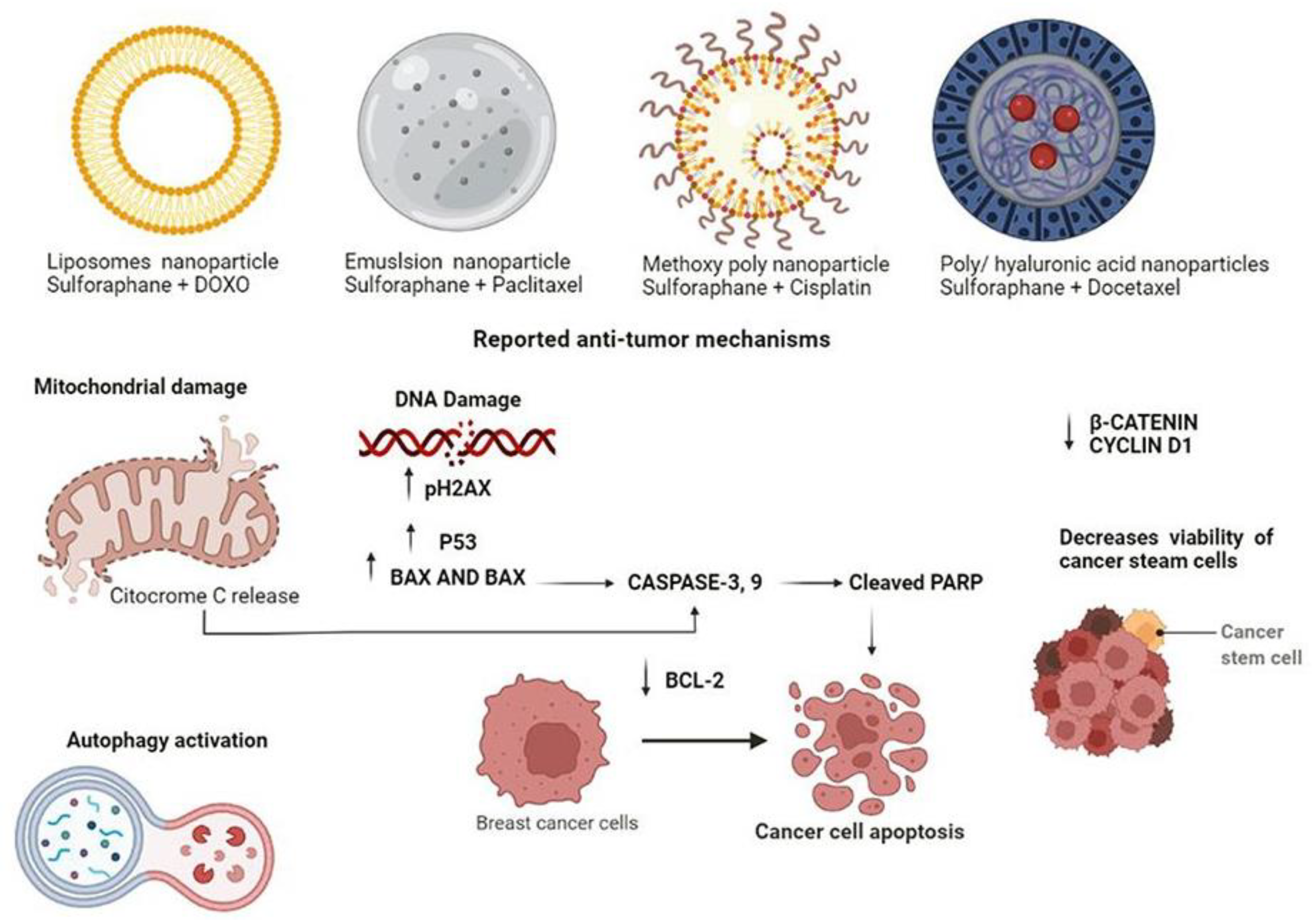
6. Anti-Tumour Effects of Sulforaphane Nanoparticles: Promising Chemotherapeutic Adjuvants in Breast Cancer

Nanotechnology has been actively researched and used in cancer therapy as nanoparticles potentially play an important role as a drug delivery method. Nanoparticle-based drug delivery provides distinct benefits over traditional drug delivery, including greater stability and biocompatibility, increased permeability and retention effect, and precision targeting. The implementation of nanoparticles for sulforaphane delivery systems might overcome its primary limitations, which are its poor solubility in water and oral bioavailability. Not surprisingly, new perspectives of sulforaphane administration through nano-composite-based therapies have promising results. For instance, the use of nanoparticles of selenium, tellurium, gold-coated Fe3O4, PEGylated Fe3O4, monomethoxypol, and based on Fe2+ and Fe3+ for the administration of sulforaphane in breast cancer, has recently been documented [66,67,73,74,75]. According to the findings of these studies, the anti-tumour effects of nanoparticle treatment are capable of decreasing the viability of tumour cells such as MDA-MB-231, MCF-7, and SKBR-3. Interestingly, most of these studies support the activation of apoptosis in tumour cells by promoting the upregulation of pro-apoptotic BAX and Bak and the downregulation of anti-apoptotic bcl-2 and bcl-xL. Furthermore, sulforaphane-coated selenium and tellurium nanoparticles appear to specifically target tumour cells, whereas normal cells, MCF-10A, have a better tolerance to treatment, as observed in the MTT assay [61,66,67]. Sulforaphane nanoparticle therapy in breast cancer has consistently anti-tumour effects and therefore its adjuvant administration in combination with other chemotherapeutic drugs through nanoparticles offers great potential.

Potential Effects of Sulforaphane Nanoparticles as Adjuvant

According to the findings described in breast cancer lines like MDA-MB-231 and MCF7, the combination of sulforaphane + DOX was entrapped in liposomes nanoparticles at 9.375 mg/g of DOX, while for sulforaphane, it was 487.5 mg/g. Cell viability was statistically lower after the application of the DOX/sulforaphane nanoparticle combination. In addition, cell viability is reduced by 50% when sulforaphane and DOX are combined and seem to be effective in terms of their ability to inhibit tumour cell growth (Figure 2). Additionally, sulforaphane + DOX nanoparticles induce described anti-tumour mechanisms such as ROS generation, mitochondrial damage, and autophagy [61]. Sulforaphane micro-emulsion nanoparticle is another technique employed in combination with PTX. When utilized at high doses (8.7 µM of sulforaphane and 500 nM of PTX), the sulforaphane emulsion promotes the solubilization of PTX and improves its activity; moreover, a substantial growth suppression was found in PTX-sulforaphane micro-emulsion treated MDA-MB-231 and MCF-7 cell lines (Figure 2). PTX is a highly potent chemotherapeutic drug against breast cancer cells, however, it contains excipients, which cause several of the adverse effects. Sulforaphane, on the other hand, decreases the content of excipients utilized in commercial PTX preparations [62]. On the other hand, it has been reported that the methoxy poly (ethylene glycol)-poly (lactide-co-glycolide) nanoparticle loaded with sulforaphane at 48.97 µM increases the chemosensitivity of cisplatin at 72.59 µM by enhancing platinum binding to DNA. Cisplatin induced moderate apoptosis in MCF-7 cells, with a 15.2% apoptotic rate. In contrast, the combination of sulforaphane + cisplatin nanoparticles increased the apoptotic rate to 39.5%. Furthermore, the sulforaphane + cisplatin nanoparticle increased the expression of p-H2AX, p53, cleaved PARP, and promoted the decrease of Bcl-2 expression, indicating that apoptosis is the primary mechanism of action (Figure 2). In vivo models showed a 74.1% reduction in tumour growth treated with sulforaphane + cisplatin nanoparticle [63]. Previous research has shown that poly (D, L-lactideco-glycolide)/hyaluronic acid nanoparticles loaded with 5 nM DTX and 350 µg sulforaphane have a novel effect on breast cancer stem cells. After 72 h of treatment, sulforaphane + DTX nanoparticle decreased the viability of MCF-7 MS cells with CD44 + CD24-epithelial-specific antigen phenotype. Both b-catenin and cyclin D1 levels were reduced, leading to cell growth inhibition (Figure 2).
A decrease in tumour growth from day 24 to day 36 was observed in the treated group, and in conjunction with these results, the nanoparticle showed anticancer activity in stem cells, with approximately 140 mammospheres per 2000 cells formed per 2000 cells compared to 210 mammospheres formed per 2000 cells in the untreated groups [64]. The research by Keshandehghan et al. describes that carbon nanoparticles loaded with sulforaphane, and metformin reduced markers associated with breast cancer stem cells such as WNT-1, -catenin, and CD44. Surprisingly, sulforaphane + metformin-induced apoptosis enhanced BAX levels. This might explain why MCF-7 and BT-474 cells are sensitive to sulforaphane + metformin nanoparticles at 48 and 42 nM, respectively, resulting in cell viability decrease. Apoptosis increased in 46% of the treated cells after 8 h of treatment [65]. Other investigation performed by Gu et al. described the potential anticarcinogenic sulforaphane effect in in vivo models. They prepared a nanocarrier with a higher capacity (33.64%) to load sulforaphane, their nanocarrier consisted of a mineralized hyaluronic acid-SS-tetradecyl, after 8 h in in vitro studies the system was capable of releasing most of the sulforaphane (>60%) at acid conditions. In vivo analyses showed the rapid accumulation and sulforaphane release in situ after 6 h, the localization increased after 24 h post injection. Furthermore, the sulforaphane concentration was higher at the excised tumour in comparison with other organs, demonstrating CD44 targeting specificity and a low toxicity system [76].

7. Conclusions

Sulforaphane is a relatively accessible and easy-to-acquire molecule, compared to other organic components that can be used to treat cancer. Since its discovery, it has been identified as a potent phytopharmaceutical capable of altering the resistance of carcinogenic cells. Although more studies are still needed to fully understand the molecular processes that this molecule triggers, the experimental evidence generated and compiled in this review demonstrates that in addition to being a compound that is consumed indirectly in the diet, it is a component with a high specificity for cancer cells. The experimental evidence generated and compiled in this review demonstrates that in addition to being a compound that is consumed indirectly in the diet, it is a component that presents a high specificity for cancer cells, thus proving to be a potential candidate for the treatment of different cancer types. Additionally, its efficiency when complexed with various types of nanoparticles improves the prospects for its use in the development of targeted and specialized therapies for each patient. Apart from this evidence, it has also been observed that the use of sulforaphane in conjunction with strategies currently implemented in the treatment of cancer enhances their therapeutic effects, also improving the tolerability of the side effects in these therapies. In this regard the clinical studies that exist regarding the use of sulforaphane in the treatment of breast cancer, have not yet obtained a nanoparticulate as a candidate for phase 3 studies and subsequent therapy. However, overall, the experimental studies demonstrate a high safety and bioavailability of the nanocomplexes studied by different authors, each with different perspectives and different chemical and physical characteristics that allow new perspectives for the use of these nanoparticles to be generated. These can be focused not only on the controlled release of sulforaphane but also in approaches that involve treatment in conjunction with physical methods. The research of new molecular targets and signalling pathways of sulforaphane will allow the development of its clinical applications for the prevention and treatment of breast cancer. In the evidence described in this review, we have emphasized the antioncogenetic sulforaphane role and its possible use in cancer treatment as adjuvant or complexed with different nanomaterials.

Author Contributions

M.Z.S.-L. and M.B.S.-C. conceived and designed the review; E.J.-A., J.P.-R. and M.B.S.-C. performed the literature review and were involved in the writing and revision of the manuscript; E.J.-A. and M.B.S.-C. prepared all the figures. All authors have read and agreed to the published version of the manuscript.

Funding

This research received no external funding.

Institutional Review Board Statement

Not applicable.

Informed Consent Statement

Not applicable.

Data Availability Statement

Not applicable.

Acknowledgments

We thank the Universidad Autónoma de San Luis Potosí and Universidad Autónoma de la Ciudad de México for their support.

Conflicts of Interest

The authors declare no conflict of interest.

References

  1. Reuter, S.; Gupta, S.C.; Chaturvedi, M.M.; Aggarwal, B.B. Oxidative Stress, Inflammation, and Cancer: How Are They Linked? Free Radic. Biol. Med. 2010, 49, 1603–1616. [Google Scholar] [CrossRef] [PubMed] [Green Version]
  2. Malumbres, M.; Carnero, A. Cell Cycle Deregulation: A Common Motif in Cancer. Prog. Cell Cycle Res. 2003, 5, 5–18. [Google Scholar] [PubMed]
  3. Klaunig, J.E. Oxidative Stress and Cancer. Curr. Pharm. Des. 2019, 24, 4771–4778. [Google Scholar] [CrossRef] [PubMed]
  4. Resnik, D.B. Responsibility for Health: Personal, Social, and Environmental. J. Med. Ethics 2007, 33, 444–445. [Google Scholar] [CrossRef]
  5. Chen, Y.; Michalak, M.; Agellon, L.B. Importance of Nutrients and Nutrient Metabolism on Human Health. Yale J. Biol. Med. 2018, 91, 95–103. [Google Scholar]
  6. Wiseman, M.J. Nutrition and Cancer: Prevention and Survival. Br. J. Nutr. 2019, 122, 481–487. [Google Scholar] [CrossRef] [Green Version]
  7. Baena Ruiz, R.; Salinas Hernández, P. Diet and Cancer: Risk Factors and Epidemiological Evidence. Maturitas 2014, 77, 202–208. [Google Scholar] [CrossRef]
  8. Kensler, T.W. Chemoprevention by Inducers of Carcinogen Detoxication Enzymes. Environ. Health Perspect. 1997, 105, 965–970. [Google Scholar] [CrossRef]
  9. Moloney, J.N.; Cotter, T.G. ROS Signalling in the Biology of Cancer. Semin. Cell Dev. Biol. 2018, 80, 50–64. [Google Scholar] [CrossRef]
  10. Ramsey, A.T.; Baker, T.B.; Pham, G.; Stoneking, F.; Smock, N.; Colditz, G.A.; James, A.S.; Liu, J.; Bierut, L.J.; Chen, L.-S. Low Burden Strategies Are Needed to Reduce Smoking in Rural Healthcare Settings: A Lesson from Cancer Clinics. Int. J. Environ. Res. Public Health 2020, 17, 1728. [Google Scholar] [CrossRef] [Green Version]
  11. Surman, M.; Janik, M.E. Stress and Its Molecular Consequences in Cancer Progression. Postepy Hig. Med. Dosw. Online 2017, 71, 485–499. [Google Scholar] [CrossRef] [PubMed]
  12. Fiolet, T.; Srour, B.; Sellem, L.; Kesse-Guyot, E.; Allès, B.; Méjean, C.; Deschasaux, M.; Fassier, P.; Latino-Martel, P.; Beslay, M.; et al. Consumption of Ultra-Processed Foods and Cancer Risk: Results from NutriNet-Santé Prospective Cohort. BMJ 2018, 360, k322. [Google Scholar] [CrossRef] [PubMed] [Green Version]
  13. Bober, P.; Alexovič, M.; Tomková, Z.; Kilík, R.; Sabo, J. RHOA and MDia1 Promotes Apoptosis of Breast Cancer Cells via a High Dose of Doxorubicin Treatment. Open Life Sci. 2019, 14, 619–627. [Google Scholar] [CrossRef] [PubMed]
  14. Tantamango-Bartley, Y.; Jaceldo-Siegl, K.; Fan, J.; Fraser, G. Vegetarian Diets and the Incidence of Cancer in a Low-Risk Population. Cancer Epidemiol. Biomark. Prev. 2013, 22, 286–294. [Google Scholar] [CrossRef] [Green Version]
  15. Samanta, S.K.; Choudhury, P.; Sarma, P.P.; Gogoi, B.; Gogoi, N.; Devi, R. Dietary Phytochemicals/Nutrients as Promising Protector of Breast Cancer Development: A Comprehensive Analysis. Pharmacol. Rep. 2022, 74, 583–601. [Google Scholar] [CrossRef]
  16. Abdull Razis, A.F.; Noor, N.M. Cruciferous Vegetables: Dietary Phytochemicals for Cancer Prevention. Asian Pac. J. Cancer Prev. 2013, 14, 1565–1570. [Google Scholar] [CrossRef] [Green Version]
  17. Higdon, J.; Delage, B.; Williams, D.; Dashwood, R. Cruciferous Vegetables and Human Cancer Risk: Epidemiologic Evidence and Mechanistic Basis. Pharmacol. Res. 2007, 55, 224–236. [Google Scholar] [CrossRef] [PubMed] [Green Version]
  18. Gray, A.R. Taxonomy and Evolution of Broccoli (Brassica oleracea Var. Italica). Econ. Bot. 1982, 36, 397–410. [Google Scholar] [CrossRef]
  19. Pickering, R.J.; Rosado, C.J.; Sharma, A.; Buksh, S.; Tate, M.; de Haan, J.B. Recent Novel Approaches to Limit Oxidative Stress and Inflammation in Diabetic Complications. Clin. Transl. Immunol. 2018, 7, e1016. [Google Scholar] [CrossRef]
  20. Fischer, R.; Maier, O. Interrelation of Oxidative Stress and Inflammation in Neurodegenerative Disease: Role of TNF. Oxid. Med. Cell. Longev. 2015, 2015, 1–18. [Google Scholar] [CrossRef] [Green Version]
  21. Lynch, R.; Diggins, E.L.; Connors, S.L.; Zimmerman, A.W.; Singh, K.; Liu, H.; Talalay, P.; Fahey, J.W. Sulforaphane from Broccoli Reduces Symptoms of Autism: A Follow-up Case Series from a Randomized Double-Blind Study. Glob. Adv. Health Med. 2017, 6. [Google Scholar] [CrossRef] [PubMed]
  22. Sivapalan, T.; Melchini, A.; Saha, S.; Needs, P.W.; Traka, M.H.; Tapp, H.; Dainty, J.R.; Mithen, R.F. Bioavailability of Glucoraphanin and Sulforaphane from High-Glucoraphanin Broccoli. Mol. Nutr. Food Res. 2018, 62, 1700911. [Google Scholar] [CrossRef] [PubMed] [Green Version]
  23. Lin, H.-H.; Lin, K.-H.; Chen, S.-C.; Shen, Y.-H.; Lo, H.-F. Proteomic Analysis of Broccoli (Brassica oleracea) under High Temperature and Waterlogging Stresses. Bot. Stud. 2015, 56, 18. [Google Scholar] [CrossRef] [Green Version]
  24. Natarajan, S.; Thamilarasan, S.; Park, J.-I.; Chung, M.-Y.; Nou, I.-S. Molecular Modeling of Myrosinase from Brassica Oleracea: A Structural Investigation of Sinigrin Interaction. Genes 2015, 6, 1315–1329. [Google Scholar] [CrossRef] [PubMed] [Green Version]
  25. Ludikhuyze, L.; Rodrigo, L.; Hendrickx, M. The Activity of Myrosinase from Broccoli (Brassica oleracea L. Cv. Italica): Influence of Intrinsic and Extrinsic Factors. J. Food Prot. 2000, 63, 400–403. [Google Scholar] [CrossRef] [PubMed]
  26. Kensler, T.W.; Egner, P.A.; Agyeman, A.S.; Visvanathan, K.; Groopman, J.D.; Chen, J.-G.; Chen, T.-Y.; Fahey, J.W.; Talalay, P. Keap1–Nrf2 Signaling: A Target for Cancer Prevention by Sulforaphane. In Natural Products in Cancer Prevention and Therapy; Pezzuto, J.M., Suh, N., Eds.; Topics in Current Chemistry; Springer: Berlin/Heidelberg, Germany, 2012; Volume 329, pp. 163–177. ISBN 978-3-642-34574-6. [Google Scholar]
  27. Cottaz, S.; Rollin, P.; Driguez, H. ChemInform Abstract: Synthesis of 2-Deoxy-2-Fluoro-Glucotropaeolin, a Thioglucosidase Inhibitor. ChemInform 2010, 28. [Google Scholar] [CrossRef]
  28. de Oliveira, M.R. Sulforaphane and Its Modulation of Brain Redox Status: The Mitochondria as a Target. In Oxidative Stress and Dietary Antioxidants in Neurological Diseases; Academic Press: Cambridge, MA, USA, 2020; pp. 377–391. [Google Scholar] [CrossRef]
  29. Holst, B.; Williamson, G. A Critical Review of the Bioavailability of Glucosinolates and Related Compounds. Nat. Prod. Rep. 2004, 21, 425. [Google Scholar] [CrossRef]
  30. Faulkner, K. Selective Increase of the Potential Anticarcinogen 4- Methylsulphinylbutyl Glucosinolate in Broccoli. Carcinogenesis 1998, 19, 605–609. [Google Scholar] [CrossRef] [Green Version]
  31. Preobrazhenskaya, M.N.; Korolev, A.M. Indole Derivatives in Vegetables of the Cruciferae Family. Russ. J. Bioorganic Chem. 2000, 26, 85–97. [Google Scholar] [CrossRef]
  32. Kong, X.Y.; Kissen, R.; Bones, A.M. Characterization of Recombinant Nitrile-Specifier Proteins (NSPs) of Arabidopsis Thaliana: Dependency on Fe(II) Ions and the Effect of Glucosinolate Substrate and Reaction Conditions. Phytochemistry 2012, 84, 7–17. [Google Scholar] [CrossRef]
  33. Foo, H.L.; Grønning, L.M.; Goodenough, L.; Bones, A.M.; Danielsen, B.-E.; Whiting, D.A.; Rossiter, J.T. Purification and Characterisation of Epithiospecifier Protein from Brassica Napus: Enzymic Intramolecular Sulphur Addition within Alkenyl Thiohydroximates Derived from Alkenyl Glucosinolate Hydrolysis. FEBS Lett. 2000, 468, 243–246. [Google Scholar] [CrossRef] [Green Version]
  34. Pradhan, N.; Kar, S.; Parbin, S.; Sengupta, D.; Deb, M.; Das, L.; Patra, S.K. Epigenetic Dietary Interventions for Prevention of Cancer. In Epigenetics of Cancer Prevention; Elsevier: Amsterdam, The Netherlands, 2019; pp. 23–48. ISBN 978-0-12-812494-9. [Google Scholar]
  35. Bauman, J.E.; Zang, Y.; Sen, M.; Li, C.; Wang, L.; Egner, P.A.; Fahey, J.W.; Normolle, D.P.; Grandis, J.R.; Kensler, T.W.; et al. Prevention of Carcinogen-Induced Oral Cancer by Sulforaphane. Cancer Prev. Res. 2016, 9, 547–557. [Google Scholar] [CrossRef] [PubMed] [Green Version]
  36. Rai, R.; Gong Essel, K.; Mangiaracina Benbrook, D.; Garland, J.; Daniel Zhao, Y.; Chandra, V. Preclinical Efficacy and Involvement of AKT, MTOR, and ERK Kinases in the Mechanism of Sulforaphane against Endometrial Cancer. Cancers 2020, 12, 1273. [Google Scholar] [CrossRef] [PubMed]
  37. Hudecova, S.; Markova, J.; Simko, V.; Csaderova, L.; Stracina, T.; Sirova, M.; Fojtu, M.; Svastova, E.; Gronesova, P.; Pastorek, M.; et al. Sulforaphane-Induced Apoptosis Involves the Type 1 IP3 Receptor. Oncotarget 2016, 7, 61403–61418. [Google Scholar] [CrossRef] [Green Version]
  38. Yasuda, S.; Horinaka, M.; Sakai, T. Sulforaphane Enhances Apoptosis Induced by Lactobacillus Pentosus Strain S-PT84 via the TNFα Pathway in Human Colon Cancer Cells. Oncol. Lett. 2019, 18, 4253–4261. [Google Scholar] [CrossRef] [Green Version]
  39. Myzak, M.C.; Hardin, K.; Wang, R.; Dashwood, R.H.; Ho, E. Sulforaphane Inhibits Histone Deacetylase Activity in BPH-1, LnCaP and PC-3 Prostate Epithelial Cells. Carcinogenesis 2006, 27, 811–819. [Google Scholar] [CrossRef]
  40. Mokhtari, R.B.; Qorri, B.; Baluch, N.; Sparaneo, A.; Fabrizio, F.P.; Muscarella, L.A.; Tyker, A.; Kumar, S.; Cheng, H.-L.M.; Szewczuk, M.R.; et al. Next-Generation Multimodality of Nutrigenomic Cancer Therapy: Sulforaphane in Combination with Acetazolamide Actively Target Bronchial Carcinoid Cancer in Disabling the PI3K/Akt/MTOR Survival Pathway and Inducing Apoptosis. Oncotarget 2021, 12, 1470–1489. [Google Scholar] [CrossRef]
  41. Hahm, E.-R.; Singh, K.B.; Kim, S.-H.; Powolny, A.A.; Singh, S.V. The Role of Lysosome-Associated Membrane Protein 2 in Prostate Cancer Chemopreventive Mechanisms of Sulforaphane. Cancer Prev. Res. 2020, 13, 661–672. [Google Scholar] [CrossRef]
  42. Sundaram, M.K.; Khan, M.A.; Alalami, U.; Somvanshi, P.; Bhardwaj, T.; Pramodh, S.; Raina, R.; Shekfeh, Z.; Haque, S.; Hussain, A. Phytochemicals Induce Apoptosis by Modulation of Nitric Oxide Signaling Pathway in Cervical Cancer Cells. Eur. Rev. Med. Pharmacol. Sci. 2020, 24, 11827–11844. [Google Scholar] [CrossRef]
  43. Myzak, M.C.; Dashwood, R.H. Chemoprotection by Sulforaphane: Keep One Eye beyond Keap1. Cancer Lett. 2006, 233, 208–218. [Google Scholar] [CrossRef] [Green Version]
  44. Hao, Q.; Wang, M.; Sun, N.; Zhu, C.; Lin, Y.; Li, C.; Liu, F.; Zhu, W. Sulforaphane Suppresses Carcinogenesis of Colorectal Cancer through the ERK/Nrf2-UDP Glucuronosyltransferase 1A Metabolic Axis Activation. Oncol. Rep. 2020, 43, 1067–1080. [Google Scholar] [CrossRef] [PubMed]
  45. Singh, S.V.; Herman-Antosiewicz, A.; Singh, A.V.; Lew, K.L.; Srivastava, S.K.; Kamath, R.; Brown, K.D.; Zhang, L.; Baskaran, R. Sulforaphane-Induced G2/M Phase Cell Cycle Arrest Involves Checkpoint Kinase 2-Mediated Phosphorylation of Cell Division Cycle 25C. J. Biol. Chem. 2004, 279, 25813–25822. [Google Scholar] [CrossRef] [PubMed] [Green Version]
  46. Shen, G.; Xu, C.; Chen, C.; Hebbar, V.; Kong, A.-N.T. P53-Independent G1 Cell Cycle Arrest of Human Colon Carcinoma Cells HT-29 by Sulforaphane Is Associated with Induction of P21CIP1 and Inhibition of Expression of Cyclin D1. Cancer Chemother. Pharmacol. 2006, 57, 317–327. [Google Scholar] [CrossRef] [PubMed]
  47. Traka, M.; Gasper, A.V.; Smith, J.A.; Hawkey, C.J.; Bao, Y.; Mithen, R.F. Transcriptome Analysis of Human Colon Caco-2 Cells Exposed to Sulforaphane. J. Nutr. 2005, 135, 1865–1872. [Google Scholar] [CrossRef] [PubMed] [Green Version]
  48. Parnaud, G.; Li, P.; Cassar, G.; Rouimi, P.; Tulliez, J.; Combaret, L.; Gamet-Payrastre, L. Mechanism of Sulforaphane-Induced Cell Cycle Arrest and Apoptosis in Human Colon Cancer Cells. Nutr. Cancer 2004, 48, 198–206. [Google Scholar] [CrossRef]
  49. Kung, C.-P.; Budina, A.; Balaburski, G.; Bergenstock, M.K.; Murphy, M. Autophagy in Tumor Suppression and Cancer Therapy. Crit. Rev. Eukaryot. Gene Expr. 2011, 21, 71–100. [Google Scholar] [CrossRef] [Green Version]
  50. Lu, Z.; Ren, Y.; Yang, L.; Jia, A.; Hu, Y.; Zhao, Y.; Zhao, W.; Yu, B.; Zhao, W.; Zhang, J.; et al. Inhibiting Autophagy Enhances Sulforaphane-Induced Apoptosis via Targeting NRF2 in Esophageal Squamous Cell Carcinoma. Acta Pharm. Sin. B 2021, 11, 1246–1260. [Google Scholar] [CrossRef]
  51. Alzahrani, A.S. PI3K/Akt/MTOR Inhibitors in Cancer: At the Bench and Bedside. Semin. Cancer Biol. 2019, 59, 125–132. [Google Scholar] [CrossRef]
  52. Liu, P.; Atkinson, S.J.; Akbareian, S.E.; Zhou, Z.; Munsterberg, A.; Robinson, S.D.; Bao, Y. Sulforaphane Exerts Anti-Angiogenesis Effects against Hepatocellular Carcinoma through Inhibition of STAT3/HIF-1α/VEGF Signalling. Sci. Rep. 2017, 7, 12651. [Google Scholar] [CrossRef] [Green Version]
  53. Davis, R.; Singh, K.P.; Kurzrock, R.; Shankar, S. Sulforaphane Inhibits Angiogenesis through Activation of FOXO Transcription Factors. Oncol. Rep. 2009, 22, 1473–1478. [Google Scholar] [CrossRef] [Green Version]
  54. Yao, H.; Wang, H.; Zhang, Z.; Jiang, B.; Luo, J.; Shi, X. Sulforaphane Inhibited Expression of Hypoxia-inducible Factor-1α in Human Tongue Squamous Cancer Cells and Prostate Cancer Cells. Int. J. Cancer 2008, 123, 1255–1261. [Google Scholar] [CrossRef] [PubMed]
  55. Castro, N.P.; Rangel, M.C.; Merchant, A.S.; MacKinnon, G.; Cuttitta, F.; Salomon, D.S.; Kim, Y.S. Sulforaphane Suppresses the Growth of Triple-Negative Breast Cancer Stem-like Cells In Vitro and In Vivo. Cancer Prev. Res. 2019, 12, 147–158. [Google Scholar] [CrossRef] [PubMed] [Green Version]
  56. Royston, K.J.; Paul, B.; Nozell, S.; Rajbhandari, R.; Tollefsbol, T.O. Withaferin A and Sulforaphane Regulate Breast Cancer Cell Cycle Progression through Epigenetic Mechanisms. Exp. Cell Res. 2018, 368, 67–74. [Google Scholar] [CrossRef]
  57. Lewinska, A.; Adamczyk-Grochala, J.; Deregowska, A.; Wnuk, M. Sulforaphane-Induced Cell Cycle Arrest and Senescence Are Accompanied by DNA Hypomethylation and Changes in MicroRNA Profile in Breast Cancer Cells. Theranostics 2017, 7, 3461–3477. [Google Scholar] [CrossRef] [PubMed]
  58. Pore, S.K.; Hahm, E.-R.; Kim, S.-H.; Singh, K.B.; Nyiranshuti, L.; Latoche, J.D.; Anderson, C.J.; Adamik, J.; Galson, D.L.; Weiss, K.R.; et al. A Novel Sulforaphane-Regulated Gene Network in Suppression of Breast Cancer–Induced Osteolytic Bone Resorption. Mol. Cancer Ther. 2020, 19, 420–431. [Google Scholar] [CrossRef] [PubMed]
  59. Zhang, Y.; Lu, Q.; Li, N.; Xu, M.; Miyamoto, T.; Liu, J. Sulforaphane Suppresses Metastasis of Triple-Negative Breast Cancer Cells by Targeting the RAF/MEK/ERK Pathway. NPJ Breast Cancer 2022, 8, 40. [Google Scholar] [CrossRef] [PubMed]
  60. Yasunaga, A.; Ono, M.; Takeshima, M.; Nakano, S. Sulforaphane Suppresses the Growth of EGFR-overexpressing MDA-MB-468 Triple-negative Breast Cancer Cells in Vivo and in Vitro. Int. J. Funct. Nutr. 2022, 3, 3. [Google Scholar] [CrossRef]
  61. Mielczarek, L.; Krug, P.; Mazur, M.; Milczarek, M.; Chilmonczyk, Z.; Wiktorska, K. In the Triple-Negative Breast Cancer MDA-MB-231 Cell Line, Sulforaphane Enhances the Intracellular Accumulation and Anticancer Action of Doxorubicin Encapsulated in Liposomes. Int. J. Pharm. 2019, 558, 311–318. [Google Scholar] [CrossRef]
  62. Kamal, M.M.; Nazzal, S. Novel Sulforaphane-Enabled Self-Microemulsifying Delivery Systems (SFN-SMEDDS) of Taxanes: Formulation Development and in Vitro Cytotoxicity against Breast Cancer Cells. Int. J. Pharm. 2018, 536, 187–198. [Google Scholar] [CrossRef]
  63. Xu, Y.; Han, X.; Li, Y.; Min, H.; Zhao, X.; Zhang, Y.; Qi, Y.; Shi, J.; Qi, S.; Bao, Y.; et al. Sulforaphane Mediates Glutathione Depletion via Polymeric Nanoparticles to Restore Cisplatin Chemosensitivity. ACS Nano 2019, 13, 13445–13455. [Google Scholar] [CrossRef]
  64. Huang, J.; Tao, C.; Yu, Y.; Yu, F.; Zhang, H.; Gao, J.; Wang, D.; Chen, Y.; Gao, J.; Zhang, G.; et al. Simultaneous Targeting of Differentiated Breast Cancer Cells and Breast Cancer Stem Cells by Combination of Docetaxel- and Sulforaphane-Loaded Self-Assembled Poly(D, L-Lactide-Co-Glycolide)/Hyaluronic Acid Block Copolymer-Based Nanoparticles. J. Biomed. Nanotechnol. 2016, 12, 1463–1477. [Google Scholar] [CrossRef] [PubMed]
  65. Keshandehghan, A.; Nikkhah, S.; Tahermansouri, H.; Heidari-Keshel, S.; Gardaneh, M. Co-Treatment with Sulforaphane and Nano-Metformin Molecules Accelerates Apoptosis in HER2+ Breast Cancer Cells by Inhibiting Key Molecules. Nutr. Cancer 2020, 72, 835–848. [Google Scholar] [CrossRef] [PubMed]
  66. Krug, P.; Mielczarek, L.; Wiktorska, K.; Kaczyńska, K.; Wojciechowski, P.; Andrzejewski, K.; Ofiara, K.; Szterk, A.; Mazur, M. Sulforaphane-Conjugated Selenium Nanoparticles: Towards a Synergistic Anticancer Effect. Nanotechnology 2019, 30, 065101. [Google Scholar] [CrossRef] [PubMed]
  67. Krug, P.; Wiktorska, K.; Kaczyńska, K.; Ofiara, K.; Szterk, A.; Kuśmierz, B.; Mazur, M. Sulforaphane-assisted preparation of tellurium flower-like nanoparticles. Nanotechnology 2019, 31, 055603. [Google Scholar] [CrossRef] [PubMed]
  68. Milczarek, M.; Wiktorska, K.; Mielczarek, L.; Koronkiewicz, M.; Dąbrowska, A.; Lubelska, K.; Matosiuk, D.; Chilmonczyk, Z. Autophagic Cell Death and Premature Senescence: New Mechanism of 5-Fluorouracil and Sulforaphane Synergistic Anti-cancer Effect in MDA-MB-231 Triple Negative Breast Cancer Cell Line. Food Chem. Toxicol. 2018, 111, 1–8. [Google Scholar] [CrossRef]
  69. Bose, C.; Awasthi, S.; Sharma, R.; Beneš, H.; Hauer-Jensen, M.; Boerma, M.; Singh, S.P. Sulforaphane Potentiates Anticancer Effects of Doxorubicin and Attenuates Its Cardiotoxicity in a Breast Cancer Model. PLoS ONE 2018, 13, e0193918. [Google Scholar] [CrossRef]
  70. Rong, Y.; Huang, L.; Yi, K.; Chen, H.; Liu, S.; Zhang, W.; Yuan, C.; Song, X.; Wang, F. Co-Administration of Sulforaphane and Doxorubicin Attenuates Breast Cancer Growth by Preventing the Accumulation of Myeloid-Derived Suppressor Cells. Cancer Lett. 2020, 493, 189–196. [Google Scholar] [CrossRef]
  71. Burnett, J.P.; Lim, G.; Li, Y.; Shah, R.B.; Lim, R.; Paholak, H.J.; McDermott, S.P.; Sun, L.; Tsume, Y.; Bai, S.; et al. Sulforaphane Enhances the Anticancer Activity of Taxanes against Triple Negative Breast Cancer by Killing Cancer Stem Cells. Cancer Lett. 2017, 394, 52–64. [Google Scholar] [CrossRef]
  72. Kim, S.-H.; Park, H.-J.; Moon, D.-O. Sulforaphane Sensitizes Human Breast Cancer Cells to Paclitaxel-Induced Apoptosis by Downregulating the NF-ΚB Signaling Pathway. Oncol. Lett. 2017, 13, 4427–4432. [Google Scholar] [CrossRef] [Green Version]
  73. Danafar, H.; Sharafi, A.; Kheiri, S.; Kheiri Manjili, H. Co -Delivery of Sulforaphane and Curcumin with PEGylated Iron Oxide-Gold Core Shell Nanoparticles for Delivery to Breast Cancer Cell Line. Iran. J. Pharm. Res. IJPR 2018, 17, 480–494. [Google Scholar]
  74. Danafar, H.; Sharafi, A.; Kheiri Manjili, H.; Andalib, S. Sulforaphane Delivery Using MPEG–PCL Co-Polymer Nanoparticles to Breast Cancer Cells. Pharm. Dev. Technol. 2017, 22, 642–651. [Google Scholar] [CrossRef] [PubMed]
  75. Kheiri Manjili, H.; Ma’mani, L.; Tavaddod, S.; Mashhadikhan, M.; Shafiee, A.; Naderi-Manesh, H. D, L-Sulforaphane Loaded Fe3O4@ Gold Core Shell Nanoparticles: A Potential Sulforaphane Delivery System. PLoS ONE 2016, 11, e0151344. [Google Scholar] [CrossRef] [PubMed]
  76. Gu, H.; Ren, F.; Mao, X.; Du, M. Mineralized and GSH-Responsive Hyaluronic Acid Based Nano-Carriers for Potentiating Repressive Effects of Sulforaphane on Breast Cancer Stem Cells-like Properties. Carbohydr. Polym. 2021, 269, 118294. [Google Scholar] [CrossRef] [PubMed]
Figure 1. Molecular pathways related to sulforaphane treatment. Molecular targets of sulforaphane could improve the response of actual cancer treatments and triggered autophagy, apoptosis, DNA damage, and/or cell cycle arrest.
Medicina 58 01377 g001
Figure 2. Reported anti-tumour mechanisms of sulforaphane as adjuvant. Mechanisms reported for liposome nanoparticles microemulsion, methoxy poly (ethylene glycol)-poly (lactide-co-glycolide) and poly (D, L-lactideco-glycolide)/hyaluronic acid nanoparticles loaded with sulforaphane and chemotherapeutic agents such as DOX, PTX, cisplatin, and DTX. Among the biological mechanisms of these nanoparticles are the activation of apoptosis through the mitochondrial pathway, autophagy, and the decrease in the viability of cancer steam cells.
Medicina 58 01377 g002
Table 1. Precursors and products of sulforaphane.
PrecursorMolecule ProductsConditions or Necessary EnzymeReference
GlucoraphaninThiohydroxamate-O-sulfonate and glucoseMyrosinaseKensler, et al. [26]
Cottaz, et al. [27]
de Oliveira [28]
Stable Isothiocyanates
Thiohydroxamate-O-sulfonateSulforaphanepH 6–7, microbial thioglucosidaseKensler, et al. [26]
Holst [29]
Faulkner [30]
Erucin
Iberin
Unstable isothiocyanates
β-hydroxylated IsothiocyanateGoitrinpH 6–7 and spontaneous cyclisationLudikhuyze, et al. [25]
Indolylmethyl-isothiocyanate3,3-DiindolylmethaneTrimeres, tetramers, etc. of Indole-3-carbinolProbrazhenskaya [31]
AscorbigenAscorbic acid presenceFaulkner [30]
Preobrazhenskaya [31]
Nitrile aliphatic and aromatic, indole and β-OH-nitriles
Thiohydroxamate-O-sulfonateCrambenepH 3–7 and the presence of nitrile-forming factorKong, et al. [32]
Others
Thiohydroxamate-O-sulfonateEpithioalkylnitrilePresence of double bound at Radical, epithiospecifer protein and Fe2+Cottaz, et al. [27]
Kong, et al. [32]
Foo, et al. [33]
ThiocyanateNot clearPradhan [34]
Table 2. Sulforaphane effect in breast cancer.
SubjectsSulforaphane DosageAnticancer EffectGenes TargetsReference
MDA-MB 231.40 µmApoptosis and necrosisZEB1, Claudin, and FibronectinMokhtari et al. [40]
BalbC/nude mice50 mg/kgApoptosis and decrease in tumour volume.ALDH1A1, NANOG, GDF3 and FOXD3Castro et al. [55]
MC7-cells and MDA-MB-2315.0 μMCell cycle arrestCCND1 and CDK4Royston et al. [56]
MCF-7 and MDA-MB-2314.05 M, 19.35 M, and 16.64 MCell cycle arrestcell cycle arrestLewinska et al. [57]
MDA-MB-231, SK-BR-3, and MCF-720 ng/mLCell cycle, suppression of osteolytic bone resorptionRUNX2 and NF-κB1Pore et al. [58]
MDA-MB-231, SK-BR-3, and MCF-77.5–30 µMMigration and invasion of breast cancer cellsRAF/MEK/ERK pathwayZhang et al. [59]
MDA-MB-4681.8 µMCell cycle arrestEGFR, BCL2, BAX and Akt/mTOR pathwayYasunaga et al. [60]
Nanoparticles in sulforaphane treatment
SubjectsSulforaphane dosageAnticancer EffectGenes TargetsNanoparticlesReferences
MDA-MB-231 and MCF7487.5 mg/g + 9.375 mg/g of DOXInhibit tumour cell growth, ROS generation, mitochondrial damage and autophagyDid not reportLiposome nanoparticlesMielczarek et al. [61]
MDA-MB-231 and MCF-78.7 µM and 500 nM of paclitaxel (PTX)Growth suppressionDid not reportMicro-emulsion nanoparticleKamal et al. [62]
MCF-748.97 µM and cisplatin at 72.59 µMInduction of apoptosis through DNA damage.p-H2AX, p53, PARP and Bcl-2Methoxy poly (ethylene glycol)-poly nanoparticleXu et al. [63]
Breast cancer CD44+ CD24-350 µg and docetaxel (DTX) 5 nMCell growth inhibitionB-catenin and Cyclin D1Poly (D, L-lactideco-glycolide)/hyaluronic acid nanoparticlesHuang et al. [64]
MCF-7 and BT-47448 nM and metformin 42 nMCell viability decrease and apoptosisWNT-1, B-catenin, and CD44.Carbon nanoparticlesKeshandehghan et al. [65]
Adult male Wistar rats10 mg/kgCell viability decrementNrf2 pathwaySelenium nanoparticleKrug et al. [66]
Adult male Wistar rats10 mg/kgInduction of ROS productionDid not reportflower-like tellurium nanoparticlesKrug et al. [67]
Publisher’s Note: MDPI stays neutral with regard to jurisdictional claims in published maps and institutional affiliations.

