

Diagnostic Mock-Up as a Surgical Reduction Guide for Crown Lengthening: Technique Description and Case Report

 ,
, 
Abstract
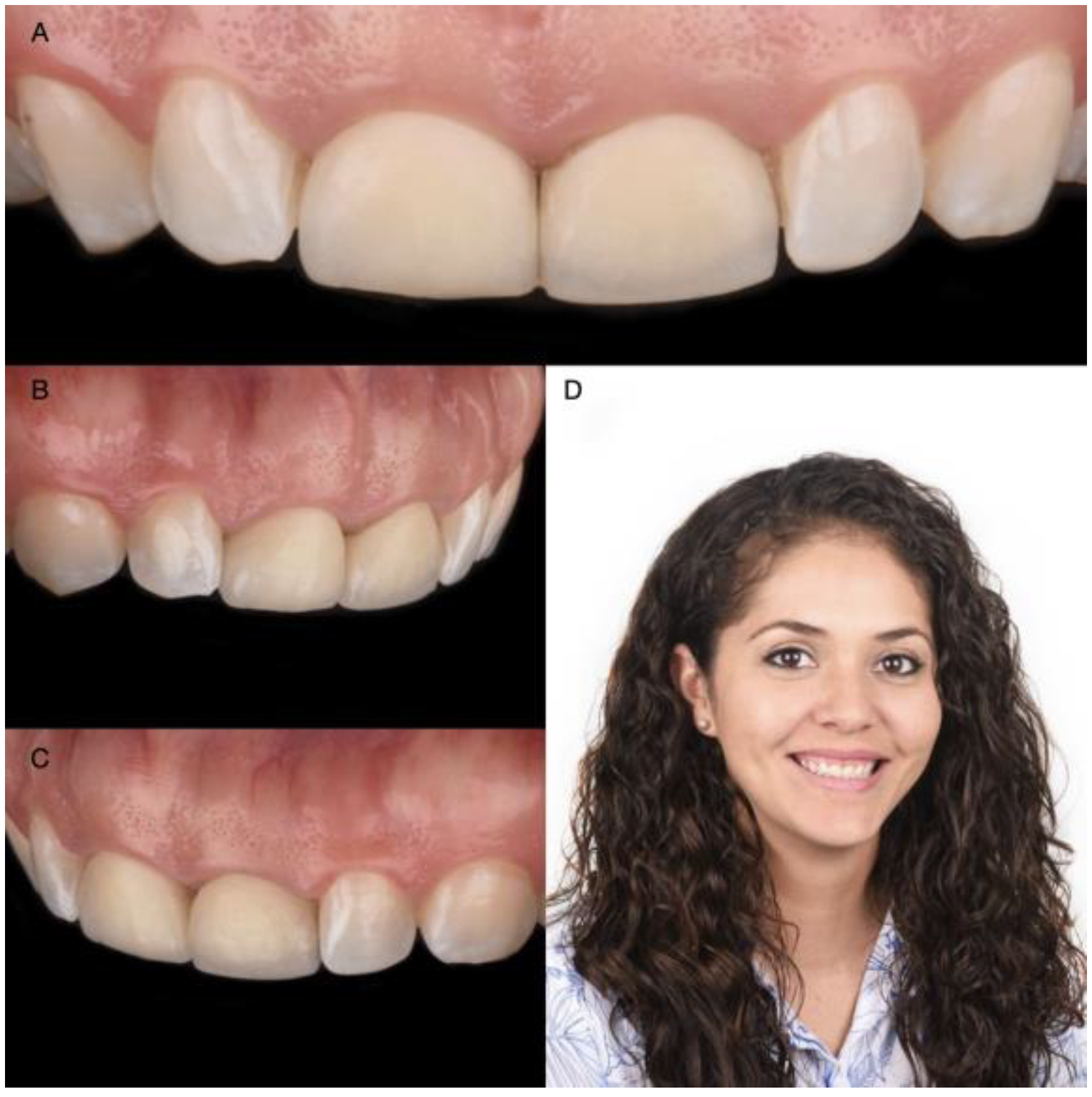
:1. Introduction
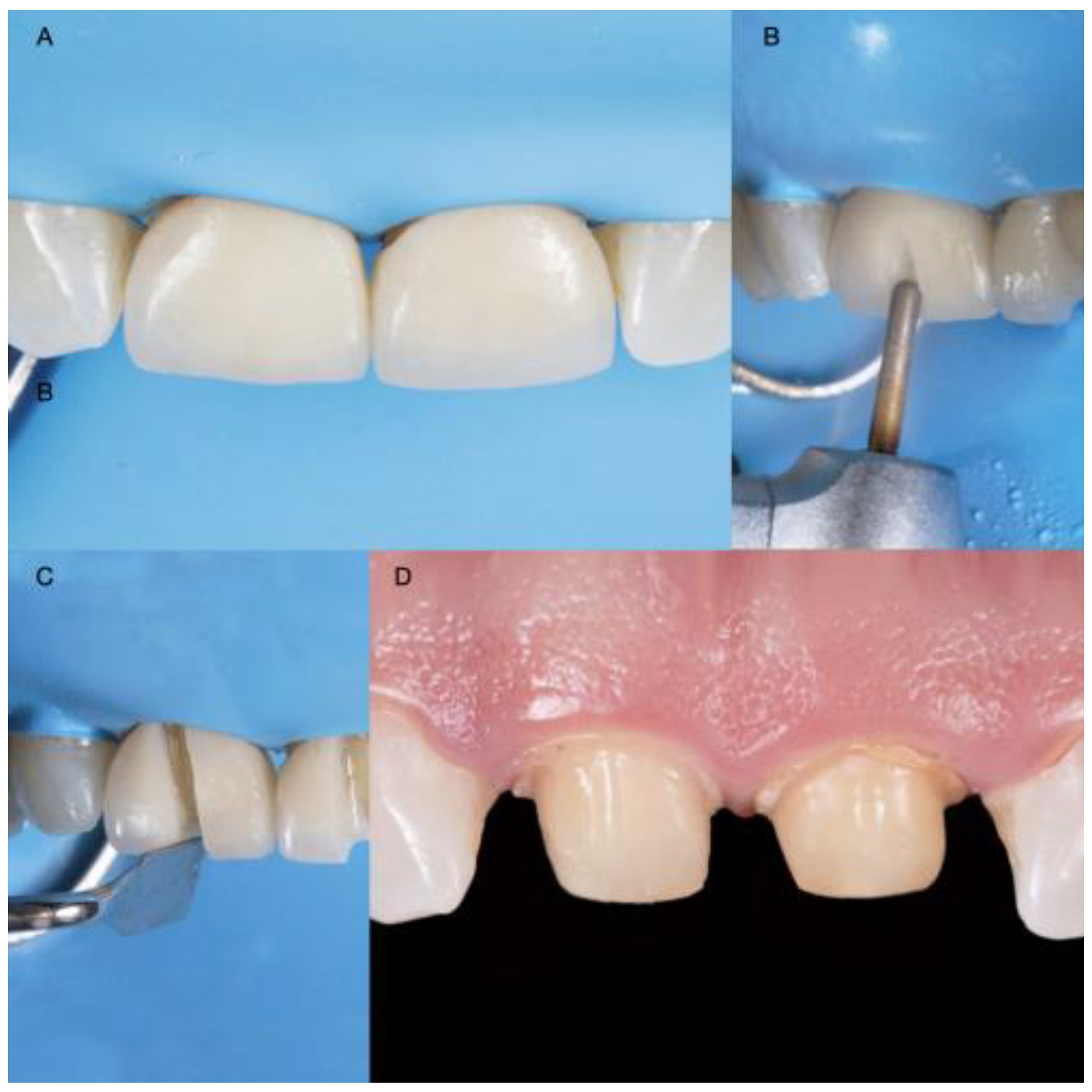
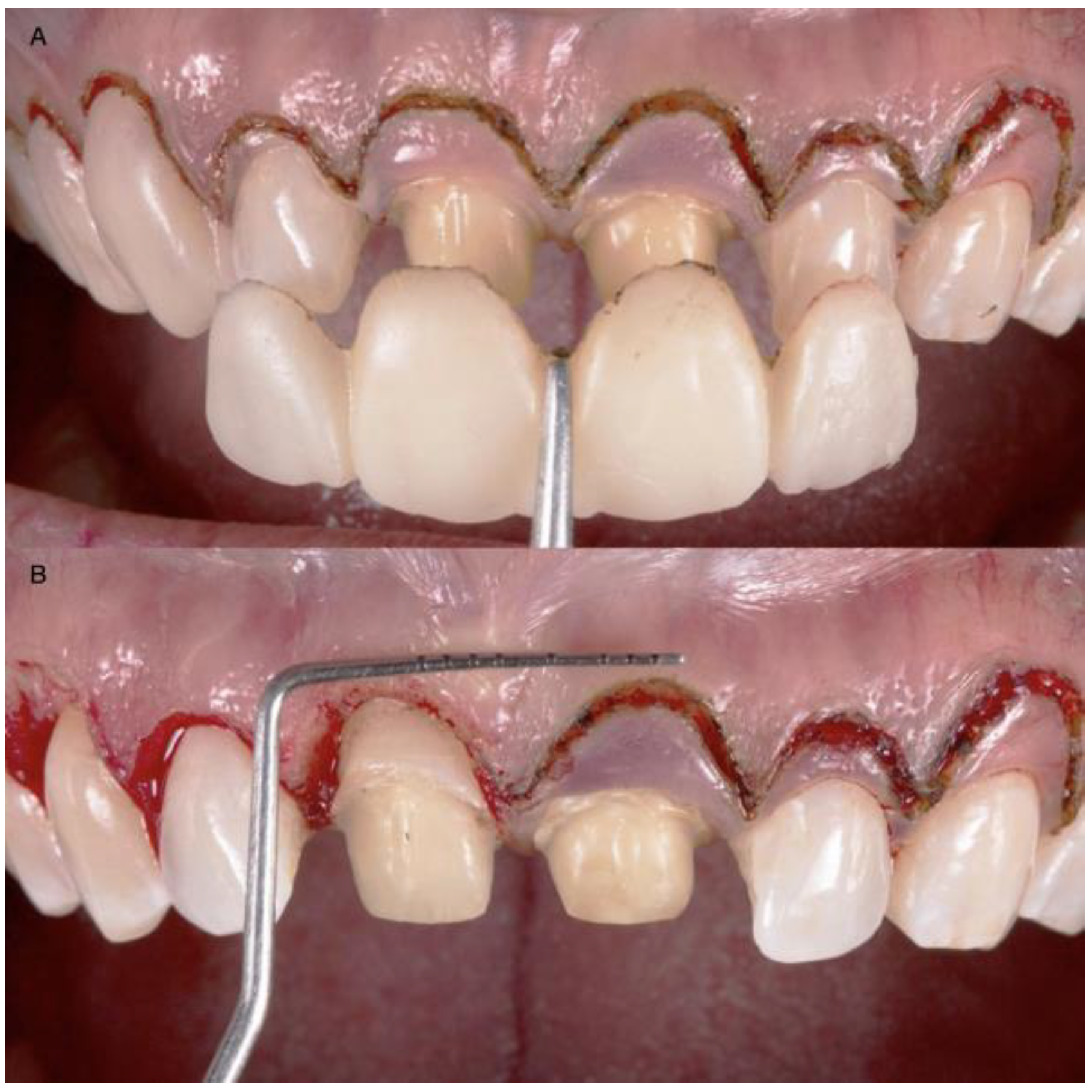
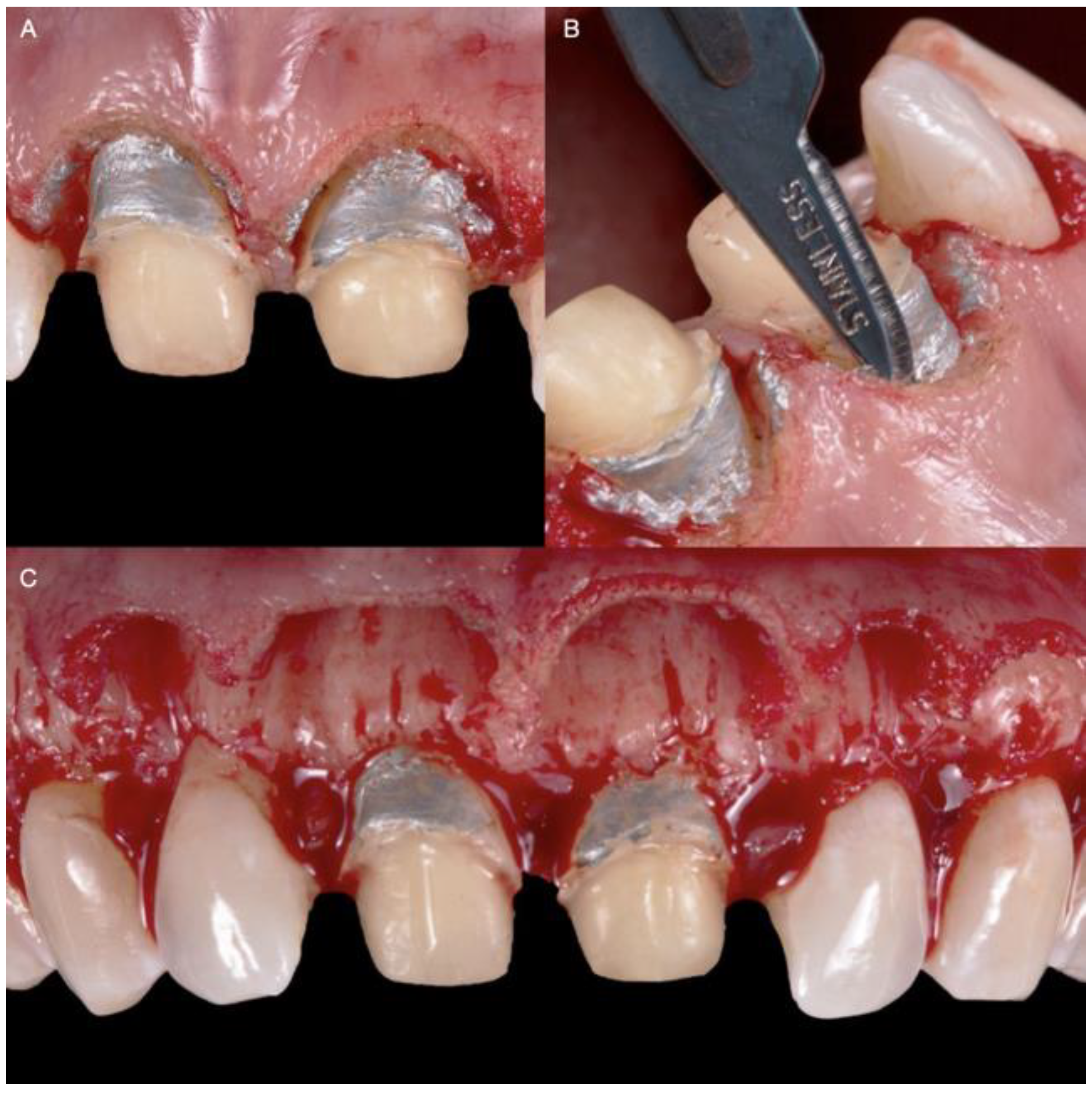
2. Materials and Methods
3. Discussion
4. Conclusions
Author Contributions
Funding
Institutional Review Board Statement
Informed Consent Statement
Data Availability Statement
Acknowledgments
Conflicts of Interest
References
- Jurado, C.A.; Tinoco, J.V.; Tsujimoto, A.; Barkmeier, W.; Fischer, N.; Markham, M. Clear matrix use for composite resin core fabrication. Int. J. Esthet. Dent. 2020, 15, 108–117. [Google Scholar] [PubMed]
- Afrashtehfar, K.I.; Assery, M.K.A.; Bryant, S.R. Aesthetic Parameters and patient-perspective assessment tools for maxillary anterior single implants. Int. J. Dent. 2021, 2021, 6684028. [Google Scholar] [CrossRef] [PubMed]
- Del Monte, S.; Afrashtehfar, K.I.; Emami, E.; Abi Nader, S.; Tamimi, F. Lay preferences for dentogingival esthetic parameters: A systematic review. J. Prosthet. Dent. 2017, 118, 717–724. [Google Scholar] [CrossRef] [PubMed]
- Miranda, M.E.; Olivieri, K.A.; Rigolin, F.J.; de Vasconcellos, A.A. Esthetic challenges in rehabilitating the anterior maxilla: A case report. Oper. Dent. 2016, 41, 2–7. [Google Scholar] [CrossRef] [PubMed]
- Afrashtehfar, K.I.; Assery, M.K. Five considerations in cosmetic and esthetic dentistry. J. New Jersey Dent. Assoc. 2014, 85, 14–15. [Google Scholar]
- Afrashtehfar, K.I.; Assery, M.K.A.; Bryant, S.R. Patient Satisfaction in Medicine and Dentistry. Int. J. Dent. 2020, 2020, 6621848. [Google Scholar] [CrossRef]
- Alikhasi, M.; Yousefi, P.; Afrashtehfar, K.I. Smile Design: Mechanical Considerations. Dent. Clin. North Am. 2022, 66, 477–487. [Google Scholar] [CrossRef]
- Afrashtehfar, K.I.; Bryant, S.R. Understanding the lived experience of north american dental patients with a single-tooth implant in the upper front region of the mouth: Protocol for a qualitative Study. JMIR Res. Protoc. 2021, 10, e25767. [Google Scholar] [CrossRef]
- Jurado, C.A.; Tsujimoto, A.; Guzman, L.G.; Fischer, N.G.; Markham, M.D.; Barkmeier, W.W.; Latta, M.A. Implant therapy with monolithic translucent zirconia restorations in the esthetic zone. Gen. Dent. 2020, 68, 46–49. [Google Scholar]
- Marzadori, M.; Stefanini, M.; Sangiorgi, M.; Mounssif, I.; Monaco, C.; Zucchelli, G. Crown lengthening and restorative procedures in the esthetic zone. Periodontol. 2000 2018, 77, 84–92. [Google Scholar] [CrossRef]
- Simon, H.; Magne, P. Clinically based diagnostic wax-up for optimal esthetics: The diagnostic mock-up. J. Calif. Dent. Assoc. 2008, 36, 355–362. [Google Scholar] [PubMed]
- Jurado, C.; Watanabe, H.; Tinoco, J.V.; Valenzuela, H.U.; Perez, G.G.; Tsujimoto, A. A conservative approach to ceramic veneers: A case report. Oper. Dent. 2020, 45, 229–234. [Google Scholar] [CrossRef] [PubMed]
- Afrashtehfar, K.I.; Igarashi, K.; Bryant, S.R. Canadian Dental Patients with a Single-Unit Implant-Supported Restoration in the Aesthetic Region of the Mouth: Qualitative and Quantitative Patient-Reported Outcome Measures (PROMs). Data 2021, 6, 90. [Google Scholar] [CrossRef]
- Afrashtehfar, K.I. Conventional free-hand, dynamic navigation and static guided implant surgery produce similar short-term patient-reported outcome measures and experiences. Evid.-Based Dent. 2021, 22, 143–145. [Google Scholar] [CrossRef]
- Longo, E.; Frosecchi, M.; Marradi, L.; Signore, A.; de Angelis, N. Guided periodontal surgery: A novel approach for the treatment of gummy smile. A case report. Int. J. Esthet. Dent. 2019, 14, 384–392. [Google Scholar]
- Gurrea, J.; Bruguera, A. Wax-up and mock-up. A guide for anterior periodontal and restorative treatments. Int. J. Esthet. Dent. 2014, 9, 146–162. [Google Scholar] [PubMed]
- Liu, X.; Yu, J.; Zhou, J.; Tan, J. A digitally guided dual technique for both gingival and bone resection during crown lengthening surgery. J. Prosthet. Dent. 2018, 119, 345–349. [Google Scholar] [CrossRef]
- Kongkiatkamon, S.; Rokaya, D. Full digital workflow in the esthetic dental restoration. Case Rep Dent. 2022, 2022, 8836068. [Google Scholar] [CrossRef]
- Mendoza-Azpur, G.; Cornejo, H.; Villanueva, M.; Alva, R.; Barbisan de Souza, A. Periodontal plastic surgery for esthetic crown lengthening by using data merging and a CAD-CAM surgical guide. J. Prosthet. Dent. 2022, 127, 556–559. [Google Scholar] [CrossRef]
- Jurado, C.A.; Tsujimoto, A.; Watanabe, H.; Villalobos-Tinoco, J.; Garaicoa, J.L.; Markham, M.D.; Barkmeier, W.W.; Latta, M.A. Chair-side CAD/CAM fabrication of a single-retainer resin bonded fixed dental prosthesis: A case report. Restor. Dent. Endod. 2020, 45, e15. [Google Scholar] [CrossRef]
- Alazmi, S.O. Three dimensional digitally designed surgical guides in esthetic crown lengthening: A clinical case report with 12 months follow up. Clin. Cosmet Investig. Dent. 2022, 14, 55–59. [Google Scholar] [CrossRef] [PubMed]
- Takei, H.H.; Bevilacqua, F.; Cooney, J. Surgical crown lengthening of the maxillary anterior dentition: Aesthetic considerations. Pract. Periodontics Aesthetic Dent. 1999, 11, 639–644. [Google Scholar]
- Hempton, T.J.; Dominici, J.T. Contemporary crown-lengthening therapy: A review. J. Am. Dent. Assoc. 2010, 141, 647–655. [Google Scholar] [CrossRef] [PubMed]
- Pontoriero, R.; Carnevale, G. Surgical crown lengthening: A 12-month clinical wound healing study. J. Periodontol. 2001, 72, 841–848. [Google Scholar] [CrossRef]
- Kois, J.C. Altering gingival levels: The restorative connection part I: Biologic variables. J. Esthet. Restor. Dent. 1994, 6, 3–7. [Google Scholar] [CrossRef]
- Deas, D.E.; Moritz, A.J.; McDonnell, H.T.; Powell, C.A.; Mealey, B. Osseous surgery for crown lengthening: A 6-month clinical study. J. Periodontol. 2004, 75, 1288–1294. [Google Scholar] [CrossRef] [PubMed]
- Afrashtehfar, K.I.; Moshaverinia, A. Five things to know about regenerative periodontal therapies in dental medicine. J. New Jersey Dent. Assoc. 2015, 86, 12–13. [Google Scholar]


Publisher’s Note: MDPI stays neutral with regard to jurisdictional claims in published maps and institutional affiliations. |
© 2022 by the authors. Licensee MDPI, Basel, Switzerland. This article is an open access article distributed under the terms and conditions of the Creative Commons Attribution (CC BY) license (https://creativecommons.org/licenses/by/4.0/).
Share and Cite
Jurado, C.A.; Parachuru, V.; Villalobos Tinoco, J.; Guzman-Perez, G.; Tsujimoto, A.; Javvadi, R.; Afrashtehfar, K.I. Diagnostic Mock-Up as a Surgical Reduction Guide for Crown Lengthening: Technique Description and Case Report. Medicina 2022, 58, 1360. https://doi.org/10.3390/medicina58101360
Jurado CA, Parachuru V, Villalobos Tinoco J, Guzman-Perez G, Tsujimoto A, Javvadi R, Afrashtehfar KI. Diagnostic Mock-Up as a Surgical Reduction Guide for Crown Lengthening: Technique Description and Case Report. Medicina. 2022; 58(10):1360. https://doi.org/10.3390/medicina58101360
Chicago/Turabian StyleJurado, Carlos A., Venkata Parachuru, Jose Villalobos Tinoco, Gerardo Guzman-Perez, Akimasa Tsujimoto, Ramya Javvadi, and Kelvin I. Afrashtehfar. 2022. "Diagnostic Mock-Up as a Surgical Reduction Guide for Crown Lengthening: Technique Description and Case Report" Medicina 58, no. 10: 1360. https://doi.org/10.3390/medicina58101360