Article

Immediate Effects of Diaphragmatic Breathing with Cervical Spine Mobilization on the Pulmonary Function and Craniovertebral Angle in Patients with Chronic Stroke

1
Department of Physical Therapy, Dongnam Health University, 50, Cheoncheon-ro 74beon-gil, Jangan-gu, Suwon-si 16328, Gyeonggi-do, Korea
2
Department of Physical Therapy, Graduate School, Yongin University, 134, Yongindaehak-ro, Cheoin-gu, Yongin-si 17092, Gyeonggi-do, Korea
3
Department of Physical Therapy, Suwon Women’s University, 1098, Juseok-ro, Bongdam-eup, Hwaseong-si 18333, Gyeonggi-do, Korea
*
Author to whom correspondence should be addressed.
Medicina 2021, 57(8), 826; https://doi.org/10.3390/medicina57080826
Submission received: 15 July 2021 / Revised: 3 August 2021 / Accepted: 13 August 2021 / Published: 16 August 2021
(This article belongs to the Special Issue Treatment of Stroke, Dementia and Atrial Fibrillation)

Abstract

:
Background and Objectives: Patients with stroke have a forward neck posture due to neurological damage and often have impaired pulmonary function. This study investigated the effect of diaphragmatic breathing with cervical mobilization to improve pulmonary function cervical alignments. Materials and Methods: This study used a one-group pre-test–post-test design including 20 patients with stroke. Two types of cervical joint mobilization techniques, consisting of left and right lateral glide mobilization and posterior–anterior mobilization, were utilized. During joint mobilization, the patients performed diaphragmatic breathing. The measurements were performed immediately after the intervention. Pulmonary function was evaluated using a spirometer to measure the forced expiratory volume in 1 s (FEV1), forced vital capacity (FVC), and peak expiratory flow (PEF). The craniovertebral angle (CVA) was measured using lateral photographs. Results: After diaphragm breathing with cervical joint mobilization, subjects had significantly increased FEV1, FVC, PEF and CVA. Conclusion: Diaphragm breathing with cervical joint mobilization are possible interventions to increase pulmonary function and improve the craniovertebral angle in patients with stroke. However, a complete conclusion can be reached only after a follow-up study has been conducted with a comparison of more subjects and controls.

1. Introduction

Stroke patients have respiratory disturbances due to weakening of the diaphragm, responsible for inhalation, and the abdominal muscles, responsible for exhalation [1,2,3]. The nature of these disorders can cause abnormalities in respiratory mechanics and abnormal gas exchange, which can lead to respiratory complications [4,5]. Respiratory complications experienced by stroke patients include sleep apnea, venous thromboembolism, swallowing abnormalities, aspiration, and pneumonia [6].
In the case of patients with stroke, respiratory rehabilitation is necessary for the inhalation muscles because the more difficult it is for a patient to walk, the worse the effect on inhalation muscles is [7,8]. Threshold inspiratory muscle training was used as a method to improve inspiratory function in stroke patients [9]. This exercise is a way to improve inspiratory ability by using the diaphragm as well as accessory muscles of inspiration. Unlike threshold inspiratory muscle training, the diaphragm exercise relaxes the shoulders, head and neck, allowing only the rise of the abdomen while inhaling deeply through the nose [10].
Therefore, the importance of diaphragm exercise has been emphasized in respiratory rehabilitation for patients with stroke [11,12,13,14]. Diaphragm exercise is a method that improves the ability to inhale by strengthening the diaphragm muscles [10]. This exercise method has been proven to be capable of increasing pulmonary function and respiratory muscle strength in patients with stroke in several studies [11,12,13,14].
Diaphragm exercise is proposed to increase diaphragm contraction in patients with stroke. However, patients with stroke often have a forward neck posture due to a failure to adjust their posture [15], which can cause entrapment of the phrenic nerve [16,17,18]. Maitland joint mobilization is an manual orthopedic treatment method that uses brick wall theory as a basic concept [19]. It is a technique that applies movement to painful joints or those with limited movement [19]. This method involves passive movements applied by a therapist and is safe because the patient can stop mobilization at any time if uncomfortable when applying this approach. Maitland joint mobilization applied to the cervical vertebra reduces upper limb muscle tone and stiffness in patients with stroke [20], and when applied to the rib cage, it increases respiratory muscle tone and lung function [21,22]. In addition, manual correction applied to the cervical vertebra increases the inhalation and exhalation volumes in healthy people [23]. Although diaphragm exercise and cervical joint mobilization for diaphragm contraction in patients with stroke provide well-suited interventions for improving pulmonary function, the effects of these two methods have not yet been confirmed.
Therefore, the purpose of this study was to investigate the effects of diaphragm exercise and cervical joint mobilization in improving pulmonary function and the craniovertebral angle in stroke patients.

2. Methods

2.1. Design

This study was conducted as a one-group pre-test–post-test of quasi-experimental design. This study was conducted in accordance with the ethical principles of the Declaration of Helsinki and was approved by the institutional review board of Yong In University (No: 2-1040966-AB-N-01). Informed consent was obtained from all subjects before the pre-test. The pre-test was a pulmonary function and cervical alignments. Intervention was diaphragmatic breathing with cervical joint mobilization for stroke patients. The post-test was immediate changes in pulmonary function and cervical alignment after intervention.
No subjects had ever experienced cervical joint mobilization and were informed that this study was investigating the effects of respiratory function and cervical alignments. Subjects could not see the CVA lateral photographs, or FVC, FEV1, or PEF displays; thus, they were not provided feedback on the assessment. The evaluator, a person unrelated to this study, measured the pre-test and post-test in an independent space without knowing that this study was investigating the therapeutic effect of joint mobilization. Physical therapists who applied cervical joint mobilization were not informed that this study would confirm the effect of this procedure on respiratory function.
First, the experimental group was educated on diaphragmatic breathing for 30 min. Then, one physical therapist applied two types of cervical joint mobilization techniques (lateral sliding mobilization and anterior–posterior mobilization) for 18 min. During cervical joint mobilization, the subject was asked to perform diaphragmatic breathing. The data were collected through assessment immediately after intervention.

2.2. Sample Size Calculation

In this study, the sample size was calculated based on calculations through G-power 3.1.9 software (Heinrich Heine University, Dusseldorf, Germany) according to a t test of Cohen’s d [24]. Prior to calculating the total sample size, a pilot study was conducted on 6 stroke patients. According to the pilot study, the effect sizes were 1.33 (FEV1), 1.38 (FVC), 0.77 (PEF), and 0.89 (CVA). The total sample sizes was 7 (FEV1), 7 (FVC), 16 (PEF), and 12 (CVA) with each effect size; the power of the test was 0.80; and an alpha error probability of 0.05 were input parameters. The total sample size of this study was 20.

2.3. Participants and Recruitment
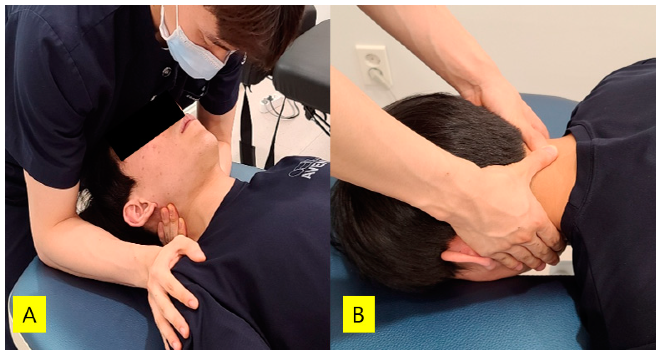
In this study, among adult patients hospitalized for stroke under the care of a rehabilitation medicine doctor at a rehabilitation hospital located in Gyeonggi-do Province, those whose diagnosis was made more than 6 months previously, those whose forced vital capacity was 10% lower than the normal predicted value [25], those with a craniovertebral angle of less than 50° [26], those who scored more than 24 points in the Korean mini-mental state examination [27], those with no given comorbidities, and those who expressed their willingness to participate in this study were selected as subjects (n = 20). All subjects who participated in this study were receiving a neurodevelopmental treatment (NDT) program from a physical therapist after hospitalization. The NDT programs typically included mat and gait training.

2.4. Intervention

Diaphragmatic Breathing with Joint Mobilization

Diaphragmatic breathing with cervical joint mobilization was performed with the patient in a supine position. Grade Ⅲ cervical joint mobilization was applied according to the Maitland classification, and during joint mobilization, the patients performed diaphragmatic breathing with controls on breathing [10]. One of the two type of cervical joint mobilization techniques (left and right lateral glide mobilization, posterior–anterior mobilization) was performed for 1 min per set, for a total of three sets [28]. Specifically, cervical joint mobilization took a total of 9 sets of 9 min, with a rest period of 1 min between sets. For two types of cervical mobilization, left and right lateral glide mobilization and posterior–anterior mobilization were performed on the C4-5 segment (Figure 1). During left glide lateral mobilization, the physiotherapist fixed the subject’s head using the chest, and the shoulder was fixed to prevent the shoulder from rising with the opposite hand while pulling the side surrounding the cervical vertebra using the index finger of one hand [29]. The right glide lateral mobilization was applied in the same way as the left glide lateral mobilization technique. Posterior–anterior mobilization was performed by the physiotherapist standing above the head of the patients, placing the thumb of one hand on the spinous process, supporting it with the thumb of the other hand, and extending the arm in a vertical straight line [19].

2.5. Assessments

2.5.1. Pulmonary Function

A spirometer (MicroLab spirometer ML3500 MK6, Kent, UK) was used to evaluate pulmonary function. The study subjects sat on a chair with a backrest, looked to the front, inhaled as much as possible, and then exhaled air through a spirometer for 6 s. Forced expiratory volume in 1 s, forced vital capacity, and peak expiratory flow were selected as the measurement variables. All items were measured three times and the average value was recorded. A spirometer was used to measure lung function. Pulmonary function measurements were conducted according to ATS/ERS Standardization of Spirometry guidelines for pulmonary function testing [30].

2.5.2. Craniovertebral Angle

ImageJ (National Institute of Health, NIH Version, 1.32J, Bethesda, ML, USA) software for Windows was used to measure the cervical alignment in stroke patients. The study subjects sat on a chair with a backrest. Stickers were attached to the tragus of the ear and the seventh spinous process of the cervical vertebra to obtain the angle between a horizontal line and the line between these two points [31]. Lateral photographs were taken with a camera positioned vertically with the camera lens 80 cm away from the shoulder using a smartphone (iPhone, Apple Inc., Cupertino, CA, USA).

2.6. Statistical Analyses

The data collected in this study were statistically analyzed using the SPSS 21.0 program (SPSS Inc., Chicago, IL, USA). The Shapiro–Wilk test confirmed whether the distribution followed a normal distribution. The Cohen’s d effect size was obtained using the G * power program. A paired t-test was used to determine the significance of the change in pulmonary function and cervical alignment following diaphragmatic breathing and cervical joint mobilization. The data are presented as means and standard deviations. The statistical significance level was set at α = 0.05, a two-tailed test.

3. Results

3.1. General Characteristics of Subjects

Twenty participants who performed diaphragmatic breathing with cervical mobilization were analyzed in this study. The results of the analysis of the general characteristics of the study participants are shown in Table 1. The subjects included 14 men and 6 women, and the paralysis was right hemiparesis and left hemiparesis in 12 and 8 patients, respectively. The average disease duration of patients was 11.50 ± 3.33 months, the average age of patients was 64.25 ± 11.47 years, the average height was 165.68 ± 8.35 cm, the average weight was 64.86 ± 9.43 kg, the average BMI was 23.51 ± 1.90 kg/m2, the average K-MMSE was 25.70 ± 1.78 points, and the average K-NIHSS was 10.00 ± 3.48 points.

3.2. Changes in Pulmonary Function, Craniovertebral Angle

The effects of diaphragmatic breathing with cervical joint mobilization on pulmonary function and craniovertebral angle in subjects are shown in Table 2. FEV1 increased from 2.15 ± 0.62 ℓ before intervention to 2.33 ± 0.64 ℓ after intervention (p < 0.01); FVC increased from 2.64 ± 0.55 ℓ before intervention to 2.91 ± 0.64 ℓ after intervention (p < 0.01); PEF increased from 213.30 ± 88.52 ℓ/min to 254.00 ± 98.34 ℓ/min (p < 0.01); and FEV1/FVC changed from 80.00 ± 9.96% to 79.00 ± 8.51% (p > 0.05). The craniovertebral angle increased significantly, from 45.39 ± 6.26° before intervention to 46.04 ± 6.14° after intervention (p < 0.01).

4. Discussion

In this study, we applied diaphragmatic breathing with cervical joint mobilization to patients with stroke to assess whether they improved pulmonary function and the craniovertebral angle. Our findings were consistent with a previous study, showing that the cervical spine angle improved after cervical joint mobilization [32]. In addition, this study was able to confirm an increase in respiratory function.
Patients with stroke often sit in a slumped position due to poor postural control [15]. In addition, having to always use a wheelchair leads to a worse posture; the patients with stroke participating in this study were all hospitalized who relied on wheelchairs for mobility [33]. When a slumped position is maintained, kyphosis occurs, and the lumbar spine appears flat [15]. This eventually leads to forward neck posture and changes the alignment of the cervical vertebra [15]. Abnormal alignment of the cervical vertebra can capture or block the path of the spinal or phrenic nerve extending from the vertebrae [16,17,18]. Manual therapy of the C3 vertebra increased the maximum inspiratory pressure and maximum expiratory pressure in healthy people [23,34]. However, prior studies were conducted in healthy people, and manual therapy was applied to cervical vertebra manipulation [23,34]. Manipulation is a fast application method with high velocity and low amplitude; thus, there is a disadvantage in that the patient cannot stop before feeling abnormal [35]. In this study, cervical joint mobilization was applied that allowed patients to stop the movement at any time when they experienced an abnormality. Cervical joint mobilization applied to stroke patients immediately reduces muscle tone and stiffness [20]. In previous studies, this was the result of the expansion of the spinal nerve originating from the vertebrae through re-alignment of the cervical vertebra. Therefore, it is believed that joint mobilization applied to the cervical vertebra promotes the phrenic nerve and increases diaphragm contraction by affecting the activity of the descending pathway. Cervical spine correction can improve postural alignment, which can increase rib movement.
Patients with a forward neck posture have limitations in the movement of the rib cage [36]. Joint mobilization applied to the cervical and thoracic spine increases pulmonary function through increasing rib cage movement by re-aligning the vertebrae [37]. The joint mobilization site applied in this study was also a C3-C4 cervical vertebra; therefore, it was hypothesized that it affected the re-alignment of the vertebrae and had a positive effect on respiratory function through increased rib cage movement. In this study, while applying cervical joint mobilization, the patient was asked to maintain awareness of their respiration. The recognition of respiration in this study is noteworthy in that it used diaphragmatic breathing [38]. The phrenic nerve is located at C3-C4 [39]. Cervical joint mobilization activated the phrenic nerve through mechanical stimulation [23,28,34], and diaphragmatic breathing resulted in an immediate increase in the saturation of peripheral oxygen because diaphragmatic contractions were more activated [38].
Despite these results, this study has several limitations. First, because diaphragm movements and cervical joint mobilization were applied at the same time, it was not possible to determine which of the two had more influence on the respiratory function. Secondly, this study lacks internal and external validity because there was no control group due to the single-group pre–post design. Thirdly, the number of subjects in this study was very small, and the findings may not be generalizable to all stroke patients. Additionally, the short intervention period could have prevented interference with other treatments, but the long-term effects have not been confirmed. Finally, in this study, respiratory muscle strength could not be objectively confirmed. PEF has a strong correlation with maximal inspiratory pressure and maximal expiratory pressure, i.e., respiratory muscle strength [40,41]. This study measured the pulmonary function to check the flow of air because of diaphragmatic breathing, which is an inhalation and exhalation exercise. Even if PEF is strongly correlated with MIP and MEP, it is necessary to confirm direct respiratory muscle strength. In addition, various measures such as joint stiffness, ventilatory capacity measures, and perceptual measures are still needed to complete the results.
However, this study has clinical significance in that it is the first study to apply both diaphragmatic movement exercise and cervical joint mobilization. Joint mobilization is a safe method, and diaphragmatic exercise has the advantage that any physical therapist can utilize it. In addition, it is expected that follow-up studies will be conducted based on the effect size identified in this study. Nevertheless, the single group and small number of subjects remain a problem. This issue should be addressed by a randomized study design with a control group.

5. Conclusions

The purpose of this study was to investigate the effect of diaphragmatic breathing with cervical joint mobilization on pulmonary function and cervical alignment in 20 stroke patients. It was found that diaphragmatic breathing with cervical joint mobilization was effective in improving pulmonary function and cervical alignment in patients with stroke. However, based on this study, it is difficult to draw conclusions about the difference in value due to the absence of a control group. In future studies, it will be necessary to compare the effect of diaphragmatic breathing with joint mobilization with changes in a control group in a larger group of subjects.

Author Contributions

Conceptualization, A.Y.K. and S.J.P.; investigation, A.Y.K. and H.J.A.; data curation, A.Y.K.; writing—original draft preparation, S.J.P. and H.J.A.; writing—review and editing, S.J.P. and H.J.A. All authors have read and agreed to the published version of the manuscript.

Funding

This study received no external funding.

Institutional Review Board Statement

The study was conducted according to the guidelines of the Declaration of Helsinki, and approved by the Institutional Review Board of Yong In University on 26 March 2021 (approval No. 2-1040966-AB-N-01).

Informed Consent Statement

Informed consent was obtained from all subjects involved in the study.

Data Availability Statement

Study data are available upon request from corresponding author.

Conflicts of Interest

The authors declare no conflict of interest.

References

  1. Menezes, K.K.; Nascimento, L.; Ada, L.; Polese, J.C.; Avelino, P.R.; Teixeira-Salmela, L.F. Respiratory muscle training increases respiratory muscle strength and reduces respiratory complications after stroke: A systematic review. J. Physiother. 2016, 62, 138–144. [Google Scholar] [CrossRef] [Green Version]
  2. Teixeira-Salmela, L.F.; Parreira, V.F.; Britto, R.R.; Brant, T.C.; Inácio, P.; Alcântara, T.O.; Carvalho, I.F. Respiratory Pressures and Thoracoabdominal Motion in Community-Dwelling Chronic Stroke Survivors. Arch. Phys. Med. Rehabil. 2005, 86, 1974–1978. [Google Scholar] [CrossRef]
  3. Jung, J.-H.; Kim, N.-S. The correlation between diaphragm thickness, diaphragmatic excursion, and pulmonary function in patients with chronic stroke. J. Phys. Ther. Sci. 2017, 29, 2176–2179. [Google Scholar] [CrossRef] [Green Version]
  4. Lanini, B.; Bianchi, R.; Romagnoli, I.; Coli, C.; Binazzi, B.; Gigliotti, F.; Pizzi, A.; Grippo, A.; Scano, G. Chest Wall Kinematics in Patients with Hemiplegia. Am. J. Respir. Crit. Care Med. 2003, 168, 109–113. [Google Scholar] [CrossRef] [PubMed]
  5. Lanini, B.; Gigliotti, F.; Coli, C.; Bianchi, R.; Pizzi, A.; Romagnoli, I.; Grazzini, M.; Stendardi, L.; Scano, G.J.C.S. Dissociation between respiratory effort and dyspnoea in a subset of patients with stroke. Clin. Sci. 2002, 103, 467–473. [Google Scholar] [CrossRef]
  6. Rochester, C.L.; Mohsenin, V. Respiratory Complications of Stroke. Semin. Respir. Crit. Care Med. 2002, 23, 248–260. [Google Scholar] [CrossRef] [PubMed]
  7. Pinheiro, M.B.; Polese, J.C.; Faria, C.; Machado, G.; Parreira, V.F.; Britto, R.R.; Teixeira-Salmela, L.F. Inspiratory muscular weakness is most evident in chronic stroke survivors with lower walking speeds. Eur. J. Phys. Rehabil. Med. 2014, 50, 301–307. [Google Scholar]
  8. Zhang, X.; Zheng, Y.; Dang, Y.; Wang, L.; Cheng, Y.; Zhang, X.; Mao, M.; Lu, X. Can inspiratory muscle training benefit patients after stroke? A systematic review and meta-analysis of randomized controlled trials. Clin. Rehabil. 2020, 34, 866–876. [Google Scholar] [CrossRef]
  9. Britto, R.R.; Rezende, N.R.; Marinho, K.C.; Torres, J.L.; Parreira, V.F.; Teixeira-Salmela, L.F. Inspiratory Muscular Training in Chronic Stroke Survivors: A Randomized Controlled Trial. Arch. Phys. Med. Rehabil. 2011, 92, 184–190. [Google Scholar] [CrossRef]
  10. Kigin, C.M. Breathing exercises for the medical patient: The art and the science. Phys. Ther. 1990, 70, 700–706. [Google Scholar] [CrossRef]
  11. Seo, K.C.; Lee, H.M.; Kim, H.A. The Effects of Combination of Inspiratory Diaphragm Exercise and Exspiratory Pursed-Lip Breathing Exercise on Pulmonary Functions of Stroke Patients. J. Phys. Ther. Sci. 2013, 25, 241–244. [Google Scholar] [CrossRef] [Green Version]
  12. Shetty, N.; Samuel, S.R.; Alaparthi, G.K.; Amaravadi, S.K.; Joshua, A.M.; Pai, S. Comparison of Diaphragmatic Breathing Exercises, Volume, and Flow-Oriented Incentive Spirometry on Respiratory Function in Stroke Subjects: A Non-Randomized Study. Ann. Neurosci. 2021, 0972753121990193. [Google Scholar] [CrossRef]
  13. Lee, H.-J.; Kang, T.-W.; Kim, B.-R. Effects of diaphragm and deep abdominal muscle exercise on walking and balance ability in patients with hemiplegia due to stroke. J. Exerc. Rehabil. 2018, 14, 648–653. [Google Scholar] [CrossRef]
  14. Seo, K.; Hwan, P.S.; Park, K. The effects of inspiratory diaphragm breathing exercise and expiratory pursed-lip breathing exercise on chronic stroke patients’ respiratory muscle activation. J. Phys. Ther. Sci. 2017, 29, 465–469. [Google Scholar] [CrossRef] [PubMed] [Green Version]
  15. Iyengar, Y.; Vijayakumar, K.; Abraham, J.; Misri, Z.; Suresh, B.; Unnikrishnan, B. Relationship between postural alignment in sitting by photogrammetry and seated postural control in post-stroke subjects. NeuroRehabilitation 2014, 35, 181–190. [Google Scholar] [CrossRef]
  16. Haghighat, F.; Moradi, R.; Rezaie, M.; Yarahmadi, N.; Ghaffarnejad, F. Added Value of Diaphragm Myofascial Release on forward Head Posture and Chest Expansion in Patients with Neck Pain: A Randomized Controlled Trial. 2020. Available online: https://assets.researchsquare.com/files/rs-53279/v1/e2455ee7-327f-43ab-a291-4b303dd5bcdb.pdf?fbclid=IwAR0xwROyiiX44msrom6gZ1cPCi3NUl4Kf9WWGMnLH-8fYpaVh0sKmQ3rDrA (accessed on 15 July 2021).
  17. Zafar, H.; Albarrati, A.; Alghadir, A.H.; Iqbal, Z.A. Effect of Different Head-Neck Postures on the Respiratory Function in Healthy Males. BioMed Res. Int. 2018, 2018, 4518269. [Google Scholar] [CrossRef] [Green Version]
  18. Triangto, K.; Widjanantie, S.C.; Nusdwinuringtyas, N. Biomechanical Impacts of forward Head Posture on the Respiratory Function. Indones. J. Phys. Med. Rehabil. 2019, 8, 50–64. [Google Scholar] [CrossRef]
  19. Maitland, G.D. Vertebral Manipulation; Butterworth-Heinemann: Oxford, UK, 2013. [Google Scholar]
  20. Park, S.-J. The Immediate Effect of Maitland Cervical Spine Mobilization on Tone and Stiffness of Upper Limb Muscles in Chronic Stroke Patients. Phys. Ther. Korea 2018, 25, 13–21. [Google Scholar] [CrossRef]
  21. Park, S.-J.; Kim, S.-H.; Min, K.-O. The immediate effects of rib cage joint mobilization and chest wall stretch on muscle tone and stiffness of respiratory muscles and chest expansion ability in patients with chronic stroke. J. Phys. Ther. Sci. 2017, 29, 1960–1963. [Google Scholar] [CrossRef] [Green Version]
  22. Park, S.J. Effects of Inspiratory Muscles Training Plus Rib Cage Mobilization on Chest Expansion, Inspiratory Accessory Muscles Activity and Pulmonary Function in Stroke Patients. Appl. Sci. 2020, 10, 5178. [Google Scholar] [CrossRef]
  23. Behne, G.; Silva, P.; Ré, D.d.; Vazatta, M.; Carvalho, A.R. Maximal respiratory pressures: Comparison between immediate manipulation effect and mobilization of the third cervical vertebra. Arq. Ciências Saúde da UNIPAR 2013, 17, 69–76. [Google Scholar]
  24. Cohen, J. Statistical Power Analysis for the Behavioral Sciences; Academic Press: Cambridge, MA, USA, 2013. [Google Scholar]
  25. Do, J.G.; Park, C.-H.; Lee, Y.-T.; Yoon, K.J. Association between underweight and pulmonary function in 282,135 healthy adults: A cross-sectional study in Korean population. Sci. Rep. 2019, 9, 14308. [Google Scholar] [CrossRef] [PubMed]
  26. Sheikhhoseini, R.; Shahrbanian, S.; Sayyadi, P.; O’Sullivan, K. Effectiveness of Therapeutic Exercise on forward Head Posture: A Systematic Review and Meta-Analysis. J. Manip. Physiol. Ther. 2018, 41, 530–539. [Google Scholar] [CrossRef] [PubMed]
  27. Kang, Y.; Na, D.L.; Hahn, S.H. A validity study on the Korean Mini-Mental State Examination (K-MMSE) in dementia patients. J. Korean Neurol. Assoc. 1997, 15, 300–308. [Google Scholar]
  28. Do Bonfim, R.V.F.; de Moura Rocha, G.M.; da Silveira Sousa, A.M.; Rodrigues, B.F.O.; Almeida, V.R. Effect of Vertebral Mobilizations on Respiratory Pressure. In Proceedings of the XXVI Brazilian Congress on Biomedical Engineering, Vitória, Brazil, 26–30 October 2020; pp. 201–205. [Google Scholar]
  29. Vicenzino, B.; Cartwright, T.; Collins, D.; Wright, A. Cardiovascular and respiratory changes produced by lateral glide mobilization of the cervical spine. Man. Ther. 1998, 3, 67–71. [Google Scholar] [CrossRef]
  30. Miller, M.R.; Crapo, R.; Hankinson, J.; Brusasco, V.; Burgos, F.; Casaburi, R.; Coates, A.; Enright, P.; van der Grinten, C.P.M.; Gustafsson, P.; et al. General considerations for lung function testing. Eur. Respir. J. 2005, 26, 153–161. [Google Scholar] [CrossRef] [PubMed] [Green Version]
  31. Salahzadeh, Z.; Maroufi, N.; Ahmadi, A.; Behtash, H.; Razmjoo, A.; Gohari, M.; Parnianpour, M. Assessment of forward head posture in females: Observational and photogrammetry methods. J. Back Musculoskelet. Rehabil. 2014, 27, 131–139. [Google Scholar] [CrossRef] [PubMed]
  32. Park, S.J.; Park, S.E. Effect of upper thoracic mobilization on cervical alignment in stroke patients with forward head posture: A case study. J. Int. Acad. Phys. Ther. Res. 2018, 9, 1513–1516. [Google Scholar] [CrossRef] [Green Version]
  33. Park, S.J.; Kim, S.H. Effects of Kinesio Taping on the Pulmonary Function in Patients with Stroke during Manual Wheelchairs. J. Int. Acad. Phys. Ther. Res. 2018, 9, 1533–1536. [Google Scholar] [CrossRef]
  34. Da Silva, P.H.L.; de Ré, D.; Behne, G.R.; Vazatta, M.P.; de Carvalho, A.R. Maximum respiratory pressure alterations after spinal manipulation. Eur. J. Physiother. 2013, 15, 64–69. [Google Scholar] [CrossRef]
  35. John, S.; Tavee, J. Bilateral diaphragmatic paralysis due to cervical chiropractic manipulation. Neurologist 2015, 19, 65–67. [Google Scholar] [CrossRef] [PubMed]
  36. Koseki, T.; Kakizaki, F.; Hayashi, S.; Nishida, N.; Itoh, M. Effect of forward head posture on thoracic shape and respiratory function. J. Phys. Ther. Sci. 2019, 31, 63–68. [Google Scholar] [CrossRef] [Green Version]
  37. Jang, S.-H.; Bang, H.-S. Effect of thoracic and cervical joint mobilization on pulmonary function in stroke patients. J. Phys. Ther. Sci. 2016, 28, 257–260. [Google Scholar] [CrossRef] [PubMed] [Green Version]
  38. Wang, J.-S.; Cho, K.-H.; Park, S.-J. The immediate effect of diaphragm taping with breathing exercise on muscle tone and stiffness of respiratory muscles and SpO2 in stroke patient. J. Phys. Ther. Sci. 2017, 29, 970–973. [Google Scholar] [CrossRef] [Green Version]
  39. Gandevia, S.; Rothwell, J.C. Activation of the human diaphragm from the motor cortex. J. Physiol. 1987, 384, 109–118. [Google Scholar] [CrossRef] [PubMed]
  40. Sriboonreung, T.; Leelarungrayub, J.; Yankai, A.; Puntumetakul, R. Correlation and Predicted Equations of MIP/MEP from the Pulmonary Function, Demographics and Anthropometrics in Healthy Thai Participants aged 19 to 50 Years. Clin. Med. Insights Circ. Respir. Pulm. Med. 2021, 15, 11795484211004494. [Google Scholar] [CrossRef]
  41. Jo, M.-R.; Kim, N.-S. The correlation of respiratory muscle strength and cough capacity in stroke patients. J. Phys. Ther. Sci. 2016, 28, 2803–2805. [Google Scholar] [CrossRef] [PubMed] [Green Version]
Figure 1. Two type of cervical joint mobilization techniques: (A) lateral glide mobilization; (B) posterior–anterior mobilization.
Medicina 57 00826 g001
Table 1. General characteristics of the subjects.
Experimental Group (n = 20)
Gender(male/female)14/6
Affected side(left/right)12/8
Pathogenesis (hemorrhage/infarction)5/15
Disease duration (months)11.50 ± 3.33
Age (years)64.25 ± 11.47
Height (cm)165.68 ± 8.35
Weight (kg)64.86 ± 9.43
BMI (kg/m2)23.51 ± 1.90
K-MMSE (points)25.70 ± 1.78
K-NIHSS (points)10.00 ± 3.48
All values are denoted as mean ± standard deviations. BMI: Body mass index. K-NHSS: Korean version of the National Institutes of Health Stroke Scale. K-MMSE: Korean version of mini-mental state examination.
Table 2. Changes in pulmonary function, craniovertebral angle after intervention.
ClassificationExperimental Group (n = 20)Within Group Change atp%Effect
Size
Pre-TestPost-Test
FEV1 (ℓ)2.15 ± 0.622.33 ± 0.640.18 ± 0.19
(0.10, 0.27)
4.4050.001 *8.370.95
FVC (ℓ)2.64 ± 0.552.91 ± 0.640.26 ± 0.22
(0.16, 0.37)
5.4240.001 *10.231.23
FEV1/FVC (%)80.00 ± 9.9679.00 ± 8.51−1.00 ± 5.87
(−3.75, 1.75)
−0.7620.455−1.250.17
PEF (ℓ/min)213.30 ± 88.52254.00 ± 98.3440.70 ± 46.85
(18.78, 62.62)
3.8850.001 *19.080.87
CVA (°)45.39 ± 6.2646.04 ± 6.140.64 ± 0.72
(0.31, 0.98)
4.0310.001 *1.430.90
All values are denoted as mean ± SD, a Values are the mean (95% CI). * p < 0.05. FEV1: Forced expiratory volume in 1 s. FVC: Forced vital capacity. PEF: Peak expiratory flow. CVA: Craniovertebral angle. Experimental group: Diaphragmatic breathing with joint mobilization.
Publisher’s Note: MDPI stays neutral with regard to jurisdictional claims in published maps and institutional affiliations.

