
Primary Mucosal Melanoma Presenting with a Unilateral Nasal Obstruction of the Left Inferior Turbinate

 , ,
, ,  , , and
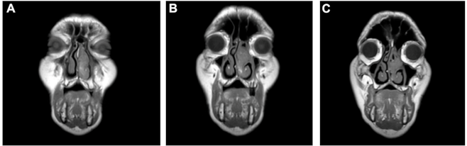
Abstract
:1. Introduction
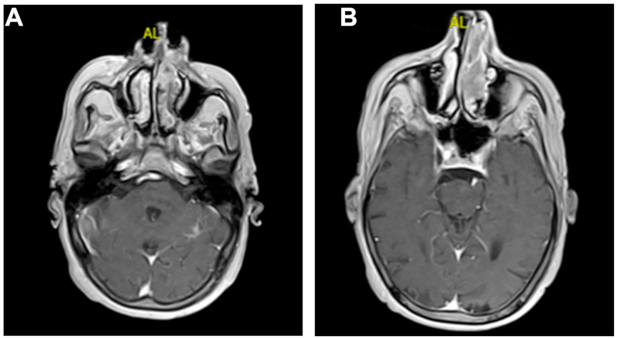
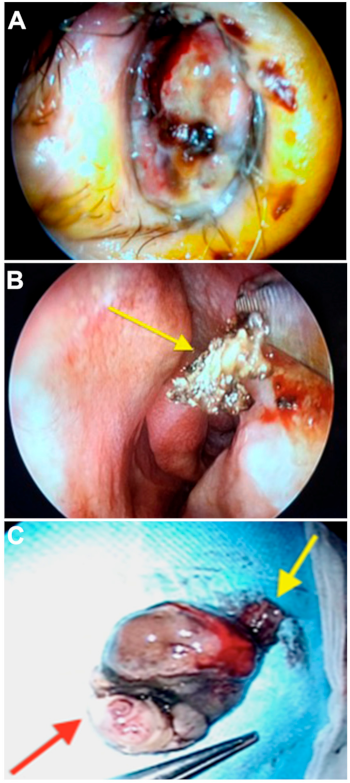
2. Case Report
3. Discussion
4. Conclusions
Funding
Institutional Review Board Statement
Informed Consent Statement
Data Availability Statement
Conflicts of Interest
References
- Matthews, N.H.; Li, W.Q.; Qureshi, A.A.; Weinstock, M.A.; Cho, E. Epidemiology of Melanoma. In Cutaneous Melanoma: Etiology and Therapy [Internet]; Ward, W.H., Farma, J.M., Eds.; Codon Publications: Brisbane, Australia, 2017; Chapter 1. Available online: https://www.ncbi.nlm.nih.gov/books/NBK481862/ (accessed on 29 December 2020). [CrossRef]
- Ascierto, P.A.; Accorona, R.; Botti, G.; Farina, D.; Fossati, P.; Gatta, G.; Gogas, H.; Lombardi, D.; Maroldi, R.; Nicolai, P.; et al. Mucosal melanoma of the head and neck. Crit. Rev. Oncol. Hematol. 2017, 112, 136–152. [Google Scholar] [CrossRef] [PubMed]
- Crippen, M.M.; Kilic, S.; Eloy, J.A. Updates in the management of sinonasal mucosal melanoma. Curr. Opin. Otolaryngol. Head Neck Surg. 2018, 26, 52–57. [Google Scholar] [CrossRef] [PubMed]
- Gilain, L.; Houette, A.; Montalban, A.; Mom, T.; Saroul, N. Mucosal melanoma of the nasal cavity and paranasal sinuses. Eur. Ann. Otorhinolaryngol. Head Neck Dis. 2014, 131, 365–369. [Google Scholar] [CrossRef] [PubMed] [Green Version]
- Patrick, R.J.; Fenske, N.A.; Messina, J.L. Primary mucosal melanoma. J. Am. Acad. Dermatol. 2007, 56, 828–834. [Google Scholar] [CrossRef] [PubMed]
- Allam, J.P.; Niederhagen, B.; Bucheler, M.; Appel, T.; Betten, H.; Bieber, T.; Berge, S.; Novak, N. Comparative analysis of nasal and oral mucosa dendritic cells. Allergy 2006, 61, 166–172. [Google Scholar] [CrossRef] [PubMed]
- Behranwala, R.; Loku Waduge, B.H.; Teo, B. Nasal mucosal melanoma as a cause of epistaxis. BMJ Case Rep. 2019, 12, e228640. [Google Scholar] [CrossRef] [PubMed]
- Chang, W.; Wu, L.; Han, D. Imaging progress of sinonasal mucosal malignant melanoma. Lin Chung Er Bi Yan Hou Tou Jing Wai Ke Za Zhi 2018, 32, 960–962. [Google Scholar] [CrossRef] [PubMed]
- Nardi, C.; Vignoli, C.; Vannucchi, M.; Pietragalla, M. Magnetic resonance features of sinonasal melanotic mucosal melanoma. BMJ Case Rep. 2019, 12, e229790. [Google Scholar] [CrossRef] [PubMed]
- Houette, A.; Gilain, L.; Mulliez, A.; Mom, T.; Saroul, N. Prognostic value of two tumour staging classifications in patients with sinonasal mucosal melanoma. Eur. Ann. Otorhinolaryngol. Head Neck Dis. 2016, 133, 313–317. [Google Scholar] [CrossRef] [PubMed]
- Kim, S.H. Mucosal malignant melanoma presenting with huge, fleshy, dark-red polyp in the nasal cavity. Br. J. Hosp. Med. (Lond.) 2017, 78, 297. [Google Scholar] [CrossRef] [PubMed]
- Wahid, N.W.; Meghji, S.; Barnes, M. Nasal mucosal melanoma. Lancet Oncol. 2019, 20, e284. [Google Scholar] [CrossRef]
- Pittaka, M.; Kardamakis, D.; Spyropoulou, D. Comparison of International Guidelines on Mucosal Melanoma of the Head and Neck: A Comprehensive Review of the Role of Radiation Therapy. In Vivo 2016, 30, 165–170. [Google Scholar] [PubMed]
- Topic, B.; Masic, T.; Radovic, S.; Lincender, I.; Muhic, E. Primary Oral Mucosal Melanomas—Two Case Reports and Comprehensive Literature Review. Acta Clin. Croat. 2017, 56, 323–330. [Google Scholar] [CrossRef] [PubMed]
- Schaefer, T.; Satzger, I.; Gutzmer, R. Clinics, prognosis and new therapeutic options in patients with mucosal melanoma: A retrospective analysis of 75 patients. Medicine 2017, 96, e5753. [Google Scholar] [CrossRef] [PubMed]
- Axell, T.; Hedin, C.A. Epidemiologic study of excessive oral melanin pigmentation with special reference to the influence of tobacco habits. Scand. J. Dent. Res. 1982, 90, 434–442. [Google Scholar] [CrossRef] [PubMed]
- Kim, S.S.; Han, M.H.; Kim, J.E.; Lee, C.H.; Chung, H.W.; Lee, J.S.; Chang, K.H. Malignant melanoma of the sinonasal cavity: Explanation of magnetic resonance signal intensities with histopathologic characteristics. Am. J. Otolaryngol. 2000, 21, 366–378. [Google Scholar] [CrossRef] [PubMed]
- Lynch, S.C.; Lee, A.G.; Graham, S.M.; Kirby, P.A. Primary melanoma of the sphenoid sinus presenting with a third cranial nerve palsy. J. Neuroophthalmol. 2005, 25, 289–292. [Google Scholar] [CrossRef] [PubMed] [Green Version]
- Al-Haseni, A.; Vrable, A.; Qureshi, M.M.; Mathews, S.; Pollock, S.; Truong, M.T.; Sahni, D. Survival outcomes of mucosal melanoma in the USA. Future Oncol. 2019, 15, 3977–3986. [Google Scholar] [CrossRef] [PubMed]
- Altieri, L.; Eguchi, M.; Peng, D.H.; Cockburn, M. Predictors of mucosal melanoma survival in a population-based setting. J. Am. Acad. Dermatol. 2019, 81, 136–142.e132. [Google Scholar] [CrossRef] [PubMed]


Publisher’s Note: MDPI stays neutral with regard to jurisdictional claims in published maps and institutional affiliations. |
© 2021 by the authors. Licensee MDPI, Basel, Switzerland. This article is an open access article distributed under the terms and conditions of the Creative Commons Attribution (CC BY) license (https://creativecommons.org/licenses/by/4.0/).
Share and Cite
Lombardo, N.; Della Corte, M.; Pelaia, C.; Piazzetta, G.; Lobello, N.; Del Duca, E.; Bennardo, L.; Nisticò, S.P. Primary Mucosal Melanoma Presenting with a Unilateral Nasal Obstruction of the Left Inferior Turbinate. Medicina 2021, 57, 359. https://doi.org/10.3390/medicina57040359
Lombardo N, Della Corte M, Pelaia C, Piazzetta G, Lobello N, Del Duca E, Bennardo L, Nisticò SP. Primary Mucosal Melanoma Presenting with a Unilateral Nasal Obstruction of the Left Inferior Turbinate. Medicina. 2021; 57(4):359. https://doi.org/10.3390/medicina57040359
Chicago/Turabian StyleLombardo, Nicola, Marcello Della Corte, Corrado Pelaia, Giovanna Piazzetta, Nadia Lobello, Ester Del Duca, Luigi Bennardo, and Steven Paul Nisticò. 2021. "Primary Mucosal Melanoma Presenting with a Unilateral Nasal Obstruction of the Left Inferior Turbinate" Medicina 57, no. 4: 359. https://doi.org/10.3390/medicina57040359