
Antioxidant Flavonoid Diosmetin Is Cardioprotective in a Rat Model of Myocardial Infarction Induced by Beta 1-Adrenergic Receptors Activation


Abstract
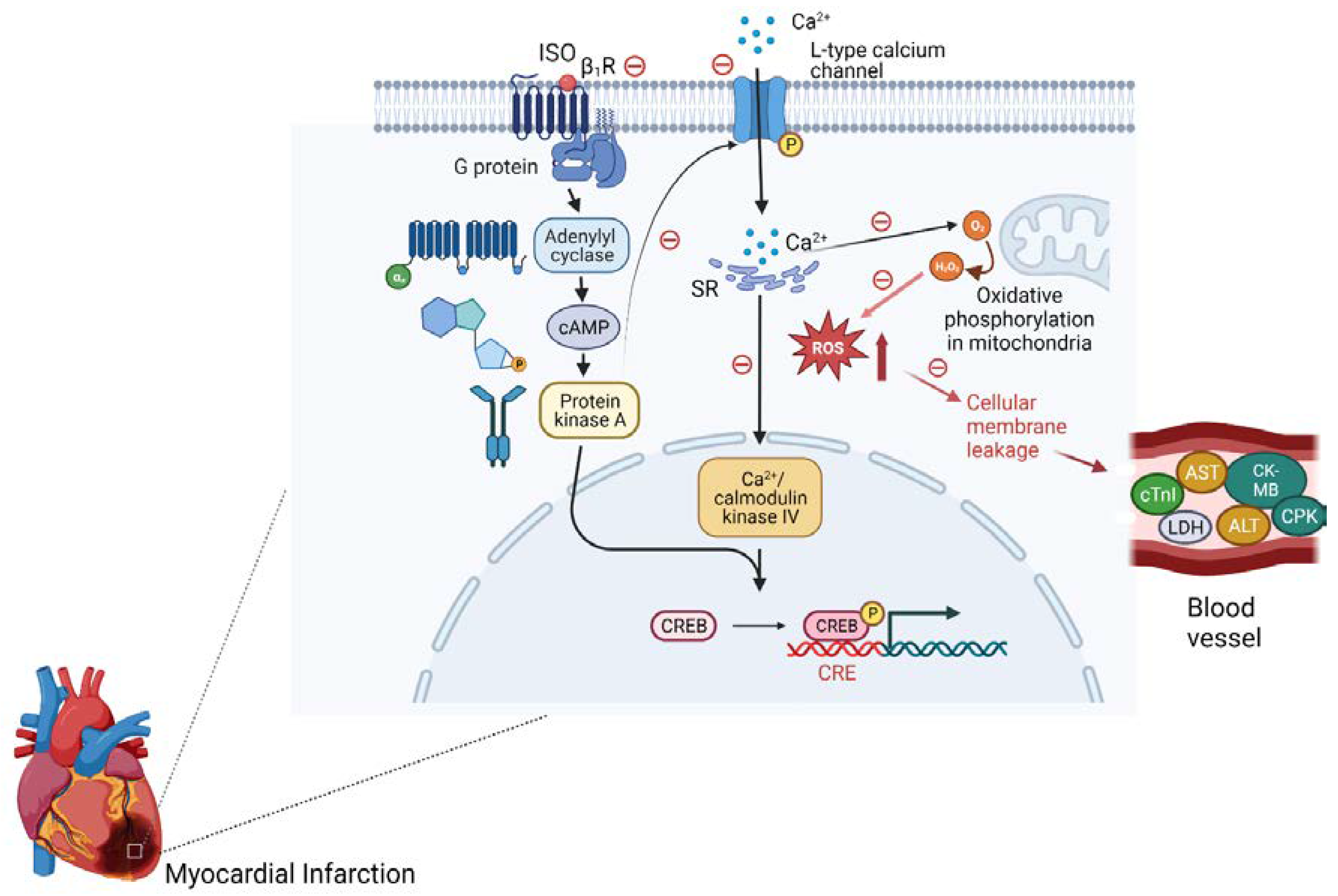
:1. Introduction
2. Methods
2.1. Animals Used
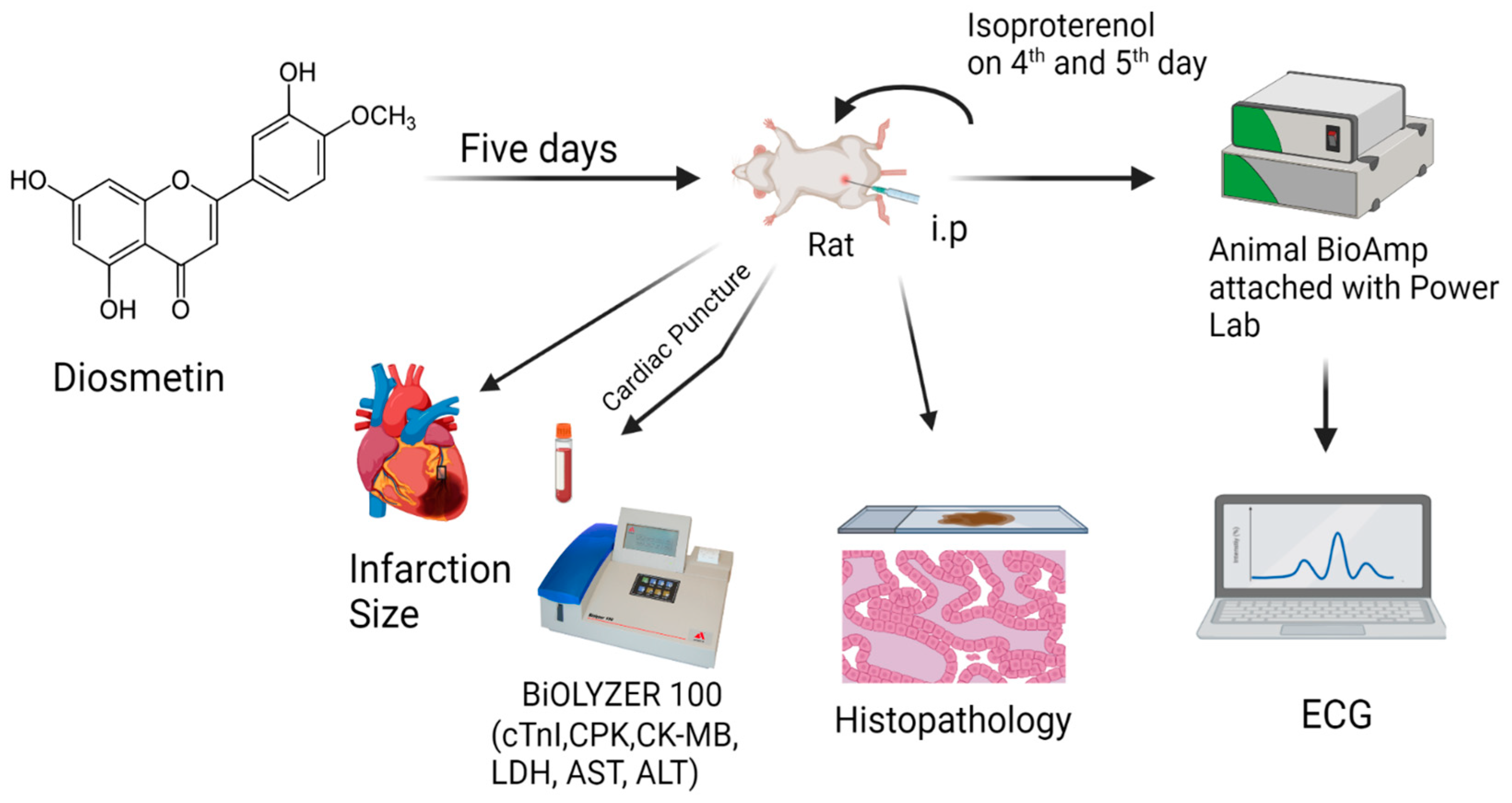
2.2. Experimental Protocol
Diosmetin Response against the MI Induced by β1-Adrenergic Receptors Activation
2.3. Statistical Significance Analysis
3. Results
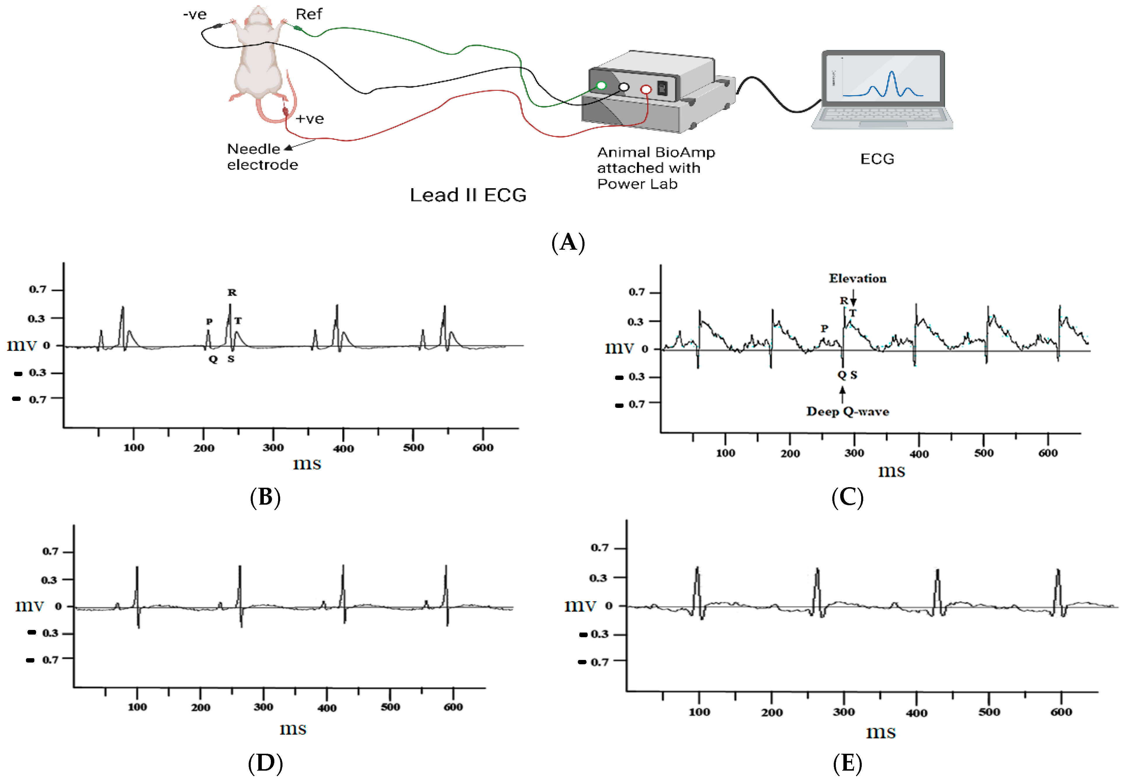
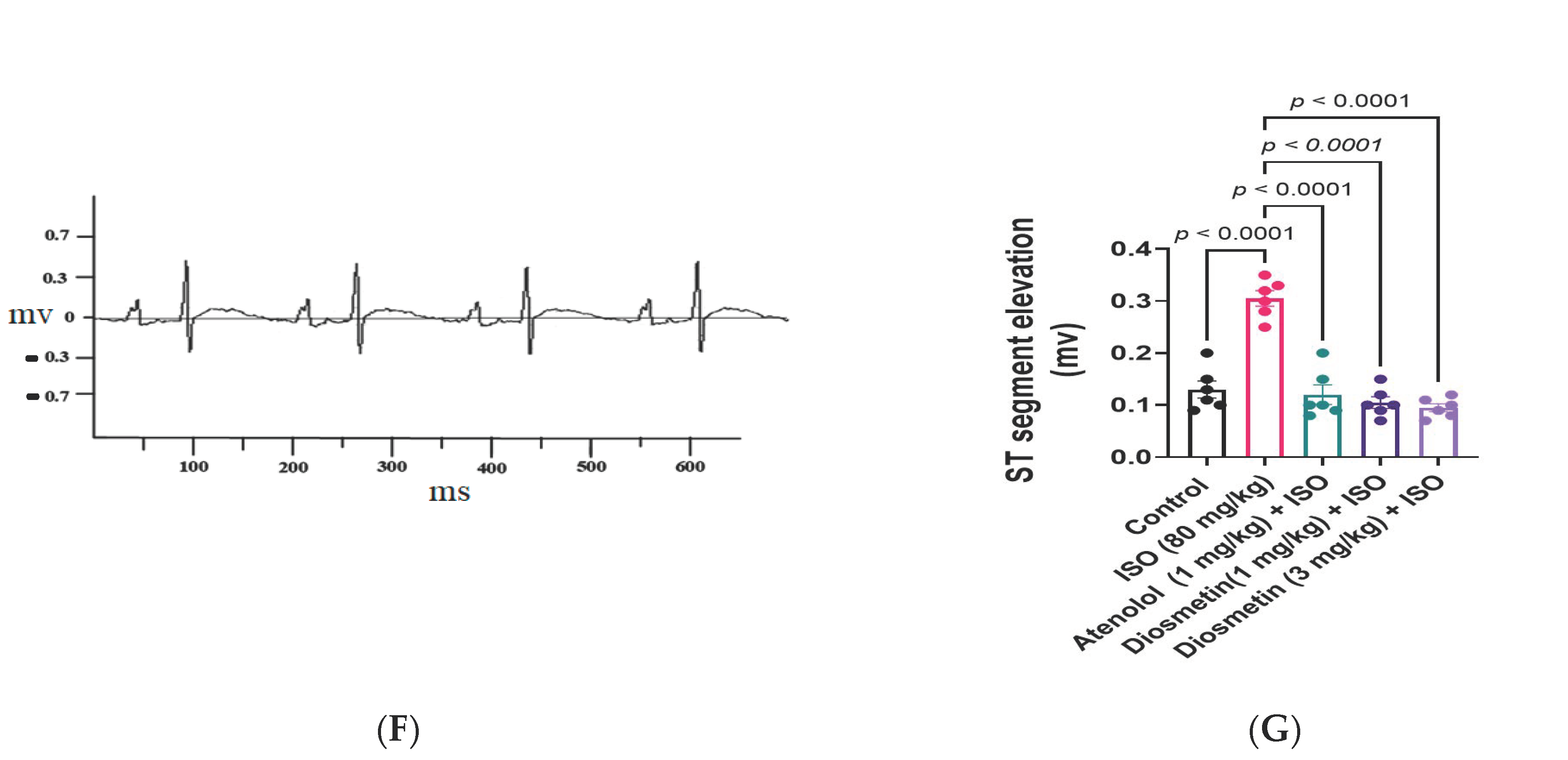
3.1. Electrocardiograph (ECG) Analysis of Different Groups of Experimental Rats
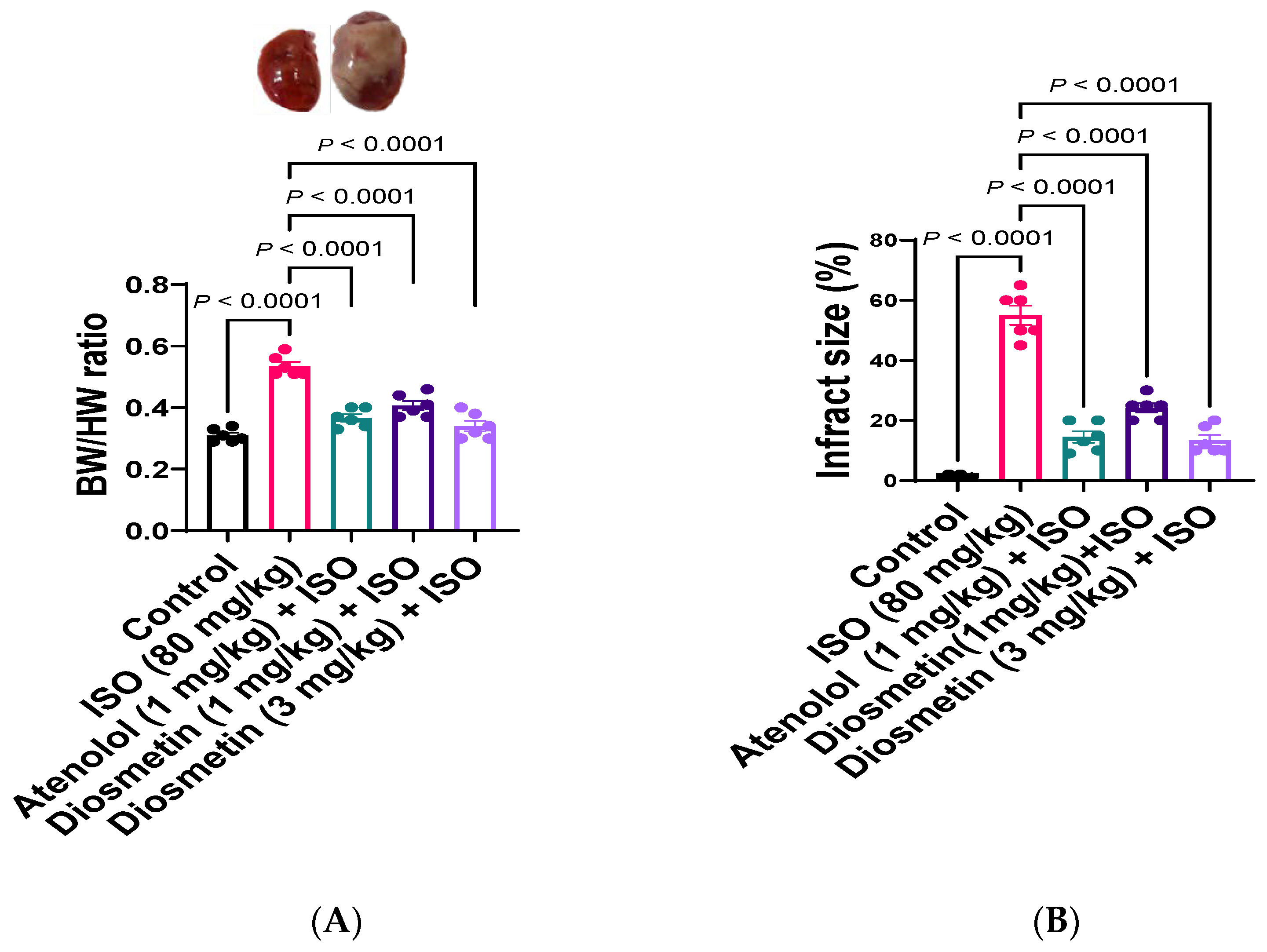
3.2. Heart to Body Weight Ratio
3.3. Anatomical and Histopathological Analysis
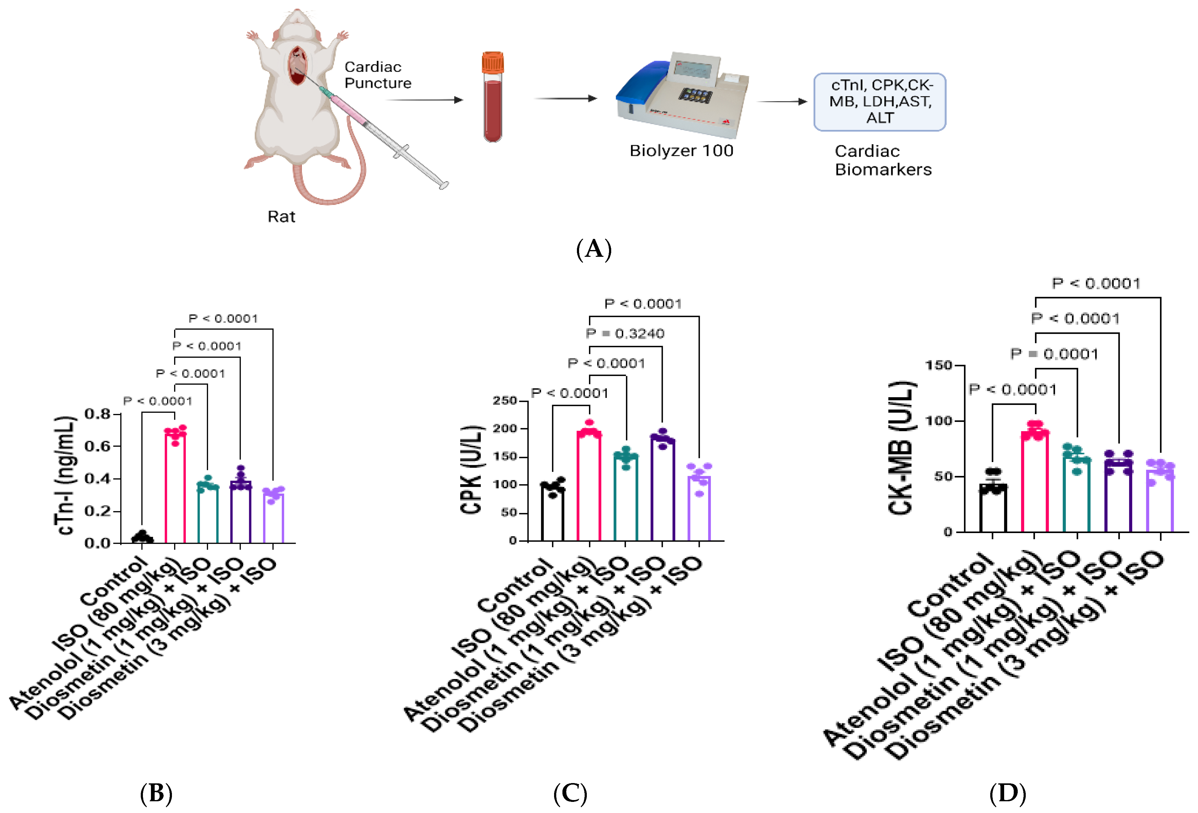
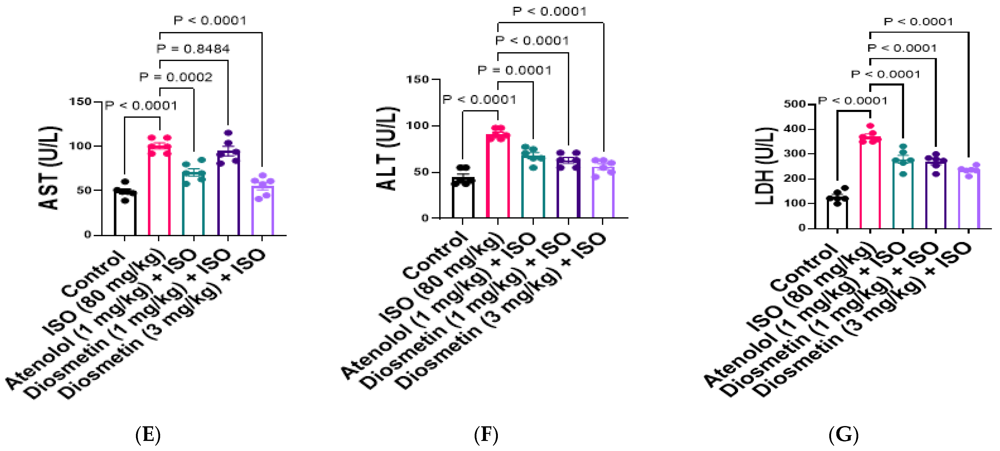
3.4. Protective Response of Diosmetin against Alteration in Cardiac Biomarkers
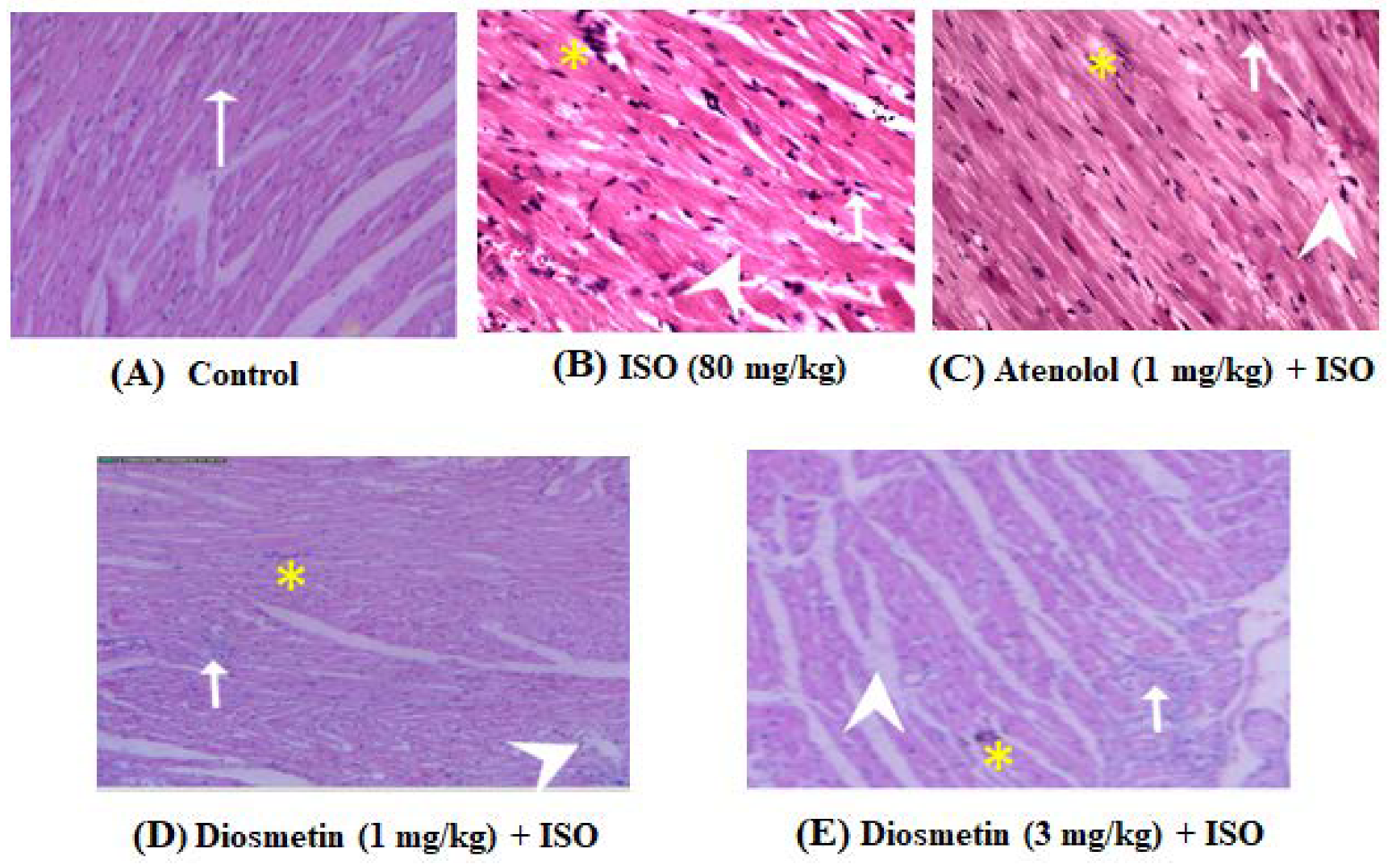
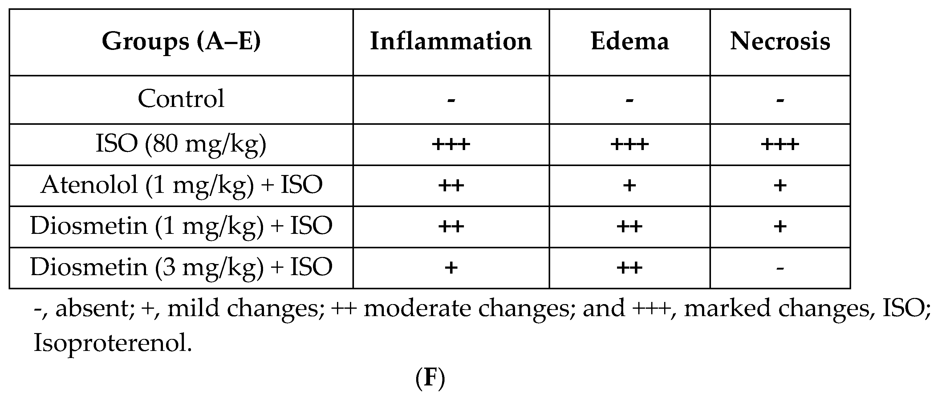
3.5. Diosmetin Prevent Histopathological Changes Induced by ISO
4. Discussion
Author Contributions
Funding
Institutional Review Board Statement
Informed Consent Statement
Data Availability Statement
Acknowledgments
Conflicts of Interest
References
- Anversa, P.; Sonnenblick, E.H. Ischemic Cardiomyopathy: Pathophysiologic Mechanisms. Prog. Cardiovasc. Dis. 1990, 33, 49–70. [Google Scholar] [CrossRef] [PubMed]
- Nowbar, A.N.; Gitto, M.; Howard, J.P.; Francis, D.P.; Al-Lamee, R. Mortality from Ischemic Heart Disease. Circ. Cardiovasc. Qual. Outcomes 2019, 12, e005375. [Google Scholar] [CrossRef] [PubMed]
- Gaziano, T.A.; Bitton, A.; Anand, S.; Abrahams-Gessel, S.; Murphy, A. Growing Epidemic of Coronary Heart Disease in Low- and Middle-Income Countries. Curr. Probl. Cardiol. 2010, 35, 72–115. [Google Scholar] [CrossRef] [PubMed]
- Hegstad, A.C.; Ytrehus, K.; Myklebust, R.; Jørgensen, L. Ultrastructural Changes in the Myocardial Myocytic Mitochondria: Crucial Step in the Development of Oxygen Radical-Induced Damage in Isolated Rat Hearts? Basic Res. Cardiol. 1994, 89, 128–138. [Google Scholar] [CrossRef]
- Misra, M.K.; Sarwat, M.; Bhakuni, P.; Tuteja, R.; Tuteja, N. Oxidative Stress and Ischemic Myocardial Syndromes. Med. Sci. Monit. 2009, 15, RA209–RA219. [Google Scholar]
- Wu, J.; Hecker, J.G.; Chiamvimonvat, N. Antioxidant Enzyme Gene Transfer for Ischemic Diseases. Adv. Drug Deliv. Rev. 2009, 61, 351–363. [Google Scholar] [CrossRef]
- Rezende, P.C.; Ribas, F.F.; Serrano, C.V.; Hueb, W. Clinical Significance of Chronic Myocardial Ischemia in Coronary Artery Disease Patients. J. Thorac. Dis. 2019, 11, 1005–1015. [Google Scholar] [CrossRef]
- Libby, P.; Ridker, P.M.; Maseri, A. Inflammation and Atherosclerosis. Circulation 2002, 105, 1135–1143. [Google Scholar] [CrossRef]
- Cunningham, K.S.; Gotlieb, A.I. The Role of Shear Stress in the Pathogenesis of Atherosclerosis. Lab. Investig. J. Tech. Methods Pathol. 2005, 85, 9–23. [Google Scholar] [CrossRef]
- Baker, J.E.; Su, J.; Fu, X.; Hsu, A.; Gross, G.J.; Tweddell, J.S.; Hogg, N. Nitrite Confers Protection against Myocardial Infarction: Role of Xanthine Oxidoreductase, NADPH Oxidase and K(ATP) Channels. J. Mol. Cell. Cardiol. 2007, 43, 437–444. [Google Scholar] [CrossRef]
- Brunton, L.L.; Hilal-Dandan, R.; Bjorn, K.C. Goodman and Gillman’s The Pharmacological Basis of Therapeutics, 13th ed.; McGraw-Hill Education: New York, NY, USA, 2018. [Google Scholar]
- Talukder, M.A.H.; Zweier, J.L.; Periasamy, M. Targeting Calcium Transport in Ischaemic Heart Disease. Cardiovasc. Res. 2009, 84, 345–352. [Google Scholar] [CrossRef] [PubMed]
- Kim, J.-E.; Choi, B.-K.; Choi, J.-Y.; Ryu, T.; Roh, W.S.; Song, S.-Y. Role of Calcium Channels Responsible for Phenylephrine-Induced Contraction in Rat Aorta 3 Days after Acute Myocardial Infarction. Korean J. Anesthesiol. 2014, 66, 143–152. [Google Scholar] [CrossRef] [PubMed]
- Akila, P.; Vennila, L. Chlorogenic Acid a Dietary Polyphenol Attenuates Isoproterenol Induced Myocardial Oxidative Stress in Rat Myocardium: An in Vivo Study. Biomed. Pharmacother. 2016, 84, 208–214. [Google Scholar] [CrossRef] [PubMed]
- Ighodaro, O.M.; Akinloye, O.A. First Line Defence Antioxidants-Superoxide Dismutase (SOD), Catalase (CAT) and Glutathione Peroxidase (GPX): Their Fundamental Role in the Entire Antioxidant Defence Grid. Alex. J. Med. 2018, 54, 287–293. [Google Scholar] [CrossRef]
- Mythili, S.; Malathi, N. Diagnostic Markers of Acute Myocardial Infarction. Biomed. Rep. 2015, 3, 743–748. [Google Scholar] [CrossRef]
- Kubler, W.; Haass, M. Cardioprotection: Definition, Classification, and Fundamental Principles. Heart 1996, 75, 330–333. [Google Scholar] [CrossRef]
- Egred, M.; Shaw, S.; Mohammad, B.; Waitt, P.; Rodrigues, E. Under-Use of Beta-Blockers in Patients with Ischaemic Heart Disease and Concomitant Chronic Obstructive Pulmonary Disease. QJM Mon. J. Assoc. Physicians 2005, 98, 493–497. [Google Scholar] [CrossRef]
- Khan, M.; Gilani, A.-H. Studies on Blood Pressure Lowering, Vasodilator and Cardiac Suppressant Activities of Vitex Negundo: Involvement of K+ Channel Activation and Ca++ Channel Blockade. Available online: https://scialert.net/abstract/?doi=ijp.2015.137.142 (accessed on 1 April 2021).
- Afsheen, N.; Khalil-ur-Rehman; Jahan, N.; Ijaz, M.; Manzoor, A.; Khan, K.M.; Hina, S. Cardioprotective and Metabolomic Profiling of Selected Medicinal Plants against Oxidative Stress. Oxid. Med. Cell. Longev. 2018, 2018, 9819360. [Google Scholar] [CrossRef]
- Patwardhan, B.; Vaidya, A.D.B.; Chorghade, M. Ayurveda and Natural Products Drug Discovery. Curr. Sci. 2004, 86, 789–799. [Google Scholar]
- Lahlou, M. The Success of Natural Products in Drug Discovery. Pharmacol. Pharm. 2013, 4, 17–31. [Google Scholar] [CrossRef]
- Brüll, V.; Burak, C.; Stoffel-Wagner, B.; Wolffram, S.; Nickenig, G.; Müller, C.; Langguth, P.; Alteheld, B.; Fimmers, R.; Naaf, S.; et al. Effects of a Quercetin-Rich Onion Skin Extract on 24 h Ambulatory Blood Pressure and Endothelial Function in Overweight-to-Obese Patients with (Pre-)Hypertension: A Randomised Double-Blinded Placebo-Controlled Cross-over Trial. Br. J. Nutr. 2015, 114, 1263–1277. [Google Scholar] [CrossRef] [PubMed]
- Panche, A.N.; Diwan, A.D.; Chandra, S.R. Flavonoids: An Overview. J. Nutr. Sci. 2016, 5, E47. [Google Scholar] [CrossRef] [PubMed]
- Meng, Z.; Zhao, J.; Duan, H.; Guan, Y.; Zhao, L. Green and Efficient Extraction of Four Bioactive Flavonoids from Pollen Typhae by Ultrasound-Assisted Deep Eutectic Solvents Extraction. J. Pharm. Biomed. Anal. 2018, 161, 246–253. [Google Scholar] [CrossRef]
- Choy, K.W.; Murugan, D.; Leong, X.-F.; Abas, R.; Alias, A.; Mustafa, M.R. Flavonoids as Natural Anti-Inflammatory Agents Targeting Nuclear Factor-Kappa B (NFκB) Signaling in Cardiovascular Diseases: A Mini Review. Front. Pharmacol. 2019, 10, 1295. [Google Scholar] [CrossRef] [PubMed]
- Maleki, S.J.; Crespo, J.F.; Cabanillas, B. Anti-Inflammatory Effects of Flavonoids. Food Chem. 2019, 299, 125124. [Google Scholar] [CrossRef]
- Doostdar, H.; Burke, M.D.; Mayer, R.T. Bioflavonoids: Selective Substrates and Inhibitors for Cytochrome P450 CYP1A and CYP1B1. Toxicology 2000, 144, 31–38. [Google Scholar] [CrossRef]
- Spanakis, M.; Kasmas, S.; Niopas, I. Simultaneous Determination of the Flavonoid Aglycones Diosmetin and Hesperetin in Human Plasma and Urine by a Validated GC/MS Method: In Vivo Metabolic Reduction of Diosmetin to Hesperetin. Biomed. Chromatogr. BMC 2009, 23, 124–131. [Google Scholar] [CrossRef]
- Meirinhos, J.; Silva, B.M.; Valentão, P.; Seabra, R.M.; Pereira, J.A.; Dias, A.; Andrade, P.B.; Ferreres, F. Analysis and Quantification of Flavonoidic Compounds from Portuguese Olive (Olea Europaea L.) Leaf Cultivars. Nat. Prod. Res. 2005, 19, 189–195. [Google Scholar] [CrossRef]
- Boshtam, M.; Naderi, G.A.; Moshtaghian, J.; Asgary, S.; Jafari, N. Effects of Citrus Limon Burm. f. on Some Atherosclerosis Risk Factors in Rabbits with Atherogenic Diet. Atherosclerosis 2009, 5, 88–93. [Google Scholar]
- Riaz, A.; Khan, R.A.; Mirza, T.; Mustansir, T.; Ahmed, M. In Vitro/in Vivo Effect of Citrus Limon (L. Burm. f.) Juice on Blood Parameters, Coagulation and Anticoagulation Factors in Rabbits. Pak. J. Pharm. Sci. 2014, 27, 907–915. [Google Scholar]
- Tripoli, E.; La Guardia, M.; Giammanco, S.; Di Majo, D.; Giammanco, M. Citrus Flavonoids: Molecular Structure, Biological Activity and Nutritional Properties: A Review. Food Chem. 2007, 104, 466–479. [Google Scholar] [CrossRef]
- Kato, Y.; Domoto, T.; Hiramitsu, M.; Katagiri, T.; Sato, K.; Miyake, Y.; Aoi, S.; Ishihara, K.; Ikeda, H.; Umei, N.; et al. Effect on Blood Pressure of Daily Lemon Ingestion and Walking. J. Nutr. Metab. 2014, 2014, 912684. [Google Scholar] [CrossRef]
- Rašković, A.; Milanović, I.; Pavlović, N.; Ćebović, T.; Vukmirović, S.; Mikov, M. Antioxidant Activity of Rosemary (Rosmarinus officinalis L.) Essential Oil and Its Hepatoprotective Potential. BMC Complement. Altern. Med. 2014, 14, 225. [Google Scholar] [CrossRef] [PubMed]
- Morel, I.; Lescoat, G.; Cogrel, P.; Sergent, O.; Pasdeloup, N.; Brissot, P.; Cillard, P.; Cillard, J. Antioxidant and Iron-Chelating Activities of the Flavonoids Catechin, Quercetin and Diosmetin on Iron-Loaded Rat Hepatocyte Cultures. Biochem. Pharmacol. 1993, 45, 13–19. [Google Scholar] [CrossRef] [PubMed]
- Liao, W.; Ning, Z.; Chen, L.; Wei, Q.; Yuan, E.; Yang, J.; Ren, J. Intracellular Antioxidant Detoxifying Effects of Diosmetin on 2,2-Azobis(2-Amidinopropane) Dihydrochloride (AAPH)-Induced Oxidative Stress through Inhibition of Reactive Oxygen Species Generation. J. Agric. Food Chem. 2014, 62, 8648–8654. [Google Scholar] [CrossRef]
- Andreeva, O.A.; Ivashev, M.N.; Ozimina, I.I.; Maslikova, G.V. Diosmetin Glycosides from Caucasian Vetch: Isolation and Study of Biological Activity. Pharm. Chem. J. 1998, 32, 595–597. [Google Scholar] [CrossRef]
- Ge, A.; Liu, Y.; Zeng, X.; Kong, H.; Ma, Y.; Zhang, J.; Bai, F.; Huang, M. Effect of Diosmetin on Airway Remodeling in a Murine Model of Chronic Asthma. Acta Biochim. Biophys. Sin. 2015, 47, 604–611. [Google Scholar] [CrossRef]
- Chen, Y.; Wang, Y.; Liu, M.; Zhou, B.; Yang, G. Diosmetin Exhibits Anti-Proliferative and Anti-Inflammatory Effects on TNF-α-Stimulated Human Rheumatoid Arthritis Fibroblast-like Synoviocytes through Regulating the Akt and NF-ΚB Signaling Pathways. Phytother. Res. PTR 2020, 34, 1310–1319. [Google Scholar] [CrossRef]
- Manivannan, J.; Silambarasan, T.; Kadarkarairaj, R.; Raja, B. Systems Pharmacology and Molecular Docking Strategies Prioritize Natural Molecules as Cardioprotective Agents. RSC Adv. 2015, 5, 77042–77055. [Google Scholar] [CrossRef]
- Mo, G.; He, Y.; Zhang, X.; Lei, X.; Luo, Q. Diosmetin Exerts Cardioprotective Effect on Myocardial Ischaemia Injury in Neonatal Rats by Decreasing Oxidative Stress and Myocardial Apoptosis. Clin. Exp. Pharmacol. Physiol. 2020, 47, 1713–1722. [Google Scholar] [CrossRef]
- Wang, S.; Tian, S.; Yang, F.; Yang, H.; Yang, X.; Du, G. Cardioprotective Effect of Salvianolic Acid A on Isoproterenol-Induced Myocardial Infarction in Rats. Eur. J. Pharmacol. 2009, 615, 125–132. [Google Scholar] [CrossRef] [PubMed]
- Li, H.; Xie, Y.-H.; Yang, Q.; Wang, S.-W.; Zhang, B.-L.; Wang, J.-B.; Cao, W.; Bi, L.-L.; Sun, J.-Y.; Miao, S.; et al. Cardioprotective Effect of Paeonol and Danshensu Combination on Isoproterenol-Induced Myocardial Injury in Rats. PLoS ONE 2012, 7, e48872. [Google Scholar] [CrossRef] [PubMed]
- Sahu, B.D.; Anubolu, H.; Koneru, M.; Kumar, J.M.; Kuncha, M.; Rachamalla, S.S.; Sistla, R. Cardioprotective Effect of Embelin on Isoproterenol-Induced Myocardial Injury in Rats: Possible Involvement of Mitochondrial Dysfunction and Apoptosis. Life Sci. 2014, 107, 59–67. [Google Scholar] [CrossRef] [PubMed]
- Kim, Y.-J.; Ahn, J.K.; Shin, K.-A.; Kim, C.-H.; Lee, Y.-H.; Park, K.-M. Correlation of Cardiac Markers and Biomarkers With Blood Pressure of Middle-Aged Marathon Runners. J. Clin. Hypertens. 2015, 17, 868–873. [Google Scholar] [CrossRef]
- Ahmed, S.; Gul, S.; Ze Jaafar, H.; Moga, M.; Zia-Ul-Haq, M.; Dima, L. Anti-Platelet Effects of Nimesulide in Isoproterenol-Induced Myocardial Ischaemia and Infarction in Rabbits. Acta Cardiol. 2015, 70, 401–408. [Google Scholar] [CrossRef]
- Wexler, B.C. Myocardial Infarction in Young vs Old Male Rats: Pathophysiologic Changes. Am. Heart J. 1978, 96, 70–80. [Google Scholar] [CrossRef]
- Geng, B.; Chang, L.; Pan, C.; Qi, Y.; Zhao, J.; Pang, Y.; Du, J.; Tang, C. Endogenous Hydrogen Sulfide Regulation of Myocardial Injury Induced by Isoproterenol. Biochem. Biophys. Res. Commun. 2004, 318, 756–763. [Google Scholar] [CrossRef]
- Sambhi, M.P.; White, F.N. The Electrocardiogram of the Normal and Hypertensive Rat. Circ. Res. 1960, 8, 129–134. [Google Scholar] [CrossRef]
- Rajadurai, M.; Prince, P.S.M. Preventive Effect of Naringin on Cardiac Mitochondrial Enzymes during Isoproterenol-Induced Myocardial Infarction in Rats: A Transmission Electron Microscopic Study. J. Biochem. Mol. Toxicol. 2007, 21, 354–361. [Google Scholar] [CrossRef]
- Suchalatha, S.; Shyamala Devi, C.S. Protective Effect of Terminalia Chebula against Experimental Myocardial Injury Induced by Isoproterenol. Indian J. Exp. Biol. 2004, 42, 174–178. [Google Scholar]
- Thygesen, K.; Alpert, J.S.; Jaffe, A.S.; Chaitman, B.R.; Bax, J.J.; Morrow, D.A.; White, H.D. Executive Group on behalf of the Joint European Society of Cardiology (ESC)/American College of Cardiology (ACC)/American Heart Association (AHA)/World Heart Federation (WHF) Task Force for the Universal Definition of Myocardial Infarction Fourth Universal Definition of Myocardial Infarction (2018). Circulation 2018, 138, e618–e651. [Google Scholar] [CrossRef] [PubMed]
- Sabeena Farvin, K.H.; Anandan, R.; Kumar, S.H.S.; Shiny, K.S.; Sankar, T.V.; Thankappan, T.K. Effect of Squalene on Tissue Defense System in Isoproterenol-Induced Myocardial Infarction in Rats. Pharmacol. Res. 2004, 50, 231–236. [Google Scholar] [CrossRef] [PubMed]
- Rajadurai, M.; Prince, P.S.M. Preventive Effect of Naringin on Cardiac Markers, Electrocardiographic Patterns and Lysosomal Hydrolases in Normal and Isoproterenol-Induced Myocardial Infarction in Wistar Rats. Toxicology 2007, 230, 178–188. [Google Scholar] [CrossRef] [PubMed]
- Panda, V.S.; Naik, S.R. Cardioprotective Activity of Ginkgo Biloba Phytosomes in Isoproterenol-Induced Myocardial Necrosis in Rats: A Biochemical and Histoarchitectural Evaluation. Exp. Toxicol. Pathol. Off. J. Ges. Toxicol. Pathol. 2008, 60, 397–404. [Google Scholar] [CrossRef] [PubMed]
- Roy, S.J.; Prince, P.S.M. Protective Effects of Sinapic Acid on Cardiac Hypertrophy, Dyslipidaemia and Altered Electrocardiogram in Isoproterenol-Induced Myocardial Infarcted Rats. Eur. J. Pharmacol. 2013, 699, 213–218. [Google Scholar] [CrossRef]
- Body, R.; McDowell, G.; Carley, S.; Wibberley, C.; Ferguson, J.; Mackway-Jones, K. A FABP-Ulous “rule out” Strategy? Heart Fatty Acid Binding Protein and Troponin for Rapid Exclusion of Acute Myocardial Infarction. Resuscitation 2011, 82, 1041–1046. [Google Scholar] [CrossRef]
- Karras, D.J.; Kane, D.L. Serum Markers in the Emergency Department Diagnosis of Acute Myocardial Infarction. Emerg. Med. Clin. N. Am. 2001, 19, 321–337. [Google Scholar] [CrossRef]
- Baird, M.F.; Graham, S.M.; Baker, J.S.; Bickerstaff, G.F. Creatine-Kinase- and Exercise-Related Muscle Damage Implications for Muscle Performance and Recovery. J. Nutr. Metab. 2012, 2012, 960363. [Google Scholar] [CrossRef]
- Peer, P.A.; Trivedi, P.C.; Nigade, P.B.; Ghaisas, M.M.; Deshpande, A.D. Cardioprotective Effect of Azadirachta Indica A. Juss. on Isoprenaline Induced Myocardial Infarction in Rats. Int. J. Cardiol. 2008, 126, 123–126. [Google Scholar] [CrossRef]
- Shen, J.; Zhang, J.; Wen, J.; Ming, Q.; Zhang, J.; Xu, Y. Correlation of Serum Alanine Aminotransferase and Aspartate Aminotransferase with Coronary Heart Disease. Int. J. Clin. Exp. Med. 2015, 8, 4399–4404. [Google Scholar]
- Meephat, S.; Prasatthong, P.; Potue, P.; Bunbupha, S.; Pakdeechote, P.; Maneesai, P. Diosmetin Ameliorates Vascular Dysfunction and Remodeling by Modulation of Nrf2/HO-1 and p-JNK/p-NF-ΚB Expression in Hypertensive Rats. Antioxidants 2021, 10, 1487. [Google Scholar] [CrossRef]
- Padmanabhan, M.; Prince, P.S.M. Preventive Effect of S-Allylcysteine on Lipid Peroxides and Antioxidants in Normal and Isoproterenol-Induced Cardiotoxicity in Rats: A Histopathological Study. Toxicology 2006, 224, 128–137. [Google Scholar] [CrossRef] [PubMed]
- Bendary, E.; Francis, R.R.; Ali, H.M.G.; Sarwat, M.I.; El Hady, S. Antioxidant and Structure–Activity Relationships (SARs) of Some Phenolic and Anilines Compounds. Ann. Agric. Sci. 2013, 58, 173–181. [Google Scholar] [CrossRef]
- Safizadeh, M.R.; Rahemi, M.; Aminlari, M. Effect of Postharvest Calcium and Hot-Water Dip Treatments on Catalase, Peroxidase and Superoxide Dismutase in Chilled Lisbon Lemon Fruit. Int. J. Agric. Res. 2007, 2, 440–449. [Google Scholar] [CrossRef]
- Rasoul, A.; Maryam, H.G.; Taghi, G.M.; Taghi, L.; Dehghan, R. asle Antioxidant Activity of Oral Administration of Rosmarinus Officinalis Leaves Extract on Rat’s Hippocampus Which Exposed to 6-Hydroxydopamine. Braz. Arch. Biol. Technol. 2016, 59, 354. [Google Scholar] [CrossRef]
- Soussi, R.; Hfaiedh, N.; Sakly, M.; Rhouma, K.B. The Aqueous Extract of Olea Europaea Leaves Protects from Haematotoxicity and Kidney Damage Induced by Diclofenac in Swiss Albino Mice. RSC Adv. 2019, 9, 23352–23361. [Google Scholar] [CrossRef] [PubMed]
- Godfraind, T. Antioxidant Effects and the Therapeutic Mode of Action of Calcium Channel Blockers in Hypertension and Atherosclerosis. Philos. Trans. R. Soc. B Biol. Sci. 2005, 360, 2259–2272. [Google Scholar] [CrossRef] [PubMed]
- Reddy, B.S.; Rao, C.V. Novel Approaches for Colon Cancer Prevention by Cyclooxygenase-2 Inhibitors. J. Environ. Pathol. Toxicol. Oncol. Off. Organ. Int. Soc. Environ. Toxicol. Cancer 2002, 21, 155–164. [Google Scholar] [CrossRef]
- Si, Q.; Shi, Y.; Huang, D.; Zhang, N. Diosmetin Alleviates Hypoxia-Induced Myocardial Apoptosis by Inducing Autophagy through AMPK Activation. Mol. Med. Rep. 2020, 22, 1335–1341. [Google Scholar] [CrossRef]
- Ahmad, T.; Shah, A.J.; Khan, T.; Roberts, R. Mechanism Underlying the Vasodilation Induced by Diosmetin in Porcine Coronary Artery. Eur. J. Pharmacol. 2020, 884, 173400. [Google Scholar] [CrossRef]
- Ahmad, T.; Javed, A.; Khan, T.; Althobaiti, Y.S.; Ullah, A.; Almutairi, F.M.; Shah, A.J. Investigation into the Antihypertensive Effects of Diosmetin and Its Underlying Vascular Mechanisms Using Rat Model. Pharmaceuticals 2022, 15, 951. [Google Scholar] [CrossRef] [PubMed]


| S.NO. | Groups | Procedure |
|---|---|---|
| 1 | Control |
|
| 2 | ISO |
|
| 3 | Atenolol + ISO |
|
| 4 | Diosmetin + ISO |
|
Disclaimer/Publisher’s Note: The statements, opinions and data contained in all publications are solely those of the individual author(s) and contributor(s) and not of MDPI and/or the editor(s). MDPI and/or the editor(s) disclaim responsibility for any injury to people or property resulting from any ideas, methods, instructions or products referred to in the content. |
© 2023 by the authors. Licensee MDPI, Basel, Switzerland. This article is an open access article distributed under the terms and conditions of the Creative Commons Attribution (CC BY) license (https://creativecommons.org/licenses/by/4.0/).
Share and Cite
Ahmad, T.; Khan, T.; Kirabo, A.; Shah, A.J. Antioxidant Flavonoid Diosmetin Is Cardioprotective in a Rat Model of Myocardial Infarction Induced by Beta 1-Adrenergic Receptors Activation. Curr. Issues Mol. Biol. 2023, 45, 4675-4686. https://doi.org/10.3390/cimb45060297
Ahmad T, Khan T, Kirabo A, Shah AJ. Antioxidant Flavonoid Diosmetin Is Cardioprotective in a Rat Model of Myocardial Infarction Induced by Beta 1-Adrenergic Receptors Activation. Current Issues in Molecular Biology. 2023; 45(6):4675-4686. https://doi.org/10.3390/cimb45060297
Chicago/Turabian StyleAhmad, Taseer, Taous Khan, Annet Kirabo, and Abdul Jabbar Shah. 2023. "Antioxidant Flavonoid Diosmetin Is Cardioprotective in a Rat Model of Myocardial Infarction Induced by Beta 1-Adrenergic Receptors Activation" Current Issues in Molecular Biology 45, no. 6: 4675-4686. https://doi.org/10.3390/cimb45060297