Article

Whole-Genome Analysis of Antimicrobial-Resistant Salmonella enterica Isolated from Duck Carcasses in Hanoi, Vietnam

1
Department of Food Microbiology and Genetically Modified Food, National Institute for Food Control, Cau Giay, Hanoi 100000, Vietnam
2
Division of Bacteriology, Department of Infection and Immunity, School of Medicine, Jichi Medical University, Tochigi 329-0498, Japan
3
Center for Genetic and Reproductive Health, Faculty of Medicine, Vietnam National University, Ho Chi Minh City 700000, Vietnam
4
Vinmec Research Institute of Stemcell and Gene Technology, Hai Ba Trung, Hanoi 100000, Vietnam
*
Author to whom correspondence should be addressed.
Curr. Issues Mol. Biol. 2023, 45(3), 2213-2229; https://doi.org/10.3390/cimb45030143
Submission received: 18 January 2023 / Revised: 16 February 2023 / Accepted: 22 February 2023 / Published: 8 March 2023
(This article belongs to the Section Molecular Microbiology)

Abstract

:
Salmonella enterica is one of the most dangerous foodborne pathogens listed by the World Health Organization. In this study, whole-duck samples were collected at wet markets in five districts in Hanoi, Vietnam, in October 2019 to assess their Salmonella infection rates and evaluate the susceptibility of the isolated strains to antibiotics currently used in the prophylaxis and treatment of Salmonella infection. Based on the antibiotic resistance profiles, eight multidrug resistance strains were whole-genome-sequenced, and their antibiotic resistance genes, genotypes, multi-locus sequence-based typing (MLST), virulence factors, and plasmids were analyzed. The results of the antibiotic susceptibility test indicate that phenotypic resistance to tetracycline and cefazolin was the most common (82.4%, 28/34 samples). However, all isolates were susceptible to cefoxitin and meropenem. Among the eight sequenced strains, we identified 43 genes associated with resistance to multiple classes of antibiotics such as aminoglycoside, beta-lactam, chloramphenicol, lincosamide, quinolone, and tetracycline. Notably, all strains carried the blaCTX-M-55 gene, which confers resistance to third-generation antibiotics including cefotaxime, cefoperazone, ceftizoxime, and ceftazidime, as well as resistance genes of other broad-spectrum antibiotics used in clinical treatment such as gentamicin, tetracycline, chloramphenicol, and ampicillin. Forty-three different antibiotic resistance genes were predicted to be present in the isolated Salmonella strains’ genomes. In addition, three plasmids were predicted in two strains, 43_S11 and 60_S17. The sequenced genomes also indicated that all strains carried SPI-1, SPI-2, and SPI-3. These SPIs are composed of antimicrobial resistance gene clusters and thus represent a potential threat to public health management. Taken together, this study highlights the extent of multidrug-resistant Salmonella contamination in duck meat in Vietnam.

1. Introduction

Foodborne pathogens such as non-typhoid Salmonella are of great concern globally [1,2,3]. According to the European Food Safety Authority (EFSA), Salmonella alone induces over 91,000 foodborne salmonellosis cases each year, resulting in an economic burden of human salmonellosis reaching up to EUR 3 billion per year. In the US, Salmonella is the cause of 1.35 million infections, 26,500 cases of hospitalization, and 420 deaths annually [4]. As a result, Salmonella infections continue to be a global concern, with millions of cases reported every year, which creates difficulty in identifying the sources and causal organisms involved. Within the Salmonella genus, Salmonella enterica is the leading factor that causes foodborne outbreaks. This species consists of six subspecies: enterica, arizonae, diarizonae, salamae, houtenae, and indica; an estimated 2659 serovars have been identified to date. Moreover, S. enterica subspecies enterica has 1547 serovars, of which 99% are infectious to humans and animals [5].
Data collected from around the world have shown that chicken meat and chicken egg are the main sources of Salmonella infection [3,5,6]. Today, with the increase in the human consumption of poultry, duck is raised in industrial conditions as chicken. Consequently, the risk of exposure to antimicrobial resistance (AMR) genes is the same as that for chickens [7]. Tran et al. observed that 8.7% of duck fecal samples in the Mekong delta region were positive for Salmonella [8]. In Korea, research in 2013 revealed that the rate of Salmonella infection in duck flocks was 43.4% [9]. In recent years, the antibiotic resistance characteristics of Salmonella have become a significant concern for the public [4,10,11,12,13]. In low- and middle-income countries, antimicrobials are usually used in large amounts to treat and prevent bacterial infections, increasing productivity in animal farming [14,15,16]. We have previously found that domestic animals, such as pigs, chickens, and ducks, from the Mekong Delta harbor Salmonella at a high rate [17]. It is therefore important to determine the rate of Salmonella contamination also in retail meat, since meat that originates from domestic animals has been known to be an important source of human infection by Salmonella [18]. This can be a serious problem, especially in the countryside where there is little knowledge about the hygienic handling of meat and no refrigeration. However, inappropriate antibiotic use in agricultural and veterinary practices has resulted in the emergence of multidrug-resistant (MDR) bacteria and transferrable genetic loci, which together represent a serious worldwide public health problem, particularly the spread of pathogens and antimicrobial resistance genes (ARG) [19]. A recent study on the prevalence of endemic Salmonella in raw meat collected from traditional markets in Ho Chi Minh City (Vietnam) revealed that 37.89% of all isolated strains were resistant to at least one antibiotic, and up to 8.7% of strains were resistant to more than six antibiotics [20]. In addition, Tsai and Hsiang’s research in Taiwan indicated that 40% of Salmonella strains isolated from duck were resistant to ampicillin, chloramphenicol, tetracycline, and sulfamethoxazole/trimethoprim [21]. Little is known, however, about the prevalence of Salmonella in duck meat and duck production in Vietnam.
Although several studies of MDR Salmonella in animal-derived products have been published, data on duck meat contamination in Vietnam are still lacking. There have been several traditional molecular-typing approaches used to explore the subsequence transmission of antibiotic-resistant Salmonella in humans, animals, and the environment, including pulsed-field gel electrophoresis (PFGE) [22] and multi-locus sequence-based typing (MLST) [23]. However, the disadvantage of these approaches seems to be a lack of the discriminating power needed to separate closely related Salmonella isolates in epidemic investigations and to distinguish between intraserovar isolates from different hosts. In order to obtain a better understanding of antimicrobial-resistant pathogen molecular epidemiology, the application of whole-genome sequencing (WGS) has significantly influenced research in recent times [24]. Numerous projects utilizing WGS have been reported in comprehensive studies on foodborne pathogens, especially those in foodborne outbreaks, while also giving more insight about controlling antimicrobial resistance [24,25,26,27]. Pornsukarom et al. reported that multiple virulence genes were identified among the several Salmonella serovars across different sources based on WGS [28]. These genes were associated with various essential Salmonella transmission and infection mechanisms, including adhesion, the type III secretion system (T3SS), and host recognition/invasion [28]. Ultimately, due to the lack of utilization of WGS in the genomic research of Salmonella serovars distributed in Vietnam, this study aims to analyze the rate of Salmonella infection and identify ARG in various serovars collected from infected duck samples.

2. Materials and Methods

2.1. Sample Collection

A total of 47 duck carcass samples were collected in wet markets during October 2019 from five districts in Hanoi, Vietnam, including Ba Dinh (n = 8), Cau Giay (n = 3), Dong Da (n = 12), Hoang Mai (n = 11), and Thanh Xuan (n = 13). For each sample, a whole duck was purchased and individually placed in a sterilized plastic bag. All samples were preserved in sample transport containers filled with dry ice and sent to the laboratory within the same day for analysis.

2.2. Salmonella Isolation

Isolation of Salmonella was performed according to the United States Department of Agriculture (USDA) standard methods for rinsing whole-bird samples [29]. Each sample was aseptically placed in a sterile plastic bag containing 400 mL of buffered peptone water (BPW, Difco, Detroit, MI, USA). The whole duck was then rinsed by shaking for 2 min. Next, 30 mL rinsed fluid of each sample was vortex-mixed in 30 mL of BPW for 15 s, followed by incubation at 37 °C for 18–24 h. Then, 0.1 mL portions of BPW enrichment broth were transferred to 10 mL of Rappaport–Vassiliadis broth (RV; BD, USA), and 0.5 mL portions subjected to pre-enrichment were transferred into 10 mL of tetrathionate broths (TT; BD, Franklin Lakes, NJ, USA) and continued to be incubated for 24 h at 41.5 °C. The selective cultures were streaked on xylose–lysine–desoxycholate agar (XLD; BD, Franklin Lakes, NJ, USA) and bismuth sulfite agar (BSA; BD, Franklin Lakes, NJ, USA) plates, followed by incubation at 37 °C for 24 h. Typical colonies were selected for biochemical tests, and polyvalent antisera for O and H antigens (BD, Franklin Lakes, NJ, USA) in order to identify Salmonella isolates. Salmonella ATTC 14028, Salmonella ATCC 13076, and Escherichia coli ATCC 8389 were used as the quality control standards for the isolation procedure. All isolated Salmonella strains were stored at −80 °C for further analyses [30].

2.3. Antibiotic Susceptibility Test

The antimicrobial susceptibility of each Salmonella isolate was tested using the disk diffusion method, which was introduced by Bauer and Kirby in 1956 [31]; then, criteria for the classification of each MDR strain were followed according to Clinical and Laboratory Standards Institute (CLSI) guidelines (Wayne, PA, USA) [32]. Antibiotic susceptibility was determined using Liofilchem discs (Roseto degli Abruzzi (TE), Italy) with the following antibiotics: cefuroxime (CXM, 30 µg); ceftriaxone (CRO, 30 µg); cefoxitin (FOX, 30 µg); cefazolin (CZ, 30 µg); cefotaxime (CTX, 30 µg); ceftazidime (CAZ, 30 µg); an ESBL disc kit (acc. to CLSI) containing cefotaxime (CTX, 30 µg), cefotaxime + clavulanic acid (CTL, 30 + 10 µg), and ceftazidime (CAZ, 30 µg); ceftazidime + clavulanic acid (CAL, 30 + 10 µg); an AmpC disc kit containing cefotaxime (CTX, 30 µg), cefotaxime 30 μg + cloxacillin (CTC), and ceftazidime (CAZ, 30 µg); ceftazidime 30 μg + cloxacillin (CAC); gentamicin (CN, 10 µg); tetracycline (TE, 30 µg); ciprofloxacin (CIP, 5 µg); chloramphenicol (C, 10 µg); ampicillin (AMP, 10 µg); meropenem (MRP, 10 µg); imipenem (IMI 10 µg); nalidixic acid (NA, 30 µg); and trimethoprim (TM, 5 µg).
Briefly, Salmonella was grown in Tryptic Soy Agar plates (TSA; BD, Franklin Lakes, NJ, USA) overnight, and a suspension with a concentration of 1.0 × 10⁶ cfu/mL was prepared. We then used a sterile cotton swab to evenly spread the bacterial suspension on Mueller Hinton agar plate. Next, we placed antibiotic disks on the surfaces of the inoculated and dried plates and incubated the plates in an inverted position at 37 °C for 16–18 h. Escherichia coli (ATCC 25922) was used as the quality control standard. Salmonella spp. that showed resistance to more than three classes and more than one antibiotic in a single class was designated as the MDR strain.
Antibiotic susceptibility test was repeated three times; only average results are shown.

2.4. Genomic DNA Extraction, Whole-Genome Sequencing, and De Novo Assembly

In total, 8 strains were selected for whole-genome sequencing (WGS) based on the antibiotic resistance profiles among the 34 tested isolates. Genomic DNA was extracted from 1 mL of an overnight culture grown in Brain Heart Infusion broth (BHI; BD, Franklin Lakes, NJ, USA) using a PureLink™ Genomic DNA Mini Kit (Invitrogen, Thermofisher scientific, Waltham, MA, USA) according to the manufacturer’s protocol. A library was prepared for sequencing, and WGS sequencing was performed using an Illumina MiSeq system (Illumina, San Diego, CA, USA), as described by the respective manufacturers.
Read trimming was carried out using Trimmomatic tool (v 0.32, Julich, Germany) to remove Nextera adapter sequence and poor-quality basecalls [33]. Quality control was conducted with FastQC (v 0.11.9) (https://www.bioinformatics.babra-ham.ac.uk/projects/fastqc/, accessed on 1 July 2020). De novo assembly was performed using SPAdes (v 3.15.3, Saint Petersburg, Russia) [34]. Contigs were ordered against the Salmonella enterica sbsps. enterica serovar Typhimurium strain ATCC 14028 using ABACAS v1.3.1 (Austin, TX, USA) with the -dmbc setting [35].

2.5. Annotation

The raw sequenced reads were analyzed using the Salmonella In Silico Typing Resource for serovar identification [36]. The assembled contigs were screened for ARG using Abricate [37], plasmid replicons, and virulence genes. The antibiotic resistance genes were determined by screening the draft genome against ResFinder [38], the Comprehensive Antibiotic Resistance Database (CARD) [11], and the Antibiotic Resistance Gene-ANNOTation (ARG-ANNOT) [39] database. The search for plasmid replicons was performed by screening the draft genome against the PlasmidFinder database [40]. The presence of virulence genes was identified using Virulence Factor Database (VFDB) [41]. Mobile genetic elements were detected using the mobile element finder tool [42]. MLST profiles were determined using the software package MLST v2.16.1 from the draft assemblies [23].

3. Results

3.1. Prevalence of Salmonella spp.

The positive rate of Salmonella in the 47 duck carcass samples was 72.34% (34/47 samples), in which the infection rate was different district-wise, with the highest infection rate found in the Cau Giay district (100%, 3/3 samples), followed by the Dong Da district (75.00%, 9/12 samples), Hoang Mai district (72.73%, 8/11 samples), Thanh Xuan district (69.23%, 9/13 samples), and Ba Dinh district (62.50%, 5/8 samples).
The results of the core-genome multi-locus sequence typing (cgMLST) analysis showed that the MDR Salmonella strains isolated from different areas were clustered into different sequence types and phenotypically different in terms of serovars, serogroups, and the presence of H and O antigens (Table 1).
Within these eight isolates, two MLSTs were identified, of which seven were classified as MLST 321. These seven isolates were identified as the serovar Muenster based on the presence of O antigens 3,{10}{15}{15,34} and H antigens i, 1, 5. Muenster was also the most prevalent serovar in this study. Another serotype found in this study was Kentucky (n = 1).

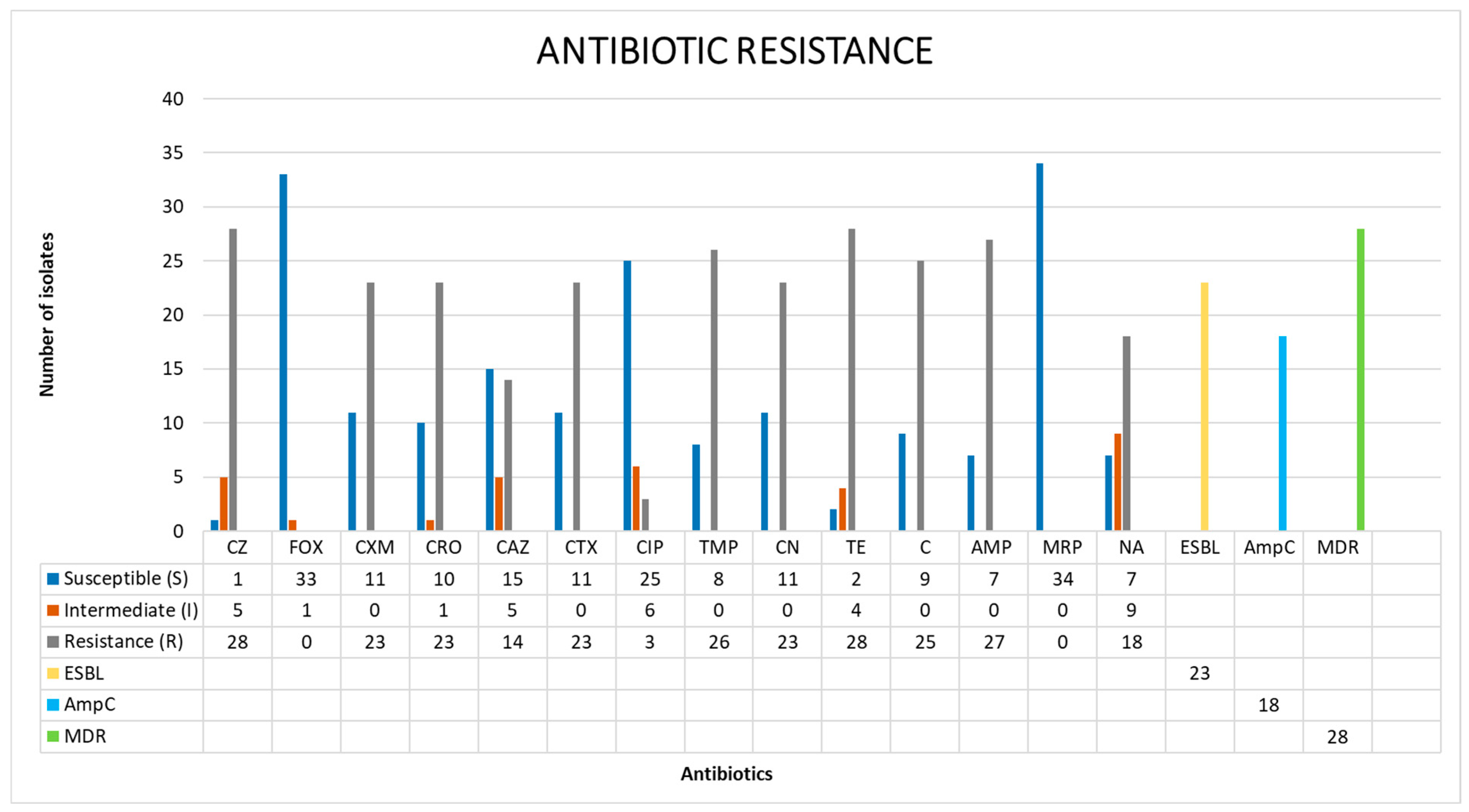
3.2. Antibiotic Resistance Profiles of the Salmonella Isolates

The antibiotic resistance profiles of all isolates are shown in Figure 1. Among the 34 Salmonella strains isolated and tested in this study, 97.06% (33/34 strains) presented a resistance phenotype to at least one of the 15 antimicrobials in the tested panel (Table A1).
The results of the antibiotic susceptibility test indicated that phenotypic resistances to tetracycline and cefazolin were the most common (82.35%, 28/34), followed by ampicillin (79.41%, 27/34), trimethoprim (76.47%, 26/34), chloramphenicol (73.53%, 25/34), cefuroxime (67.65%, 23/34), ceftriaxone (67.65%, 23/34), cefotaxime (67.65%, 23/34), gentamicin (67.65%, 23/34), nalidixic acid (52.94%, 18/34), ceftazidime (41.18%, 14/34), and ciprofloxacin (8.82%, 3/34). However, all isolates were susceptible to cefoxitin and meropenem (Figure 1).
Among the 34 isolates, 18 (52.94%) had the ability to synthesize the AmpC β-lactamase enzyme, and 67.65% of all tested strains were identified as ESBL strains (23/34). In total, 28/34 strains (82.35%) were considered multidrug-resistant strains.

3.2.1. Whole-Genome Sequencing and Genome Characteristics

For a better understanding of genotypic antibiotic resistance, eight strains were whole-genome sequenced using the Illumina platform, including isolate numbers 31_S7, 42_S10, 43_S11, 45_S12, 51_S13, 57_S16, 60_S17, and 68_S20. After de novo assembly using the SPAdes algorithm, the number of contigs was found to range from 305 to 493 contigs, while the N50 values ranged from 22,011 to 36,997 bp. The GC (%) content of the genomes ranged from 52.15% to 52.51% (average 52.36%). All sequencing data were deposited in Genbank with the SRA accession numbers listed below (Table 2).

3.2.2. Antibiotic Resistance Gene Profile

Using in silico prediction, the sequenced genomes of MRD isolates were predicted to carry 43 different ARG in total (Table 3 and Table A2), all of which belong to different gene families. Genotypic predictions of those ARM genes fully matched the phenotypic results yielded in the antibiotic susceptibility test above.

Aminoglycoside

All eight strains were resistant to gentamicin, which is an antibiotic belonging to the aminoglycoside class. The sequenced genomes carried a diverse range of aminoglycoside resistance genes. Among these, coding genes for aminoglycoside acetyltransferase (aac(3)-Iia, aac(3)-IId_1, aac(6)-Iaa_1, and aac(6)-Iy), a protein frequently found in S. Enteritidis and S. Enterica, were identified in all of the sequenced isolates. Genes that confer resistance to aminoglycoside also included ant(3)-Ia_1, aadA17, aadA22, and aadA7_1; these genes encode for aminoglycoside nucleotidyltransferase (found in all eight strains). In the aph group, aph(3)-Ia_3 and aph(6)-Id_1 were found to encode for aminoglycoside phosphotransferases (found in six strains). Another important gene, rmtB-1, was detected in isolate 68_S20; this gene encodes for 16S rRNA methyltransferase.

Beta-Lactam

The antibiotic susceptibility results indicated that all eight isolates were resistant to third-generation cephalosporins. These phenotypes were later confirmed to feature alongside beta-lactam resistance-related genes. blaCTX-M-55 is a notable gene in this group involved in resistance to various antibiotics, including amoxicillin, ampicillin, aztreonam, cefepime, cefotaxime, ceftazidime, ceftriaxone, piperacillin, and ticarcillin. In addition, seven out of eight isolates were predicted to carry the gene blaTEM-1B_1. Some isolates carried several beta-lactam resistance genes. In particular, isolate 68_S20 had one contig that predicted five genes (blaCTX-M-55, blaTEM-1B, blaTEM-206, blaTEM-141, and blaTEM-214), and isolate 51_S13 carried 11 beta-lactam resistance-related genes in total, including blaLAP-2, blaTEM-214, blaTEM-206, blaTEM-33, blaTEM-1B, blaTEM-216, blaTEM-209, blaCTX-M-55, blaTEM-34, blaTEM-210, and blaTEM-141. Notably, isolate 51_S13 featured a contig predicted to contain the beta-lactam resistance genes mentioned above, excluding blaCTX-M-55.

Quinolone

In eight ciprofloxacin and nalidixic acid phenotypically resistant strains, seven out of them contained the qnrS1_1 gene, which is involved in the mechanism of quinolone resistance. The mutations associated with quinolone resistance were as follows: strain No. 68_S20 was related to four mutations, including the parC: p. T57S mutation (ACC to AGC mutation encoding the amino acid T to S); parC:p.S80I (AGC to ATC mutation encoding the amino acid S to I); the gyrA:p.S83F mutation (TCC to TTC mutation encoding the amino acid S to F); and the gyrA:p.D87N mutation (GAC to AAC mutation encoding the amino acid D to N). Seven out of eight strains carried the parC:p.T57S mutation. Furthermore, in one sample (sample 31_S7), qnrS_1 was found to be located on the insertion sequence ISKpn19, which belongs to the ISKra4 family.

Other Genes

We found that six out of eight strains carried the gene floR-2, which encodes for chloramphenicol acetyltransferase. Interestingly, the 5/6-sample fluorine-2 gene was observed in the insertion sequence ISVs3 of the IS91 family (five out of eight samples).
Only one strain carried the gene mph(A)_2, which encodes for the enzyme macrolide phosphotransferase. Seven out of eight strains contained the gene tet(A)_6, which is involved in resistance to the tetracycline group; four out of eight strains carried genes (sul1_5 or sul3_2) related to resistance by replacing the antibiotic target of sulfonamide; and one out of eight isolates carried the gene fosA3_1, encoding the gene for osfomycin thiol transferase. These genes are involved in antibiotic resistance to osfomycin. The genomes of all eight isolates appeared to carry the dfrA14_5 gene, this gene is involved in trimethoprim resistance through the formation of trimethoprim-resistant dihydrofolate reductase dfr. All eight strains contained the gene arr-3_4, which encodes for Rifampin ADP-ribosyltransferase. All eight strains contained the gene lnu(F)_1 (equivalent to lin(F)), which is the gene that encodes for integron-mediated nucleotidyltransferase, resulting in resistance to lincomycin and lindamycin. All strains carried genes associated with multidrug resistance (golS; mdsA; mdsB; mdsC; mdtK; sdiA; Mrx).

3.3. Plasmid Replicons and Virulence Genes

Detailed results of the resistant plasmids are shown in Table 4. Three plasmids were detected in two out of eight Salmonella strains. In the strain 43_S11 genome, we found plasmids IncHI2_1 and IncHI2A_1, while in isolate 60_S17, we detected plasmid IncL/M(pMU407) 1_pMU407.
In addition, to determine the presence of virulent genes, we analyzed the assembled genomes using VFDB with Abricate. The analysis results showed that all eight isolates carried between 72 and 84 virulence genes and contained 20–24 virulent factors (VFs). Furthermore, all strains carried genes encoding for the invasion of host cells (InviA-J). The SPIFinder-2.0 prediction findings indicated the widespread presence of SPI-1, SPI-2, SPI-3, SPI-5, SPI-9, SPI-13, and SPI-14, of which 100% strains had SPI-1, SPI-2, and SPI-3. The most abundant classified serovar was Muenster, with seven out of eight strains belonging to it. Moreover, three out of seven Muenster serovars carried C63PI, an iron transport system in SPI-1.
The mobile element finder (version v.1.0.3, database v.1.0.2) revealed a wide range of plasmid and mobile genetic elements. IncHI2, IncHI2A, and IncL/M are listed among the predicted plasmids (three out of eight strains). The blaCTX-M-55 gene, which confers resistance to cefotaxime and ceftriaxone, was frequently found in IncHI2.

4. Discussion

In this study, 72.34% of whole-duck samples were found to be positive for Salmonella. This percentage of contaminated duck carcasses was significantly higher compared to that in previous studies [8,9,17,21,43,44]. According to Zhengquan’s study, the ratio of Salmonella-positive results in Southern Chinese retail markets was 41.4% [45], while another study by Li et al. in Sichuan Province (Southwestern Chin) determined that 26.9% of samples at a local market were positive for Salmonella [46]. The variety in the Salmonella prevalence rate might be attributed to differences in sample location, sample collection time, sampling methods, and Salmonella detection methods. However, the outcome of our study on duck carcasses was similar to the results of previous studies that experimented on other types of poultry samples, including chicken. A 2018 study by Zhang et al. in China illustrated the contamination of Salmonella in chicken meat at a rate of 63.6% (n = 475) [47]. The Salmonella-positive rate was 65.7% (n = 105) in Thailand in 2017 [48] and 2015. In Ho Chi Minh City, Vu et al. reported a 77.63% (n = 76) Salmonella-positive rate in chicken meat [49]. In addition, the prevalence of Salmonella in our study varied between 69% and 74% and differed in each district. In detail, Ba Dinh had the lowest rate of 69%, while Cau Giay reached the highest amount compared to other districts. This result suggests that the poor hygiene of family-run slaughterhouses might be responsible for the different levels of Salmonella in duck meat. Thus, strategies to improve food safety should be implemented to strengthen the supervision of retail markets, improve the market management system (stall sales, tool cleaning, and regular disinfection), and ensure high standards of environmental hygiene (cleaning and drying retail stands) to protect public health.
Our findings revealed that 97.06% (33/34 strains) of whole-strain samples phenotypically expressed resistance to 15 tested antimicrobials. In detail, MDR Salmonella was most commonly (28/33 strains, 84.85%) found to have an antimicrobial resistance profile in retail duck meat. This result indicates the significant antibiotic resistance capabilities of Salmonella isolates compared to other isolates tested for resistance to Salmonella in duck meat. Chen et al., in 2020, also reported that more than 88.1% (133/151 strains) of isolates in duck meat were multidrug-resistant [45]. Based on other published studies on the resistance of Salmonella, we found that this rate of duck samples critically surpassed that in other animals including chicken, pork, beef, and shellfish. Van et al. in 2007 demonstrated that 50.5% (n = 18) of Salmonella isolates resisted at least one drug, and multidrug resistance was found in all food types [50].
The results of our study correspond to the results of numerous other studies around the world on the antibiotic sensitization of Salmonella globally, as reported by Castro-Vargas et al. in 2020. In this author’s research, reports on current multi-resistance were found in 45/46 studies of Salmonella in poultry. Salmonella strains found in the food chain had high rates of resistance to antibiotics such as nalidixic acid (26.8–86.6%), ampicillin (14.9–68%), ampicillin (14.9–68%), and trimethoprim/sulfamethoxazole (16–54.2%) and were not treatable with carbapenems belonging to families such as imipenem and meropenem [12]. However, our study also showed that the prevalence of Salmonella compared to other antibiotics was higher than that reported by Castro-Vargas et al. for cephalosporins belonging to a resistant family (cefazolin, cefuroxime, cefotaxime, ceftazidime, and ceftriaxone), aminoglycosides (gentamicin), and phenicols (chloramphenicol) [13]. Han et al. reported the rate of Salmonella isolates from a duck slaughter line (fecal and carcass samples) that resisted ampicillin (59.6%), tetracycline (51.3%), ciprofloxacin (27.6%), ceftriaxone (25.6%), and gentamicin (14.1%) [51]. These results are lower than those obtained in our study.
An important reinforcement of the antibiotic resistance test yielded eight selected strains carrying 43 ARG. All strains carried a variety of aminoglycoside-class ARG (aac(3)-Iia, aac(3)-IId_1, aac(6)-Iaa_1….). Moreover, strain No. 68_S20 carrying the rmtB gene encoding for 16S RNA methyltransferase was found to be resistant to all aminoglycoside antibiotic classes, which is an extremely important antibiotic group in animal husbandry and treatment in humans [52]. Furthermore, it is quite surprising that strain 68_S20 with the five beta-lactam family ARG was in the same contig as all eight strains with ESBL phenotypes carrying the blaCTX-M-55 gene (seven Muenster and one Kentucky serovar) (blaTEM-1B; blaCTX-M-55; blaTEM-206; blaTEM-214; and blaTEM-141). Additionally, sample 51_S13 consisted of 11 genes associated with beta-lactam resistance, especially 10/11 genes located in one contig (blaLAP-2; blaTEM-214; blaTEM-206; blaTEM-33; blaTEM-1B; blaTEM-216; blaTEM-209; blaCTX-M-55; blaTEM-34; blaTEM-210; blaTEM-141). This is the first report on this gene cluster in Vietnam. The existence of large clusters of genes resistant to antibiotics could help address the potential threat of AMR gene transmission between different strains and species. Notably, analyzing the genomes of eight strains with quinolone antibiotics containing two genes, floR and qnrS1_1, showed that these genes all carried at least one mutation parC:p.T57S. Especially, strain No. 68_S20 carried four mutations (parC:p.S80I; parC:p.T57S; gyrA:p.S83F; and gyrA:p.D87N). These mutations resulted in resistance to nalidixic acid and ciprofloxacin and hence could enhance and complicate the quinolone family’s antibiotic resistance; we presume these widely predicted mutations possibly because this group of antibiotics is widely used in agriculture. Another finding of interest in this study was the existence of floR-resistant chloramphenicol and florfenicol. This gene is often based on a mobile genetic factor that exacerbates antibiotic resistance due to transverse, vertical, or variable traits, resulting in the very quick and easy transmission of ARG, even for strains that do not exist under the pressure of that antibiotic. Remarkably, seven genes associated with multidrug resistance (golS; mdsA; mdsB; mdsC; mdtK; sdiA; Mrx) were present in all strains.
The WGS data showed that the Salmonella serovar Muenster was the dominant serovar isolated in duck carcass, with three different plasmid replicons in Salmonella isolates (IncHI2_1, IncHI2A_1, IncL/M(pMU407)_1_pMU407). The plasmid replicons were found to be harbored by Salmonella Muenster. Interestingly, IncHI2 and IncHI2A plasmids were harbored by different isolates originating from chickens, ducks, and Muscovy ducks collected from wet markets, which indicated wide dissemination of these plasmids among the other hosts and across distinct geographic regions. These plasmids represent the most significant plasmid lineage implicated in the transmission of antibiotic resistance in Salmonella, particularly in S. Typhimurium strains. β-lactam (blaOXA-1 and blaTEM-1) and quinolone-resistant genes (qnrA and acc(6′)-ib-cr) were horizontally transferred by the IncHI2 plasmid [53].
In total, 72–84 virulence genes implicated in different mechanisms were recorded using the WGS technique. Notably, our results showed that all eight isolates carried Salmonella pathogenicity island 1 (SPI-1) and Salmonella pathogenicity island 3 (SPI-3). SPI-1 plays a significant role in the Salmonella pathogen by invading epithelial cells. SPI-3 contains the mgtCB operon that encodes the MgtC (macrophage survival protein) and MgtB (Mg2+ transporter), thereby enhancing the pathogenicity of Salmonella [54]. However, these strains contained distinct pathogenic islands, virulent factors, and virulence genes due to the differences in their collection locations.
This study showed that ducks sold in the market are a high source of Salmonella enterica infection with very high levels of resistance to many antibiotics and a high diversity of ARG. Therefore, this is a public health issue that deserves public attention.

Author Contributions

Conceptualization, T.T.N., D.P.X., H.T.T.T. and H.H.L.T.; data curation, T.T.N.; formal analysis, T.T.N. and H.V.L.; funding acquisition, T.T.N.; investigation, T.T.N., H.V.T.H. and T.N.T.; methodology, T.T.N., H.V.L., H.V.T.H. and T.N.T.; project administration, T.T.N.; resources, T.T.N., H.V.L. and H.H.L.T.; software, T.T.N., H.V.L., T.N.T. and H.T.T.T.; supervision, D.P.X., H.T.T.T. and H.H.L.T.; validation, T.T.N., D.P.X. and H.T.T.T.; visualization, T.T.N.; writing—original draft, T.T.N., H.V.L. and T.N.T.; writing—review and editing, T.T.N., H.M.N., D.P.X., H.T.T.T. and H.H.L.T. All authors have read and agreed to the published version of the manuscript.

Funding

We would like to acknowledge the National Institute for Food Control, Ministry of Health in Vietnam, for funding this project under the Specific Task Program 2019 (Vietnam Ministry of Health, numbered 149/QD-BYT; project title: Genotyping of Salmonella serovars prevalent in northern Vietnam).

Institutional Review Board Statement

Not applicable.

Informed Consent Statement

Not applicable.

Data Availability Statement

Not applicable.

Acknowledgments

We thank the members of our laboratories for the meaningful discussion and technical assistance.

Conflicts of Interest

The authors declare no conflict of interest.

Appendix A

Table A1. Antibiotic resistance profiles of Salmonella isolates (zone of inhibition expressed in millimeters).
Isolation of SamplesCXM
(mm)
CRO
(mm)
FOX
(mm)
CZ
(mm)
CTX
(mm)
CAZ
(mm)
TM
(mm)
TE
(mm)
C
(mm)
CN
(mm)
NA
(mm)
CIP
(mm)
AMP
(mm)
MRP
(mm)
Resistance Number
Ba Đinh DistrictR
(8)
R
(6)
S
(25)
R
(6)
R
(6)
R
(6)
R
(6)
R
(8)
S
(26)
R
(6)
I
(18)
S
(27)
R
(6)
S
(35)
9
S
(22)
I
(21)
S
(24)
I
(20)
S
(33)
S
(25)
S
(33)
I
(12)
R
(8)
S
(20)
S
(24)
S
(39)
S
(21)
S
(37)
1
R
(9)
R
(6)
S
(24)
R
(6)
R
(6)
R
(6)
R
(6)
R
(11)
R
(6)
R
(6)
S
(20)
S
(31)
R
(6)
S
(35)
10
R
(6)
R
(9)
S
(27)
R
(6)
R
(6)
R
(6)
R
(6)
R
(6)
R
(6)
S
(12)
I
(17)
S
(25)
R
(6)
S
(30)
10
S
(27)
S
(32)
S
(27)
S
(23)
S
(38)
S
(27)
S
(32)
S
(18)
S
(30)
S
(20)
I
(18)
I
(28)
S
(21)
S
(25)
0
Cau Giay DistrictR
(10)
R
(10)
S
(21)
R
(6)
R
(8)
R
(13)
R
(6)
R
(6)
R
(6)
R
(12)
R
(6)
S
(30)
R
(6)
S
(34)
11
R
(10)
R
(6)
S
(18)
R
(6)
R
(6)
R
(6)
R
(6)
R
(6)
R
(6)
R
(6)
R (12)S
(27)
R
(6)
S
(37)
11
R
(6)
R
(6)
S
(26)
R
(6)
R
(6)
R
(6)
R
(6)
R
(10)
R
(6)
R
(6)
S (20)S
(35)
R
(6)
S
(35)
10
Dong Da DistrictS
(22)
S
(34)
S
(23)
I
(21)
S
(31)
S
(23)
R
(6)
R
(11)
R
(6)
R
(6)
S
(21)
S
(29)
S
(19)
S
(32)
4
R
(6)
R
(6)
S
(25)
R
(6)
R
(6)
R
(6)
R
(6)
R
(6)
S
(25)
R
(8)
I
(17)
I
(23)
R
(6)
S
(30)
9
S
(26)
S
(37)
S
(28)
I
(21)
S
(34)
S
(27)
S
(33)
R
(11)
S
(30)
S
(21)
S
(27)
S
(41)
S
(21)
S
(26)
1
R
(6)
R
(8)
S
(19)
R
(6)
R
(10)
I
(20)
R
(6)
R
(6)
R
(6)
R
(11)
R
(6)
S
(30)
R
(6)
S
(32)
10
R
(6)
R
(11)
S
(25)
R
(6)
R
(16)
S
(22)
R
(6)
R
(6)
R
(6)
R
(12)
R
(6)
S
(32)
R
(6)
S
(35)
10
R
(6)
R
(12)
S
(23)
R
(6)
R
(19)
S
(27)
R
(6)
R
(6)
R
(6)
R
(11)
R
(6)
S
(31)
R
(6)
S
(35)
10
R
(6)
R
(8)
S
(29)
R
(6)
R
(7)
I
(19)
R
(6)
R
(6)
R
(6)
R
(10)
R
(6)
S
(34)
R
(6)
S
(37)
10
R
(6)
R
(12)
S
(25)
R
(6)
R
(19)
S
(21)
S
(30)
R
(6)
R
(6)
R
(12)
R
(6)
S
(32)
R
(6)
S
(35)
9
R
(6)
R
(9)
I
(17)
R
(6)
R
(9)
R
(16)
S
(28)
R
(6)
R
(6)
R
(10)
R
(6)
S
(27)
R
(6)
S
(31)
10
Hoang Mai DistrictS (22)S (32)S (24)R (17)S (34)S (25)R (6)R (8)S (27)S (20)I (15)S (34)R (6)S (35)4
R (6)R (6)S (24)R (6)R (6)R (15)R (6)R (11)R (6)R (9)R (6)R (13)R (6)S (35)10
R (6)R (8)S (21)R (6)R (8)R (12)R (6)R (6)R (6)RR (6)R (8)R (6)S (36)10
R (6)R (6)S (33)R (6)R (8)S (26)R (6)I (12)R (6)R (6)R (6)R (18)R (6)S (30)8
R (6)R (8)S (21)R (6)R (9)S (21)R (6)R (6)R (6)R (12)R (6)S (32)R (6)S (36)9
S (21)S (35)S (19)R (15)S (36)S (25)R (6)R (8)R (6)S (17)R (11)I (23)R (6)S (30)5
S (25)S (38)S (27)I (21)S (39)S (27)S (36)R (10)S (32)S (22)I (16)S (34)S (22)S (26)1
S (22)S (33)S (25)I (20)S (35)S (27)R (6)I (14)S (29)S (20)I (15)S (28)S (22)S (23)1
Thanh Xuan DistrictS
(21)
S
(31)
S
(23)
R
(17)
S
(32)
I
(20)
R
(6)
R
(8)
R
(6)
S
(17)
I
(15)
S
(30)
R
(6)
S
(30)
5
R
(9)
R
(9)
S
(23)
R
(6)
R
(9)
R
(13)
R
(6)
R
(11)
S
(24)
R
(12)
I
(15)
I
(24)
R
(6)
S
(29)
9
R
(6)
R
(6)
S
(20)
R
(6)
R
(6)
R
(6)
R
(6)
R
(6)
R
(6)
R
(6)
R
(12)
I
(25)
R
(6)
S
(34)
11
R
(10)
R
(6)
S
(21)
R
(6)
R
(6)
R
(6)
R
(6)
I
(14)
R
(6)
R
(6)
R
(6)
S
(26)
R
(6)
S
(37)
10
S
(22)
S
(31)
S
(24)
R
(17)
S
(33)
S
(23)
R
(6)
R
(8)
R
(6)
S
(17)
R
(11)
I
(23)
R
(6)
S
(30)
6
R
(6)
R
(11)
S
(30)
R
(6)
R
(9)
R
(6)
R
(6)
R
(10)
R
(6)
R
(6)
S
(20)
S
(30)
R
(6)
S
(35)
10
R
(6)
R
(13)
S
(23)
R
(6)
R
(12)
I
(20)
R
(6)
R
(6)
R
(6)
R
(12)
R
(6)
S
(29)
R
(6)
S
(32)
10
R
(6)
R
(9)
S
(20)
R
(6)
R
(13)
I
(19)
S
(28)
R
(11)
R
(6)
S
(20)
R
(6)
S
(31)
R
(6)
S
(33)
8
S
(20)
S
(31)
S
(20)
R
(18)
S
(30)
S
(23)
S
(30)
S
(19)
S
(27)
S
(19)
S
(22)
S
(36)
S
(18)
S
(35)
1
E. coli ATCC 25922 (negative control)S
(28)
S (27)S (26)S (34)S (35)S (29)S (30)S (30)S (32)S (30)S (29)S (31)S (32)S (30)-
Table A2. Antimicrobial resistance genes in Salmonella isolates.
Drug Classes
Antibiotic ResistanceCode StrainStrains AminoglycosideBeta-LactamChloramphenicolQuinoloneMacrolidesTetracyclineSulfonamides FosfomycinDiaminopyrimidineRifampinLincosamidePolypeptideMultidrug Classes
CMX-CRO-CZ-CTX-CAZ-TM-CN-TE-AMP68_S20Kentuckyaac(3)-Iia;
aac(3)-Id;
aac(3)-IId_1;
aac(6)-Iaa_1; aadA17; aadA7_1; ant(3)-Ia_1; aph(3)-Ia_3;
rmtB_1;
blaCTX-M-55;
blaTEM-206;
blaTEM-1B;
blaTEM-141;
blaTEM-214
floR_2qnrS1_1mph(A)-2Tet(A)_6;
TetR
sul1_5; fosA3_1dfrA14_5ARR-3_4; ARR-2;lnu(F)_1;
linG;
golS;
mdsA;
mdsB;
mdsC;
mdtK;
sdiA;
Mrx;
CMX-CRO-CZ-CTX-CAZ-TM-CN-TE-C-AMP43_S11Muensteraac(3)-Iia; aac(3)-IId_1; aac(6)-Iaa_1;
aac(6)-Iy; aadA22; ant(3)-Ia_1; aph(3)-Ia_3; aph(6)-Id_1;
blaCTX-M-55; blaTEM-1B_1;
blaLAP-2
floR_2qnrS1_1 Tet(A)_6;
TetR
sul3_2; dfrA14_5ARR-3_4; ARR-2;lnu(F)_1;
linG;
golS;
mdsA;
mdsB;
mdsC;
mdtK;
sdiA;
CMX-CRO-CZ-CTX-CAZ-TM-CN-TE-C-AMP45_S12Muensteraac(3)-Iia; aac(3)-IId_1; aac(6)-Iaa_1;
aac(6)-Iy; aadA22; ant(3)-Ia_1; aph(6)-Id_1;
blaCTX-M-55; blaTEM-1B_1;
blaLAP-2
floR_2qnrS1_1 Tet(A)_6;
TetR
sul3_2; dfrA14_5ARR-3_4; ARR-2;lnu(F)_1;
linG;
golS;
mdsA;
mdsB;
mdsC;
mdtK;
sdiA;
CMX-CRO-CZ-CTX-CAZ-TM-CN-TE-C-AMP51_S13Muensteraac(3)-Iia; aac(3)-IId_1; aac(6)-Iaa_1;
aac(6)-Iy; aadA22; ant(3)-Ia_1; aph(6)-Id_1;
blaCTX-M-55; blaTEM-1B_1;
blaLAP-2;
blaTEM-214;
blaTEM-206;
blaTEM-33;
blaTEM-216;
blaTEM-209;
blaTEM-34;
blaTEM-210;
blaTEM-141
qnrS1_1 Tet(A)_6;
TetR
sul3_2; dfrA14_5ARR-3_4; ARR-2;lnu(F)_1;
linG;
golS;
mdsA;
mdsB;
mdsC;
mdtK;
sdiA;
CMX-CRO-CZ-CTX-CAZ-TM-CN-TE-AMP31_S7Muensteraac(3)-Iia; aac(3)-IId_1; aac(6)-Iaa_1;
aac(6)-Iy; aadA22; ant(3)-Ia_1; aph(6)-Id_1;
blaCTX-M-55; blaTEM-1B_1; blaLAP-2 qnrS1_1 Tet(A)_6;
TetR
sul3_2; dfrA14_5ARR-3_4; ARR-2;lnu(F)_1;
linG;
golS;
mdsA;
mdsB;
mdsC;
mdtK;
sdiA;
CMX-CRO-CZ-CTX-CAZ-TM-CN-TE-C-AMP42_S10Muensteraac(3)-Iia; aac(3)-IId_1; aac(6)-Iaa_1;
aac(6)-Iy; aadA22; ant(3)-Ia_1; aph(6)-Id_1;
blaCTX-M-55; blaTEM-1B_1;
blaLAP-2
floR_2qnrS1_1 Tet(A)_6;
TetR
sul3_2; dfrA14_5ARR-3_4; ARR-2;lnu(F)_1;
linG;
golS;
mdsA;
mdsB;
mdsC;
mdtK;
sdiA;
CMX-CRO-CZ-CTX-CAZ-TM-CN-C-AMP57_S16Muensteraac(3)-Iia; aac(3)-IId_1; aac(6)-Iaa_1;
aac(6)-Iy; aadA22; ant(3)-Ia_1; aph(3)-Ia_3; aph(6)-Id_1;
blaCTX-M-55; blaTEM-1B_1floR_2qnrS1_1 Tet(A)_6;
TetR
sul3_2; dfrA14_5ARR-3_4; ARR-2;lnu(F)_1;
linG;
golS;
mdsA;
mdsB;
mdsC;
mdtK;
sdiA;
CMX-CRO-CZ-CTX-CAZ-TM-CN-TE-C-AMP60_S17Muensteraac(3)-Iia; aac(3)-IId_1; aac(6)-Iaa_1;
aac(6)-Iy; aadA22; ant(3)-Ia_1; aph(3)-Ia_3;
blaCTX-M-55; blaTEM-1B_1;
blaLAP-2
floR_2qnrS1_1 sul3_2; dfrA14_5ARR-3_4; ARR-2;lnu(F)_1;
linG;
golS;
mdsA;
mdsB;
mdsC;
mdtK;
sdiA;

References

  1. FAO; WHO. M I C R O B I O Salmonella and Campylobacter in Chicken Meat; Center for Emerging: Geneva, Switzerland, 2009. [Google Scholar]
  2. EFSA; ECDC. The European Union Summary Report on Trends and Sources of Zoonoses, Zoonotic Agents and Food-Borne Outbreaks in 2017. EFSA J. 2018, 16, e05500. [Google Scholar] [CrossRef] [Green Version]
  3. Center for Emerging; CDCN; Infectious Diseases, Z. National Enteric Disease Surveillance: Salmonella Annual Report 2016; Centers for Disease Control and Prevention: Atlanta, GA, USA, 2016.
  4. Centers for Disease Control and Prevention. Antibiotic Resistance Threats in the United States 2019; Centers for Disease Control and Prevention: Atlanta, GA, USA, 2019. [CrossRef] [Green Version]
  5. Ferrari, R.G.; Rosario, D.K.A.; Cunha-Neto, A.; Mano, S.B.; Figueiredo, E.E.S.; Conte-Juniora, C.A. Worldwide Epidemiology of Salmonella Serovars in Animal-Based Foods: A Meta-Analysis. Appl. Environ. Microbiol. 2019, 85, e00591-19. [Google Scholar] [CrossRef] [PubMed] [Green Version]
  6. EFSA; ECDC. The European Union One Health 2020 Zoonoses Report. EFSA J. 2021, 19, e06971. [Google Scholar] [CrossRef]
  7. Appleby, M.C. Whom Should We Eat? Why Veal Can Be Better for Welfare than Chicken; Appleby, M.C., Weary, D.M., Sandoe, P., Eds.; CABI: Wallingford, UK, 2014. [Google Scholar] [CrossRef]
  8. Tran, T.P.; Ly, T.L.K.; Nguyen, T.T.; Akiba, M.; Ogasawara, N.; Shinoda, D.; Okatani, A.T.; Hayashidani, H. Prevalence of Salmonella Spp. in Pigs, Chickens and Ducks in the Mekong Delta, Vietnam. J. Vet. Med. Sci. 2004, 66, 1011–1014. [Google Scholar] [CrossRef] [PubMed] [Green Version]
  9. Cha, S.Y.; Kang, M.; Yoon, R.H.; Park, C.K.; Moon, O.K.; Jang, H.K. Prevalence and Antimicrobial Susceptibility of Salmonella Isolates in Pekin Ducks from South Korea. Comp. Immunol. Microbiol. Infect. Dis. 2013, 36, 473–479. [Google Scholar] [CrossRef]
  10. Nair, D.V.T.; Venkitanarayanan, K.; Johny, A.K. Antibiotic-Resistant Salmonella in the Food Supply and the Potential Role of Antibiotic Alternatives for Control. Foods 2018, 7, 167. [Google Scholar] [CrossRef] [Green Version]
  11. McArthur, A.G.; Waglechner, N.; Nizam, F.; Yan, A.; Azad, M.A.; Baylay, A.J.; Bhullar, K.; Canova, M.J.; de Pascale, G.; Ejim, L.; et al. The Comprehensive Antibiotic Resistance Database. Antimicrob. Agents Chemother. 2013, 57, 3348. [Google Scholar] [CrossRef] [Green Version]
  12. Castro-Vargas, R.E.; Herrera-Sánchez, M.P.; Rodríguez-Hernández, R.; Rondón-Barragán, I.S. Antibiotic Resistance in Salmonella Spp. Isolated from Poultry: A Global Overview. Vet. World 2020, 13, 2070–2084. [Google Scholar] [CrossRef]
  13. Hagag, A.; Naguib, D.; Mohamed, A.A.; El-Gohary, A.H. Prevalence, Virulence Genes, and Antibiotic Resistance of Escherichia Coli and Salmonella Spp. Isolated from Pigeons and Humans ARTICLE HISTORY ABSTRACT. Mansoura. Vet. Med. J. 2022, 23, 24–30. [Google Scholar] [CrossRef]
  14. Carrique-Mas, J.J.; Trung, N.V.; Hoa, N.T.; Mai, H.H.; Thanh, T.H.; Campbell, J.I.; Wagenaar, J.A.; Hardon, A.; Hieu, T.Q.; Schultsz, C. Antimicrobial Usage in Chicken Production in the Mekong Delta of Vietnam. Zoonoses Public Health 2014, 62 (Suppl. 1), 70–78. [Google Scholar] [CrossRef] [Green Version]
  15. McDermott, P.F.; Zhao, S.; Tate, H. Antimicrobial Resistance in Nontyphoidal Salmonella. Microbiol. Spectr. 2018, 6, 261–287. [Google Scholar] [CrossRef] [PubMed] [Green Version]
  16. Chu, C.; Chiu, C.-H. Forum on Antimicrobial Resistance Evolution of the Virulence Plasmids of Non-Typhoid Salmonella and Its Association with Antimicrobial Resistance. Microbes Infect. 2006, 8, 1931–1936. [Google Scholar] [CrossRef] [PubMed]
  17. Thi Phan, T.; Thi Lien Khai, L.; Ogasawara, N.; Thu Tam, N.; Tomomitsu Okatani, A.; Akiba, M.; Hayashidani, H. Contamination of Salmonella in Retail Meats and Shrimps in the Mekong Delta, Vietnam. J. Food Prot. 2005, 68, 1077–1080. [Google Scholar] [CrossRef] [PubMed]
  18. Humphrey, T. Salmonella in Domestic Animals; Wray, C., Wray, A., Eds.; CABI International: Oxford, UK, 2000. [Google Scholar]
  19. Löfström, C.; Hansen, T.; Maurischat, S.; Malorny, B. Salmonella: Salmonellosis. Encycl. Food Health 2015, 701–705. [Google Scholar] [CrossRef]
  20. Nguyen, T.K.; Nguyen, L.T.; Chau, T.T.H.; Nguyen, T.T.; Tran, B.N.; Taniguchi, T.; Hayashidani, H.; Ly, K.T.L. Prevalence and Antibiotic Resistance of Salmonella Isolated from Poultry and Its Environment in the Mekong Delta, Vietnam. Vet. World 2021, 14, 3216. [Google Scholar] [CrossRef]
  21. Tsai, H.J.; Hsiang, P.H. The Prevalence and Antimicrobial Susceptibilities of Salmonella and Campylobacter in Ducks in Taiwan. J. Vet. Med. Sci. 2005, 67, 7–12. [Google Scholar] [CrossRef] [Green Version]
  22. Scaltriti, E.; Sassera, D.; Comandatore, F.; Morganti, C.M.; Mandalari, C.; Gaiarsa, S.; Bandi, C.; Zehender, G.; Bolzoni, L.; Casadei, G.; et al. Differential Single Nucleotide Polymorphism-Based Analysis of an Outbreak Caused by Salmonella Enterica Serovar Manhattan Reveals Epidemiological Details Missed by Standard Pulsed-Field Gel Electrophoresis. J. Clin. Microbiol. 2015, 53, 1227. [Google Scholar] [CrossRef] [Green Version]
  23. Achtman, M.; Wain, J.; Weill, F.X.; Nair, S.; Zhou, Z.; Sangal, V.; Krauland, M.G.; Hale, J.L.; Harbottle, H.; Uesbeck, A.; et al. Multilocus Sequence Typing as a Replacement for Serotyping in Salmonella Enterica. PLoS Pathog. 2012, 8, e1009040. [Google Scholar] [CrossRef] [Green Version]
  24. Gilchrist, C.A.; Turner, S.D.; Riley, M.F.; Petri, W.A.; Hewlett, E.L. Whole-Genome Sequencing in Outbreak Analysis. Clin. Microbiol. Rev. 2015, 28, 541. [Google Scholar] [CrossRef] [Green Version]
  25. Köser, C.U.; Ellington, M.J.; Peacock, S.J. Whole-Genome Sequencing to Control Antimicrobial Resistance. Trends Genet. 2014, 30, 401. [Google Scholar] [CrossRef] [Green Version]
  26. La, T.M.; Kim, T.; Lee, H.J.; Lee, J.B.; Park, S.Y.; Choi, I.S.; Lee, S.W. Whole-Genome Analysis of Multidrug-Resistant Salmonella Enteritidis Strains Isolated from Poultry Sources in Korea. Pathogens 2021, 10, 1615. [Google Scholar] [CrossRef] [PubMed]
  27. Deng, X.; den Bakker, H.C.; Hendriksen, R.S. Genomic Epidemiology: Whole-Genome-Sequencing-Powered Surveillance and Outbreak Investigation of Foodborne Bacterial Pathogens. Annu. Rev. Food Sci. Technol. 2016, 7, 16–17. [Google Scholar] [CrossRef] [PubMed] [Green Version]
  28. Pornsukarom, S.; van Vliet, A.H.M.; Thakur, S. Whole Genome Sequencing Analysis of Multiple Salmonella Serovars Provides Insights into Phylogenetic Relatedness, Antimicrobial Resistance, and Virulence Markers across Humans, Food Animals and Agriculture Environmental Sources. BMC Genom. 2018, 19, 801. [Google Scholar] [CrossRef] [Green Version]
  29. USDA. Isolation and Identification of Salmonella from Meat, Poultry, Pasteurized Egg, and Siluriformes (Fish) Products and Carcass and Environmental Sponges. Revision 10. 2019. Available online: https://content.govdelivery.com/accounts/USFSIS/bulletins/2ed04a9 (accessed on 24 August 2022).
  30. Hasman, H.; Agersø, Y.; Hendriksen, R.; Cavaco, L.M.; Guerra-Roman, B. Isolation of ESBL-, AmpC-and Carbapenemase-Producing E. coli from Caecal Samples. LAB. PROTOCOL. 2019, 1–18. [Google Scholar]
  31. Bauer, A.W.; Kirby, W.M.; Sherris, J.C.; Turck, M. Antibiotic Susceptibility Testing by a Standardized Single Disk Method. Am. J. Clin. Pathol. 1966, 45, 493–496. [Google Scholar] [CrossRef]
  32. CLSI. Performance Standards for Antimicrobial Susceptibility. Clin. Lab. Stand. Inst. 2022, 32, 1–16. [Google Scholar]
  33. Bolger, A.M.; Lohse, M.; Usadel, B. Trimmomatic: A Flexible Trimmer for Illumina Sequence Data. Bioinformatics 2014, 30, 2114. [Google Scholar] [CrossRef] [Green Version]
  34. Bankevich, A.; Nurk, S.; Antipov, D.; Gurevich, A.A.; Dvorkin, M.; Kulikov, A.S.; Lesin, V.M.; Nikolenko, S.I.; Pham, S.; Prjibelski, A.D.; et al. SPAdes: A New Genome Assembly Algorithm and Its Applications to Single-Cell Sequencing. J. Comput. Biol. 2012, 19, 455–477. [Google Scholar] [CrossRef] [Green Version]
  35. Assefa, S.; Keane, T.M.; Otto, T.D.; Newbold, C.; Berriman, M. ABACAS: Algorithm-Based Automatic Contiguation of Assembled Sequences. Bioinformatics 2009, 25, 1968–1969. [Google Scholar] [CrossRef] [Green Version]
  36. Yoshida, C.E.; Kruczkiewicz, P.; Laing, C.R.; Lingohr, E.J.; Gannon, V.P.J.; Nash, J.H.E.; Taboada, E.N. The Salmonella in Silico Typing Resource (SISTR): An Open Web-Accessible Tool for Rapidly Typing and Subtyping Draft Salmonella Genome Assemblies. PLoS ONE 2016, 11, e0147101. [Google Scholar] [CrossRef] [Green Version]
  37. Seemann, T. ABRicate: Mass Screening of Contigs for Antiobiotic Resistance Genes. Available online: https://github.com/tseemann/abricate (accessed on 24 August 2022).
  38. Zankari, E.; Hasman, H.; Cosentino, S.; Vestergaard, M.; Rasmussen, S.; Lund, O.; Aarestrup, F.M.; Larsen, M.V. Identification of Acquired Antimicrobial Resistance Genes. J. Antimicrob. Chemother. 2012, 67, 2640–2644. [Google Scholar] [CrossRef] [PubMed]
  39. Gupta, S.K.; Padmanabhan, B.R.; Diene, S.M.; Lopez-Rojas, R.; Kempf, M.; Landraud, L.; Rolain, J.M. ARG-ANNOT, a New Bioinformatic Tool to Discover Antibiotic Resistance Genes in Bacterial Genomes. Antimicrob. Agents Chemother. 2014, 58, 212–220. [Google Scholar] [CrossRef] [PubMed] [Green Version]
  40. Carattoli, A.; Zankari, E.; Garciá-Fernández, A.; Larsen, M.V.; Lund, O.; Villa, L.; Aarestrup, F.M.; Hasman, H. In Silico Detection and Typing of Plasmids Using Plasmidfinder and Plasmid Multilocus Sequence Typing. Antimicrob. Agents Chemother. 2014, 58, 3895–3903. [Google Scholar] [CrossRef] [PubMed] [Green Version]
  41. Chen, L.; Yang, J.; Yu, J.; Yao, Z.; Sun, L.; Shen, Y.; Jin, Q. VFDB: A Reference Database for Bacterial Virulence Factors. Nucleic Acids Res. 2005, 33, D325–D328. [Google Scholar] [CrossRef] [PubMed] [Green Version]
  42. Johansson, M.H.K.; Bortolaia, V.; Tansirichaiya, S.; Aarestrup, F.M.; Roberts, A.P.; Petersen, T.N. Detection of Mobile Genetic Elements Associated with Antibiotic Resistance in Salmonella Enterica Using a Newly Developed Web Tool: MobileElementFinder. J. Antimicrob. Chemother. 2021, 76, 101–109. [Google Scholar] [CrossRef]
  43. Flament, A.; Soubbotina, A.; Mainil, J.; Marlier, D. Prevalence of Salmonella Serotypes in Male Mule Ducks in Belgium. Vet. Rec. 2012, 170, 311. [Google Scholar] [CrossRef]
  44. Adzitey, F.; Huda, N.; Rahmat Ali, G.R. Prevalence and Antibiotic Resistance of Campylobacter, Salmonella, and L. Monocytogenes in Ducks: A Review. Foodborne Pathog. Dis. 2012, 9, 498–505. [Google Scholar] [CrossRef]
  45. Chen, Z.; Bai, J.; Wang, S.; Zhang, X.; Zhan, Z.; Shen, H.; Zhang, H.; Wen, J.; Gao, Y.; Liao, M.; et al. Prevalence, Antimicrobial Resistance, Virulence Genes and Genetic Diversity of Salmonella Isolated from Retail Duck Meat in Southern China. Microorganisms 2020, 8, 444. [Google Scholar] [CrossRef] [Green Version]
  46. Li, R.; Lai, J.; Wang, Y.; Liu, S.; Li, Y.; Liu, K.; Shen, J.; Wu, C. Prevalence and Characterization of Salmonella Species Isolated from Pigs, Ducks and Chickens in Sichuan Province, China. Int. J. Food Microbiol. 2013, 163, 14–18. [Google Scholar] [CrossRef]
  47. Zhang, L.; Fu, Y.; Xiong, Z.; Ma, Y.; Wei, Y.; Qu, X.; Zhang, H.; Zhang, J.; Liao, M. Highly Prevalent Multidrug-Resistant Salmonella From Chicken and Pork Meat at Retail Markets in Guangdong, China. Front. Microbiol. 2018, 9, 2104. [Google Scholar] [CrossRef] [Green Version]
  48. Trongjit, S.; Angkititrakul, S.; Tuttle, R.E.; Poungseree, J.; Padungtod, P.; Chuanchuen, R. Prevalence and Antimicrobial Resistance in Salmonella Enterica Isolated from Broiler Chickens, Pigs and Meat Products in Thailand–Cambodia Border Provinces. Microbiol. Immunol. 2017, 61, 23–33. [Google Scholar] [CrossRef] [PubMed] [Green Version]
  49. Vu, T.H.A.; Hai, C.V.; Ha, H.Y.; Tu, N.H.K. Antibiotic Resistance in Salmonella Isolated from Ho Chi Minh City (Vietnam) and Difference of Sulfonamide Resistance Gene Existence in Serovars. J. Pure. Appl. Microbiol. 2021, 15, 2244–2251. [Google Scholar] [CrossRef]
  50. Van, T.T.H.; Moutafis, G.; Istivan, T.; Tran, L.T.; Coloe, P.J. Detection of Salmonella Spp. in Retail Raw Food Samples from Vietnam and Characterization of Their Antibiotic Resistance. Appl. Environ. Microbiol. 2007, 73, 6885. [Google Scholar] [CrossRef] [PubMed] [Green Version]
  51. Han, X.; Peng, J.; Guan, X.; Li, J.; Huang, X.; Liu, S.; Wen, Y.; Zhao, Q.; Huang, X.; Yan, Q.; et al. Genetic and Antimicrobial Resistance Profiles of Salmonella Spp. Isolated from Ducks along the Slaughter Line in Southwestern China. Food Control 2020, 107, 106805. [Google Scholar] [CrossRef]
  52. Wang, J.; Wang, Z.Y.; Wang, Y.; Sun, F.; Li, W.; Wu, H.; Shen, P.C.; Pan, Z.M.; Jiao, X. Emergence of 16S RRNA Methylase Gene RmtB in Salmonella Enterica Serovar London and Evolution of RmtB-Producing Plasmid Mediated by IS26. Front. Microbiol. 2021, 11, 604278. [Google Scholar] [CrossRef]
  53. Zhou, D.; Li, X.-Z.; Canada, H.; Pina Fratamico, C.; Chen, W.; Fang, T.; Zhou, X.; Zhang, D.; Shi, X.; Shi, C. IncHI2 Plasmids Are Predominant in Antibiotic-Resistant Salmonella Isolates. Front. Microbiol. 2016, 7, 1566. [Google Scholar] [CrossRef] [Green Version]
  54. Atrice Blanc-Potard, A.-B.; Solomon, F.; Kayser, J.; Groisman, E.A. The SPI-3 Pathogenicity Island of Salmonella Enterica. J. Bacteriol. 1999, 181, 998–1004. [Google Scholar] [CrossRef] [Green Version]
Figure 1. Antibiotic resistance profiles of duck carcass samples: cefazolin (CZ), cefoxitin (FOX), cefuroxime (CXM), ceftriaxone (CRO), ceftazidime (CAZ), cefotaxime (CTX), ciprofloxacin (CIP), trimethoprim (TMP), gentamicin (CN), tetracycline (TE), chloramphenicol (C), ampicillin (AMP), meropenem (MRP), nalidixic acid (NA), extended-spectrum beta-lactam (ESBL), AmpC β-lactamase (AmpC), and multidrug resistance (MDR).
Cimb 45 00143 g001
Table 1. Serotyping and cgMLST results.
SampleSerovarSerogroupH1H2O AntigenMLST
31_S7Muenster-e,h1,53,{10}{15}{15,34}321
42_S10Muenster-e,h1,53,{10}{15}{15,34}321
43_S11Muenster-e,h1,53,{10}{15}{15,34}321
45_S12Muenster-e,h1,53,{10}{15}{15,34}321
51_S13MuensterE1e,h1,53,{10}{15}{15,34}321
57_S16Muenster-e,h1,53,{10}{15}{15,34}321
60_S17Muenster-e,h1,53,{10}{15}{15,34}321
68_S20KentuckyC2-C3iz68,20198
Table 2. Characteristics of sequenced and assembled genomic data.
SampleReadsAverage Read Length
(bp)
Contigs Genome Length
(bp)
Average Contig Length
(bp)
N50
(bp)
GC
(%)
SRA Accession Number
31_S7808,7522383054,701,119129,32134,94252.29SRR21051813
42_S10683,2582374254,671,790103,64728,05052.45SRR21051784
43_S11677,2482384934,905,076101,19623,43752.15SRR21051783
45_S12749,1402393344,744,687160,34537,07452.27SRR21051781
51_S13783,1242393294,766,796157,72936,99752.32SRR21051780
57_S16591,7342373824,655,893156,59730,15352.41SRR21051777
60_S17655,3102384934,682,79787,05422,01152.51SRR21051776
68_S20910,6442374644,804,148181,02024,43652.47SRR21051782
Table 3. Distribution of ARG in Salmonella serovars based on in silico predictions.
Samples
68_S2043_S1145_S1251_S1331_S742_S1057_S1660_S17
SerovarKentuckyMuensterMuensterMuensterMuensterMuensterMuensterMuenster
Genes/Number of genes identified3327263325262624
Drug classesRifampinarr-3_411111111
arr211111111
Aminoglycosideaac(3)-Iia11111111
aac(3)-IId_111111111
aac(3)-Id_11 Absence (Negative)
aac(6)-Iaa_111111111
aac(6)-Iy 1111111
aadA171
aadA22 1111111
aadA7_11
ant(3)-Ia_111111111
aph(3)-Ia_311 11
aph(6)-Id_1 111111
rmtB_11
Beta-lactamblaCTX-M-55_111111111
blaLAP-2 11111 1
blaTEM-1411 1
blaTEM-1B_111111111
blaTEM-2061 1
blaTEM-209 1
blaTEM-210 1
blaTEM-2141 1
blaTEM-216 1
blaTEM-33 1 presence (Positive)
blaTEM-34 1
FosfomycinfosA3_11
ChloramphenicolfloR_2111 111
DiaminopyrimidinedfrA14_511111111
LincosamidelinG11111111
Inu(F)_111111111
QuinoloneqnrS1_111111111
Macrolidesmph(A)-21
Sulfonamidessul1_51
sul3_2 1111111
Tetracyclintet(A)_61111111
tetR1111111
Multi-drug classesgolS11111111
mdsA11111111
mdsB11111111
mdsC11111111
mdtK11111111
Mrx1
sdiA11111111
Table 4. Plasmids, virulence factors, and SPI results.
StrainsSerotypePlasmidNumber of Virulence FactorsNumber of Virulence GenesSPI
31_S7Muenster 2482SPI-1, SPI-2, SPI-3, SPI-5, SPI-9, SPI-13, SPI-14
42_S10Muenster 2379C63PI, SPI-1, SPI-2, SPI-3, SPI-5, SPI-9, SPI-13, SPI-14
43_S11MuensterIncHI2_1
IncHI2A_1
2075Not_named, SPI-1, SPI-2, SPI-3, SPI-9, SPI-13, SPI-14
45_S12Muenster 2179SPI-1, SPI-2, SPI-3, SPI-5, SPI-13, SPI-14
51_S13Muenster 2384C63PI, SPI-1, SPI-2, SPI-3, SPI-5, SPI-9, SPI-13
57_S16Muenster 2381C63PI, SPI-1, SPI-2, SPI-3, SPI-13
60_S17MuensterIncL/M(pMU407)_1_pMU4072172Not_named, SPI-1, SPI-2, SPI-3, SPI-13, SPI-14
68_S20Kentucky 2383SPI-1, SPI-2, SPI-3, SPI-9
Disclaimer/Publisher’s Note: The statements, opinions and data contained in all publications are solely those of the individual author(s) and contributor(s) and not of MDPI and/or the editor(s). MDPI and/or the editor(s) disclaim responsibility for any injury to people or property resulting from any ideas, methods, instructions or products referred to in the content.

