

Antibacterial Properties and Computational Insights of Potent Novel Linezolid-Based Oxazolidinones
 , ,
, ,
Abstract
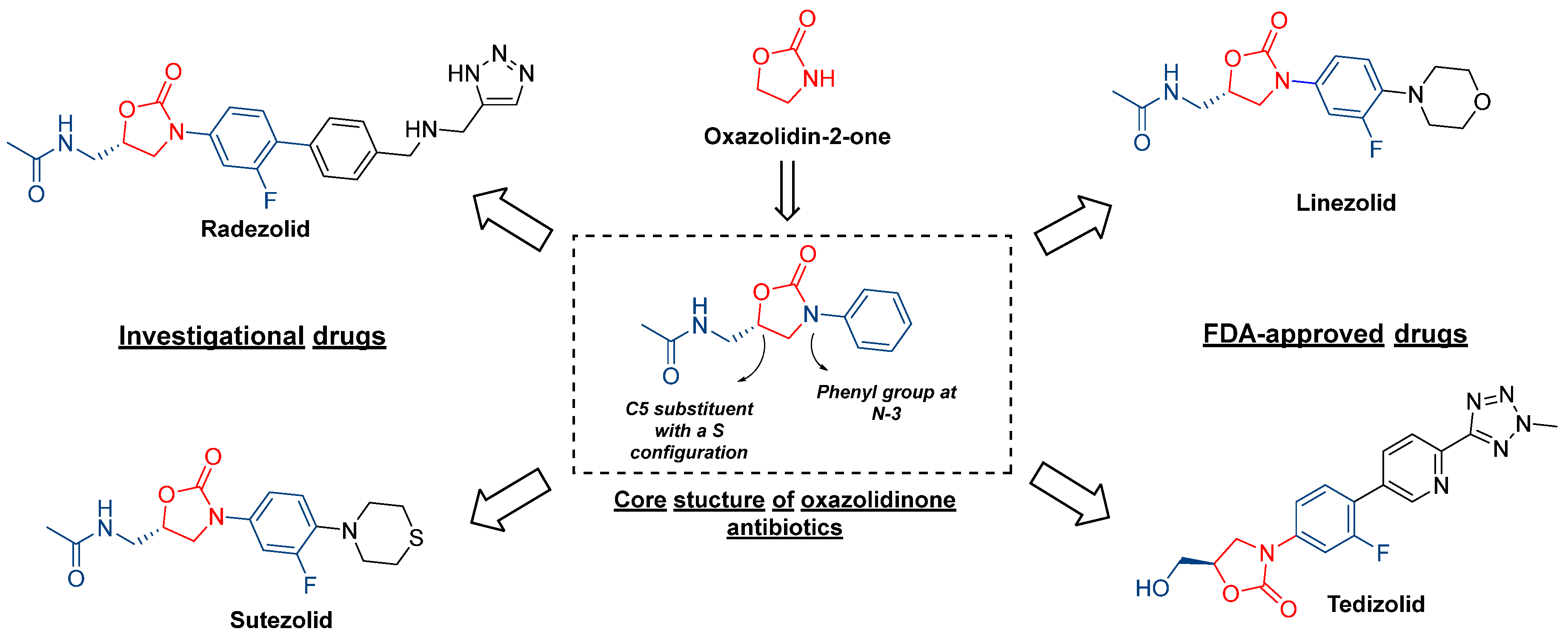
:1. Introduction
2. Results and Discussion
2.1. Antibacterial Evaluation
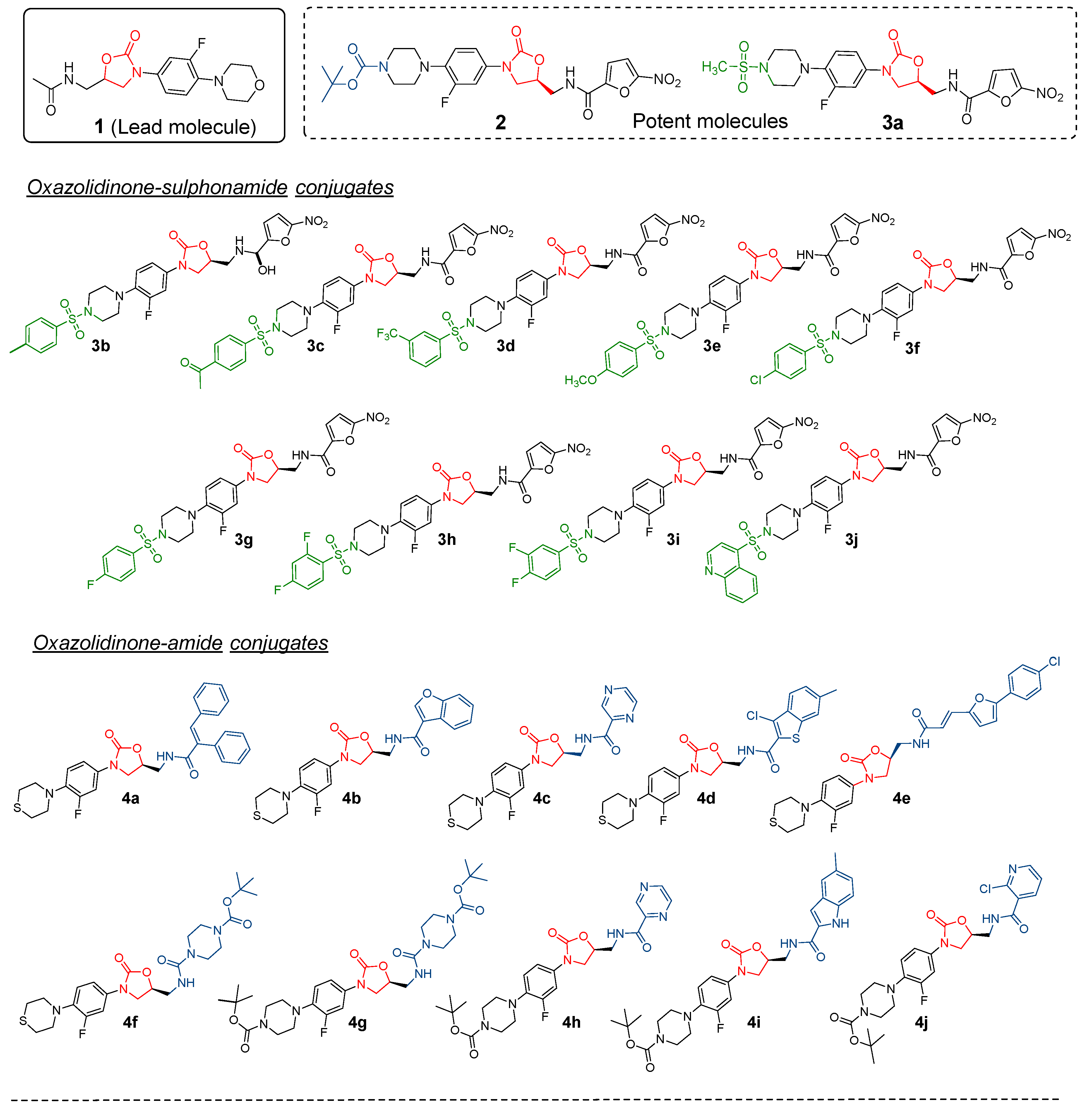
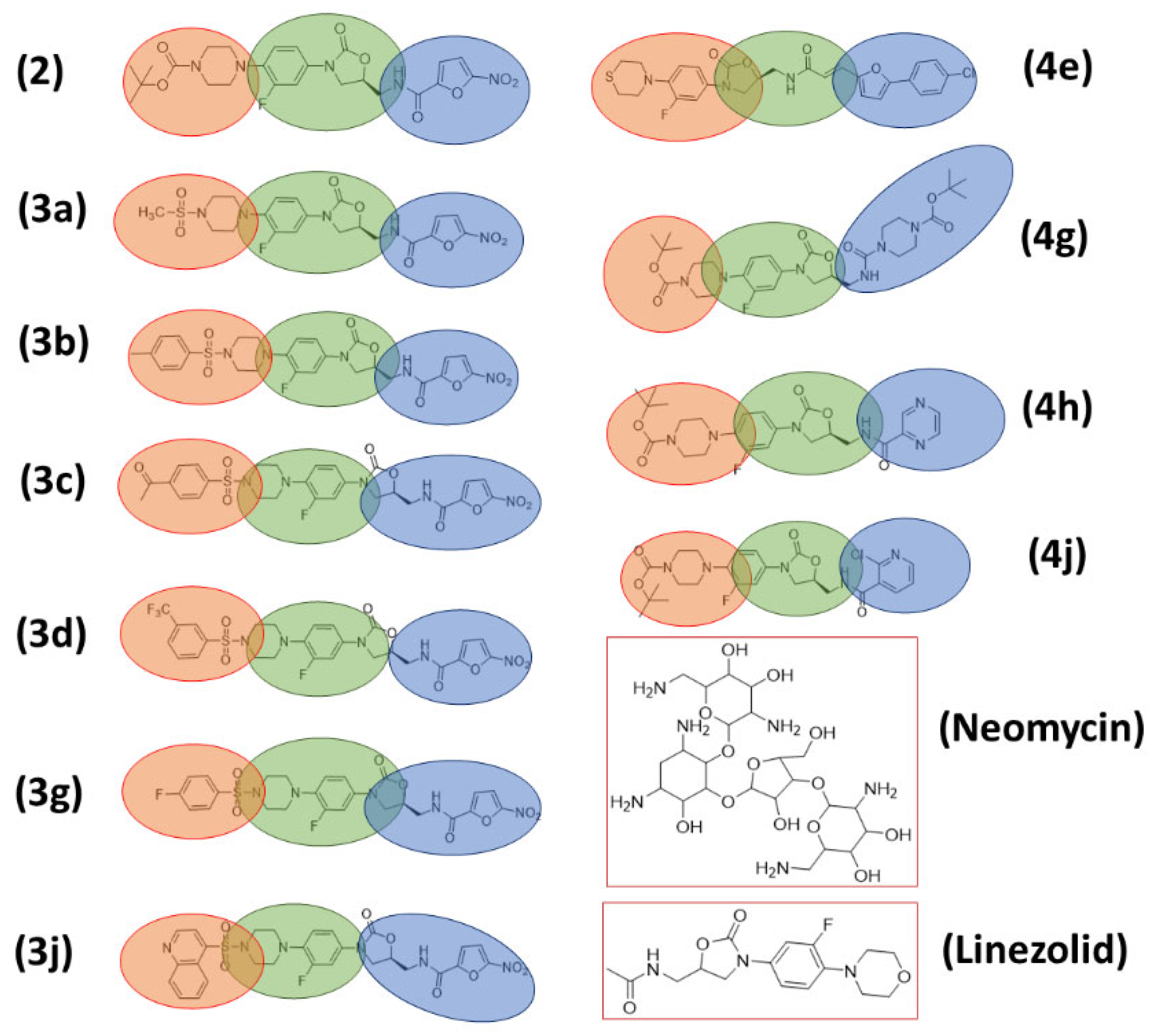
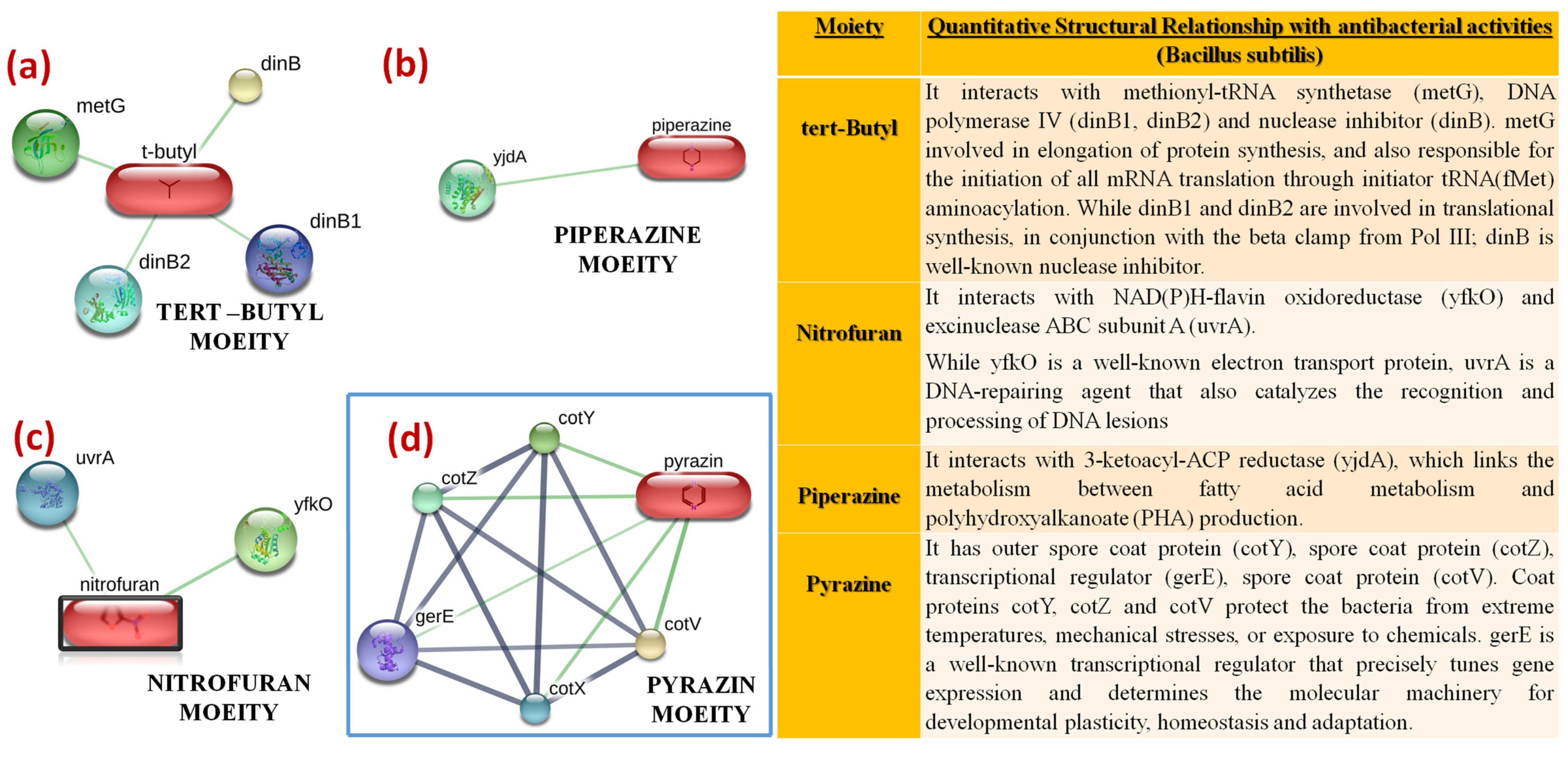
2.2. Structure–Activity Relationship
2.3. Antibiofilm Evaluation
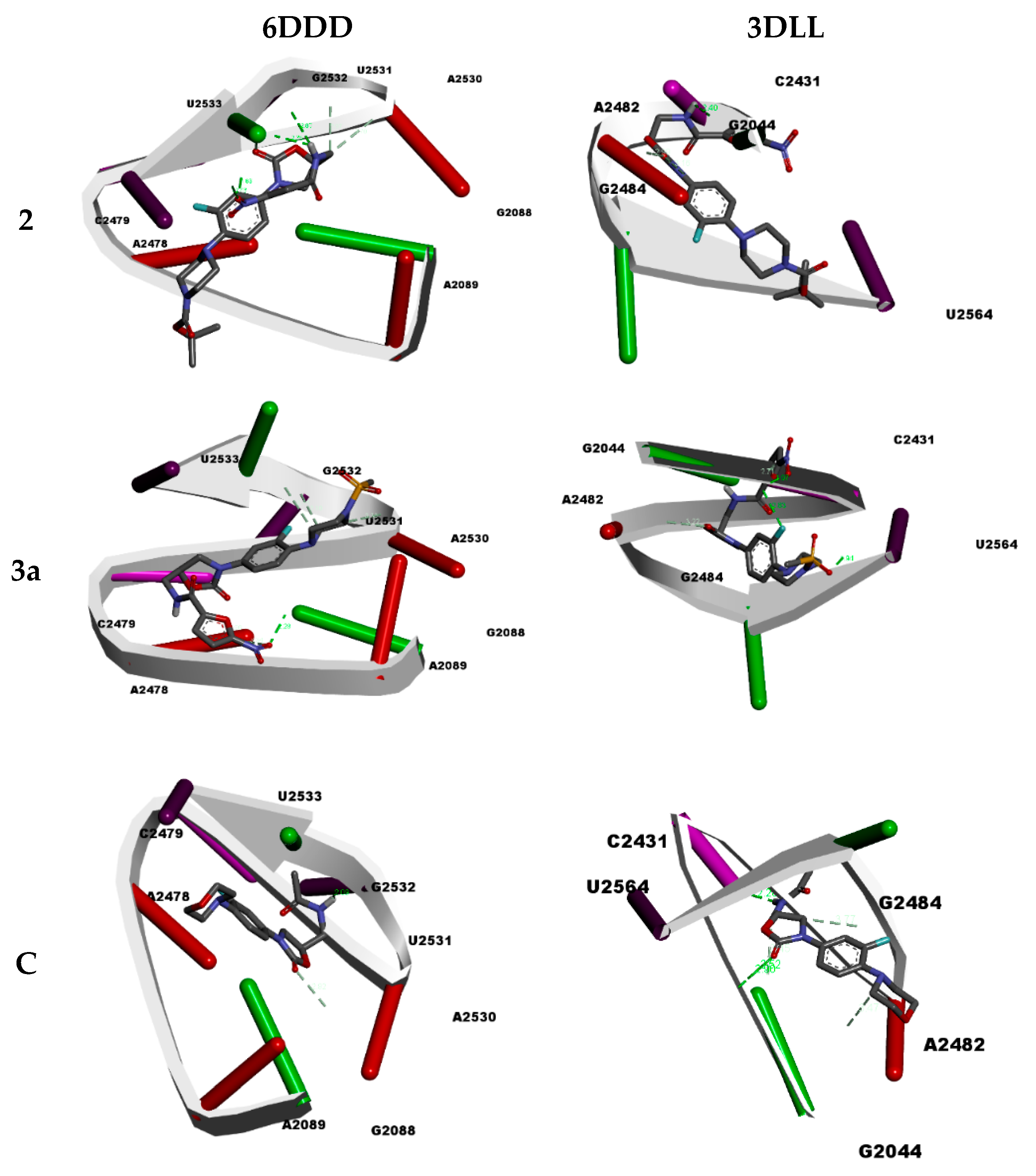
2.4. Molecular Docking Studies
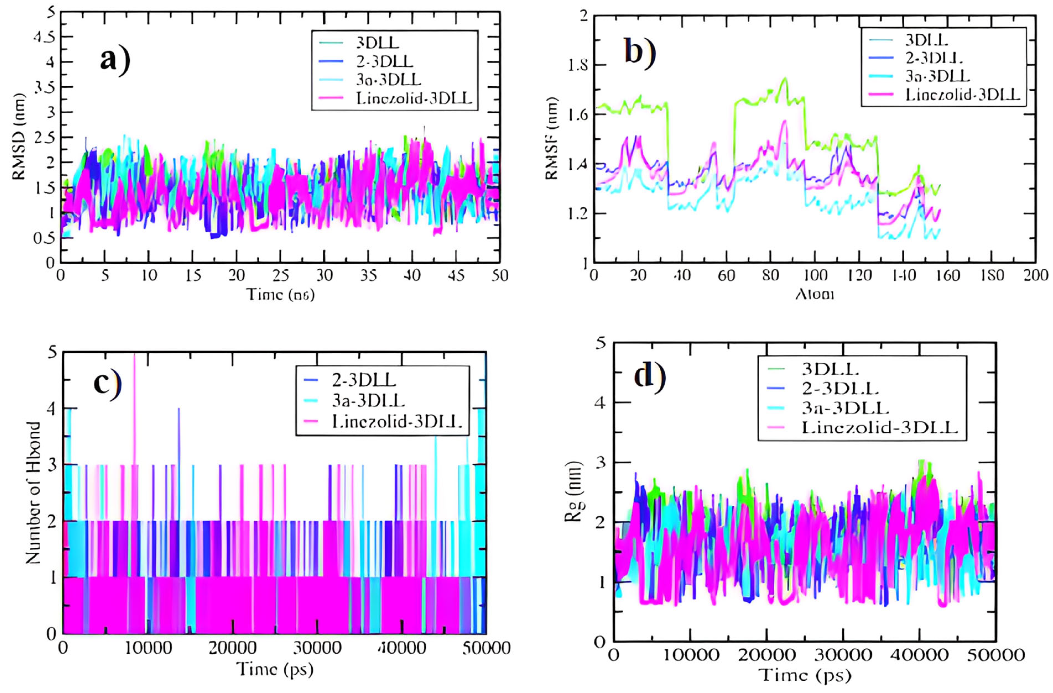
2.5. Molecular Dynamics Simulations
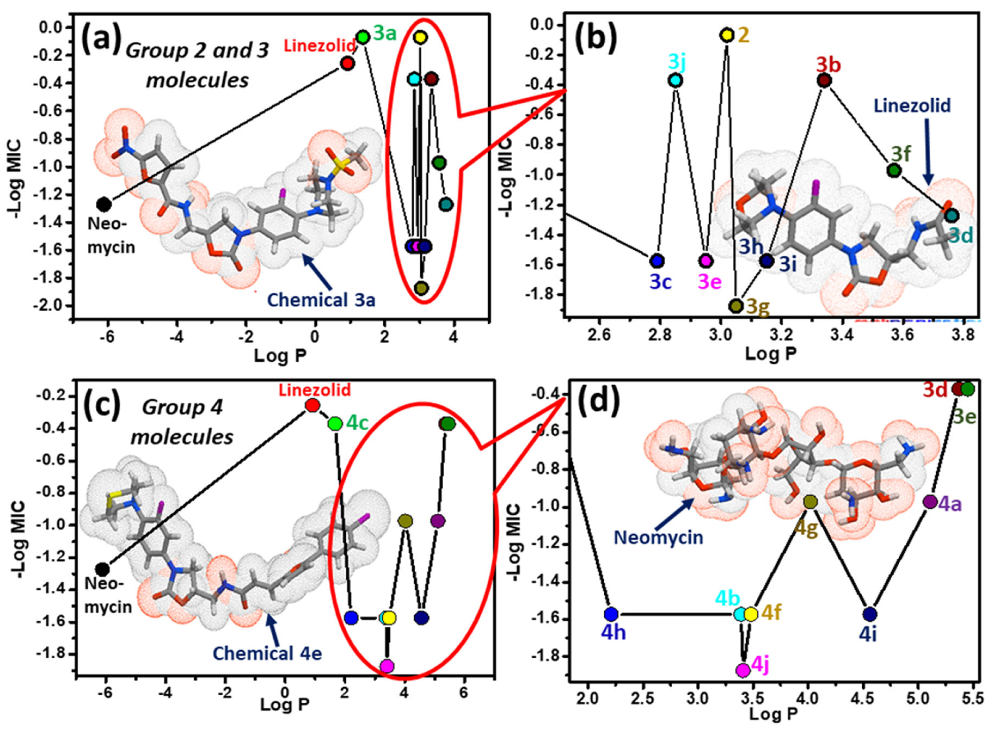
2.6. One-Descriptor (log P) Analysis
2.7. ADME-T and Drug Likeness Analyses
3. Experimental Section
3.1. Biological Evaluation
3.1.1. Antibacterial Assay
3.1.2. MBC Assay
3.1.3. Biofilm Inhibition Assay
3.2. Computational Studies
3.2.1. Molecular Docking
3.2.2. Molecular Dynamics Simulations
3.2.3. Descriptor (log P) Analysis, ADME-T and Drug Likeness Analyses
4. Conclusions
Supplementary Materials
Author Contributions
Funding
Institutional Review Board Statement
Informed Consent Statement
Data Availability Statement
Acknowledgments
Conflicts of Interest
References
- Murray, C.J.L.; Ikuta, K.S.; Sharara, F.; Swetschinski, L.; Robles Aguilar, G.; Gray, A.; Han, C.; Bisignano, C.; Rao, P.; Wool, E.; et al. Global burden of bacterial antimicrobial resistance in 2019: A systematic analysis. Lancet 2022, 399, 629–655. [Google Scholar] [CrossRef] [PubMed]
- Ventola, C.L. The antibiotic resistance crisis: Part 1: Causes and threats. Pharm. Ther. 2015, 40, 277–283. [Google Scholar]
- Swedberg, G.; Sköld, O. Characterization of different plasmid-borne dihydropteroate synthases mediating bacterial resistance to sulfonamides. J. Bacteriol. 1980, 142, 1–7. [Google Scholar] [CrossRef] [PubMed] [Green Version]
- Abushaheen, M.A.; Muzaheed; Fatani, A.J.; Alosaimi, M.; Mansy, W.; George, M.; Acharya, S.; Rathod, S.; Divakar, D.D.; Jhugroo, C.; et al. Antimicrobial resistance, mechanisms and its clinical significance. Dis.-A-Mon. DM 2020, 66, 100971. [Google Scholar] [CrossRef]
- Foti, C.; Piperno, A.; Scala, A.; Giuffrè, O. Oxazolidinone Antibiotics: Chemical, Biological and Analytical Aspects. Molecules 2021, 26, 4280. [Google Scholar] [CrossRef]
- Fernandes, G.; Scarim, C.B.; Kim, S.H.; Wu, J.; Castagnolo, D. Oxazolidinones as Versatile Scaffolds in Medicinal Chemistry. RSC Med. Chem. 2023. [Google Scholar] [CrossRef]
- Zhao, Q.; Xin, L.; Liu, Y.; Liang, C.; Li, J.; Jian, Y.; Li, H.; Shi, Z.; Liu, H.; Cao, W. Current Landscape and Future Perspective of Oxazolidinone Scaffolds Containing Antibacterial Drugs. J. Med. Chem. 2021, 64, 10557–10580. [Google Scholar] [CrossRef]
- Barbachyn, M.R.; Ford, C.W. Oxazolidinone structure-activity relationships leading to linezolid. Angew. Chem. (Int. Ed. Engl.) 2003, 42, 2010–2023. [Google Scholar] [CrossRef]
- Leach, K.L.; Brickner, S.J.; Noe, M.C.; Miller, P.F. Linezolid, the first oxazolidinone antibacterial agent. Ann. N. Y. Acad. Sci. 2011, 1222, 49–54. [Google Scholar] [CrossRef]
- von der Lippe, B.; Sandven, P.; Brubakk, O. Efficacy and safety of linezolid in multidrug resistant tuberculosis (MDR-TB)—A report of ten cases. J. Infect. 2006, 52, 92–96. [Google Scholar] [CrossRef]
- Carena, A.A.; Stryjewski, M.E. Tedizolid (torezolid) for the treatment of complicated skin and skin structure infections. Expert Rev. Clin. Pharmacol. 2020, 13, 577–592. [Google Scholar] [CrossRef]
- Wyskiel, C. To be or not to be exclusive: The sutezolid story. Lancet. Glob. Health 2016, 4, e91. [Google Scholar] [CrossRef] [PubMed]
- Zheng, J.; Chen, Z.; Lin, Z.; Sun, X.; Bai, B.; Xu, G.; Chen, J.; Yu, Z.; Qu, D. Radezolid Is More Effective Than Linezolid Against Planktonic Cells and Inhibits Enterococcus faecalis Biofilm Formation. Front. Microbiol. 2020, 11, 196. [Google Scholar] [CrossRef] [PubMed]
- Black, T.A.; Buchwald, U.K. The pipeline of new molecules and regimens against drug-resistant tuberculosis. J. Clin. Tuberc. Other Mycobact. Dis. 2021, 25, 100285. [Google Scholar] [CrossRef] [PubMed]
- Sangshetti, J.N.; Khan, F.A.; Patil, R.H.; Marathe, S.D.; Gade, W.N.; Shinde, D.B. Biofilm inhibition of linezolid-like Schiff bases: Synthesis, biological activity, molecular docking and in silico ADME prediction. Bioorg. Med. Chem. Lett. 2015, 25, 874–880. [Google Scholar] [CrossRef]
- Gadekar, P.K.; Roychowdhury, A.; Kharkar, P.S.; Khedkar, V.M.; Arkile, M.; Manek, H.; Sarkar, D.; Sharma, R.; Vijayakumar, V.; Sarveswari, S. Design, synthesis and biological evaluation of novel azaspiro analogs of linezolid as antibacterial and antitubercular agents. Eur. J. Med. Chem. 2016, 122, 475–487. [Google Scholar] [CrossRef]
- Guo, B.; Fan, H.; Xin, Q.; Chu, W.; Wang, H.; Yang, Y. Synthesis and biological evaluation of novel benzoxazinyl-oxazolidinones as potential antibacterial agents. Bioorg. Med. Chem. Lett. 2013, 23, 3697–3699. [Google Scholar] [CrossRef]
- Lyons, A.; Kirkham, J.; Blades, K.; Orr, D.; Dauncey, E.; Smith, O.; Dick, E.; Walker, R.; Matthews, T.; Bunt, A.; et al. Discovery and structure-activity relationships of a novel oxazolidinone class of bacterial type II topoisomerase inhibitors. Bioorg. Med. Chem. Lett. 2022, 65, 128648. [Google Scholar] [CrossRef]
- Faazil, S.; Malik, M.S.; Ahmed, S.A.; Jamal, Q.M.S.; Basha, S.T.; Al-Rooqi, M.M.; Obaid, R.J.; Qurban, J.; Shaikh, I.N.; Asghar, B.H.; et al. New quinoline-thiolactone conjugates as potential antitubercular and antibacterial agents. J. Mol. Struct. 2023, 1271, 134099. [Google Scholar] [CrossRef]
- Hussein, E.M.; Malik, M.S.; Alsantali, R.I.; Asghar, B.H.; Morad, M.; Ansari, M.A.; Jamal, Q.M.S.; Alsimaree, A.A.; Abdalla, A.N.; Algarni, A.S.; et al. Bioactive fluorenes. Part IV: Design, synthesis, and a combined in vitro, in silico anticancer and antibacterial evaluation of new fluorene-heterocyclic sulfonamide conjugates. J. Mol. Struct. 2021, 1246, 131232. [Google Scholar] [CrossRef]
- Malik, M.S.; Asghar, B.H.; Syed, R.; Alsantali, R.I.; Morad, M.; Altass, H.M.; Moussa, Z.; Althagafi, I.I.; Jassas, R.S.; Ahmed, S.A. Novel Pyran-Linked Phthalazinone-Pyrazole Hybrids: Synthesis, Cytotoxicity Evaluation, Molecular Modeling, and Descriptor Studies. Front. Chem. 2021, 9, 666573. [Google Scholar] [CrossRef] [PubMed]
- Malik, M.S.; Farooq Adil, S.; Moussa, Z.; Altass, H.M.; Althagafi, I.I.; Morad, M.; Ansari, M.A.; Sajid Jamal, Q.M.; Obaid, R.J.; Al-Warthan, A.A.; et al. Rational Design and Synthesis of Naphthalene Diimide Linked Bis-Naphthalimides as DNA Interactive Agents. Front. Chem. 2021, 9, 630357. [Google Scholar] [CrossRef] [PubMed]
- Seddigi, Z.S.; Malik, M.S.; Saraswati, A.P.; Ahmed, S.A.; Babalghith, A.O.; Lamfon, H.A.; Kamal, A. Recent advances in combretastatin based derivatives and prodrugs as antimitotic agents. MedChemComm 2017, 8, 1592–1603. [Google Scholar] [CrossRef] [PubMed]
- Faazil, S.; Malik, M.S.; Ahmed, S.A.; Alsantali, R.I.; Yedla, P.; Alsharif, M.A.; Shaikh, I.N.; Kamal, A. Novel linezolid-based oxazolidinones as potent anticandidiasis and antitubercular agents. Bioorg. Chem. 2022, 126, 105869. [Google Scholar] [CrossRef]
- Zhuge, L.; Wang, Y.; Wu, S.; Zhao, R.L.; Li, Z.; Xie, Y. Furazolidone treatment for Helicobacter Pylori infection: A systematic review and meta-analysis. Helicobacter 2018, 23, e12468. [Google Scholar] [CrossRef]
- Yin, W.; Wang, Y.; Liu, L.; He, J. Biofilms: The Microbial “Protective Clothing” in Extreme Environments. Int. J. Mol. Sci. 2019, 20, 3423. [Google Scholar] [CrossRef] [PubMed] [Green Version]
- Yan, J.; Bassler, B.L. Surviving as a Community: Antibiotic Tolerance and Persistence in Bacterial Biofilms. Cell Host Microbe 2019, 26, 15–21. [Google Scholar] [CrossRef] [PubMed]
- Wilson, D.N. Ribosome-targeting antibiotics and mechanisms of bacterial resistance. Nat. Rev. Microbiol. 2014, 12, 35–48. [Google Scholar] [CrossRef]
- Belousoff, M.J.; Venugopal, H.; Wright, A.; Seoner, S.; Stuart, I.; Stubenrauch, C.; Bamert, R.S.; Lupton, D.W.; Lithgow, T. cryoEM-Guided Development of Antibiotics for Drug-Resistant Bacteria. ChemMedChem 2019, 14, 527–531. [Google Scholar] [CrossRef]
- Wilson, D.N.; Schluenzen, F.; Harms, J.M.; Starosta, A.L.; Connell, S.R.; Fucini, P. The oxazolidinone antibiotics perturb the ribosomal peptidyl-transferase center and effect tRNA positioning. Proc. Natl. Acad. Sci. USA 2008, 105, 13339–13344. [Google Scholar] [CrossRef] [Green Version]
- Horowitz, S.; Trievel, R.C. Carbon-oxygen hydrogen bonding in biological structure and function. J. Biol. Chem. 2012, 287, 41576–41582. [Google Scholar] [CrossRef] [PubMed] [Green Version]
- De Vivo, M.; Masetti, M.; Bottegoni, G.; Cavalli, A. Role of Molecular Dynamics and Related Methods in Drug Discovery. J. Med. Chem. 2016, 59, 4035–4061. [Google Scholar] [CrossRef] [PubMed]
- Yu, W.; MacKerell, A.D., Jr. Computer-Aided Drug Design Methods. Methods Mol. Biol. 2017, 1520, 85–106. [Google Scholar] [CrossRef] [PubMed] [Green Version]
- Verma, J.; Khedkar, V.M.; Coutinho, E.C. 3D-QSAR in drug design—A review. Curr. Top. Med. Chem. 2010, 10, 95–115. [Google Scholar] [CrossRef]
- Bergström, C.A. In silico predictions of drug solubility and permeability: Two rate-limiting barriers to oral drug absorption. Basic Clin. Pharmacol. Toxicol. 2005, 96, 156–161. [Google Scholar] [CrossRef] [PubMed]
- Al-Fahemi, J.H. Structural descriptors for the correlation of human blood:air partition coefficient of volatile organic molecules by QSPRs. Struct. Chem. 2013, 24, 2155–2161. [Google Scholar] [CrossRef]
- Riley, R.J.; Parker, A.J.; Trigg, S.; Manners, C.N. Development of a generalized, quantitative physicochemical model of CYP3A4 inhibition for use in early drug discovery. Pharm. Res. 2001, 18, 652–655. [Google Scholar] [CrossRef]
- Alavijeh, M.S.; Chishty, M.; Qaiser, M.Z.; Palmer, A.M. Drug metabolism and pharmacokinetics, the blood-brain barrier, and central nervous system drug discovery. NeuroRx J. Am. Soc. Exp. NeuroTherapeutics 2005, 2, 554–571. [Google Scholar] [CrossRef] [Green Version]
- Ertl, P.; Rohde, B.; Selzer, P. Fast calculation of molecular polar surface area as a sum of fragment-based contributions and its application to the prediction of drug transport properties. J. Med. Chem. 2000, 43, 3714–3717. [Google Scholar] [CrossRef]
- Palm, K.; Stenberg, P.; Luthman, K.; Artursson, P. Polar molecular surface properties predict the intestinal absorption of drugs in humans. Pharm. Res. 1997, 14, 568–571. [Google Scholar] [CrossRef]
- Krarup, L.H.; Christensen, I.T.; Hovgaard, L.; Frokjaer, S. Predicting drug absorption from molecular surface properties based on molecular dynamics simulations. Pharm. Res. 1998, 15, 972–978. [Google Scholar] [CrossRef] [PubMed]
- Clark, D.E. Rapid calculation of polar molecular surface area and its application to the prediction of transport phenomena. 2. Prediction of blood-brain barrier penetration. J. Pharm. Sci. 1999, 88, 815–821. [Google Scholar] [CrossRef] [PubMed]
- He, Z.; Zhang, J.; Shi, X.-H.; Hu, L.-L.; Kong, X.; Cai, Y.-D.; Chou, K.-C. Predicting Drug-Target Interaction Networks Based on Functional Groups and Biological Features. PLoS ONE 2010, 5, e9603. [Google Scholar] [CrossRef]
- Lin, J.; Sahakian, D.C.; de Morais, S.M.; Xu, J.J.; Polzer, R.J.; Winter, S.M. The role of absorption, distribution, metabolism, excretion and toxicity in drug discovery. Curr. Top. Med. Chem. 2003, 3, 1125–1154. [Google Scholar] [CrossRef] [PubMed]
- Prachayasittikul, V.; Worachartcheewan, A.; Shoombuatong, W.; Prachayasittikul, V.; Nantasenamat, C. Classification of P-glycoprotein-interacting compounds using machine learning methods. EXCLI J. 2015, 14, 958–970. [Google Scholar] [CrossRef]
- Beck, T.C.; Beck, K.R.; Morningstar, J.; Benjamin, M.M.; Norris, R.A. Descriptors of Cytochrome Inhibitors and Useful Machine Learning Based Methods for the Design of Safer Drugs. Pharmaceuticals 2021, 14, 472. [Google Scholar] [CrossRef]
- Manikandan, P.; Nagini, S. Cytochrome P450 Structure, Function and Clinical Significance: A Review. Curr. Drug Targets 2018, 19, 38–54. [Google Scholar] [CrossRef]
- Morris, G.M.; Goodsell, D.S.; Halliday, R.S.; Huey, R.; Hart, W.E.; Belew, R.K.; Olson, A.J. Automated docking using a Lamarckian genetic algorithm and an empirical binding free energy function. J. Comput. Chem. 1998, 19, 1639–1662. [Google Scholar] [CrossRef]
- Jamal, Q.M.S.; Ahmad, V.; Alharbi, A.H.; Ansari, M.A.; Alzohairy, M.A.; Almatroudi, A.; Alghamdi, S.; Alomary, M.N.; AlYahya, S.; Shesha, N.T.; et al. Therapeutic development by repurposing drugs targeting SARS-CoV-2 spike protein interactions by simulation studies. Saudi J. Biol. Sci. 2021, 28, 4560–4568. [Google Scholar] [CrossRef]
- Van Der Spoel, D.; Lindahl, E.; Hess, B.; Groenhof, G.; Mark, A.E.; Berendsen, H.J. GROMACS: Fast, flexible, and free. J. Comput. Chem. 2005, 26, 1701–1718. [Google Scholar] [CrossRef]
- Daina, A.; Michielin, O.; Zoete, V. iLOGP: A simple, robust, and efficient description of n-octanol/water partition coefficient for drug design using the GB/SA approach. J. Chem. Inf. Model. 2014, 54, 3284–3301. [Google Scholar] [CrossRef] [PubMed]
- Kuhn, M.; von Mering, C.; Campillos, M.; Jensen, L.J.; Bork, P. STITCH: Interaction networks of chemicals and proteins. Nucleic Acids Res. 2008, 36, D684–D688. [Google Scholar] [CrossRef] [PubMed] [Green Version]
- Szklarczyk, D.; Santos, A.; von Mering, C.; Jensen, L.J.; Bork, P.; Kuhn, M. STITCH 5: Augmenting protein-chemical interaction networks with tissue and affinity data. Nucleic Acids Res. 2016, 44, D380–D384. [Google Scholar] [CrossRef] [PubMed]


| Compound | B. s 2 | S. m 2 | S. a 2 | M. l 2 | P. a 3 | K. p 3 | E. c 3 |
|---|---|---|---|---|---|---|---|
| 2 | 1.17 (2.34) 4 | 1.17 (2.34) 4 | 125 | >125 | 1.17 (4.68) 4 | >125 | >125 |
| 3a | 1.17 (4.68) 4 | 4.68 (9.36) 4 | 18.75 | >125 | 1.17 (4.68) 4 | >125 | >125 |
| 3b | 2.34 | 18.75 | 37.5 | >125 | 4.68 | >125 | >125 |
| 3c | 37.5 | 4.68 | 37.5 | >125 | 4.68 | >125 | >125 |
| 3d | 18.75 | 75 | 125 | >125 | 75 | >125 | >125 |
| 3e | 37.5 | >125 | 4.68 | >125 | >125 | >125 | >125 |
| 3f | 9.36 | 9.36 | 4.68 | >125 | 4.68 | >125 | >125 |
| 3g | 125 | 37.5 | 125 | >125 | 75 | >125 | >125 |
| 3h | 37.5 | 18.75 | 37.5 | >125 | 18.75 | >125 | >125 |
| 3i | 37.5 | 18.75 | 37.5 | >125 | 18.75 | >125 | >125 |
| 3j | 2.34 | 9.36 | >125 | >125 | 4.68 | >125 | >125 |
| Nmyn 5 | 18.75 | 18.75 | 18.75 | 18.75 | 18.75 | 18.75 | 18.75 |
| Compound | B. s 2 | S. m 2 | S. a 2 | M. l 2 | P. a 3 | K. p 3 | E. c 3 |
|---|---|---|---|---|---|---|---|
| 4a | 9.36 | 75 | 18.75 | >125 | 3.37 | >125 | >125 |
| 4b | 37.5 | 18.75 | 37.5 | >125 | 18.75 | >125 | >125 |
| 4c | 2.34 | 4.68 | 18.75 | >125 | 4.68 | >125 | >125 |
| 4d | 2.34 | 18.75 | 37.5 | >125 | 4.68 | >125 | >125 |
| 4e | 2.34 | 4.68 | 37.5 | >125 | 4.68 | >125 | >125 |
| 4f | 37.5 | 18.75 | 37.5 | >125 | 18.75 | >125 | >125 |
| 4g | 9.36 | 75 | >125 | >125 | 75 | >125 | >125 |
| 4h | 37.5 | 18.75 | 37.5 | >125 | 18.75 | >125 | >125 |
| 4i | 37.5 | 4.68 | 37.5 | >125 | 4.68 | >125 | >125 |
| 4j | >125 | >125 | >125 | >125 | >125 | >125 | >125 |
| Nmyn 4 | 18.75 | 18.75 | 18.75 | 18.75 | 18.75 | 18.75 | 18.75 |
| S. No. | Compound | IC50 Values (in μg/mL) | ||
|---|---|---|---|---|
| Bacillus subtilis MTCC 121 | Staphylococcus aureus MLS16 MTCC 2940 | Pseudomonas aeruginosa MTCC 2453 | ||
| 1 | 2 | 0.58 ± 0.18 | 1.21 ± 0.32 | 0.58 ± 0.08 |
| 2 | 3a | 1.24 ± 0.09 | 2.32 ± 0.28 | 2.34 ± 0.26 |
| 3 | Erythromycin | 0.22 ± 0.14 | 0.25 ± 0.12 | 0.19 ± 0.22 |
| S. No. | Compound | Molecular Target | Final Intermolecular Energy (kcal/mol) | Inhibition Constant | Hydrogen Bonds * | H-Bond Length (Å) | Other Interactions |
|---|---|---|---|---|---|---|---|
| 1. | 2 | 6DDD | −6.36 | 187.94 uM | 1:G2532:H3–:UNK0:O7 1:G2532:H21–:UNK0:O7 :UNK0:H1–1:G2532:O5’ :UNK0:H1–1:G2532:O4’ 1:G2532:C4’–:UNK0:O1 :UNK0:C1–1:A2530:O2’ :UNK0:C1–1:G2532:OP2 | 1.82 2.43 2.79 2.35 3.38 3.69 2.96 | 1:G2088 = π-sigma π-π stacking |
| 2. | 3a | 6DDD | −6.01 | 557.88 uM | 1:G2088:H22–:UNK0:O7 1:A2478:C2–:UNK0:O5 1:A2478:C2–:UNK0:O7 :UNK0:C12–1:G2532:O5’ :UNK0:C11–1:A2530:O2’ :UNK0:C9–1:G2532:O4’ | 2.28 2.96 3.34 3.10 3.36 3.74 | 1:A2530 = π-alkyl |
| 3. | Linezolid (C) | 6DDD | −5.41 | 182.26 uM | :UNK0:H–1:G2532:O5’ 1:A2530:C8–:UNK0:O | 2.08 2.91 | 1:U2533 = π-alkyl 1:A2578 = π-lone pair |
| 4. | 2 | 3DLL | −6.37 | 170.07 uM | X:G2044:H1–:UNK0:O5 :UNK0:H1–X:G2044:O6 X:A2482:C1’–:UNK0:O1 X:A2482:C8–:UNK0:O1 | 1.80 2.40 2.98 3.15 | C:X2431 π-π T-shaped |
| 5. | 3a | 3DLL | −6.61 | 297.30 uM | X:G2044:H22–:UNK0:F X:G2044:H22–:UNK0:O6 X:U2564:HO2’–UNK0:O3 :UNK0:H1–X:G2044:O6 X:A2482:C8–:UNK0:O1 X:G2044:H1–:UNK0 | 2.83 1.97 1.93 1.95 3.21 2.70 | C:X2431 π-π T-shaped |
| 6. | Linezolid (C) | 3DLL | −4.92 | 377.31 uM | X:G2044:H22–:UNK0:O X:G2044:H1–:UNK0:O :UNK0:H–X:C2431:N3 :UNK0:C–X:G2484:OP2 :UNK0:C–X:G2044:O6 :UNK0:C–X:A2482:N7 | 2.89 2.52 2.28 3.77 3.45 3.47 | A:X2482 = π-sigma, π-alkyl G:X2044 = π-π T-shaped |
| Compounds | GI Absorption | BBB Permeant | Pgp Substrate | CYP1A2 Inhibitor | CYP2C19 Inhibitor | CYP2C9 Inhibitor | CYP2D6 Inhibitor | CYP3A4 Inhibitor |
|---|---|---|---|---|---|---|---|---|
| 2 | Low | No | Yes | No | Yes | Yes | No | Yes |
| 3a | Low | No | Yes | No | Yes | Yes | No | Yes |
| 3b | Low | No | Yes | No | No | Yes | No | Yes |
| 3c | Low | No | Yes | No | No | Yes | No | Yes |
| 3d | Low | No | Yes | No | No | Yes | No | Yes |
| 3e | Low | No | Yes | No | No | Yes | No | Yes |
| 3f | Low | No | Yes | No | No | Yes | No | Yes |
| 3g | Low | No | Yes | No | No | Yes | No | Yes |
| 3h | Low | No | Yes | No | No | Yes | No | Yes |
| 3i | Low | No | Yes | No | No | Yes | No | Yes |
| 3j | Low | No | Yes | No | No | Yes | No | Yes |
| 4a | High | No | Yes | No | Yes | Yes | Yes | Yes |
| 4b | High | No | Yes | Yes | Yes | Yes | Yes | Yes |
| 4c | High | No | Yes | No | Yes | No | Yes | Yes |
| 4d | Low | No | Yes | Yes | Yes | Yes | Yes | Yes |
| 4e | High | No | Yes | No | Yes | Yes | Yes | Yes |
| 4f | High | No | Yes | No | Yes | No | Yes | Yes |
| 4g | High | No | Yes | No | Yes | Yes | Yes | Yes |
| 4h | High | No | Yes | No | Yes | Yes | No | Yes |
| 4i | High | No | Yes | No | Yes | Yes | Yes | Yes |
| 4j | High | No | Yes | No | Yes | Yes | Yes | Yes |
Disclaimer/Publisher’s Note: The statements, opinions and data contained in all publications are solely those of the individual author(s) and contributor(s) and not of MDPI and/or the editor(s). MDPI and/or the editor(s) disclaim responsibility for any injury to people or property resulting from any ideas, methods, instructions or products referred to in the content. |
© 2023 by the authors. Licensee MDPI, Basel, Switzerland. This article is an open access article distributed under the terms and conditions of the Creative Commons Attribution (CC BY) license (https://creativecommons.org/licenses/by/4.0/).
Share and Cite
Malik, M.S.; Faazil, S.; Alsharif, M.A.; Sajid Jamal, Q.M.; Al-Fahemi, J.H.; Banerjee, A.; Chattopadhyay, A.; Pal, S.K.; Kamal, A.; Ahmed, S.A. Antibacterial Properties and Computational Insights of Potent Novel Linezolid-Based Oxazolidinones. Pharmaceuticals 2023, 16, 516. https://doi.org/10.3390/ph16040516
Malik MS, Faazil S, Alsharif MA, Sajid Jamal QM, Al-Fahemi JH, Banerjee A, Chattopadhyay A, Pal SK, Kamal A, Ahmed SA. Antibacterial Properties and Computational Insights of Potent Novel Linezolid-Based Oxazolidinones. Pharmaceuticals. 2023; 16(4):516. https://doi.org/10.3390/ph16040516
Chicago/Turabian StyleMalik, M. Shaheer, Shaikh Faazil, Meshari A. Alsharif, Qazi Mohammad Sajid Jamal, Jabir H. Al-Fahemi, Amrita Banerjee, Arpita Chattopadhyay, Samir Kumar Pal, Ahmed Kamal, and Saleh A. Ahmed. 2023. "Antibacterial Properties and Computational Insights of Potent Novel Linezolid-Based Oxazolidinones" Pharmaceuticals 16, no. 4: 516. https://doi.org/10.3390/ph16040516