Review

Tetanus Toxin Fragment C: Structure, Drug Discovery Research and Production

1
Analytical Sciences, Sanofi, 31/33 Quai Armand Barbès, 69250 Neuville-sur-Saône, France
2
Small Molecules for Biological Targets Team, Centre de Recherche en Cancérologie de Lyon, Centre Léon Bérard, CNRS 5286, INSERM 1052, Université Claude Bernard Lyon 1, Univ Lyon, 69373 Lyon, France
3
Institute of Pharmaceutical and Medicinal Chemistry, PharmaCampus, Westfälische Wilhelms-Universtität Münster, 48149 Münster, Germany
4
UMR 5086, Molecular Microbiology and Structural Biochemistry, CNRS/Université Lyon 1, 69367 Lyon, France
*
Authors to whom correspondence should be addressed.
Pharmaceuticals 2022, 15(6), 756; https://doi.org/10.3390/ph15060756
Submission received: 20 May 2022 / Revised: 9 June 2022 / Accepted: 13 June 2022 / Published: 17 June 2022
(This article belongs to the Section Biopharmaceuticals)

Abstract

:
Tetanus toxoid (TTd) plays an important role in the pharmaceutical world, especially in vaccines. The toxoid is obtained after formaldehyde treatment of the tetanus toxin. In parallel, current emphasis in the drug discovery field is put on producing well-defined and safer drugs, explaining the interest in finding new alternative proteins. The tetanus toxin fragment C (TTFC) has been extensively studied both as a neuroprotective agent for central nervous system disorders owing to its neuronal properties and as a carrier protein in vaccines. Indeed, it is derived from a part of the tetanus toxin and, as such, retains its immunogenic properties without being toxic. Moreover, this fragment has been well characterized, and its entire structure is known. Here, we propose a systematic review of TTFC by providing information about its structural features, its properties and its methods of production. We also describe the large uses of TTFC in the field of drug discovery. TTFC can therefore be considered as an attractive alternative to TTd and remarkably offers a wide range of uses, including as a carrier, delivery vector, conjugate, booster, inducer, and neuroprotector.

Graphical Abstract

1. Introduction

Tetanus caused by the tetanus toxin (TT) is a fatal illness, which despite the existence of a vaccine, led to an estimated 34,684 deaths in 2019 [1]. TT is a neurotoxin produced by Clostridium tetani, a Gram-positive pathogenic bacterium, mainly found in soil and the gastrointestinal tracts of animals. TT induces the inhibition of neurotransmitter release, leading to spastic paralysis in a four-step process [2]. First, TT binds to specific receptors, mainly composed of lipids and gangliosides, found at the neuromuscular junction (NMJ) [3]. Another receptor is reached by TT after these first bindings: a protein receptor responsible for its internalization (second step). This double receptor binding is responsible for the high affinity between TT and nerve cells. Third, TT is then transported into the cell body via axonal retrograde transport. In the last step, the proteolytic cleavage by TT of the VAMP/synaptobrevin, a neuronal substrate, leads to the inhibition of neurotransmitter release [4]. All of these biological properties can be distinct associated parts of the TT structure. TT is a 150.7 kDa protein composed of a 52.4 kDa light chain and a 98.3 kDa heavy chain linked by a disulfide bond [5,6].
The light chain, also named fragment A, has a zinc-dependent proteolytic activity responsible for synaptobrevin cleavage [3]. The heavy chain is composed of the fragments B and C (Figure 1). The 46.7 kDa fragment B is located at the N-terminus of the heavy chain and is responsible for toxin cell penetration. Finally, TT protein is completed by the 51.6 kDa fragment C, located at the C-terminus of its heavy chain. This latter is involved in neuronal cell binding.
The three fragments were discovered in 1974 during a study in which TT was enzymatically digested with papain, yielding fragments B and C [7]. Based on this study, several authors decided to test the properties of each fragment in order to identify their role in the TT protein. Tetanus toxin fragment C (TTFC) was rapidly in the limelight as it appeared to be non-toxic and presented interesting immunological features and neuronal binding properties. The usual terms and abbreviations used for TTFC are listed in Table 1.
In view of the numerous potential therapeutic applications of TTFC, especially in the fields of vaccination and neurology, different methods of production, characterization and evaluation were developed. This review, therefore, offers an overview of TTFC by describing its structure and its numerous properties regarding immunology and neuroprotection but also reflecting the different methods used for producing it. Several examples of therapeutic applications of TTFC and closely related products (e.g., conjugates, fusion proteins) are also reported.

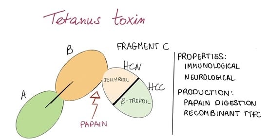
2. Structure and Association with Receptors

The native structure of TTFC is available at the Protein Data Bank under the identifier 1AF9 [8]. It is composed of the last 452 amino acid residues of TT from K865 to D1315 and contains two adjacent domains, which display a lectin-like jelly roll motif and a β-trefoil motif, respectively (Figure 2). Since the publication of this first structure in 1997, other structures obtained by X-ray diffraction have also been published (1A8D, 1D0H, 1DFQ, 1DIW, 1DLL, 1FV3, 1FV2, 7CE2) [9].
The amino-terminal jelly roll domain is composed of two curved seven-stranded β-sheets wrapped together to form a convex solvent-exposed face (strands β2, 1, 16, 7, 12, 13, 14) and a concave one (strands β3, 6, 15, 8, 9, 10, 11) (Figure 2A). The carboxyl-terminal β-trefoil domain is composed of strands β17, 21, 22, 26, 27, 30 for the β barrel and strands β18, 19a, 23, 25, 28, 29 for the β hairpin triplet (Figure 2B). This second motif can be observed in proteins involved in recognition or binding mechanisms such as histactophilin, interleukin 1α and 1β or fibroblast growth factors [8]. Site-directed mutagenesis as well as crystallization experiments made by soaking TTFC with various carbohydrates have shown that the β-trefoil domain plays a predominant role in the attachment to ganglioside receptors [2,10]. These complex molecules are sialic acid-containing glycosphingolipids highly present at the outer surface of neuronal cell membranes. The accessible moiety of ganglioside is made of an oligosaccharide of 2 to 4 hexoses to which one or more sialic acids are attached. Two distinct ganglioside binding sites have been identified at the surface of the β-trefoil domain, i.e., (i) a lactose binding site named W-pocket where the oligosaccharide core structure can bind [2,10,11,12,13,14] and (ii) a sialic acid binding site named R-pocket, which can accommodate a di- or a trisialic acid [15]. The two pockets are approximately 15 Å apart.
The R-pocket is delineated by segments 1143–1148, 1213–1216, 1226–1235 and 1271–1282. Asp1147 and Arg1226 are considered to be key binding residues [12]. The W-pocket contains binding residues Asp1222, His1271, Trp1289, Tyr1290, His1293, Phe1218 and Thr1270 [2,14,16], Trp1289 being essential for maintaining the shape of the pocket [12].
TTFC binds to gangliosides from the G1b series, which are predominant in the brain [12,17]. The tetrasaccharide core of these gangliosides is composed of Gal(β1–3)GalNac(β1–4)(NeuAc(α2–3))Gal(β1–4)Glc(β1), with Glc(β1) linked to membrane-embedded ceramide, to which at least two sialic acids are attached. A disialic acid linked to the internal galactose seems to be important for the association. Surface plasmon resonance experiments have revealed that TTFC strongly binds to disialylganglioside GD1b and slightly less to trisialylganglioside GT1b [17,18]. Affinity was not significant for GQ1b, which has a diasilic acid attached to each galactose (Figure 3). Further investigations using glycan array and solid-phase binding analysis suggest that TTFC can have a dual attachment mode and can concomitantly bind to one ganglioside at the R-pocket and one at the W-pocket [14].
In addition, it is hypothesized that the binding of TTFC to neurons could involve both gangliosides and a protein receptor. A 15–20 kDa putative receptor (p15) has been identified and different molecules have been tested for their affinity to bind TTFC [19]. In particular, a peptide Tyr-Glu-Trp and doxorubicin have shown strong interactions with TTFC [12,13]. The tripeptide binds to the R-pocket in a competitive manner with disialyllactose, suggesting that this site can interact with both proteins and carbohydrates.
In conclusion, structural data are key to understanding TTFC binding to neurons and its internalization into the central nervous system (CNS).

3. TTFC Properties and Uses: CNS Delivery and Immunogenicity

3.1. TTFC Neurological Properties

3.1.1. Permeability and CNS Delivery

TTFC was reported to bind to motor neurons (MN) [19]. TTFC contains distinct binding sites, which are recognized by neuronal receptors. This is the first step of its transport into the CNS. It especially binds to polysialylgangliosides GD1b and GT1b (Figure 3), which are present in the CNS. Their role is to regulate protein activities at the lipid membrane [20]. After ganglioside recognition, TTFC is internalized by MN at the NMJ [21]. As it has the ability to form pores in lipid vesicles, the third step of its transfer is its translocation through the lipid membrane. This translocation depends on the ganglioside content but also on the pH of the membrane [11,22]. It finally reaches the neuronal perikaryon in the CNS, where it can target the recognition and the catalytic cleavage of neuronal substrates [11]. Owing to its ability to reach the CNS and to specifically affect neurons, TTFC seems to be a promising candidate as a vector for neuronal disease treatment [22].
Indeed, used as a fusion protein or carrier after chemical linkage, TTFC may be useful to deliver either proteins or drugs into the CNS to cure neurodegenerative diseases. After intramuscular injection or systemic administration, such chemically altered TTFC were shown to enhance protein delivery in neurons by 1000-fold in a mouse model [23]. TTFC was also reported to improve both the neuronal uptake and the distribution of brain-directed therapeutics [23,24]. As the blood–brain barrier (BBB) is relatively difficult to cross, drugs are directly injected into the brain or into cerebrospinal fluid. Using TTFC as a vector for protein delivery could simplify the route of injection. Indeed, a fusion protein administrated by intramuscular injection was detected on endosomal and synaptic vesicles [24]. These two locations are interesting as the synaptic membrane can be targeted for neurotransmitter-related protein or trophic factor delivery, and endosomal and lysosomal locations can be targeted for metabolic enzyme delivery [23]. This method has been used successfully to deliver proteins to MN [24]. Importantly, several studies have shown that a TTFC/cytokine fusion had no effect on neuronal properties or cytokine protection properties [25].
Fusion proteins with TTFC can also be used to study synaptic connections. Fusion with a labeled protein (e.g., GFP, green fluorescence protein) allows transportation of the protein through synapses in a non-toxic way. Such a tool can be used to study the mechanisms of TTFC internalization and to map the neuronal network [26]. Indeed, TTFC is able to fuse with other molecules without altering its trans-synaptic and retrograde properties. This type of fusion has led to the successful study of intra- and inter-neuronal trafficking in vivo [27].
The unique properties of TTFC have also been exploited to construct a TT-derived peptide (Tet1-peptide, sequence HLNILSTLWKYR) as a new approach to delivering therapeutics to the CNS, as illustrated by the efficient delivery of small molecules into the CNS after intramuscular injection. This synthetic analog could thus also be used as a vector [28].

3.1.2. Intrinsic Neuronal Protection

In addition to its carrier properties, TTFC has also been reported to display neuroprotective activity. Indeed, retrograde and trans-synaptic transport of TTFC into the CNS after muscular injection is similar to pathways followed by trophic factors [29]. Different studies have demonstrated that TTFC had a trophic action in the brain and that it mimicked growth factors involved in survival pathways [30]. Furthermore, TTFC prevents cell death by apoptosis via several routes. Briefly, apoptosis occurs after cytochrome c release and caspase-3 activation following induction by the apoptotic promoter Bax. This promoter, in turn, is activated upon reversion of Bcl-Xl activity (an anti-apoptotic protein) through the dephosphorylation of the Bad gene [31]. Caspases are involved in major apoptotic events such as DNA digestion, chromatin condensation or membrane blebbing. TTFC disturbs these events by inhibiting caspase-3 activity, thus preventing apoptotic cell death [32]. TTFC also protects cells from apoptotic death by inhibiting Bad dissociation and association with Bcl-Xl. TTFC was thus shown to exert anti-apoptotic effects by inhibiting cell death pathways activated by Bad and by blocking pro-caspase-3 activation [33].
Moreover, TTFC is also involved in the activation of survival signaling pathways. Trk receptors transmit their signals from axons to neuronal cell bodies in a retrograde manner. Neurotrophins binding to these receptors allow protein kinase activation and tyrosine residue auto-phosphorylation. These phosphorylated tyrosine residues are then recognized by intracellular signaling proteins activating a kinase cascade. For example, Trk activation can be the trigger for phosphoinositide 3-kinase (PI3K) pathway activation, which in turn activates protein kinase B (also known as Akt), which is necessary and sufficient for eukaryotic cell survival [34]. TTFC stimulates signaling pathways Akt/PI3K, ERK and Raf/MEK/ERK [30,32,34], which play a role in cell survival. For example, PI3K is involved in the prevention of low potassium-induced apoptotic death. TTFC can provide cell protection through its action on survival signaling pathways. TTFC also protects cells by activating p21Ras protein, which is essential for the inhibition of cell death pathways [31,32]. In addition, TTFC treatment affects calcium-related gene expression, suggesting that TTFC can impact anti-apoptotic pathways through calcium-related mechanisms [29].
Another type of protection provided by TTFC concerns oxidative stress. Oxidative damage is observed in neurodegenerative diseases such as Parkinson’s disease (PD). Generation of reactive oxygen species can also contribute to MPP(+)-induced oxidative stress that activates a series of cellular factors that initiate apoptotic cell death [31]. Cubí et al. showed that after the administration of TTFC, the ceramide content and the nSMase activity increased in cerebellar granule neurons and NGF-differentiated PC12 cells [35]. It is well-known that an increase in ceramide level is related to neuron survival. Hence, as TTFC treatment increases ceramide contents, this provides neurons with a better protection against oxidation. TTFC is thus able to protect cells against neuronal oxidative stress and prevent oxidative damage.
Owing to its neuroprotective properties, TTFC has been widely tested in animal models of PD, cerebral ischemia, amyotrophic lateral sclerosis (ALS) or animals treated with molecules inducing apoptosis (Table 2, presented in Section 3.1.3). TTFC was particularly reported to induce protection against dopamine (DA) loss and improved MN behavior [30]. DA plays a key role in controlling locomotion and is the main neurotransmitter affected in PD. DA receptors are involved in the activation of Trk receptors, and it seems that the trkB pathway may be crucial for DA neuron survival. As mentioned above, TTFC has a neuroprotective effect, and that may be due to its known contribution to the activation of Trk receptors, thus improving mechanisms of DA neurotransmission [33]. TTFC had also been identified as a neuroprotective agent in ALS animal models as it prevents anti-apoptotic effects [30]. TTFC fusion proteins were tested in mice with ALS disease and displayed a combination of TTFC neuroprotective and carrier properties. Improvements in motor function activity were observed in TTFC fusion-treated mice, curbing disease progression and increasing the number of surviving MN [29]. In parallel to MN survival, a reduction in microglial reactivity was detected, stopping disease progression. Another example worth mentioning was the use of naked DNA encoding TTFC (nDNA-TTFC) in the treatment of cerebral ischemia. This disease is characterized by a decrease in blood supply to the brain. This dysfunction can lead to permanent disability and death. There is currently no treatment to counter these serious effects. The efficacy of the treatment partly relies on the ability of the therapeutic treatment to reach the neurons after crossing the BBB. Every oxidative stress parameter was reduced after nDNA-TTFC treatment in all brains tested, protecting them from oxidative damage [36]. This treatment may thus represent a non-invasive and non-viral therapeutic approach to treating cerebral ischemia. The unique properties of TTFC as a carrier and neuroprotector may pave the way for many applications to cure neurodegenerative diseases.

3.1.3. Overview of the Uses of TTFC for Its Neurological Properties

Based on all its properties, TTFC has already been used under different forms (e.g., alone, in fusion protein, conjugated) for neuronal applications, including in vivo in several studies, confirming its neurological properties. Examples of these applications are presented in Table 2.

3.2. Immunological Properties

3.2.1. Immunological Properties against Tetanus

Having presented the neurological properties of TTFC, we will now review its immunological properties. TTFC contains four universal epitopes of the 11 present in the TT sequence: 52 to 68 [52], 83 to 103 [52,53,54,55,56], 290 to 309/310 to 325 [57] (these two peptide sequences may represent the same epitope) and 409 to 420 [52] in the TTFC sequence. An immuno-informatics analysis performed by Nezafat et al. [58] unveiled four regions of the TTFC as helper epitopes (53–69, 84–108, 220–247, 361–386).
According to other studies, TTFC also contains 13 of the 28 epitopes recognized by more than 75% of patients [52,53,54,55,56,57,59]. This means that nearly half of TT epitopes are contained in the TTFC sequence. Ghafari et al. [60] suggested that TTFC may be the immunodominant part of the toxin, i.e., that the fragment may elicit the humoral immune response. Moreover, TT neutralizing antibodies were shown to be mainly directed against TTFC in mouse and human models (100% for mouse models and 75% in a human model) [61]. It is important to note that the many neutralizing antibodies described since the 1980s [62,63,64] cannot individually neutralize TT in vivo. The development of an active and safe monoclonal antibody (mAb) remains a prospect for upcoming years [65]. Hence, TTFC has become a very interesting candidate from an immunological point of view, as it could protect against tetanus. Ghotloo et al. [66] gathered the state of knowledge on epitope mapping of TT and, more particularly, the epitopes localized on the TTFC by mAbs.
Keeping its immunological properties in mind, TTFC was then tested in vivo against tetanus. The first study conducted by Fairweather et al. tested the last 121 amino acids of the B fragment associated with TTFC in mice. One microgram of the fragment was injected without adjuvant and provided complete protection against TT, as all mice survived the TT challenge [67]. Other studies were conducted on TTFC alone showing that it could induce a 13-month protection against TT when expressed in bacteria, yeasts, plant cells or insect cells [68].
Further studies then compared its immunogenicity to the antigen present in the current vaccine (tetanus toxoid, TTd) [69,70]. Depending on the injection mode, the antibody response changed. No difference between TTd and TTFC was observed after the first transcutaneous delivery (injection of either 30 µg of each protein or molar equivalent doses). However, after the third injection, TTFC induced a 2 to 12 times higher antibody response (mainly IgG1 subclasses responses, IgA were not detected). Kinetics of antibody response of both proteins also seemed to differ. Using TTFC, a constant increase in protective response was observed, whereas using TTd, the level of protective response remained constant event after new injections. Using subcutaneous injection, contradictory results were obtained. In one study, a 100 times higher dose of TTd was required to induce the same protection as TTFC [69], and in another, TTFC was 100 times less potent than TTd [70]. The origin of these differences has not been fully investigated, but the induced protection seems to be linked to the mode of injection, which must be considered when developing new tetanus vaccines. Other modes of administration confirmed the capacity of TTFC to offer protection against tetanus: sublingual immunization using Bacillus subtilis as an antigen delivery system showed full protection in mice [71]. The capacity of TTFC to be used as an antigen in a tetanus vaccine has been demonstrated, but further investigations on the impact of the mode of injection must be conducted.
Because TTFC contains many epitopes, its use as a carrier to deliver molecules to the CNS may be delicate in vaccinated individuals. Studies have been conducted to analyze the immunogenic side effects of the administration of TTFC in vaccinated animals [72]. They revealed that the amount of injected TTFC as a carrier (molar equivalent) generally exceeds 10,000 times the lethal dose of TT in a mouse model, and neutralization of all the TTFC injected was unlikely even for a vaccinated animal. This allowed a small part of TTFC to be internalized and to reach the MN. Results have shown that vaccinated and unvaccinated animals presented detectable retrograde transport of TTFC using fluorimetry [72]. This demonstrated that at a high dose, TTFC could be used as a carrier for delivery of bioactive molecules into CNS even in vaccinated individuals. Another explanation could be linked to the mode of injection, as after an intramuscular injection, TTFC was not blocked by antibodies developed after immunization against TT. This could be due to the rapid uptake of the protein after intramuscular injection [21].
Finally, as an attractive alternative to commercial ELISA assays, dipstick tests were developed using gold-conjugated TTFC [73]. This conjugate was used to bind anti-tetanus antibodies in whole blood and plasma samples, with no cold chain requirement. The results obtained were highly promising, with a specificity greater than 0.9. The use of this device could thus be integrated into monitoring programs for populations at risk (non-immune or low titer individuals).

3.2.2. TTFC as a Fusion Protein: Enhancement of Immunogenicity

TTFC did not only induce protection against tetanus, it also enhanced immunogenicity of its partner when fused. DNA-based vaccines have shown their efficacy against infectious diseases but revealed disappointing results when used in tumor models. To overcome this problem, fusion with antigen helpers was developed. TTFC displays functional characteristics of helper antigens and was thus tested in several DNA fusion proteins [74]. In particular, it improved T-cell immunity of viral oncogenes HPV-16 E6 and E7 (viral oncogenes targeted by tumor suppressor proteins p53 and pRb) used in DNA vaccines to protect against papillomavirus [75]. The fusion with TTFC enhanced CD8+ T cell response against the protein of interest. Indeed, the mounting of a robust E7-specific T-cell response was observed after the administration of a TTFC fusion protein [76]. This induced tumor regressing and prolonged survival of all mice tested. E7 stability was also improved by the fusion protein, as E7 accumulation was higher in cells treated with the fusion. This could be explained as TTFC contains universal epitopes for human CD4+ T lymphocytes: the CD4+ T-cell helper response produced after epitope recognition may promote CD8+ T-cell responses. In addition, TTFC has a stimulatory effect on IFN-γ and CD69 production, both involved in modulating the immune response. Enhancement of the immunogenicity using TTFC fusion proteins may thus be due to the action of these proteins [60].
The fragment was also shown to enhance bacterial polysaccharide antigenicity in conjugate vaccines. As for TTd, TTFC has been tested as a carrier protein in conjugate vaccines, albeit because the production of TTFC does not require a formaldehyde detoxification step, its conjugation sites are easier to characterize [77]. Several authors chose to use this protein to synthetize well-defined glycoconjugates to highlight the link between the conjugate structure and the induced immune response [78,79,80,81]. TTFC was shown to play a normal protein carrier role as its conjugation with the bacterial polysaccharides induced a higher and long-lasting immune response.

3.2.3. In Silico Design of Epitope-Based Vaccines

In the last decade, in silico methods have been used to design vaccines based on TTFC epitopes associated with other epitopes. For example, in the case of atherosclerosis, a multi-epitope construct was designed by fusing epitopes from TTFC with other immunogenic molecules such as calreticulin, heat shock protein 60, and cholera toxin B [82]. Bioinformatics analysis allowed the authors to build a stable chimeric protein with the potential to shift the immune response and to reduce atherosclerosis. In silico approaches were also used to design anti-infective vaccines against brucellosis [83] and leptospirosis [84] in order to induce a strong immune response mediated by T- and B-cells. Immuno-informatics was also helpful to design a vaccine against melanoma [85], by fusing several antigens, including TTFC. This multi-epitope vaccine (MEV) approach showed high immunogenicity, providing hope for cancer immunotherapy. In another study [58], different algorithms and servers were used to design a novel MEV against cancer. The selected cytolytic T lymphocyte (CTL) epitopes were linked together to enhance epitope presentation. In parallel, different helper epitopes (including TTFC) were conjugated to stimulate helper T lymphocyte (HTL) immunity. All these epitopes were associated with heparin-binding hemagglutinin used as an adjuvant. The final protein was thus able to stimulate both cellular and humoral immune responses. An in vivo study was conducted to produce the corresponding vaccine in E. coli in order to evaluate antitumor efficacy against the HPV-16 E7-expressing murine tumor cell line TC-1. The results obtained revealed a significantly higher IgG secretion with MEV containing TTFC epitopes compared to the E7 protein vaccine [86].

3.2.4. TTFC Uses for Its Immunological Properties
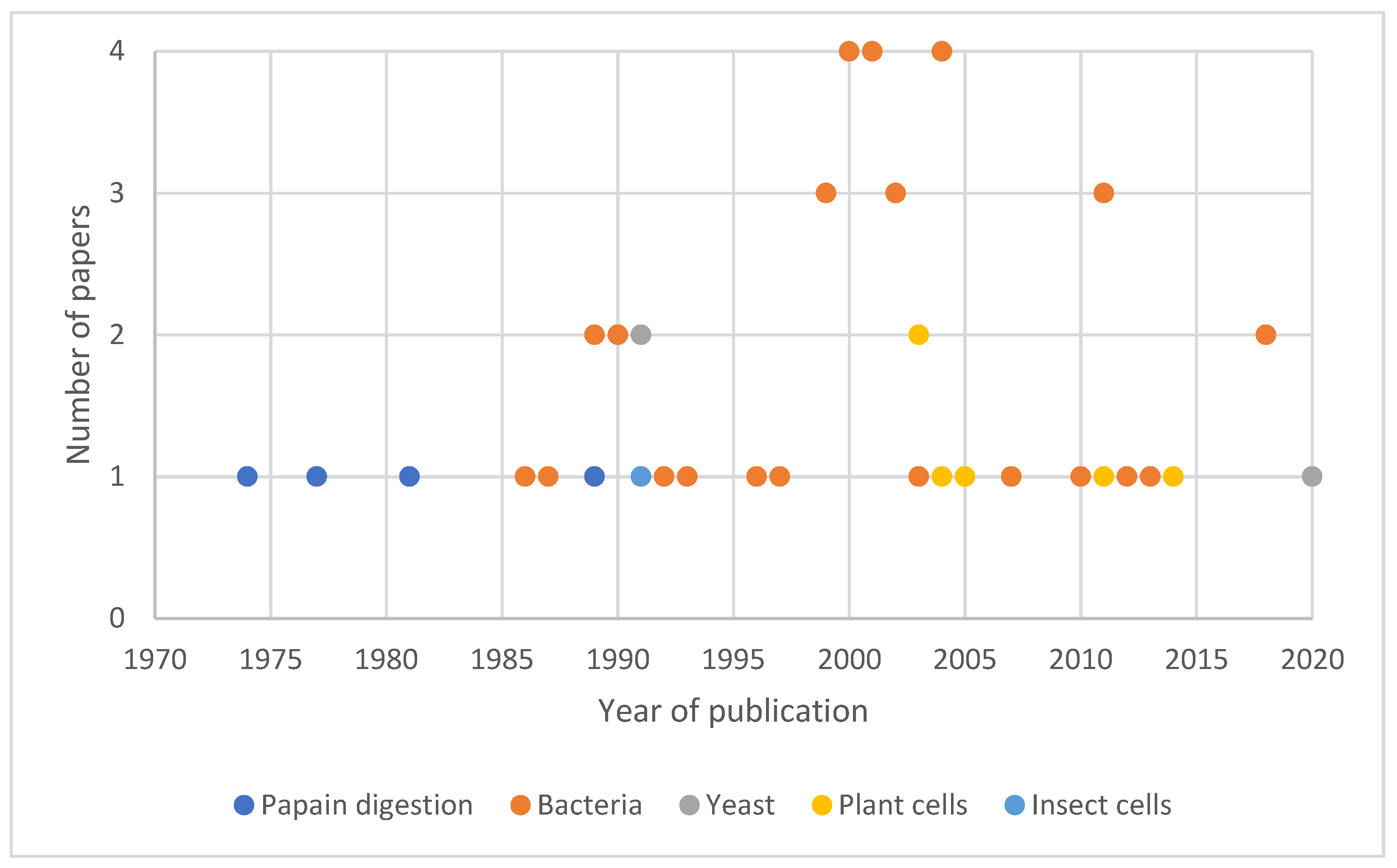
As for its neurological properties, the immunological properties of TTFC have inspired many groups of research. Again, TTFC was used under different forms (e.g., alone, in fusion proteins, in conjugate vaccines) for diverse vaccine applications. Some examples of these applications are presented in Table 3.

4. TTFC Production

Since the discovery of TTFC, different methods of production have been developed (Figure 4). The first studies were conducted using papain digestion of TT and, as a consequence, TTFC was first obtained using this technique. Nevertheless, the hazardous character of this manipulation has led researchers to develop recombinant systems to express the protein. Bacterial recombinant hosts are currently the most widely used to produce the protein, but other systems such as yeasts, plant cells and insect cells have also been used. In this part, we describe the most common methods to produce TTFC and provide information to enable the comparison of the production methods described.

4.1. Papain Digestion

One of the simplest ways of obtaining TTFC is to enzymatically digest TT. This can be achieved using the protease papain present in papaya fruits. Papain cleaves TT between Ser864 and Lys865 residues resulting in two fragments: TTFC and fragment A-B (Figure 1). Papain was first used for this purpose by Helting et al. in 1974 [7]. At that time, structural and functional studies of TT were difficult to ascertain because of the lability of the whole protein after purification. This is why researchers decided to split the protein into smaller fragments, trying to obtain separate structural information. The digestion was usually performed in 0.1 M phosphate buffer containing 1 mM EDTA and cysteine hydrochloride at pH 6.5. The experimental conditions (e.g., reaction temperature, amount of material) were progressively optimized [7,100,101,102,103]. For example, in 2014, Murzello et al. [100] digested TT within 30 min at 55 °C using 10 units of papain from papaya latex (Sigma P4762) per 10 mg of TT. To stop the reaction, the enzyme was inactivated by adding L-1-chloro-3-tosylamido-7-amino-2-heptanone and by letting the batch cool to 25 °C.
In the next step, TTFC was purified from the solution obtained after papain digestion. Usually, several chromatographic separations (e.g., size-exclusion chromatography) were performed to finally yield pure TTFC. Subsequently, an affinity chromatography column using an anti-fragment [A-B] antibody could complete the purification protocol. Sometimes, TTFC needed to be rechromatographed (on the same column material under different elution conditions) to remove the remaining contaminants. Yields obtained after all purification steps varied between 15 to 28% from the TT starting material (approximately 0.13 to 10 mg of protein per mL) [7,100,101,102,103].
This method is easy to handle and was mostly used at the beginning of TTFC discovery. Since then, different recombinant systems have been employed to produce TTFC without the use of a toxic protein as a starting material.

4.2. TTFC Production in Recombinant Systems

TTFC production using papain digestion was the first to be described, but as it was hazardous and a major difficulty was to eliminate the very small amount of undigested toxin from the fragment preparation, recombinant production systems were developed [104]. These systems present some striking advantages compared to papain digestion. First, large amounts of TTFC can be efficiently produced since large numbers of host organisms (e.g., bacteria, yeast, plant cells, insect cells) can be grown in small- and large-scale cultures with standard laboratory equipment. Second, only controlled non-toxic proteins can be produced, which means that production is safer and toxic contaminations are easy to avoid [105].

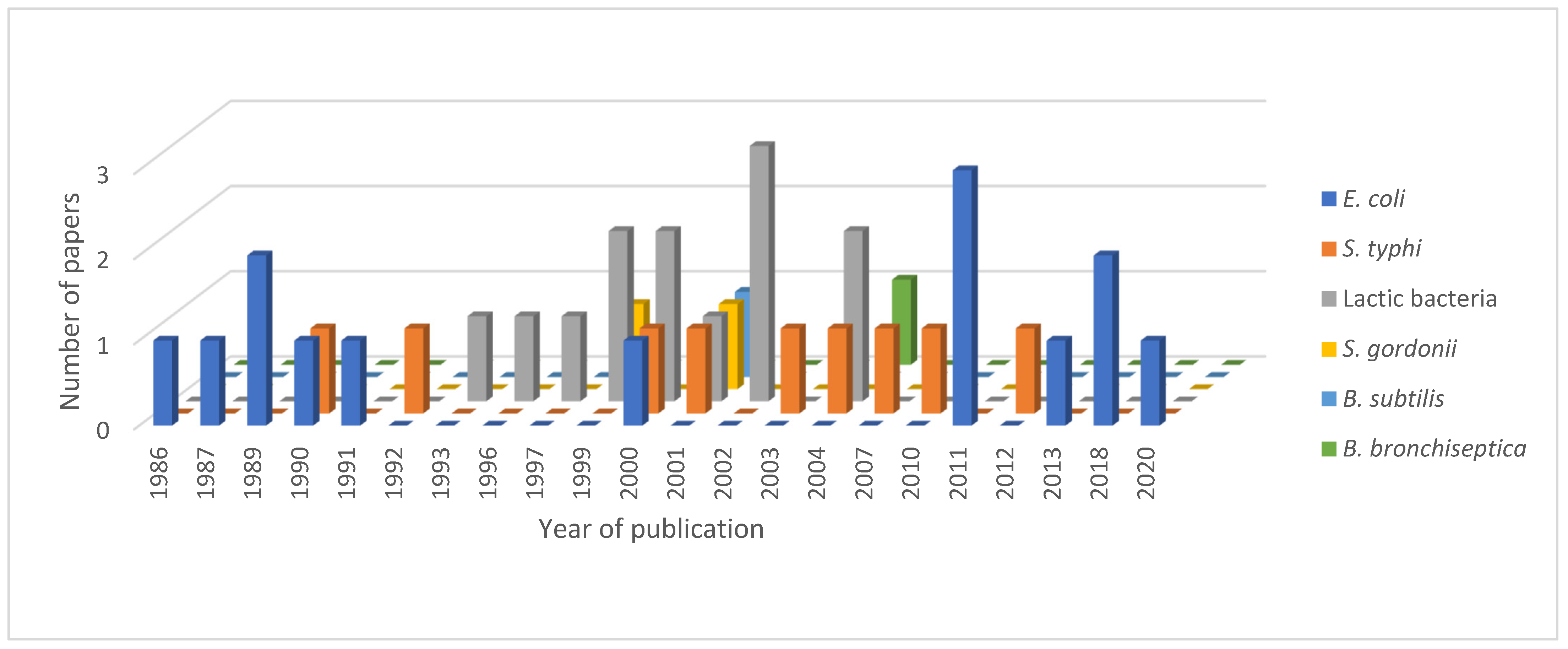
4.2.1. Escherichia coli as a Host for TTFC Production

In 1986, the cloning of the DNA encoding the TTFC from C. tetani CN3911 strain was published [106]. From this first study, several groups started to work on the production of TTFC as a heterologous protein in E. coli (Table 4) [104,105,106,107,108,109,110,111,112,113]. Low amounts (from 1 to 10 mg/L) of recombinant proteins were obtained from expression vectors containing trp or tac promoters [104,105,106]. TTFC was also produced as a fusion protein in the pMalc2x vector [109]. TFC codon analysis revealed that the gene contained a high proportion of rare codons in E. coli, and teams thus produced synthetic TTFC DNA in order to reduce rare codons and decrease the percentage of AT pairs, increasing protein production [110,111]. However, it appeared that the yield increased further when BL21 bacteria and an expression plasmid the including T7 promotor were used [107,108,112,113]. In 2011, Yu et al. [110] optimized the fermentation production of 40 L to reach 333 mg/mL recombinant TTFC. As described recently [114], the fermentation conditions were further optimized to increase the production of recombinant proteins. In the late 1990s, other bacterial expression systems were studied to produce and ensure the delivery of TTFC (Figure 5). In fact, these alternative bacterial expression systems were developed to produce oral vaccines against TT, i.e., they served both as an expression method and as a delivery system.

4.2.2. TTFC Expression and Delivery in Other Bacterial Host Strains

The production of heterologous proteins in cyanobacteria, phototrophic microorganisms with low nutrient requirements, may be an alternative. For eukaryote proteins, this is possible in the form of fusion proteins. Recently, in 2021, the TTFC protein was expressed in the cyanobacterium Synechocystic sp. PCC6803 (Synechocystis) [115]. This bacterium was initially modified to stably express the tobacco etch virus protease (TEVp). Then in the cpc operon locus, the native cpcB gene encoding the abundant β-subunit of the phycocyanin was replaced with a fusion construct comprising the cpcB and TTFC DNA sequences separated by a TEV cleavage site. In vivo cleavage led to the accumulation in the cytosol of soluble TTFC proteins and putative aggregates containing the uncleaved protein cpcB-tev-TTFC, the sum of the two proteins representing approximately 20% of the total protein content (TPC). Hidalgo Martinez et al. [116] showed that the accumulation of soluble cpcB-tev-TTFC fusion proteins could occur as they contribute to the formation of phycobilisomes, comprising the major light-harvesting antenna complex for photosynthesis.
TTFC was produced by other bacterial recombinant expression systems for different applications. All these bacteria were not only used to express TTFC but also to deliver it in oral vaccination approaches. These delivery systems were developed in order to produce safe, stable and inexpensive oral vaccines. Such vaccines are particularly appropriate in developing countries because syringes and needles used for injected vaccination may be used repeatedly and lead to the transmission of infectious agents such as HIV, hepatitis B and C viruses. Oral immunization also simplifies the administration of vaccines, allowing less qualified health workers to immunize populations. These vaccines are highly compatible with mass immunization programs as their logistics are simpler. Finally, oral administration is usually preferred by both children and adults compared to parenteral injection [117]. The objective of oral vaccines is to pass through the intestinal mucosal surfaces to reach the immune system. Mucosal surfaces play a role in nutrient uptake, and an immune response against these dietary antigens is unwanted and generally suppressed to avoid food intolerance. It is thus difficult to induce a systemic immune response after the delivery of antigens to these surfaces. The immune response can, however, be improved by associating the antigen with a bacterium [118]. Lactic acid bacteria are well adapted to deliver antigens through mucosal surfaces and are interesting since they are generally recognized as safe (GRAS) by the FDA [119]. These nonpathogenic food-grade Gram-positive bacteria can be resistant to the harsh conditions of the intestinal environment. For example, the Lactococcus lactis bacterium can prevent direct contact of antigens with gastric acid and proteolytic enzymes, conferring a higher resistance in the intestinal medium [120]. Different lactic acid bacteria were tested as expression systems for TTFC: L. plantarum, L. lactis and L. casei [118,119,120,121,122,123,124,125,126,127]. Generally, the recombinant plasmids were constructed in the E. coli DH5α host strain and were subsequently introduced into alternative bacterial host strains by electroporation. Several strains and plasmids were tested to express the protein, leading in most cases to successful intragastric immunization. Strains UCP1060 and MG1363 were most often used for L. lactis, strains NCIMB8826 for L. plantarum and strains ATCC393 for L. casei [118,119,120,121,122,123]. Plasmids pMEC46 and pMEC127 were both frequently used within these strains, and the administration of MG1363(pMEC46) and NCIMB8826(pMEC127) strains, in particular, showed a high antibody production in mice [119,121,123]. To increase the immune response, different mutations were also tested in bacterial strains. For example, L. plantarum and L. lactis (Alr) mutant strains (replacing of L-alanine by D-alanine), both producing TTFC intracellularly, were more immunogenic than their wild-type counterparts [121]. These expression systems were good alternatives to attenuate pathogenic bacterial delivery systems such as Salmonella typhimurium [128] and Mycobacterium bovis BCG [129].
For TTFC oral delivery, Streptococcus gordonii and Bacillus subtilis spores have also been used. S. gordonii is a nonpathogenic Gram-positive commensal bacterium, which is able to colonize mucosal surfaces [130]. This bears the huge advantage of continuously stimulating the immune system because such bacteria act on dendritic cell maturation [131]. Dendritic cells are linked to the induction of primary T-cell responses and undergo a maturation process to provoke such immune responses. This maturation is launched by inflammatory signals and ends upon contact with T-cells. During their maturation process, dendritic cells migrate from peripheral tissues to lymphoid organs and exhibit a strong antigen-presenting capacity. Due to their central role in the immune response, dendritic cells have been targeted for vaccine development [131]. S. gordonii appeared as a good candidate for antigen delivery in this context because it was shown to induce the maturation of dendritic cells and thus elicit an immune response. The GP1253 strain was used in two studies dealing with the production of TTFC in S. gordonii using either plasmid pSMB55 or pMSB158 [130,131]. The production resulted in the expression of TTFC in 78% of recombinant cells (100 ng/109 CFU of bacteria) and the administration of these recombinant bacteria protected mice against the TT challenge [130]. More recently, B. subtilis spores were investigated as an antigen delivery system (Figure 5). This strategy bears several advantages: spores are considered to be safe for human and animal use, and they can stimulate cytokine release and interact with antigen-presenting cells [132]. Moreover, their large-scale production is inexpensive, the genome of B. subtilis is known and is accessible for genetic manipulations. Spores also exhibit stability towards solvent exposure and extreme temperatures, which enables them to maintain integrity in extreme environments, including in the intestine [132]. TTFC was produced in all cases as a fusion with a spore coating protein (CotC or CotB). This fusion led to the expression of TTFC on the B. subtilis spore surface. Different plasmids and strains were applied, and in each case, a TTFC-specific antibody response was obtained [133,134,135]. More recently, the spore-display strategy was optimized by altering the spore coating protein and spore production temperature [136].
In other studies, attenuated S. typhimurium and Bordetella bronchiseptica Gram-negative bacteria were used for TTFC expression. These recombinant systems were developed as live bacterial vector vaccines to induce double protection within the same vaccine: one directed against the tetanus toxin and the other against the host bacterium. Both bacterial strains were used for oral vaccine development. B. bronchiseptica infects the respiratory tract and hence, could be a suitable vector for oral immunization. A plasmid encoding the TTFC (pFHAFrgC) transformed into the BBC18 strain was investigated. All immunized mice developed a protective immune response against the TT challenge and displayed high antibody titers against B. bronchiseptica [137]. Attenuated Salmonella strains used to express TTFC were used before as vaccination strains to raise an immune response against the Salmonella pathogen itself and appeared to be safe and efficient [138]. Different foreign antigens were successfully expressed in S. typhimurium, but their capacity to induce an immune response was sometimes compromised because of the instability of their plasmid. This relation between plasmid instability and the generated immune response directed against the foreign antigen was explored by different research teams. For example, Dustan et al. [138] compared the stability of different plasmids expressing TTFC in the attenuated BRD509 strain of S. typhimurium (aroA aroD mutant). In all cases, optimal antibody response was generated against S. typhimurium, but differences were observed concerning the anti-TTd antibody response. They showed that by using the unstable plasmid pIC20H/TT, no anti-TTd antibody was detected in mice, whereas by using the highly stable plasmid pTETtac4, the antibody titers were significantly higher. A third plasmid, pACYC184/TT, presenting intermediate stability, gave rise to an intermediate tetanus vaccine efficacy. This corroborates the link between the plasmid stability and the quality of the protection directed against the expressed antigen [138]. The stability of foreign antigen expression can be improved by using inducible promoter systems such as nirB, dmsA or tac [138,139,140]. In each case, the use of the promoter stabilized TTFC expression and induced higher TT antibody titers in immunized mice. Among these promoters, it was demonstrated that the pTETdmsA3 plasmid expressed in S. typhimurium generated higher titers than the pTETnir15 plasmid, expressed in the strain CVD908-htrA [139]. Another study conducted in the BRD509 strain revealed that the pTETnir15 plasmid induced stronger protection than plasmids containing the tac promoter [140]. In addition to the plasmids, the nature of the strain used as the live vector also influences the quality of the immune response directed against the foreign antigen. A study was conducted on two types of S. typhimurium mutant strains using TTFC-expressing plasmids: BRD509 and BRD807 (aroA htrA mutant). Independently of the tested plasmids, anti-TT antibody titers were always superior when mice were immunized with the BRD509 strain [141].
In summary, several bacterial production systems have been developed to produce TTFC. Different modifications have been made to the plasmids and the host strains to improve the fragment expression. Bacterial expression systems are widely used because they are cheap, their genetic manipulation is easy, and these fermentation systems are often easy to scale up. However, expressing foreign antigens in bacterial strains also has its drawbacks: Gram-negative bacteria cell walls contain lipopolysaccharides (LPS), which are toxic pyrogens. Consequently, proteins expressed in these bacteria must be carefully tested on LPS content before being used. Yeasts or plant cells exhibit no such features and could thus be attractive hosts for TTFC expression.

4.2.3. Recombinant TTFC Expression in Yeast and Plant Cells

The yeast species Saccharomyces cerevisiae and Pichia pastoris were used for recombinant TTFC expression as well as tobacco chloroplasts, representing a common plant expression system. To obtain satisfying levels of expression in yeast, a synthetic TTFC gene was constructed, containing a codon-optimized high CG gene lacking the fortuitous polyadenylation sites, which gave rise to truncated mRNA [142,143]. Around 60 to 90 mg/L of soluble protein were obtained using S. cerevisiae S150-2B strain expressing the synthetic TTFC gene (pWYG5-TET15) [142]. However, in S. cerevisiae, the recombinant TTFC secreted in the culture medium was immunogenically inactive due to glycosylation. A methylotrophic yeast, P. pastoris, produced a much higher yield of 12 g/L of soluble protein using the integrative plasmid with the promotor from the methanol-induced alcohol oxidase gene and the synthetic TTFC gene (pPIC3-TET15) [143]. When the native gene was cloned into a yeast expression plasmid (p9k-G) with the alpha factor secretion signal and expressed in P. pastoris, the secreted TTFC was glycosylated. Site-directed mutation performed on five potential N-glycosylation sites showed that the number and localization of these sites impact the expression and secretion of TTFC. Indeed, decreasing the number of N-glycosylation sites decreased TTFC secretion [144].
Conversely, plant cells did not require the use of the synthetic TTFC gene to obtain high yields of proteins. Indeed, the AT-rich bacterial gene, when expressed in a cassette consisting of a PrrnLT7g10 cassette (plasmids pJST10 and pJST11, respectively), produced twice as much protein (25% of total cell protein (TCP)) than the high-GC synthetic gene. However, this high level of TTFC is detrimental to plants, as evidenced by the chlorotic phenotype in plants [145,146]. The chloroplast expression system has inherited similarities with the prokaryotic expression systems derived from the ancestral cyanobacterium. They contain a plastid-encoded RNA polymerase, which can be compared to the bacterial RNA polymerase [147]. Two types of methods can be used to produce proteins in plant expression systems: either nuclear transgenic plants (low expression levels generally achieved) or DNA can be introduced into the chloroplast genome by particle bombardment (higher expression levels of recombinant proteins) [148]. Proteins are usually extracted from leaf tissues using an extraction buffer. In addition, chloroplasts are heat stable, cheap to produce, and they can remove undesirable selectable markers and use operons for multi-antigen expression; they produce a high level of immunogenic recombinant proteins, specifically target the gene and retain the gene product in the plastid; this system also benefits from environmental containment as a result of maternal inheritance [148]. Michoux et al. studied the production capacity of the Nt-pJST12 line obtained from a plasmid expressing the synthetic gene in a cassette consisting of a PrrnLatpB cassette. They showed that 92 g of TTFC could be produced annually, using five 250 L-bioreactors and running 20 rounds of production per reactor per year. This quantity of TTFC should be enough to launch preclinical and clinical trials [149]. Chloroplast-expressing systems were able to yield high amounts of protein but were not always efficient as vaccines. As both plant and the expressed vaccine are degraded in the intestinal tract, oral immunization was inefficient [150]. Nevertheless, intranasal immunization was protective in mice against the TT challenge [145,146].
Beyond yeast and chloroplasts, insect cells were also tested as host organisms using baculovirus expression vector p36C to co-infect Spodoptera frugipedra cells. The resulting TTFC was soluble and showed an immunogenic activity, but its secondary structure was different from the TTFC obtained in E. coli as its ganglioside binding properties were altered [151].
TTFC can be produced using a panel of different production methods. Purification [104,105,107,108,110,152] and characterization [108,110,122,133,152] steps are then usually the last ones performed in protein production to finally obtain the required quality. In 2020, Chai et al. established a simple method to purify TTFC by ion-exchange chromatography [153].

5. Conclusions

The development of new vaccines and neuroprotective agents present major issues in drug discovery. New vaccines must be well defined and better controlled, present great antigenic and immunogenic activities and be as stable as possible to allow mass vaccination campaigns in developing countries. The development of neuroprotective agents mostly focuses on the drug delivery into neuronal cells as the BBB is difficult to cross. TTFC protein can be a good candidate to overcome problems in both fields. Its structure has been characterized, allowing a better understanding of its biological properties. Recombinant TTFC is easily characterized and was shown to be sufficient for the protection of mice against tetanus. Its properties can also be used to protect against other diseases (e.g., multiple myeloma, cholera) as TTFC enhances the immunogenicity of therapeutic proteins when fused to them. This fragment is responsible for the neuronal binding of TT, meaning that it can reach neurons. This property has been widely studied for drug delivery into the CNS, as this is a key point in the cure of neurodegenerative diseases (e.g., ALS, AD, PD). These studies have shown that when conjugated or fused to a drug, TTFC retains its neuronal binding capacity and is a good carrier for drug delivery into the brain. Intrinsic neuroprotective properties were also discovered, confirming the growing interest for this protein in the neurological field. TTFC thus appears to be a promising protein for drug discovery, as its production, characterization, and properties are well described. Further studies must now be conducted on different animal models to confirm its biological relevance and hopefully will result in the development of new drugs. In addition, TTFC-based immune-informatics approaches also seem to be an attractive and effective design to access new therapeutic entities against certain zoonotic diseases (e.g., brucellosis, leptospirosis).
In conclusion, TTFC protein can be considered to be a useful drug discovery toolbox. Few proteins offer such a wide range of uses, including as a carrier, delivery vector, conjugate, booster, inducer, and neuroprotector. With the development of nanomedicines, and consideration for brain safety [154], the use of TTFC could pave the way for novel, safer applications.

Author Contributions

Software, C.B. and P.G.; investigation, C.B., N.H., S.K. and M.L.B.; writing—original draft preparation, C.B., J.P. and M.L.B.; writing—review and editing, C.B., A.M., P.G., J.J., J.P. and M.L.B.; supervision, M.L.B. All authors have read and agreed to the published version of the manuscript.

Funding

This study was sponsored by Sanofi (E1492.0002).

Institutional Review Board Statement

Not applicable.

Informed Consent Statement

Not applicable.

Data Availability Statement

Data sharing not applicable.

Acknowledgments

Marc Le Borgne and Angélique Mularoni would like to thank “Institut Convergence PLAsCAN” (ANR-17-CONV-0002). Marc Le Borgne would like to thank Brigitte Manship for editing the manuscript. The authors would like to thank Christine Janssen for conducting the graphical abstract of the article.

Conflicts of Interest

Joseph Paladino is an employee of Sanofi. The authors confirm that they have no conflict of interest with the content of this article.

Abbreviations

ADAlzheimer’s disease
ALSamyotrophic lateral sclerosis
BBBblood brain barrier
BDNFBrain-derived neurotrophic factor
CFUcolony-forming unit
CNScentral nervous system
CoaRcoagulase R domain
CTLcytotoxic T lymphocyte
DAdopamine
DOMdomain
EDLextensor digitorum longus
FlaBFlagellin B
FVBFriend virus B
GDNFglial cell line-derived neurotrophic factor
GFPgreen fluorescence protein
GRASgenerally recognized as safe
HLAhuman leukocyte antigen
HPV human papillomavirus
HTLhelper T lymphocyte
IGF-1insulin-like growth factor-1
LPSlipopolysaccharide
mAbmonoclonal antibody
METH methamphetamine
MEVmultiepitope vaccine
MNmotor neurons
NMJneuromuscular junction
NPnanoparticles
NPY-Creneuropeptide Y-Cre recombinase
OPSO-specific polysaccharide
PDParkinson’s disease
PEISHthiolated poly(ethylene imine)
PI3Kphosphoinositide 3-kinase
PSpolysaccharide
PSMAprostate-specific membrane antigen
SCspinal cord
SODsuperoxide dismutase
SOLsoleus
Tem 1tumor endothelial marker 1
TEVptobacco etch virus protease
TLR5Toll-like receptor 5
TPCtotal protein content
TTdtetanus toxoid
TTFCtetanus toxin fragment C
Trxthioredoxin

References

  1. Tetanus. Available online: https://ourworldindata.org/tetanus (accessed on 8 May 2022).
  2. Emsley, P.; Fotinou, C.; Black, I.; Fairweather, N.F.; Charles, I.G.; Watts, C.; Hewitt, E.; Isaacs, N.W. The structures of the H(C) fragment of tetanus toxin with carbohydrate subunit complexes provide insight into ganglioside binding. J. Biol. Chem. 2000, 275, 8889–8894. [Google Scholar] [CrossRef] [PubMed] [Green Version]
  3. Montecucco, C.; Schiavo, G. Mechanism of action of tetanus and botulinum neurotoxins. Mol. Microbiol. 1994, 13, 1–8. [Google Scholar] [CrossRef] [PubMed]
  4. Deinhardt, K.; Berninghausen, O.; Willison, H.J.; Hopkins, C.R.; Schiavo, G. Tetanus toxin is internalized by a sequential clathrin-dependent mechanism initiated within lipid microdomains and independent of epsin1. J. Cell Biol. 2006, 174, 459–471. [Google Scholar] [CrossRef] [PubMed] [Green Version]
  5. Eisel, U.; Jarousch, W.; Goretzki, K.; Henschen, A.; Engels, J.; Weller, U.; Hudel, M.; Habermann, E.; Niemman, H. Tetanus toxin: Primary structure, expression in E. coli, and homology with botulinum toxins. EMBO J. 1986, 5, 2495–2502. [Google Scholar] [CrossRef]
  6. Matsuda, M.; Lei, D.L.; Sugimoto, N.; Ozutsumi, K.; Okabe, T. Isolation, purification, and characterization of fragment B, the NH2-terminal half of the heavy chain of tetanus toxin. Infect. Immun. 1989, 57, 3588–3593. [Google Scholar] [CrossRef] [Green Version]
  7. Helting, T.; Zwister, O. Enzymatic breakdown of tetanus toxin. Biochem. Biophys. Res. Commun. 1974, 57, 1263–1270. [Google Scholar] [CrossRef]
  8. Umland, T.C.; Wingert, L.M.; Swaminathan, S.; Furey, W.F.; Schmidt, J.J.; Sax, M. Structure of the receptor binding fragment HC of tetanus neurotoxin. Nat. Struct. Biol. 1997, 4, 788–792. [Google Scholar] [CrossRef]
  9. RCSB Protein Data Bank. Available online: https://www.rcsb.org (accessed on 9 April 2022).
  10. Louch, H.A.; Buczko, E.S.; Woody, M.A.; Venable, R.M.; Vann, W.F. Identification of a binding site for ganglioside on the receptor binding domain of tetanus toxin. Biochemistry 2002, 41, 13644–13652. [Google Scholar] [CrossRef]
  11. Calvo, A.C.; Oliván, S.; Manzano, R.; Zaragoza, P.; Aguilera, J.; Osta, R. Fragment C of tetanus toxin: New insights into its neuronal signaling pathway. Int. J. Mol. Sci. 2012, 13, 6883–6901. [Google Scholar] [CrossRef] [Green Version]
  12. Jayaraman, S.; Eswaramoorthy, S.; Kumaran, D.; Swaminathan, S. Common binding site for disialyllactose and tri-peptide in C-fragment of tetanus neurotoxin. Proteins 2005, 61, 288–295. [Google Scholar] [CrossRef]
  13. Sinha, K.; Box, M.; Lalli, G.; Schiavo, G.; Schneider, H.; Groves, M.; Siligardi, G.; Fairweather, N.F. Analysis of mutants of tetanus toxin HC fragment: Ganglioside binding, cell binding and retrograde axonal transport properties. Mol. Microbiol. 2000, 37, 1041–1051. [Google Scholar] [CrossRef] [PubMed]
  14. Chen, C.; Fu, Z.; Kim, J.-J.P.; Barbieri, J.T.; Baldwin, M.R. Gangliosides as high affinity receptors for tetanus neurotoxin. J. Biol. Chem. 2009, 284, 26569–26577. [Google Scholar] [CrossRef] [PubMed] [Green Version]
  15. Chen, C.; Baldwin, M.R.; Barbieri, J.T. Molecular basis for tetanus toxin coreceptor interactions. Biochemistry 2008, 47, 7179–7186. [Google Scholar] [CrossRef] [PubMed]
  16. Fotinou, C.; Emsley, P.; Black, I.; Ando, H.; Ishida, H.; Kiso, M.; Sinha, K.A.; Fairweather, N.F.; Isaacs, N.W. The crystal structure of tetanus toxin Hc fragment complexed with a synthetic GT1b analogue suggests cross-linking between ganglioside receptors and the toxin. J. Biol. Chem. 2001, 276, 32274–32281. [Google Scholar] [CrossRef] [Green Version]
  17. MacKenzie, C.R.; Hirama, T.; Lee, K.K.; Altman, E.; Young, N.M. Quantitative analysis of bacterial toxin affinity and specificity for glycolipid receptors by surface plasmon resonance. J. Biol. Chem. 1997, 272, 5533–5538. [Google Scholar] [CrossRef] [Green Version]
  18. Montecucco, C. How do tetanus and botulinum toxins bind to neuronal membranes? Trends Biochem. Sci. 1986, 11, 314–317. [Google Scholar] [CrossRef]
  19. Herreros, J.; Lalli, G.; Schiavo, G. C-terminal half of tetanus toxin fragment C is sufficient for neuronal binding and interaction with a putative protein receptor. Biochem. J. 2000, 347 Pt 1, 199–204. [Google Scholar] [CrossRef]
  20. Vajn, K.; Viljetić, B.; Degmečić, I.V.; Schnaar, R.L.; Heffer, M. Differential distribution of major brain gangliosides in the adult mouse central nervous system. PLoS ONE 2013, 8, e75720. [Google Scholar] [CrossRef] [Green Version]
  21. Toivonen, J.M.; Oliván, S.; Osta, R. Tetanus toxin C-fragment: The courier and the cure? Toxins 2010, 2, 2622–2644. [Google Scholar] [CrossRef] [Green Version]
  22. Gramlich, P.A.; Remington, M.P.; Amin, J.; Betenbaugh, M.J.; Fishman, P.S. Tat-tetanus toxin fragment C: A novel protein delivery vector and its use with photochemical internalization. J. Drug Target. 2013, 21, 662–674. [Google Scholar] [CrossRef]
  23. Francis, J.W.; Bastia, E.; Matthews, C.C.; Parks, D.A.; Schwarzschild, M.A.; Brown, R.H.; Fishman, P.S. Tetanus toxin fragment C as a vector to enhance delivery of proteins to the CNS. Brain Res. 2004, 1011, 7–13. [Google Scholar] [CrossRef] [PubMed]
  24. Benn, S.C.; Ay, I.; Bastia, E.; Chian, R.-J.; Celia, S.A.; Pepinsky, R.B.; Fishman, P.S.; Brown, R.H.; Francis, J.W. Tetanus toxin fragment C fusion facilitates protein delivery to CNS neurons from cerebrospinal fluid in mice. J. Neurochem. 2005, 95, 1118–1131. [Google Scholar] [CrossRef] [PubMed]
  25. Bordet, T.; Castelnau-Ptakhine, L.; Fauchereau, F.; Friocourt, G.; Kahn, A.; Haase, G. Neuronal targeting of cardiotrophin-1 by coupling with tetanus toxin C fragment. Mol. Cell. Neurosci. 2001, 17, 842–854. [Google Scholar] [CrossRef] [PubMed]
  26. Kissa, K.; Mordelet, E.; Soudais, C.; Kremer, E.J.; Demeneix, B.A.; Brûlet, P.; Coen, L. In vivo neuronal tracing with GFP-TTC gene delivery. Mol. Cell. Neurosci. 2002, 20, 627–637. [Google Scholar] [CrossRef]
  27. Miana-Mena, F.J.; Roux, S.; Benichou, J.-C.; Osta, R.; Brûlet, P. Neuronal activity-dependent membrane traffic at the neuromuscular junction. Proc. Natl. Acad. Sci. USA 2002, 99, 3234–3239. [Google Scholar] [CrossRef] [Green Version]
  28. Kassa, R.; Monterroso, V.; David, L.L.; Tshala-Katumbay, D. Diagnostic and therapeutic potential of tetanus toxin-derivatives in neurological diseases. J. Mol. Neurosci. 2013, 51, 788–791. [Google Scholar] [CrossRef] [Green Version]
  29. Moreno-Igoa, M.; Calvo, A.C.; Penas, C.; Manzano, R.; Oliván, S.; Muñoz, M.J.; Mancuso, R.; Zaragoza, P.; Aguilera, J.; Navarro, X.; et al. Fragment C of tetanus toxin, more than a carrier. Novel perspectives in non-viral ALS gene therapy. J. Mol. Med. 2010, 88, 297–308. [Google Scholar] [CrossRef]
  30. Mendieta, L.; Bautista, E.; Sánchez, A.; Guevara, J.; Herrando-Grabulosa, M.; Moran, J.; Martínez, R.; Aguilera, J.; Limón, I.D. The C-terminal domain of the heavy chain of tetanus toxin given by intramuscular injection causes neuroprotection and improves the motor behavior in rats treated with 6-hydroxydopamine. Neurosci. Res. 2012, 74, 156–167. [Google Scholar] [CrossRef]
  31. Chaïb-Oukadour, I.; Gil, C.; Rodríguez-Alvarez, J.; Ortega, A.; Aguilera, J. Tetanus toxin HC fragment reduces neuronal MPP+ toxicity. Mol. Cell. Neurosci. 2009, 41, 297–303. [Google Scholar] [CrossRef]
  32. Chaïb-Oukadour, I.; Gil, C.; Aguilera, J. The C-terminal domain of the heavy chain of tetanus toxin rescues cerebellar granule neurones from apoptotic death: Involvement of phosphatidylinositol 3-kinase and mitogen-activated protein kinase pathways. J. Neurochem. 2004, 90, 1227–1236. [Google Scholar] [CrossRef]
  33. Mendieta, L.; Venegas, B.; Moreno, N.; Patricio, A.; Martínez, I.; Aguilera, J.; Limón, I.D. The carboxyl-terminal domain of the heavy chain of tetanus toxin prevents dopaminergic degeneration and improves motor behavior in rats with striatal MPP+-lesions. Neurosci. Res. 2009, 65, 98–106. [Google Scholar] [CrossRef] [PubMed]
  34. Gil, C.; Chaib-Oukadour, I.; Aguilera, J. C-terminal fragment of tetanus toxin heavy chain activates Akt and MEK/ERK signalling pathways in a Trk receptor-dependent manner in cultured cortical neurons. Biochem. J. 2003, 373 Pt 2, 613–620. [Google Scholar] [CrossRef]
  35. Cubí, R.; Candalija, A.; Ortega, A.; Gil, C.; Aguilera, J. Tetanus toxin Hc fragment induces the formation of ceramide platforms and protects neuronal cells against oxidative stress. PLoS ONE 2013, 8, e68055. [Google Scholar] [CrossRef] [PubMed] [Green Version]
  36. Radenovic, L.; Selakovic, V.; Olivan, S.; Calvo, A.C.; Rando, A.; Janac, B.; Osta, R. Neuroprotective efficiency of tetanus toxin C fragment in model of global cerebral ischemia in Mongolian gerbils. Brain Res. Bull. 2014, 101, 37–44. [Google Scholar] [CrossRef] [PubMed]
  37. Moreno-Martinez, L.; de la Torre, M.; Muñoz, M.J.; Zaragoza, P.; Aguilera, J.; Calvo, A.C.; Osta, R. Neuroprotective fragment C of tetanus toxin modulates IL-6 in an ALS mouse model. Toxins 2020, 12, 330. [Google Scholar] [CrossRef] [PubMed]
  38. Getachew, B.; Mendieta, L.; Csoka, A.B.; Aguilera, J.; Tizabi, Y. Antidepressant effects of C-terminal domain of the heavy chain of tetanus toxin in a rat model of depression. Behav. Brain Res. 2019, 370, 111968. [Google Scholar] [CrossRef] [PubMed]
  39. Netzahualcoyotzi, C.; Tapia, R. Tetanus toxin C-fragment protects against excitotoxic spinal motoneuron degeneration in vivo. Sci. Rep. 2018, 8, 16584. [Google Scholar] [CrossRef] [Green Version]
  40. Moreno-Galarza, N.; Mendieta, L.; Palafox-Sánchez, V.; Herrando-Grabulosa, M.; Gil, C.; Limón, D.I.; Aguilera, J. Peripheral administration of tetanus toxin Hc fragment prevents MPP+ toxicity In vivo. Neurotox. Res. 2018, 34, 47–61. [Google Scholar] [CrossRef]
  41. Patricio-Martínez, A.; Mendieta, L.; Martínez, I.; Aguilera, J.; Limón, I.D. The recombinant C-terminal fragment of tetanus toxin protects against cholinotoxicity by intraseptal injection of β-amyloid peptide (25–35) in rats. Neuroscience 2016, 315, 18–30. [Google Scholar] [CrossRef]
  42. Mendieta, L.; Granado, N.; Aguilera, J.; Tizabi, Y.; Moratalla, R. Fragment C domain of tetanus toxin mitigates methamphetamine neurotoxicity and its motor consequences in mice. Int. J. Neuropsychopharmacol. 2016, 19, pyw021. [Google Scholar] [CrossRef] [Green Version]
  43. Sánchez-González, A.; Mendieta, L.; Palafox, V.; Candalija, A.; Luna, F.; Aguilera, J.; Limón, I.D. The restorative effect of intramuscular injection of tetanus toxin C-fragment in hemiparkinsonian rats. Neurosci. Res. 2014, 84, 1–9. [Google Scholar] [CrossRef]
  44. Bohne, P.; Schwarz, M.K.; Herlitze, S.; Mark, M.D. A new projection from the deep cerebellar nuclei to the hippocampus via the ventrolateral and laterodorsal thalamus in mice. Front. Neural Circuits 2019, 13, 51. [Google Scholar] [CrossRef] [PubMed] [Green Version]
  45. Moreno-Igoa, M.; Calvo, A.C.; Jesús, C.; Muñoz, M.J.; Zaragoza, P.; Rosario, O. Non-viral gene delivery of the GDNF, either alone or fused to the C-fragment of tetanus toxin protein, prolongs survival in a mouse ALS model. Restor. Neurol. Neurosci. 2012, 30, 69–80. [Google Scholar] [CrossRef] [PubMed]
  46. Bráz, J.M.; Basbaum, A.I. Triggering genetically-expressed transneuronal tracers by peripheral axotomy reveals convergent and segregated sensory neuron-spinal cord connectivity. Neuroscience 2009, 163, 1220–1232. [Google Scholar] [CrossRef] [PubMed] [Green Version]
  47. Payne, A.M.; Messi, M.L.; Zheng, Z.; Delbono, O. Motor neuron targeting of IGF-1 attenuates age-related external Ca2+-dependent skeletal muscle contraction in senescent mice. Exp. Gerontol. 2007, 42, 309–319. [Google Scholar] [CrossRef] [PubMed] [Green Version]
  48. Perreault, M.C.; Bernier, A.P.; Renaud, J.S.; Roux, S.; Glover, J.C. C fragment of tetanus toxin hybrid proteins evaluated for muscle-specific transsynaptic mapping of spinal motor circuitry in the newborn mouse. Neuroscience 2006, 141, 803–816. [Google Scholar] [CrossRef]
  49. Lee, P.J.; Kennedy, Z.; Wang, Y.; Lu, Y.; Cefaliello, C.; Uyan, Ö.; Song, C.Q.; da Cruz Godinho, B.M.; Xu, Z.; Rusckowski, M.; et al. Imaging net retrograde axonal transport in vivo: A physiological biomarker. Ann. Neurol. 2022, 91, 716–729. [Google Scholar] [CrossRef]
  50. Lopes, C.D.; Oliveira, H.; Estevão, I.; Pires, L.R.; Pêgo, A.P. In vivo targeted gene delivery to peripheral neurons mediated by neurotropic poly(ethylene imine)-based nanoparticles. Int. J. Nanomed. 2016, 11, 2675–2683. [Google Scholar] [CrossRef] [Green Version]
  51. Larsen, K.E.; Benn, S.C.; Ay, I.; Chian, R.-J.; Celia, S.A.; Remington, M.P.; Bejarano, M.; Liu, M.; Ross, J.; Carmillo, P.; et al. A glial cell line-derived neurotrophic factor (GDNF): Tetanus toxin fragment C protein conjugate improves delivery of GDNF to spinal cord motor neurons in mice. Brain Res. 2006, 1120, 1–12. [Google Scholar] [CrossRef]
  52. Reece, J.C.; Geysen, H.M.; Rodda, S.J. Mapping the major human T helper epitopes of tetanus toxin. The emerging picture. J. Immunol. 1993, 151, 6175–6184. [Google Scholar]
  53. Diethelm-Okita, B.M.; Okita, D.K.; Banaszak, L.; Conti-Fine, B.M. Universal epitopes for human CD4+ cells on tetanus and diphtheria toxins. J. Infect. Dis. 2000, 181, 1001–1009. [Google Scholar] [CrossRef] [PubMed] [Green Version]
  54. Diethelm-Okita, B.M.; Raju, R.; Okita, D.K.; Contl-Fine, B.M. Epitope repertoire of human CD4+ T cells on tetanus toxin: Identification of immunodominant sequence segments. J. Infect. Dis. 1997, 175, 382–391. [Google Scholar] [CrossRef] [PubMed] [Green Version]
  55. Valmori, D.; Pessi, A.; Bianchi, E.; Corradin, G. Use of human universally antigenic tetanus toxin T cell epitopes as carriers for human vaccination. J. Immunol. 1992, 149, 717–721. [Google Scholar] [PubMed]
  56. Panina-Bordignon, P.; Tan, A.; Termijtelen, A.; Demotz, S.; Corradin, G.; Lanzavecchia, A. Universally immunogenic T cell epitopes: Promiscuous binding to human MHC class II and promiscuous recognition by T cells. Eur. J. Immunol. 1989, 19, 2237–2242. [Google Scholar] [CrossRef] [PubMed]
  57. Biradhar, N.; Nimmagadda, S.V.; Aavula, S.M.; Parthasarathy, S.; Sula, S.; Maithal, K. Identification and characterization of novel binding epitope of tetanus toxoid by phage display peptide library. Curr. Trends Biotechnol. Pharm. 2015, 9, 49–58. [Google Scholar]
  58. Nezafat, N.; Ghasemi, Y.; Javadi, G.; Khoshnoud, M.J.; Omidinia, E. A novel multi-epitope peptide vaccine against cancer: An in silico approach. J. Theor. Biol. 2014, 349, 121–134. [Google Scholar] [CrossRef]
  59. James, E.A.; Bui, D.; Berger, J.; Huston, L.; Roti, M.; Kwok, W.W. Tetramer-guided epitope mapping reveals broad, individualized repertoires of tetanus toxin-specific CD4+ T cells and suggests HLA-based differences in epitope recognition. Int. Immunol. 2007, 19, 1291–1301. [Google Scholar] [CrossRef] [Green Version]
  60. Ghafari-Khamene, M.; Torabi-Goudarzi, S.; Hosseini, M.; Haji-Fatahaliha, M.; Sadreddini, S.; Seyfi-Najmi, M.; Majidi, J.; Yousefi, M. Response of human T cells to tetanus neurotoxin HCC sub-domain: T cell cytokine production and activation marker induced by HCC. Iran. J. Allergy Asthma Immunol. 2015, 14, 519–525. [Google Scholar]
  61. Yousefi, M.; Younesi, V.; Bayat, A.A.; Jadidi-Niaragh, F.; Abbasi, E.; Razavi, A.; Khosravi-Eghbal, R.; Asgarin-Omran, H.; Shokri, F. Comparative human and mouse antibody responses against tetanus toxin at clonal level. J. Immunotoxicol. 2016, 13, 243–248. [Google Scholar] [CrossRef]
  62. Volk, W.A.; Bizzini, B.; Snyder, R.M.; Bernhard, E.; Wagner, R.R. Neutralization of tetanus toxin by distinct monoclonal antibodies. Binding to multiple epitopes on the toxin molecule. Infect. Immun. 1984, 45, 604–609. [Google Scholar] [CrossRef] [Green Version]
  63. Matsuda, M.; Kamei, M.; Sugimoto, N.; Ma, Y.; Hashizume, S. Characteristics of toxin-neutralization by anti-tetanus human monoclonal antibodies directed against the three functional domains [A], [B] and [C] of the tetanus toxin molecule and a reliable method for evaluating the protective effects of monoclonal antibodies. Eur. J. Epidemiol. 1992, 8, 1–8. [Google Scholar] [CrossRef] [PubMed]
  64. Wang, Y.; Wu, C.; Yu, J.; Lin, S.; Liu, T.; Zan, L.; Li, N.; Hong, P.; Wang, X.; Jia, Z.; et al. Structural basis of tetanus toxin neutralization by native human monoclonal antibodies. Cell Rep. 2021, 35, 109070. [Google Scholar] [CrossRef] [PubMed]
  65. Lukić, I.; Marinković, E.; Filipović, A.; Krnjaja, O.; Kosanović, D.; Inić-Kanada, A.; Stojanović, M. Key protection factors against tetanus: Anti-tetanus toxin antibody affinity and its ability to prevent tetanus toxin—Ganglioside interaction. Toxicon 2015, 103, 135–144. [Google Scholar] [CrossRef] [PubMed]
  66. Ghotloo, S.; Golsaz-Shirazi, F.; Amiri, M.M.; Jeddi-Tehrani, M.; Shokri, F. Epitope mapping of tetanus toxin by monoclonal antibodies: Implication for immunotherapy and vaccine design. Neurotox. Res. 2020, 37, 239–249. [Google Scholar] [CrossRef] [PubMed]
  67. Fairweather, N.F.; Lyness, V.A.; Maskell, D.J. Immunization of mice against tetanus with fragments of tetanus toxin synthesized in Escherichia coli. Infect. Immun. 1987, 55, 2541–2545. [Google Scholar] [CrossRef] [Green Version]
  68. Luo, P.; Qin, L.; Mao, X.; Chen, L.; Yu, S.; Li, Q.; Liu, W.; Zhang, W.; Gu, J.; Zou, Q. Identification of a novel linear epitope in tetanus toxin recognized by a protective monoclonal antibody: Implications for vaccine design. Vaccine 2012, 30, 6449–6455. [Google Scholar] [CrossRef]
  69. Johnston, L.; Mawas, F.; Tierney, R.; Qazi, O.; Fairweather, N.F.; Sesardic, D. Transcutaneous delivery of tetanus toxin Hc fragment induces superior tetanus toxin neutralizing antibody response compared to tetanus toxoid. Hum. Vaccin. 2009, 5, 230–236. [Google Scholar] [CrossRef] [Green Version]
  70. Qazi, O.; Sesardic, D.; Tierney, R.; Soderback, Z.; Crane, D.; Bolgiano, B.; Fairweather, N.F. Reduction of the ganglioside binding activity of the tetanus toxin HC fragment destroys immunogenicity: Implications for development of novel tetanus vaccines. Infect. Immun. 2006, 74, 4884–4891. [Google Scholar] [CrossRef] [Green Version]
  71. Amuguni, J.H.; Lee, S.; Kerstein, K.O.; Brown, D.W.; Belitsky, B.R.; Herrmann, J.E.; Keusch, G.T.; Sonenshein, A.L.; Tzipori, S. Sublingually administered Bacillus subtilis cells expressing tetanus toxin C fragment induce protective systemic and mucosal antibodies against tetanus toxin in mice. Vaccine 2011, 29, 4778–4784. [Google Scholar] [CrossRef]
  72. Fishman, P.S.; Matthews, C.C.; Parks, D.A.; Box, M.; Fairweather, N.F. Immunization does not interfere with uptake and transport by motor neurons of the binding fragment of tetanus toxin. J. Neurosci. Res. 2006, 83, 1540–1543. [Google Scholar] [CrossRef]
  73. Ramakrishnan, G.; Wright, M.; Alam, M.; Naylor, C.; Kabir, M.; Zerin, A.; Ferdous, T.; Pedersen, K.; Hennig, B.J.; Donowitz, J.R.; et al. Rapid assessment of tetanus vaccine-induced immunity in Bangladesh and the Gambia. Diagn. Microbiol. Infect. Dis. 2017, 87, 272–274. [Google Scholar] [CrossRef] [PubMed] [Green Version]
  74. Leitner, W.W.; Baker, M.C.; Berenberg, T.L.; Lu, M.C.; Yannie, P.J.; Udey, M.C. Enhancement of DNA tumor vaccine efficacy by gene gun-mediated codelivery of threshold amounts of plasmid-encoded helper antigen. Blood 2009, 113, 37–45. [Google Scholar] [CrossRef] [PubMed] [Green Version]
  75. Henken, F.E.; Oosterhuis, K.; Öhlschläger, P.; Bosch, L.; Hooijberg, E.; Haanen, J.B.A.G.; Steenbergen, R.D.M. Preclinical safety evaluation of DNA vaccines encoding modified HPV16 E6 and E7. Vaccine 2012, 30, 4259–4266. [Google Scholar] [CrossRef] [PubMed]
  76. Oosterhuis, K.; Öhlschläger, P.; Van den Berg, J.H.; Teebs, M.; Gomez, R.; Schumacher, T.N.; Haanen, J.B. Preclinical development of highly effective and safe DNA vaccines directed against HPV 16 E6 and E7. Int. J. Cancer 2011, 129, 397–406. [Google Scholar] [CrossRef] [Green Version]
  77. Jahouh, F.; Xu, P.; Vann, W.F.; Kováč, P.; Banoub, J.H. Mapping the glycation sites in the neoglycoconjugate from hexasaccharide antigen of Vibrio cholerae, serotype Ogawa and the recombinant tetanus toxin C-fragment carrier: Glycations sites of rTT-Hc neoglycoconjugates. J. Mass Spectrom. 2013, 48, 1083–1090. [Google Scholar] [CrossRef] [PubMed] [Green Version]
  78. McCarthy, P.C.; Saksena, R.; Peterson, D.C.; Lee, C.H.; An, Y.; Cipollo, J.F.; Vann, W.F. Chemoenzymatic synthesis of immunogenic meningococcal group C polysialic acid-tetanus Hc fragment glycoconjugates. Glycoconj. J. 2013, 30, 857–870. [Google Scholar] [CrossRef] [PubMed]
  79. Scott, A.E.; Ngugi, S.A.; Laws, T.R.; Corser, D.; Lonsdale, C.L.; D’Elia, R.V.; Titball, R.W.; Williamson, E.D.; Atkins, T.P.; Prior, J.L. Protection against experimental melioidosis following immunisation with a lipopolysaccharide-protein conjugate. J. Immunol. Res. 2014, 2014, 392170. [Google Scholar] [CrossRef] [Green Version]
  80. Sayeed, M.A.; Bufano, M.K.; Xu, P.; Eckhoff, G.; Charles, R.C.; Alam, M.M.; Sultana, T.; Rashu, M.R.; Berger, A.; Gonzalez-Escobedo, G.; et al. A Cholera conjugate vaccine containing O-specific polysaccharide (OSP) of V. cholerae O1 Inaba and recombinant fragment of tetanus toxin heavy chain (OSP:rTTHc) induces serum, memory and lamina proprial responses against OSP and is protective in mice. PLoS Negl. Trop. Dis. 2015, 9, e0003881. [Google Scholar] [CrossRef] [Green Version]
  81. Xu, P.; Kelly, M.; Vann, W.F.; Qadri, F.; Ryan, E.T.; Kováč, P. Conjugate vaccines from bacterial antigens by squaric acid chemistry: A closer look. ChemBioChem 2017, 18, 799–815. [Google Scholar] [CrossRef]
  82. Karkhah, A.; Saadi, M.; Nouri, H.R. In silico analyses of heat shock protein 60 and calreticulin to designing a novel vaccine shifting immune response toward T helper 2 in atherosclerosis. Comput. Biol. Chem. 2017, 67, 244–254. [Google Scholar] [CrossRef]
  83. Saadi, M.; Karkhah, A.; Nouri, H.R. Development of a multi-epitope peptide vaccine inducing robust T cell responses against brucellosis using immunoinformatics based approaches. Infect. Genet. Evol. 2017, 51, 227–234. [Google Scholar] [CrossRef] [PubMed]
  84. Validi, M.; Karkhah, A.; Prajapati, V.K.; Nouri, H.R. Immuno-informatics based approaches to design a novel multi epitope-based vaccine for immune response reinforcement against leptospirosis. Mol. Immunol. 2018, 104, 128–138. [Google Scholar] [CrossRef] [PubMed]
  85. Safavi, A.; Kefayat, A.; Abiri, A.; Mahdevar, E.; Behnia, A.H.; Ghahremani, F. In silico analysis of transmembrane protein 31 (TMEM31) antigen to design novel multiepitope peptide and DNA cancer vaccines against melanoma. Mol. Immunol. 2019, 112, 93–102. [Google Scholar] [CrossRef] [PubMed]
  86. Nezafat, N.; Sadraeian, M.; Rahbar, M.R.; Khoshnoud, M.J.; Mohkam, M.; Gholami, A.; Banihashemi, M.; Ghasemi, Y. Production of a novel multi-epitope peptide vaccine for cancer immunotherapy in TC-1 tumor-bearing mice. Biologicals 2015, 43, 11–17. [Google Scholar] [CrossRef] [PubMed]
  87. Yu, R.; Ji, C.; Xu, J.; Wang, D.; Fang, T.; Jing, Y.; Kwang-Fu Shen, C.; Chen, W. The immunogenicity of the C fragment of tetanus neurotoxin in production of tetanus antitoxin. Biomed. Res. Int. 2018, 2018, 6057348. [Google Scholar] [CrossRef] [Green Version]
  88. Qian, M.; Zhao, T.; Li, R.; Yang, Q.; Yu, R.; Yin, Y.; Zai, X.; Li, Y.; Zhang, J.; Xu, J.; et al. Targeting the R domain of coagulase by active vaccination protects mice against lethal Staphylococcus aureus infection. Microbes Infect. 2019, 21, 163–169. [Google Scholar] [CrossRef]
  89. Lee, S.E.; Nguyen, C.T.; Kim, S.Y.; Thi, T.N.; Rhee, J.H. Tetanus toxin fragment C fused to flagellin makes a potent mucosal vaccine. Clin. Exp. Vaccine Res. 2015, 4, 59–67. [Google Scholar] [CrossRef] [Green Version]
  90. McCann, K.J.; Godeseth, R.; Chudley, L.; Mander, A.; Di Genova, G.; Lloyd-Evans, P.; Kerr, J.P.; Malykh, V.B.; Jenner, M.W.; Orchard, K.H.; et al. Idiotypic DNA vaccination for the treatment of multiple myeloma: Safety and immunogenicity in a phase I clinical study. Cancer Immunol. Immunother. 2015, 64, 1021–1032. [Google Scholar] [CrossRef] [Green Version]
  91. Facciponte, J.G.; Ugel, S.; De Sanctis, F.; Li, C.; Wang, L.; Nair, G.; Sehgal, S.; Raj, A.; Matthaiou, E.; Coukos, G.; et al. Tumor endothelial marker 1–specific DNA vaccination targets tumor vasculature. J. Clin. Investig. 2014, 124, 1497–1511. [Google Scholar] [CrossRef] [Green Version]
  92. Chudley, L.; McCann, K.; Mander, A.; Tjelle, T.; Campos-Perez, J.; Godeseth, R.; Creak, A.; Dobbyn, J.; Johnson, B.; Bass, P.; et al. DNA fusion-gene vaccination in patients with prostate cancer induces high-frequency CD8+ T-cell responses and increases PSA doubling time. Cancer Immunol. Immunother. 2012, 61, 2161–2170. [Google Scholar] [CrossRef] [Green Version]
  93. Iurescia, S.; Fioretti, D.; Pierimarchi, P.; Signori, E.; Zonfrillo, M.; Tonon, G.; Fazio, V.M.; Rinaldi, M. Genetic immunization with CDR3-based fusion vaccine confers protection and long-term tumor-free survival in a mouse model of lymphoma. J. Biomed. Biotechnol. 2010, 2010, 316069. [Google Scholar] [CrossRef] [PubMed] [Green Version]
  94. Benitez, A.J.; McNair, N.; Mead, J.R. Oral immunization with attenuated Salmonella enterica serovar Typhimurium encoding Cryptosporidium parvum Cp23 and Cp40 antigens induces a specific immune response in mice. Clin. Vaccine Immunol. 2009, 16, 1272–1278. [Google Scholar] [CrossRef] [PubMed] [Green Version]
  95. Yu, R.; Xu, J.; Hu, T.; Chen, W. The pneumococcal polysaccharide-tetanus toxin native C-fragment conjugate vaccine: The carrier effect and immunogenicity. Mediators Inflamm. 2020, 2020, e9596129. [Google Scholar] [CrossRef]
  96. Yang, Y.; Yu, R.; Yang, X.; Liu, S.; Fang, T.; Song, X.; Hou, L.; Yu, C.; Xu, J.; Fu, L.; et al. Protection against Staphylococcus aureus and tetanus infections by a combined vaccine containing SasA and TeNT-Hc in mice. Mol. Med. Rep. 2017, 15, 2369–2373. [Google Scholar] [CrossRef] [PubMed] [Green Version]
  97. Chotprakaikiat, W.; Allen, A.; Bui-Minh, D.; Harden, E.; Jobsri, J.; Cavallo, F.; Gleba, Y.; Stevenson, F.K.; Ottensmeier, C.; Klimyuk, V.; et al. A plant-expressed conjugate vaccine breaks CD4(+) tolerance and induces potent immunity against metastatic Her2(+) breast cancer. Oncoimmunology 2016, 5, e1166323. [Google Scholar] [CrossRef] [Green Version]
  98. Scott, A.E.; Christ, W.J.; George, A.J.; Stokes, M.G.; Lohman, G.J.; Guo, Y.; Jones, M.; Titball, R.W.; Atkins, T.P.; Campbell, A.S.; et al. Protection against experimental melioidosis with a synthetic manno-heptopyranose hexasaccharide glycoconjugate. Bioconjug. Chem. 2016, 27, 1435–1446. [Google Scholar] [CrossRef] [Green Version]
  99. Tierney, R.; Nakai, T.; Parkins, C.J.; Caposio, P.; Fairweather, N.F.; Sesardic, D.; Jarvis, M.A. A single-dose cytomegalovirus-based vaccine encoding tetanus toxin fragment C induces sustained levels of protective tetanus toxin antibodies in mice. Vaccine 2012, 30, 3047–3052. [Google Scholar] [CrossRef]
  100. Murzello, K.; Kaundinya, J.O.; Dandekar, S. Simplified method for purification of C-fragment from tetanus toxin and toxoid by enzymatic fragmentation and chromatography. Indo Am. J. Pharm. Res. 2014, 4, 4060–4066. [Google Scholar]
  101. Weller, U.; Dauzenroth, M.E.; Meyer zu Reindorf, D.; Habermann, E. Chains and fragments of tetanus toxin. Separation, reassociation and pharmacological properties. Eur. J. Biochem. 1989, 182, 649–656. [Google Scholar] [CrossRef]
  102. Neubauer, V.; Helting, T.B. Structure of tetanus toxin: The arrangement of papain digestion products within the heavy chain-light chain framework of extracellular toxin. Biochim. Biophys. Acta 1981, 668, 141–148. [Google Scholar] [CrossRef]
  103. Helting, T.B.; Zwisler, O. Structure of tetanus toxin. I. Breakdown of the toxin molecule and discrimination between polypeptide fragments. J. Biol. Chem. 1977, 252, 187–193. [Google Scholar] [CrossRef]
  104. Makoff, A.J.; Ballantine, S.P.; Smallwood, A.E.; Fairweather, N.F. Expression of tetanus toxin fragment C in E. coli: Its purification and potential use as a vaccine. Nat. Biotechnol. 1989, 7, 1043–1046. [Google Scholar] [CrossRef]
  105. Halpern, J.L.; Habig, W.H.; Neale, E.A.; Stibitz, S. Cloning and expression of functional fragment C of tetanus toxin. Infect. Immun. 1990, 58, 1004–1009. [Google Scholar] [CrossRef] [PubMed] [Green Version]
  106. Fairweather, N.F.; Lyness, V.A.; Pickard, D.J.; Allen, G.; Thomson, R.O. Cloning, nucleotide sequencing, and expression of tetanus toxin fragment C in Escherichia coli. J. Bacteriol. 1986, 165, 21–27. [Google Scholar] [CrossRef] [PubMed] [Green Version]
  107. Yousefi, M.; Khosravi-Eghbal, R.; Hemmati, A.; Shokri, F. Production and characterization of recombinant light chain and carboxyterminal heavy chain fragments of tetanus toxin. Avicenna J. Med. Biotechnol. 2013, 5, 220–226. [Google Scholar]
  108. Yu, Y.Z.; Gong, Z.W.; Ma, Y.; Zhang, S.M.; Zhu, H.Q.; Wang, W.B.; Du, Y.; Wang, S.; Yu, W.Y.; Sun, Z.W. Co-expression of tetanus toxin fragment C in Escherichia coli with thioredoxin and its evaluation as an effective subunit vaccine candidate. Vaccine 2011, 29, 5978–5985. [Google Scholar] [CrossRef]
  109. Motamedi, H.; Seyfiabad Shapouri, M.R.; Ghorbanpour Najafabadi, M.; Arefzadeh, N. Cloning and expression of tetanus toxin C fragment (Fc) in prokaryotic vector for constructing recombinant protein based vaccine for tetanus. Iran. J. Vet. Res. 2011, 12, 107–112. [Google Scholar] [CrossRef]
  110. Yu, R.; Hou, L.; Yu, C.; Liu, S.; Ren, J.; Fang, T.; Zhang, X.; Chen, W. Enhanced expression of soluble recombinant tetanus neurotoxin Hc in Escherichia coli as a tetanus vaccine candidate. Immunobiology 2011, 216, 485–490. [Google Scholar] [CrossRef]
  111. Makoff, A.J.; Romanos, M.A.; Oxer, M.D.; Fairweather, N.F.; Ballantine, S.P. Expression of tetanus toxin fragment C in E. coli: High level expression by removing rare codons. Nucleic Acids Res. 1989, 17, 10191–10202. [Google Scholar] [CrossRef] [Green Version]
  112. Ribas, A.V.; Ho, P.L.; Tanizaki, M.M.; Raw, I.; Nascimento, A.L.T.O. High-level expression of tetanus toxin fragment C–thioredoxin fusion protein in Escherichia coli. Biotechnol. Appl. Biochem. 2000, 31, 91–94. [Google Scholar] [CrossRef]
  113. Aghayipour, K.; Teymourpour, R. High-level expression of tetanus toxin fragment C in Escherichia coli. Arch. Razi Inst. 2018, 73, 27–38. [Google Scholar] [CrossRef]
  114. Koopaei, N.N.; Khadiv-Parsi, P.; Khoshayand, M.R.; Mazlomi, M.A.; Kebriaeezadeh, A.; Moloudian, H.; Solhi, R.; Aminian, M. Optimization of rPDT fusion protein expression by Escherichia coli in pilot scale fermentation: A statistical experimental design approach. AMB Express 2018, 8, 135. [Google Scholar] [CrossRef] [PubMed]
  115. Zhang, X.; Betterle, N.; Hidalgo Martinez, D.; Melis, A. Recombinant protein stability in cyanobacteria. ACS Synth. Biol. 2021, 10, 810–825. [Google Scholar] [CrossRef] [PubMed]
  116. Hidalgo Martinez, D.; Betterle, N.; Melis, A. Phycocyanin fusion constructs for heterologous protein expression accumulate as functional heterohexameric complexes in cyanobacteria. ACS Synth. Biol. 2022, 11, 1152–1166. [Google Scholar] [CrossRef]
  117. Levine, M.M. Immunogenicity and efficacy of oral vaccines in developing countries: Lessons from a live cholera vaccine. BMC Biol. 2010, 8, 129. [Google Scholar] [CrossRef] [Green Version]
  118. Maassen, C.B.; Laman, J.D.; den Bak-Glashouwer, M.J.; Tielen, F.J.; van Holten-Neelen, J.C.; Hoogteijling, L.; Antonissen, C.; Leer, R.J.; Pouwels, P.H.; Boersma, W.J.; et al. Instruments for oral disease-intervention strategies: Recombinant Lactobacillus casei expressing tetanus toxin fragment C for vaccination or myelin proteins for oral tolerance induction in multiple sclerosis. Vaccine 1999, 17, 2117–2128. [Google Scholar] [CrossRef]
  119. Reveneau, N.; Geoffroy, M.C.; Locht, C.; Chagnaud, P.; Mercenier, A. Comparison of the immune responses induced by local immunizations with recombinant Lactobacillus plantarum producing tetanus toxin fragment C in different cellular locations. Vaccine 2002, 20, 1769–1777. [Google Scholar] [CrossRef]
  120. Robinson, K.; Chamberlain, L.M.; Schofield, K.M.; Wells, J.M.; Le Page, R.W. Oral vaccination of mice against tetanus with recombinant Lactococcus lactis. Nat. Biotechnol. 1997, 15, 653–657. [Google Scholar] [CrossRef]
  121. Grangette, C.; Muller-Alouf, H.; Hols, P.; Goudercourt, D.; Delcour, J.; Turneer, M.; Mercenier, A. Enhanced mucosal delivery of antigen with cell wall mutants of lactic acid bacteria. Infect. Immun. 2004, 72, 2731–2737. [Google Scholar] [CrossRef] [Green Version]
  122. Robinson, K.; Chamberlain, L.M.; Lopez, M.C.; Rush, C.M.; Marcotte, H.; Le Page, R.W.F.; Wells, J.M. Mucosal and cellular immune responses elicited by recombinant Lactococcus lactis strains expressing tetanus toxin fragment C. Infect. Immun. 2004, 72, 2753–2761. [Google Scholar] [CrossRef] [Green Version]
  123. Grangette, C.; Müller-Alouf, H.; Geoffroy, M.; Goudercourt, D.; Turneer, M.; Mercenier, A. Protection against tetanus toxin after intragastric administration of two recombinant lactic acid bacteria: Impact of strain viability and in vivo persistence. Vaccine 2002, 20, 3304–3309. [Google Scholar] [CrossRef]
  124. Shaw, D.M.; Gaerthé, B.; Leer, R.J.; Van Der Stap, J.G.M.M.; Smittenaar, C.; Heijne Den Bak-Glashouwer, M.J.; Thole, J.E.R.; Tielen, F.J.; Pouwels, P.H.; Havenith, C.E.G. Engineering the microflora to vaccinate the mucosa: Serum immunoglobulin G responses and activated draining cervical lymph nodes following mucosal application of tetanus toxin fragment C-expressing lactobacilli. Immunology 2000, 100, 510–518. [Google Scholar] [CrossRef] [PubMed]
  125. Norton, P.M.; Brown, H.W.; Wells, J.M.; Macpherson, A.M.; Wilson, P.W.; Le Page, R.W. Factors affecting the immunogenicity of tetanus toxin fragment C expressed in Lactococcus lactis. FEMS Immunol. Med. Microbiol. 1996, 14, 167–177. [Google Scholar] [CrossRef] [PubMed]
  126. Wells, J.M.; Wilson, P.W.; Norton, P.M.; Gasson, M.J.; Le Page, R.W. Lactococcus lactis: High-level expression of tetanus toxin fragment C and protection against lethal challenge. Mol. Microbiol. 1993, 8, 1155–1162. [Google Scholar] [CrossRef]
  127. Yang, X.Q.; Zhao, Y.G.; Chen, X.Q.; Jiang, B.; Sun, D.Y. The protective effect of recombinant Lactococcus lactis oral vaccine on a Clostridium difficile-infected animal model. BMC Gastroenterol. 2013, 13, 117. [Google Scholar] [CrossRef] [Green Version]
  128. Jang, J.I.; Kim, J.S.; Eom, J.S.; Kim, H.G.; Kim, B.H.; Lim, S.; Bang, I.S.; Park, Y.K. Expression and delivery of tetanus toxin fragment C fused to the N-terminal domain of SipB enhances specific immune responses in mice. Microbiol. Immunol. 2012, 56, 595–604. [Google Scholar] [CrossRef]
  129. Mazzantini, R.P.; Miyaji, E.N.; Dias, W.O.; Sakauchi, D.; Nascimento, A.L.T.O.; Raw, I.; Winter, N.; Gicquel, B.; Rappuoli, R.; Leite, L.C.C. Adjuvant activity of Mycobacterium bovis BCG expressing CRM197 on the immune response induced by BCG expressing tetanus toxin fragment C. Vaccine 2004, 22, 740–746. [Google Scholar] [CrossRef]
  130. Medaglini, D.; Ciabattini, A.; Spinosa, M.R.; Maggi, T.; Marcotte, H.; Oggioni, M.R.; Pozzi, G. Immunization with recombinant Streptococcus gordonii expressing tetanus toxin fragment C confers protection from lethal challenge in mice. Vaccine 2001, 19, 1931–1939. [Google Scholar] [CrossRef]
  131. Corinti, S.; Medaglini, D.; Cavani, A.; Rescigno, M.; Pozzi, G.; Ricciardi-Castagnoli, P.; Girolomoni, G. Human dendritic cells very efficiently present a heterologous antigen expressed on the surface of recombinant gram-positive bacteria to CD4+ T lymphocytes. J. Immunol. 1999, 163, 3029–3036. [Google Scholar]
  132. Amuguni, H.; Tzipori, S. Bacillus subtilis: A temperature resistant and needle free delivery system of immunogens. Hum. Vaccines Immunother. 2012, 8, 979–986. [Google Scholar] [CrossRef] [Green Version]
  133. Lee, S.; Belitsky, B.R.; Brown, D.W.; Brinker, J.P.; Kerstein, K.O.; Herrmann, J.E.; Keusch, G.T.; Sonenshein, A.L.; Tzipori, S. Efficacy, heat stability and safety of intranasally administered Bacillus subtilis spore or vegetative cell vaccines expressing tetanus toxin fragment C. Vaccine 2010, 28, 6658–6665. [Google Scholar] [CrossRef] [PubMed]
  134. Mauriello, E.M.F.; Cangiano, G.; Maurano, F.; Saggese, V.; De Felice, M.; Rossi, M.; Ricca, E. Germination-independent induction of cellular immune response by Bacillus subtilis spores displaying the C fragment of the tetanus toxin. Vaccine 2007, 25, 788–793. [Google Scholar] [CrossRef] [PubMed]
  135. Ciabattini, A.; Parigi, R.; Isticato, R.; Oggioni, M.R.; Pozzi, G. Oral priming of mice by recombinant spores of Bacillus subtilis. Vaccine 2004, 22, 4139–4143. [Google Scholar] [CrossRef] [PubMed]
  136. Petrillo, C.; Castaldi, S.; Lanzilli, M.; Saggese, A.; Donadio, G.; Baccigalupi, L.; Ricca, E.; Isticato, R. The temperature of growth and sporulation modulates the efficiency of spore-display in Bacillus subtilis. Microb. Cell Factories 2020, 19, 185. [Google Scholar] [CrossRef]
  137. Stevenson, A.; Roberts, M. Intranasal immunisation against tetanus with an attenuated Bordetella bronchiseptica vector expressing FrgC: Improved immunogenicity using a Bvg-regulated promoter to express FrgC. Vaccine 2004, 22, 4300–4305. [Google Scholar] [CrossRef]
  138. Dunstan, S.J.; Simmons, C.P.; Strugnell, R.A. In vitro and in vivo stability of recombinant plasmids in a vaccine strain of Salmonella enterica var. Typhimurium. FEMS Immunol. Med. Microbiol. 2003, 37, 111–119. [Google Scholar] [CrossRef] [Green Version]
  139. Orr, N.; Galen, J.E.; Levine, M.M. Novel use of anaerobically induced promoter, dmsA, for controlled expression of fragment C of tetanus toxin in live attenuated Salmonella enterica serovar Typhi strain CVD 908-htrA. Vaccine 2001, 19, 1694–1700. [Google Scholar] [CrossRef]
  140. Chatfield, S.N.; Charles, I.G.; Makoff, A.J.; Oxer, M.D.; Dougan, G.; Pickard, D.; Slater, D.; Fairweather, N.F. Use of the nirB promoter to direct the stable expression of heterologous antigens in Salmonella oral vaccine strains: Development of a single-dose oral tetanus vaccine. Biotechnology 1992, 10, 888–892. [Google Scholar] [CrossRef]
  141. Roberts, M.; Li, J.; Bacon, A.; Chatfield, S. Oral vaccination against tetanus: Comparison of the immunogenicities of Salmonella strains expressing fragment C from the nirB and htrA promoters. Infect. Immun. 1998, 66, 3080–3087. [Google Scholar] [CrossRef] [Green Version]
  142. Romanos, M.A.; Makoff, A.J.; Fairweather, N.F.; Beesley, K.M.; Salter, D.E.; Rayment, F.B.; Payne, M.M.; Clare, J.J. Expression of tetanus toxin fragment C in yeast: Gene synthesis is required to eliminate fortuitous polyadenylation sites in AT-rich DNA. Nucleic Acids Res. 1991, 19, 1461–1467. [Google Scholar] [CrossRef] [Green Version]
  143. Clare, J.J.; Rayment, F.B.; Ballantine, S.P.; Sreekrishna, K.; Romanos, M.A. High-level expression of tetanus toxin fragment C in Pichia pastoris strains containing multiple tandem integrations of the gene. Biotechnology 1991, 9, 455–460. [Google Scholar] [CrossRef] [PubMed]
  144. Wang, N.; Wang, K.Y.; Xu, F.; Li, G.; Liu, D. The effect of N-glycosylation on the expression of the tetanus toxin fragment C in Pichia pastoris. Protein Expr. Purif. 2020, 166, 105503. [Google Scholar] [CrossRef] [PubMed]
  145. Tregoning, J.S.; Nixon, P.; Kuroda, H.; Svab, Z.; Clare, S.; Bowe, F.; Fairweather, N.; Ytterberg, J.; van Wijk, K.J.; Dougan, G.; et al. Expression of tetanus toxin fragment C in tobacco chloroplasts. Nucleic Acids Res. 2003, 31, 1174–1179. [Google Scholar] [CrossRef] [PubMed] [Green Version]
  146. Tregoning, J.S.; Maliga, P.; Dougan, G.; Nixon, P.J. New advances in the production of edible plant vaccines: Chloroplast expression of a tetanus vaccine antigen, TetC. Phytochemistry 2004, 65, 989–994. [Google Scholar] [CrossRef] [PubMed]
  147. Yagi, Y.; Shiina, T. Recent advances in the study of chloroplast gene expression and its evolution. Front. Plant Sci. 2014, 5, 61. [Google Scholar] [CrossRef] [PubMed] [Green Version]
  148. Ma, J.K.-C.; Drake, P.M.W.; Christou, P. Genetic modification: The production of recombinant pharmaceutical proteins in plants. Nat. Rev. Genet. 2003, 4, 794–805. [Google Scholar] [CrossRef]
  149. Michoux, F.; Ahmad, N.; McCarthy, J.; Nixon, P.J. Contained and high-level production of recombinant protein in plant chloroplasts using a temporary immersion bioreactor. Plant Biotechnol. J. 2011, 9, 575–584. [Google Scholar] [CrossRef]
  150. Tregoning, J.S.; Clare, S.; Bowe, F.; Edwards, L.; Fairweather, N.F.; Qazi, O.; Nixon, P.J.; Maliga, P.; Dougan, G.; Hussell, T. Protection against tetanus toxin using a plant-based vaccine. Eur. J. Immunol. 2005, 35, 1320–1326. [Google Scholar] [CrossRef]
  151. Charles, I.G.; Rodgers, B.C.; Makoff, A.J.; Chatfield, S.N.; Salter, D.E.; Fairweather, N.F. Synthesis of tetanus toxin fragment C in insect cells by use of a baculovirus expression system. Infect. Immun. 1991, 59, 1627–1632. [Google Scholar] [CrossRef] [Green Version]
  152. Bayart, C.; Peronin, S.; Jean, E.; Paladino, J.; Talaga, P.; Borgne, M.L. The combined use of analytical tools for exploring tetanus toxin and tetanus toxoid structures. J. Chromatogr. B Analyt. Technol. Biomed. Life Sci. 2017, 1054, 80–92. [Google Scholar] [CrossRef]
  153. Chai, P.; Pu, X.; Li, J.; Xia, X.; Ge, J.; Luo, A.; Su, H.; Zhang, W.; Ma, J. Expression and purification of tetanus toxin fragment C in Escherichia coli BL21(DE3). Protein Pept. Lett. 2020, 27, 1132–1140. [Google Scholar] [CrossRef] [PubMed]
  154. Szabat-Iriaka, B.; Le Borgne, M. Brain safety concerns of nanomedicines: The need for a specific regulatory framework. Drug Discov. Today 2021, 26, 2502–2507. [Google Scholar] [CrossRef] [PubMed]
Figure 1. TT structure before and after papain digestion. Papain digests the protein by splitting it into two fragments: TTFC and fragment A-B.
Pharmaceuticals 15 00756 g001
Figure 2. TTFC jelly roll (A) and β-trefoil (B) domains. (A): in dark orange, concave solvent-exposed face; in dark purple, convex solvent-exposed face. (B): in blue, β-hairpin triplet; in green, β-barrel. Picture obtained after reprocessing PBD files 1A8D and 1AF9 using Discovery Studio® software (BIOVIA Dassault Systèmes, Discovery Studio, Release 2017, San Diego, CA, USA).
Pharmaceuticals 15 00756 g002
Figure 3. Structures of gangliosides G1b.
Pharmaceuticals 15 00756 g003
Figure 4. Diversity of TTFC production over the years. Before 1984, only papain was used to obtain TTFC (in blue); then recombinant systems were developed, the most widely used being bacteria (in orange). Some studies were also conducted on TTFC production in yeast (in grey), plant cells (in yellow) and insect cells (in light blue). The number of papers published using these methods are provided on the Y-axis.
Pharmaceuticals 15 00756 g004
Figure 5. Distribution of the expression systems for bacterial production of TTFC over the years. Escherichia coli (in blue) was the first recombinant system used to produce TTFC; between 1990 and 2012, Salmonella typhi (in orange) and lactic acid bacteria (in grey) were the most studied system of expression. Other bacteria were used less often as expression systems for TTFC (Streptococcus gordonii in yellow, Bacillus subtilis in light blue and Bordetella bronchiseptica in green).
Pharmaceuticals 15 00756 g005
Table 1. Terms and abbreviations used in association with tetanus toxin fragment C.
Key Terms UsedAbbreviations
Tetanus toxin fragment CTTFC or TTFrC or TtxFC
Tetanus toxin C fragmentTTFC or TTC or TetC or TCF or TTCF
Tetanus toxin heavy C fragment
Non-toxic carboxylic fragment of tetanus toxin
TeTx Hc or Hc-TeTx
Recombinant tetanus toxin fragment CrTT-Hc
Tetanus toxin native heavy C-fragmentTeNT-Hc
Heavy C fragment wild-typeHcWT
Carboxylic fragment of tetanus toxinHC
C-terminal fragment of tetanus toxin
Fragment C ot tetanus toxin
FrgC or FrC
Table 2. Non-exhaustive list of in vivo uses of TTFC for neurological applications between 2005 and 2022.
Medicinal
Product
Biological InterestAdministration and DoseExperimental
Model
Observed EffectsRef.
TTFC used alone
TTFCneuronal protection
(ALS)
Intramuscular
1 μg
male and female
SOD1-G93A mice
  • Modulated the levels of NLRP3 and caspase-1 in spinal cord, EDL and SOL muscles
  • Reduced IL-6 levels in tissues drastically affected by ALS
  • Potential therapeutic molecule
[37]
TTFCneuropsychiatric
disorders
(depression)
intramuscular
20–60 μg/kg
adult male
Wistar-Kyoto rats
  • Levels of hippocampal and frontal cortical BDNF increased
  • Levels of TNF-alpha in the same areas decreased
  • Potential utility of TTFC in PD-depression comorbidity
[38]
TTFCneuronal protection
(spinal MN degeneration)
direct spinal infusion
(total amount of
~42 ng/rat)
intramuscular
(total amount of
~400 ng/rat)
adult male
Wistar rats
  • Attenuated the AMPA-induced astrogliosis
  • Increased the phosphorylation of the TrkA receptor at Y490 in spinal MNs
  • Intramuscular > spinal infusion
[39]
TTFCneuronal protection
(PD)
intraperitoneal
0.5 mg/kg
male 8-week-old
Sprague–Dawley rats
  • TTFC as pre-treatment
  • Prevented decrease in DA, TH, DAT, VMAT-2
  • Uses in neuronal dysfunctions
[40]
TTFCneuronal protection
(AD, effect on learning and memory)
medial septum
(local administration)
100 ng
adult male
Wistar rats
  • Protection of the cholinergic system
  • After administration of a toxic peptide, TTFC functionally maintained memory
  • Lower level of cell degeneration
  • Maintained cell morphology
[41]
TTFCneuronal protection
(post-methamphetamine treatment)
intramuscular
40 μg/kg
adult male
C57BL/6J mice
  • Only three injections of TTFC
  • Prevented the striatal tyrosine hydroxylase (TH) and DA transporter (DAT) decrease induced by METH
  • Potential for TTFC use against the damage induced by METH
[42]
TTFCneuronal protection
(restorative effect)
intramuscular
20 µg/kg
adult male Wistar rats
  • Neurodegeneration caused by 6-OHDA
  • Prevented the progression of asymmetrical motor behavior
  • Decreased the neurodegenerative process (fewer dark cells)
  • Decreased of striatal neurodegeneration after 28 days
[43]
Naked DNA
encoding for TTFC
neuronal protection
(cerebral ischemia)
intramuscular
200 µg
adult male
Mongolian gerbils
  • Improved neurological status and survival
  • Elimination of ischemia-induced motor hyperactivity and oxidative stress
  • Reduced nitrite levels, O2-production and lipid peroxidation
  • Improved SOD activity
[36]
Naked DNA
encoding for TTFC
neuronal protection
(ALS disease)
intramuscular
300 µg
SOD1-G93A mice
  • Delayed onset of symptoms
  • Extended the mouse survival
[29]
TTFC used as a fusion protein
TTFC fused with rAAV8, CMV and eGFPtracing study
(connectivity map)
hippocampal
injection
1 µL
adult male and female
tdTomatoJ mice
  • Exploration of the sequence of cerebellar-hippocampal connections
  • Displayed eGFP positive cells in the rhinal cortex and subiculum
[44]
TTFC fused
with GDNF
neuronal protection
(ALS disease)
intramuscular
300 µg
SODG93A mice
  • Improved mice survival
  • Delayed onset symptoms
  • Improved motor function
  • Activation of survival signals in SC
  • Treatment by fusion protein was less efficient as GDNF alone
[45]
TTFC fused
with GFP
study of neuronal
network
(study of nerve injury)
/transgenic mice
(NPY-Cre, ZWX)
  • Allowed to study the consequence of an injury and especially the CNS reorganization circuits
[46]
TTFC fused
with IGF-1
neuronal protection(age related nerve
alteration)
intramuscular
10 µg
old control FVB and DBA mice
  • Prevented age-related alterations to nerve terminal at the NMJs
  • Prevented Ca2+ dependent contraction
  • No effect with TTFC alone
[47]
TTFC fused
with GFP or
β-galactosidase
study of neuronal
network
(muscle specific spinal
motor circuitry)
intramuscular
10.57–19.2 µg/mL
new born
BalbC/J mice
  • Fusion protein kept TTFC retrograde transport properties intact
  • With low injected doses, fusion protein spread on other muscles
[48]
TTFC fused
with SOD1
neuronal delivery
(protein)
intra-
cerebroventricular
adult male
C57BL6 mice
  • Enhanced protein distribution and persistence throughout the CNS
  • Injection mode difficult to manage
[24]
Other forms of TTFC (analog, complex, conjugate)
125I-TTFCretrograde transport
(spinal cord)
intramuscular
10 µg of radiolabeled TTC
transgenic mice
(C57BL6, SOD193A)
  • Quantification of the net retrograde axonal transport
  • Monitoring of a new therapy
[49]
PEISH-based NP with HCneuronal delivery
(gene therapy)
subcutaneous
150 μL of dispersion (conc. 7.5 µg pegylated HC per 2 µg of pDNA)
male 4-month old Wistar rats
  • PEISH-HC-functionalized NP
  • In day 5, GFP expressed in dorsal root ganglia neurons
  • Gene therapy strategies
[50]
Synthetic analog of TTFC, Tet1-peptideneuronal delivery
(small molecules)
intramuscular
1 µL/g of body weight)
young adult male
heterozygous rats
  • Delivery of small molecules into the CNS without toxicity
[28]
TTFC chemically coupled to GDNFneuronal delivery
(therapeutics)
intramuscular
60–100 µg
adult male mice
  • The conjugate maintained both TTFC transport and GDNF neuroprotection properties
  • Improved GDNF delivery into MN
  • GDNF persistence in spinal cord section
[51]
Table 3. Non-exhaustive list of in vivo uses of TTFC for vaccine applications between 2009 and 2020.
Medicinal
Product
Biological InterestAdministration and DoseExperimental
Model
Observed EffectsRef.
TTFC used alone
TTFCtetanus antitoxinIntramuscular
0.625–15 mg
horses
  • TTFC was safe and effective for tetanus antitoxin production
[87]
TTFCvaccine
(tetanus)
/mAbs obtained after BALB/c mice immunization with TT
  • TTFC 1155–1171 epitope has shown to protect 80% of mice against a lethal dose of TT
  • The antibody response of mice immunized with TT, evaluated with TTFC, showed that anti-TTFC and anti-TT titers were equivalent
[68]
0.1 mgBALB/c mice
TTFCvaccine
(tetanus)
transcutaneous
30 µg
BALB/c mice
  • TTFC induced higher anti-TT and anti-TTFC antibody titers than the TTd
  • TTFC more immunogenic than TTd
[69]
TTFC used as fusion protein
TTFC fused with S. aureus coagulase R domainvaccine
(S. aureus)
intramuscular
30 µg of TTFC-CoaR
BALB/c mice
  • TTFC increased immunogenicity of CoaR
  • Higher T-cell response with TTFC-CoaR vaccine than with CoaR alone
[88]
TTFC fused with several epitopescancer vaccine
(HPV-induced cancer)
subcutaneous
1.5 nmol of MEV
(100 µL)
C57BL/6 mice
  • HTL epitopes (TTFrC and HLA PADRE) and CTL epitopes (WT-1 and HPV E7)
  • Prevention: 100% of immunized mice remained tumor-free
  • Therapeutic: immunized mice had significantly smaller tumors and fewer metastases
[86]
TTFC fused to
flagellin
mucosal vaccine
(tetanus)
intranasal
2.75 μg
female BALB/c mice
  • FlaB-TTFC induced strong TLR5 stimulating activity
  • Potential candidate for the development of polyvalent vaccines
[89]
TTFC fused with DNAcancer vaccine
(multiple myeloma)
intramuscular
6 times 1 mg fusion vaccine
clinical trial—phase I
14 patients with
multiple myeloma
  • Idiotype-specific immune response was observed in 29% of patients
  • 43% of patients showed immune response to TTFC alone
[90]
TTFC fused with Tem 1 cDNAcancer vaccine
(tumor vasculature)
intramuscular
50 µg of plasmid
in saline
C57BL/6 and
BALB/c mice
  • Vaccine reduced tumor vasculature compared to control
  • Specifically induced a cellular immune response that controlled tumor progression
[91]
TTFC domain fused with DNA (PSMA27–35)cancer vaccine
(prostate)
intramuscular
5 times 400–3200 µg of fusion vaccine
clinical trial—phase I/II
32 HLA-A2+ patients and 32 HLA-A2
control patients
  • Induced DOM CD4+ specific and PSMA27-specific CD8+ T cells
  • After week 24, significant increase in CD4+ and CD8+ specific T cells
  • Safe vaccine which generated anti-PSMA responses in the majority of patients
[92]
TTFC fused with naked DNA (VHCDR3109–116)cancer vaccine
(lymphoma)
intramuscular
50 µg DNA plasmid
male C3H/HeN mice
  • Induced immune response
  • Provided strong protective anti-tumor immunity
  • Ensured completed long-term tumor free survival of mice
[93]
TTFC fused with DNADNA vaccine
(HPV 16 E6 and E7)
intradermal tattoo vaccination
20 µg
C57BL/6 mice
  • TTFC enhanced the immunogenicity of fused antigens
  • TTFC might promotes CD8+ T cells responses
[76]
TTFC fused with Cryptosporidium parvum antigensvaccine
(Cryptosporidium
parvum)
per os
single dose
5 × 10⁹ CFU
female C57BL/6 and IL18-KO mice
  • TTFC may play a stabilization role for fusion protein expression
  • Induced specific antigens: IgA detected
[94]
Other forms of TTFC (conjugate, bacteria)
TTFC conjugated to pneumococcal
polysaccharide
vaccine
(Pneumococcus)
intraperitoneal
2 µg/mL of PS per vaccine
female BALB/c mice
  • Conjugation with TTd, CRM197 and TTFC
  • TTFC increased the immunogenicity of the vaccine
  • TTFC is an efficient carrier as those previously used
[95]
TTFC and S. aureus surface protein A (SasA)combined vaccine
(tetanus and
S. aureus)
intraperitoneal
10 µg SasA + 10 µg TTFC
female BALB/c mice
  • Effective protection against both tetanus and S. aureus
[96]
TTFC conjugated to Her2 protein
fragment
cancer vaccine
(Her2+ breast cancer)
subcutaneous
50 µg of conjugate,
4 boosters of 25 µg
female BALB-neuT mice
  • 50% long-term survival rate with Her2-TTFC vaccine vs. 0% with Her2-only vaccine
[97]
TTFC conjugated to Burkholderia pseudomallei PSvaccine
(melioidosis)
intraperitoneal
66 µg of conjugate per dose
female BALB/c mice
  • The conjugate showed higher levels of IgG than the mix of PS and TTFC
  • Efficient protection against B. pseudomallei
[98]
TTFC conjugate to Vibrio cholerae OPSconjugate vaccine
(cholera)
intramuscular and intradermal
10 µg of OPS per animal (5:1 conjugate molar ratio OPS:TTFC)
female Swiss-
Webster mice
  • Induced OPS-specific memory response
  • Induced anti-OPS response
[80]
Cytomegalovirus expressing TTFCvaccine
(tetanus)
intraperitoneal
5 × 10⁶ pfu
age-matched female 129S1/SvlmJ/Cr
mice
  • A 13-month protection was induced after a single dose injection
[99]
Bacillus subtilis
expressing TTFC
vaccine
(tetanus)
sublingual and intranasal
1 × 10⁹ cells of died TTFC-expressing B. subtilis
weaned piglets
  • Stimulation of both systemic and mucosal response
  • Equal protection compared to the standard tetanus vaccine
[71]
Table 4. Expression of recombinant TTFC in E. coli.
Expression
Conditions
Fairweather
et al. 1986
[106]
Makkof
et al. 1989
[104]
Makkof
et al. 1989
[111]
Halpern
et al. 1990
[105]
Ribas
et al. 2000
[112]
Motamedi
et al. 2011
[109]
Yu et al.
2011
[108]
Yu et al.
2011
[110]
Yousefi
et al. 2013 [107]
Aghayipour
et al. 2018
[113]
hostDH1E. coliE. coliDH5αBL21DH5αBL21BL21BL21BL21pLys
TTFC DNA originC. tetanisynthetic
for end of
TTFC
synthetic (optimized codons
for TTFC)
C. tetaniC. tetaniC. tetanisynthetic synthetic
(optimized AT: 72.50% to 52.47%)
C. tetaniC. tetani
recombinant proteinTrpE-TTFC
(trpE: anthranilate synthetase)
1: met-3AA INFγ-TTFB(537–864)-TTFC(865–1315)
2: met-TTFC
met-TTFCfusion with 8AA from vector and 9AA from fragment B112AA Trx-45AA TTFC-
His-tag
MBP-TTFC
(MBP:maltose binding
protein)
Trx-TTFC-6His tagno tagCterm of TTFC (25 kDa)-6His tag6His-tagged fusion protein
plasmidpWRL507pTET-Tact1
pTET-Tact2
pTET-Tact2pTTQ8pET32apMalc2xpTIG-TrxpET32a+pET28b+pET28a
pET22a
promotortrpEtac (derived
from trp and lac UV5)
tactacT7tacT7T7T7T7
inducerindoylacrylic
acid
IPTG
60 µg/mL
IPTGIPTG
0.67 mM
IPTG
1 mM
IPTGIPTG
0.4 mM
IPTG
0.2 mM
IPTG
1 mM
IPTG
(optimized protocol)
quantitylow amount
of fusion protein
/trpE protein
2: 12 mg TTFC/L
(3–4% TPC)
11–14%
TPC (with optimized promotor)
1 mg/L
(0.5% TPC)
35 mg/Lun-specified15–30% TPC
(20–35 mg/mL after
purification)
333 mg/L
42 L
fermentor
(46% TPC)
35% TCPpET28a:
38 mg/mL

pET22a:
32 mg/mL
solubilitysoluble1: low solubilty
2: soluble
solublesolublesolublesolublesolublesolublesolublesoluble
Publisher’s Note: MDPI stays neutral with regard to jurisdictional claims in published maps and institutional affiliations.

