Article

Synthetic Design and Biological Evaluation of New p53-MDM2 Interaction Inhibitors Based on Imidazoline Core

by
Daniil R. Bazanov
1,†,
Nikolay V. Pervushin
2,†,
Egor V. Savin
2,
Michael D. Tsymliakov
1,
Anita I. Maksutova
1,
Victoria Yu. Savitskaya
1,
Sergey E. Sosonyuk
1,
Yulia A. Gracheva
1,
Michael Yu. Seliverstov
1,
Natalia A. Lozinskaya
1,*,† and
Gelina S. Kopeina
2,*,†
1
Department of Chemistry, M. V. Lomonosov Moscow State University, 119992 Moscow, Russia
2
Department of Medicine, M. V. Lomonosov Moscow State University, 119991 Moscow, Russia
*
Authors to whom correspondence should be addressed.
These authors contributed equally to this work.
Pharmaceuticals 2022, 15(4), 444; https://doi.org/10.3390/ph15040444
Submission received: 25 February 2022 / Revised: 23 March 2022 / Accepted: 26 March 2022 / Published: 2 April 2022

Abstract

:
The use of p53-MDM2 inhibitors is a prospective strategy in anti-cancer therapy for tumors expressing wild type p53 protein. In this study, we have applied a simple approach of two-step synthesis of imidazoline-based alkoxyaryl compounds, which are able to efficiently inhibit p53-MDM2 protein–protein interactions, promote accumulation of p53 and p53-inducible proteins in various cancer cell lines. Compounds 2l and 2k cause significant upregulation of p53 and p53-inducible proteins in five human cancer cell lines, one of which possesses overexpression of MDM2.

1. Introduction

p53 is a “guardian of genome”, which is important for maintenance of genomic stability in normal and pathological conditions. This multidomain protein is a transcription factor, which plays a pivotal role in regulation of programmed cell death (PCD), the cell cycle and cellular responses to various stresses [1]. TP53 is one of the most frequently mutated genes in cancer tissues. The majority of its mutations lead to the loss of oncosuppressive functions of wild-type p53 protein. Moreover, these mutations commonly cause tumor progression and chemoresistance [2,3]. The MDM2 (mouse double-minute 2) protein is a negative regulator of p53, which promotes proteasome-dependent degradation of the last thorough E3 ubiquitin ligase activity. Additionally, MDM2 can inhibit p53 via physical occlusion and activation of its nuclear export [4].
The chemical compounds, which are able to block p53-MDM2 protein–protein interaction (PPI) and induce p53 activation, have a high potential for the treatment of different tumors expressing wild-type p53 [5,6]. An accumulation of p53 leads to the cell cycle arrest, the PCD activation and an initiation of DNA repair [7]. Nutlins, cis-substituted imidazolines, were first reported as the potent and specific small molecule inhibitors of p53-MDM2 PPI [8,9]. Nowadays, after the optimization of their structure, two compounds, RG7112 and RG7388, are being evaluated in several clinical trials (NCT00623870, NCT02670044, NCT02407080) [10,11,12].
Notably, a specific feature of the p53-MDM2 PPI is the high hydrophobicity of the binding surface. Hydrophobic molecules, which are able to destroy this PPI, are hard to use in clinical applications due to undesirable pharmacological properties [13,14,15]. The core of nutlins is highly hydrophobic so the whole molecules are insoluble in water. In addition, cis-imidazoline core is able to oxidize to simple aromatic imidazole that leads to loss of activity, and methoxy groups in the Nutlin-3a are able to hydrolyze during metabolism [10]. These drawbacks were remarkably solved in the design of RG7112 molecule [11]. Nevertheless, the core has remained without significant changes in haloaryl substituents that occur due to the reluctance to change the active center of the molecule, because para-chlorophenyl substituents perfectly occupy the pockets of Leu26 and Trp23 and the synthesis of other vicinal diamines as precursors of imidazoline core is complicated.
Previously, we have shown that the replacement of halogen in the imidazoline core with a methoxy group significantly improved its water solubility and novel compounds were also able to increase the level of p53 [16]. The most promising cores among alkoxyaryl compounds were the 2,4-dimethoxyphenyl-based derivatives, although the effect of p53 stabilization remained rather weak [17]. Herein, we propose a novel modification of previously found imidazolines in order to increase their biological effect using the 2,4,5-tris(alkoxyphenyl)imidazoline core. The synthesis of 2,4,5-trimethoxyphenylimidazolines in one step from the corresponding arylaldehydes makes this approach attractive for the synthetic strategy and atom economy.

2. Results

2.1. Chemical Synthesis

There are two principal approaches to synthesize nutlin derivatives. A starting compound in both approaches is a vicinal diamine with the required erythro-configuration. In the case of nutlin-3a, it was obtained using the previously described method via the interaction of aldehydes with a source of ammonia [18]. In the case of the analogue RG7112 [10], the starting diamine was a butane derivative (Figure 1). Further, the first approach assumes the ring closure with the formation of an imidazoline ring followed by modification at the nitrogen atom. The second, in turn, is based on the selective modification of amino groups with substituents followed by cyclization of the obtained diamides. Both approaches involve different substituents at the positions of the imidazoline core but possess a low overall yield. For example, the original creation of RG7112 involves the synthesis of the imidazoline core from diamine with 18% yield. Scaling up the synthesis of the imidazoline core from diamines and the corresponding benzoic acid derivative were presented in [19].
A fundamentally different synthetic approach, described by us earlier [16,17,20], involved the direct preparation of the cis-imidazoline core from aldehydes by their reaction with ammonia in the appropriate solvents. This method allowed us to obtain diazapentadiene 5 (Figure 1D) followed by disrotatory closure in the presence of a base that led to a synthesis of the imidazoline core 6. The reaction yields at this stage reach up to 95%. The unreacted aldehyde can be regenerated. Despite the fact that this approach makes it possible to obtain only imidazolines with the same substituents, these compounds have been shown to retain the ability of nutlins to inhibit the p53-MDM2 interaction and can serve as their cheap analogs for further modification.
Previously, we synthesized a series of cis-imidazolines and determined the most active of them, the 2,4-dimethoxyphenyl derivative. The modification of the nitrogen atom by sulfonic derivatives has been carried out [16]. Here, we performed the further modification of imidazoline derivatives at the nitrogen atom using triphosgene and appropriate amines with good yields (Figure 1D, Table 1) [21,22]. To compare both synthetic features and biological activity, we synthesized not only 2,4-dimethoxyphenyl derivatives, but also a number of other alkoxy and halogen derivatives.

2.2. In Silico Studies

To clarify the binding of cis-imidazolines with amido-substituents to MDM2 protein, molecular docking of its p53 binding site was performed for 2,4-dimethoxyphenyl-based imidazolines 2im and 3,4-dimethoxyphenyl-based compound 2n (Table 1, Figure 2 and Supplementary Figures S1–S3). Molecular dynamics simulations were carried out for active compounds 2k and 2l to refine binding modes in a solvating vicinity (water filled box) and to test the stability of the obtained positions. As a result, the selected positions (Figure 2) were assessed as stable for more than 5 ns based on time-dependent convergence of coordinate RMSD (root-mean-square deviation). The binding profile of nutlin-3a to MDM2 protein (PDB 4HG7) was used for all performed computations.
All selected compounds were able to dispose in the p53 binding region of MDM2 with appropriate estimated energies (Figure 2). They occupied the same binding pockets and had roughly the same docking pose as nutlin-3a, but these slight differences allowed them to interact with other amino acid residues, such as Tyr67 or Phe91. This feature gave us an additional hydrogen bond or π-π stacking for our compounds, whereas for nutlin-3a, this interaction was constrained because of its bulky isopropyl-substituent. On the other hand, bigger substituents in position 1 and in aromatic rings in positions 4, 5 (e.g., -OMe in 2l compared to -Cl in nutlin-3a) could attenuate an important interaction with His96, increasing the distance between the stacking aromatic systems. Thus, 2l interacted with His96 through a hydrogen bond with a structural water molecule. Alkoxyaryl groups in the external part of the site allowed solvent interactions, while internal methoxyphenyl groups provided hydrophobic interactions (e.g., with Val53, Phe55 and Tyr56). Moreover, different methoxy groups and the nitrogen atom in position 3 of imidazoline ring participated in numerous hydrogen bonds with structural water that was a significant advantage of the proposed scaffold compared to nutlin-3a.
The docking poses of 2k and 2l (Figure 2) are based on an overall great geometric resemblance due to their structural similarity. Compound 2l demonstrated weaker interactions with His96 and Tyr67 at distances, which could not be considered as a full π-π or hydrogen bond. Probably, the methyl substituent in 2l was too bulky for the corresponding hydrophobic pocket near Tyr67. This feature might result in some instability of the binding site, altering the interactions of the derivate with MDM2 and slightly changing the biological activity.

2.3. Biology

To analyze the biological activity of the synthesized compounds, we have tested the ability of these molecules to stabilize p53 and induce expression of p53-dependent genes -CDKN1A and BBC3 encoding p21 and Puma (p53 upregulated modulator of apoptosis), correspondently [23,24]. p21, a cyclin-dependent kinase inhibitor, plays an important role in the cell cycle arrest and protects cells from different stressful stimuli including DNA damage [25]. Moreover, p21 takes part in regulation of apoptosis, a form of PCD. Puma, a pro-apoptotic member of the Bcl-2 family proteins, is able to inhibit anti-apoptotic proteins of this family that result in apoptosis activation via intrinsic pathway [24,26]. Importantly, Nutlin-3 interrupts p53-MDM2 PPI and leads to stabilization of p53 protein that, in turn, causes an increase in p21 and Puma levels.
We have selected 22 compounds, whose structures were delineated in Table 1, for screening of biological activity in colon carcinoma cell line RKO expressing wild-type p53 [27]. A series of derivatives containing the 2,4-dimethoxyphenyl fragment was selected for screening due to previously shown promising results [17]. Other compounds from this group were chosen to assess the structure-property relationship.
RKO cells were treated with these compounds in the concentrations of 20 μM since earlier we had found that it was the most efficient concentration for the alkoxy-derivates developed by us [17]. After 24 h incubation, p53 levels in the treated cells were estimated using Western blot (WB). Among all the compounds (Figure 3), two derivatives (2l and 2k) have demonstrated promising results. Their administration led to 4.82-(2l) and 5.12-fold (2k) increase in p53 compared to non-treated cells (Figure 3). These compounds were selected for further investigation. According to the MTT-test, 2l and 2k were comparable to Nutlin-3a IC50 [28] (Supplementary Table S1).
Next, we have analyzed the biological activity of 2l and 2k (20 μM) in RKO cells compared to Nutlin-3a (10 μM) [29] and RG7388 (5 μM), which were used at micromolar concentrations as the positive controls of p53-MDM2 inhibition.
We have found that 2l and 2k were able to effectively stimulate an increase in p53 and p21 levels in RKO cells. The statistically significant increase in p53 level (up to more than 7) was comparable to Nutlin-3 and RG7388. Moreover, 2k caused the significant accumulation of p21 (Figure 4A,B).
Furthermore, we have tested lower concentrations of 2l and 2k in RKO cells. The 2l derivative was able to stabilize p53 level in concentration range 500 nM–10 μM: from 1.31 (500 nM) to 3.8-fold (10 μM). The treatment with 2k compound led to effective accumulation of p53 only at 5 and 10 μM. Both compounds caused a growth of p21 level starting from 500 nM (Figure 5A). Additionally, we have compared these molecules with low concentrations of Nutlin-3a, RG7112 and RG7388. Importantly, RG7112 and RG7388 demonstrated high efficacy from 100 nM, while Nutlin-3a—only up to 500 nM (Figure 5B). Moreover, 500 nM of Nutlin-3a and 2l derivative demonstrated similar efficacy (Figure 5).
To evaluate whether synthesized compounds caused cell death, the cleavage of PARP, which is a well-known apoptotic marker, was estimated using WB analysis [30]. Among all tested compounds, only 2l caused a slight accumulation of the cleaved PARP fragment (Figure 3 and Figure 4). To confirm the obtained data, we performed flow cytometric analysis using double-staining with Annexin V–FITC and propidium iodide (PI) which allowed us to evaluate the population of apoptotic and necrotic cells [31]. The cell death assay using Annexin V-FITC/PI staining revealed that only 2l attenuated cell viability to some degree compared to non-treated RKO cells (Figure 4C). Similar results were demonstrated using another flow cytometric analysis, sub-G1 assay (Figure 4C), in which the percentage of sub-G1 population reflects the number of apoptotic cells [32].
It should be noted that Nutlin-3a (10 μM) and RG7388 (5 μM) did not decrease cell viability of RKO cells (Figure 4C). However, we decided to assess whether nutlins were able to induce cell death at high concentrations in RKO cells similar to 2l compound. According to WB analysis, RG7112 and RG7388 (both—at 20 μM) led to the pronounced accumulation of cleaved PARP (Supplementary Figure S4A). These data were confirmed by the flow cytometric analysis using Annexin V-FITC/PI staining: RG7388 and RG7112 caused the decrease of cell viability to 73.3% and 57.5%, respectively (Supplementary Figure S4B).
Next, we tested our compounds in two neuroblastoma cell lines, SK-N-SH and SH-SY5Y. Besides the analysis of p53 and p21, we have also evaluated the level of another p53-inducible protein, Puma. We have found that both compounds significantly upregulated levels of p53 (up to 4.11—2l), p21 (up to 5.47—2l) and Puma (up to 1.7—2k) (Figure 6A,B) in SK-N-SH cells. Moreover, we have shown that both derivatives also significantly upregulated levels of p53 (up to 6.83—2l), p21 (up to 11.2—2l) and Puma (up to 2.83—2l) (Figure 6D,E) in SH-SY5Y cells. According to the sub-G1 assay, 2l and 2k did not decrease cell viability in contrast to Nutlin-3a and RG7388 in both cell lines (Figure 6C,F).
Finally, we have analyzed the biological activity of two compounds 2l and 2k in osteosarcoma cell line SJSA-1 and prostate cancer line LNCaP. Notably, SJSA-1 is characterized by amplification of the MDM2 gene [33]. The treatment of cells with 2l derivate triggered an increase in p53 level (up to 3.1), p21 (up to 13.7) and Puma (up to 4.22) in both cancer cell lines (Supplementary Figure S5). Despite amplified MDM2 in SJSA-1 cells, 2l was able to increase p53 and its target levels. Taken together, the compound 2l was more effective than 2k and caused the high accumulation of p53, p21 and Puma levels in the studied cancer cell lines expressing wild-type p53.

3. Discussion

Thus, we have developed a simple approach for obtaining new p53-MDM2 inhibitors based on 2,4,5-tris(methoxyphenyl)imidazolines. This two-step approach allows us to synthesize the compound 2l in gram scale with a total yield of 55 percent. The binding poses with the active site of MDM2 were shown using the method of molecular modeling. We have found that the treatment with 2l caused the significant upregulation of p53 and p53-inducible proteins in 5 human cancer cell lines. Our results have demonstrated that the nutlin’s core could be modified to optimize its water solubility without loss of the biological activity. Moreover, the developed synthetic strategy allowed us to reduce the number of steps and might help to improve the pharmacological properties of nutlins. We have demonstrated that the efficacy of the hit compound 2l was comparable with Nutlin-3a in nanomolar concentrations in RKO cells. Moreover, this agent was able to stabilize p53 in SJSA-1 cell line characterized by an amplification of MDM2 gene and subsequent overexpression of the protein. This fact indicated that the compound 2l possessed essential biological activity. Thus, these agents and the synthetic strategy can be applied to a further design of new water-soluble nutlin family members.

4. Materials and Methods

4.1. Chemistry

All solvents were used as received without further purification. The reactions were monitored by thin layer chromatography (TLC) carried out on Merck TLC silica gel plates (60 F254), using UV light for visualization and basic aqueous potassium permanganate or iodine fumes as developing agent. Flash column chromatography purifications were carried out using silica gel 60 (particle size 0.040–0.060 mm).
1H and 13C NMR spectra were recorded at 298 K on Bruker Avance 300 spectrometer with operating frequencies of 400 and 100 MHz, respectively, and calibrated using residual CHCl3 (δH = 7.26 ppm) and CDCl3 (δC = 77.16 ppm) or DMSO-d5 (δH = 2.50 ppm) and DMSO-d6 (δC = 39.52 ppm) as internal references. NMR data were presented as follows: chemical shift (δ ppm), multiplicity (s = singlet, d = doublet, dd = doublet of doublet, t = triplet, q = quartet, m = multiplet, br. = broad), coupling constant (J) in Hertz (Hz), integration.
High-resolution mass spectra (HRMS) were measured on a Thermo Scientific LTQ Orbitrap instrument using nanoelectrospray ionization (nano-ESI). Aromatic aldehydes were provided by Merk. The starting imidazolines were obtained according to the previously published procedure [17].

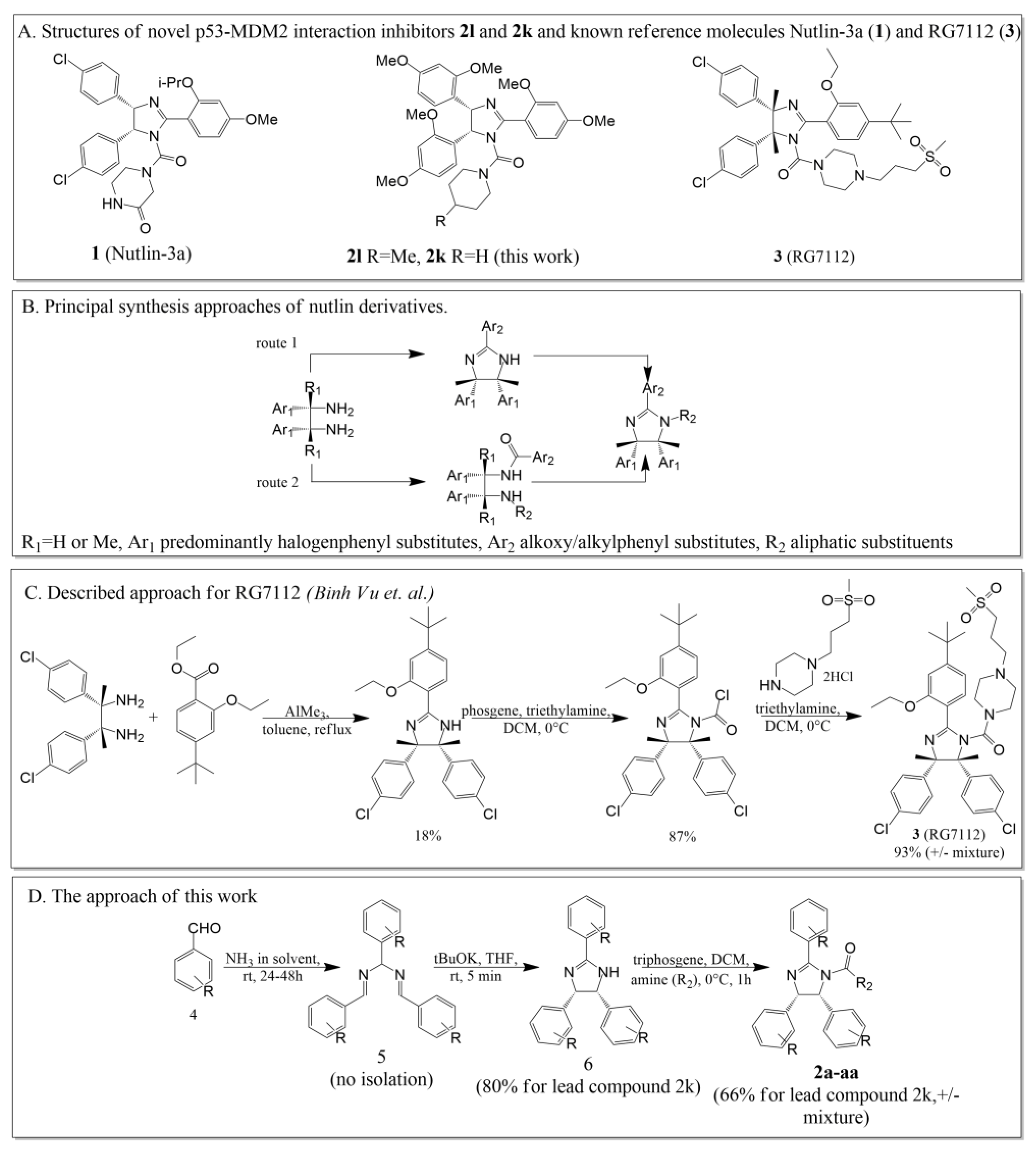
4.2. General Procedure for the Synthesis of Compounds 2a2aa

An amount of 0.7656 g (2.58 mmol) of triphosgene was dissolved in CH2Cl2 (10 mL) and slowly added dropwise to a solution of an imidazoline derivative (1 mmol) and triethylamine 0.975 mL (7.01 mmol) in CH2Cl2 (10 mL) cooled to 0 °C. The reaction mixture was stirred for 30 min, then the solvent was removed under reduced pressure. The solid residue was dissolved in CH2Cl2 (10 mL), added dropwise to a solution of amine (19.4 mmol) in CH2Cl2 (10 mL) and stirred for 15 min. The reaction mixture was then washed with NaHCO3 solution followed by water and brine. The crude product was purified by column chromatography on silica gel using EtOAc/light petroleum ether (1:1) (or EtOAc/light petroleum ether (1:2) for 3,5-dimethoxy derivatives) as eluent.
Cis-N,N-diethyl-2,4,5-tris(4-methoxyphenyl)-4,5-dihydro-1H-imidazole-1-carboxamide (2a). White solid, 37% yield. Rf EtOAc/Et3N (99:1) 0.62. 1H NMR (CDCl3, 400 MHz, mixture of two conformers, single conformer was described): δ 0.89 (t, J = 5.9 Hz, 6H), 3.04–3.13 (m, 2H), 3.22–3.31 (m, 2H), 3.65 (s, 3H), 3.67 (s, 3H), 3.82 (s, 3H), 5.42 (d, J = 9.2 Hz, 1H), 5.59 (d, J = 9.2 Hz, 1H), 6.57 (d, J = 8.8 Hz, 2H), 6.62 (d, J = 8.8 Hz, 2H), 6.76 (d, J = 8.8 Hz, 2H), 6.85 (d, J = 8.6 Hz, 2H), 6.96 (d, J = 8.6 Hz, 2H), 7.83 (d, J = 8.8 Hz, 2H). 13C NMR (CDCl3, 100 MHz): 12.60, 41.10, 55.03, 55.07, 55.35, 70.40, 113.13, 113.89, 128.73, 128.80, 130.07, 158.51, 158.81, 162.11, 163.69. ESI-HRMS (m/z): calcd. for C29H34N3O4 [M + H]+: 488.2544; found: 488.2545.
4-{[Cis-2,4,5-tris(4-methoxyphenyl)-4,5-dihydro-1H-imidazol-1-yl]carbonyl}morpholine (2b). White solid, 40% yield. Rf EtOAc/Et3N (99:1) 0.50. 1H NMR (CDCl3, 400 MHz): δ 3.19–3.24 (m, 4H), 3.30–3.38 (m, 4H), 3.68 (s, 3H), 3.69 (s, 3H), 3.86 (s, 3H), 5.48 (d, J = 9.3 Hz, 1H), 5.52 (d, J = 9.3 Hz, 1H), 6.58 (d, J = 8.8 Hz, 2H), 6.62 (d, J = 8.7 Hz, 2H), 6.73 (d, J = 8.7 Hz, 2H), 6.83 (d, J = 8.8 Hz, 2H), 6.98 (d, J = 8.8 Hz, 2H), 7.76 (d, J = 8.8 Hz, 2H). 13C NMR (CDCl3, 100 MHz): 47.08, 54.96, 55.01, 55.32, 65.96, 66.45, 70.31, 113.13, 113.28, 113.92, 127.35, 128.47, 128.66, 129.05, 129.98, 155.40, 158.51, 158.82, 162.16, 162.76. ESI-HRMS (m/z): calcd. for C29H32N3O5 [M + H]+: 502.2336; found: 502.2342.
Piperidin-1-yl(cis-2,4,5-tris(4-methoxyphenyl)-4,5-dihydro-1H-imidazol-1-yl)methanone (2c). White solid, 42% yield. Rf EtOAc/Et3N (99:1) 0.60. 1H NMR (CDCl3, 400 MHz): δ 1.22–1.30 (m, 4H), 1.39–1.45 (m, 2H), 3.15–3.17 (m, 4H), 3.68 (s, 3H), 3.69 (s, 3H), 3.85 (s, 3H), 5.47 (d, J = 9.4 Hz, 1H), 5.52 (d, J = 9.4 Hz, 1H), 6.57 (d, J = 8.8 Hz, 2H), 6.62 (d, J = 8.8 Hz, 2H), 6.74 (d, J = 8.6 Hz, 2H), 6.84 (d, J = 8.8 Hz, 2H), 6.97 (d, J = 9.0 Hz, 2H), 7.79 (d, J = 8.8 Hz, 2H). 13C NMR (CDCl3, 100 MHz): 23.91, 25.21, 46.09, 55.01, 55.07, 55.37, 70.43, 71.38, 113.15, 113.22, 113.87, 127.70, 128.52, 128.74, 129.35, 130.07, 155.34, 158.51, 158.75, 162.08, 163.03. ESI-HRMS (m/z): calcd. for C30H34N3O4 [M + H]+: 500.2544; found: 500.2533.
(4-Methylpiperidin-1-yl) (cis-2,4,5-tris(4-methoxyphenyl)-4,5-dihydro-1H-imidazol-1-yl)methanone (2d). White solid, 75% yield. Rf EtOAc/Et3N (99:1) 0.75. 1H NMR (CDCl3, 400 MHz): δ 0.80 (d, J = 6.3 Hz, 3H), 1.31–1.39 (m, 1H), 1.39–1.49 (m, 2H), 2.38–2.55 (m, 2H), 3.68 (s, 3H), 3.70 (s, 3H), 3.86 (s, 3H), 3.74–3.94 (m, 4H), 5.49 (d, J = 9.4 Hz, 1H), 5.53 (d, J = 9.4 Hz, 1H), 6.58 (d, J = 8.7 Hz, 2H), 6.63 (d, J = 8.6 Hz, 2H), 6.74 (d, J = 8.6 Hz, 2H), 6.84 (d, J = 8.6 Hz, 2H), 6.97 (d, J = 8.8 Hz, 2H), 7.80 (d, J = 8.7 Hz, 2H). 13C NMR (CDCl3, 100 MHz): 21.50, 30.37, 33.19, 33.41, 45.23, 45.68, 55.03, 55.08, 55.40, 70.47, 113.20, 113.24, 113.96, 127.29, 128.63, 128.69, 129.02, 130.25, 154.88, 158.58, 158.84, 162.33, 163.23. ESI-HRMS (m/z): calcd. for C31H36N3O4 [M + H]+: 514.2700; found: 514.2686.
Pyrrolidin-1-yl(cis-2,4,5-tris(4-methoxyphenyl)-4,5-dihydro-1H-imidazol-1-yl)methanone (2e). White solid, 40% yield. Rf EtOAc/Et3N (99:1) 0.45. 1H NMR (CDCl3, 400 MHz, mixture of two conformers, single conformer was described): δ 1.62–1.70 (m, 4H), 3.09–3.24 (m, 4H), 3.68 (s, 3H), 3.69 (s, 3H), 3.86 (s, 3H), 5.47–5.56 (m, 2H), 6.57 (d, J = 8.8 Hz, 2H), 6.62 (d, J = 8.6 Hz, 2H), 6.74 (d, J = 8.2 Hz, 2H), 6.83 (d, J = 8.0 Hz, 2H), 6.97 (d, J = 9.0 Hz, 2H), 7.82–7.85 (m, 2H). 13C NMR (CDCl3, 100 MHz): 25.46, 47.83, 55.02, 55.08, 55.38, 69.73, 71.13, 113.18, 113.22, 113.94, 127.53, 128.44, 128.76, 130.07, 154.17, 158.60, 158.74, 162.35, 163.08. ESI-HRMS (m/z): calcd. for C29H32N3O4 [M + H]+: 486.2387; found: 486.2379.
Azepan-1-yl(cis-2,4,5-tris(4-methoxyphenyl)-4,5-dihydro-1H-imidazol-1-yl)methanone (2f). White solid, 41% yield. Rf EtOAc/Et3N (99:1) 0.60. 1H NMR (CDCl3, 400 MHz): δ 1.34–1.58 (m, 8H), 3.00–3.25 (m, 4H), 3.66 (s, 3H), 3.68 (s, 3H), 3.84 (s, 3H), 5.43 (d, J = 9.2 Hz, 1H), 5.51 (d, J = 9.2 Hz, 1H), 6.56 (d, J = 8.6 Hz, 2H), 6.62 (d, J = 8.6 Hz, 2H), 6.74 (d, J = 8.6 Hz, 2H), 6.83 (d, J = 8.6 Hz, 2H), 6.94 (d, J = 8.7, 2H), 7.78 (d, J = 8.7 Hz, 2H). 13C NMR (CDCl3, 100 MHz): 27.34, 27.89, 47.61, 55.01, 55.07, 55.35, 70.25, 71.53, 113.15, 113.20, 113.92, 127.57, 128.76, 128.82, 129.37, 129.95, 158.59, 158.79, 162.16, 163.50. ESI-HRMS (m/z): calcd. for C31H36N3O4 [M + H]+: 514.2700; found: 514.2689.
tert-Butyl 4-(cis-2,4,5-tris(4-methoxyphenyl)-4,5-dihydro-1H-imidazole-1-carbonyl)piperazine-1-carboxylate (2g). White solid, 37% yield. Rf EtOAc/Et3N (99:1) 0.64. 1H NMR (CDCl3, 400 MHz): δ 1.40 (s, 9H), 3.07–3.15 (m, 4H), 3.17–3.24 (m, 4H), 3.68 (s, 3H), 3.70 (s, 3H), 3.86 (s, 3H), 5.53 (d, J = 9.5 Hz, 1H), 5.62 (d, J = 9.3 Hz, 1H), 6.57 (d, J = 8.6 Hz, 2H), 6.62 (d, J = 8.6 Hz, 2H), 6.73 (d, J = 8.4 Hz, 2H), 6.82 (d, J = 8.5 Hz, 2H), 6.97 (d, J = 8.7 Hz, 2H), 7.81 (d, J = 8.6 Hz, 2H). 13C NMR (CDCl3, 100 MHz): 28.23, 43.21, 44.99, 55.02, 55.06, 55.35, 70.41, 72.27, 80.23, 113.14, 113.34, 113.90, 127.83, 128.37, 128.75, 129.39, 129.86, 154.22, 156.04, 158.49, 158.78, 161.95, 162.40. ESI-HRMS (m/z): calcd. for C34H41N4O6 [M + H]+: 601.3021; found: 601.3014.
Piperidin-1-yl(cis-2,4,5-tris(2-methoxyphenyl)-4,5-dihydro-1H-imidazol-1-yl)methanone (2h). White solid, 37% yield. Rf EtOAc/Et3N (99:1) 0.68. 1H NMR (CDCl3, 400 MHz): δ 1.01–1.16 (m, 4H), 1.29–1.39 (m, 2H), 3.01–3.09 (m, 4H), 3.47 (s, 3H), 3.71 (s, 3H), 3.86 (s, 3H), 5.89 (d, J = 10.6 Hz, 1H), 6.22 (br. s, 1H), 6.44 (d, J = 8.1 Hz, 1H), 6.5 (br. s, 1H), 6.59 (br. s, 1H), 6.73 (br. s, 1H), 6.79 (t, J = 7.5 Hz, 1H), 6.93–6.98 (m, 2H), 7.01–7.07 (m, 2H), 7.31 (br. s, 1H), 7.44 (t, J = 8.2 Hz, 1H), 7.75 (d, J = 7.3 Hz, 1H). 13C NMR (CDCl3, 100 MHz): 23.95, 25.05, 46.41, 54.48, 55.02, 55.40, 64.89, 108.90, 109.35, 110.64, 119.01, 119.44, 120.53, 127.80, 128.12, 130.71, 158.92, 157.34. ESI-HRMS (m/z): calcd. for C30H34N3O4 [M + H]+: 500.2544; found: 500.2530.
Cis-N,N-diethyl-2,4,5-tris(2,4-dimethoxyphenyl)-4,5-dihydro-1H-imidazole-1-carboxamide (2i). White solid, 37% yield. Rf EtOAc/Et3N (99:1) 0.50. 1H NMR (CDCl3, 400 MHz): δ 0.75–0.86 (m, 6H), 2.91–3.00 (m, 2H), 3.26–3.35 (m, 4H), 3.54 (s, 3H), 3.59 (s, 3H), 3.65 (s, 3H), 3.69 (s, 3H), 3.81–3.83 (m, 6H), 5.76 (d, J = 10.0 Hz, 1H), 5.91 (d, J = 8.9 Hz, 1H), 6.08–6.14 (m, 3H), 6.24 (dd, J = 8.4 Hz, J = 2.0, 1H), 6.40–6.44 (m, 2H), 6.53 (dd, J = 8.5 Hz, J = 2.1 Hz, 1H), 6.83 (d, J = 8.7 Hz, 1H), 7.09 (br. s, 1H), 7.70 (d, J = 8.4 Hz, 1H). 13C NMR (CDCl3, 100 MHz): 0.94, 12.40, 40.73, 54.75, 54.91, 55.10, 55.17, 55.41, 55.47, 97.09, 97.20, 98.48, 103.16, 103.18, 104.76, 128.80, 131.95, 158.39, 159.66. ESI-HRMS (m/z): calcd. for C32H40N3O7 [M + H]+: 578.2861; found: 578.2867.
4-{[Cis-2,4,5-tris(2,4-dimethoxyphenyl)-4,5-dihydro-1H-imidazol-1-yl]carbonyl}morpholine (2j). White solid, 39% yield. Rf EtOAc/Et3N (99:1) 0.34. 1H NMR (CDCl3, 400 MHz): δ 3.07–3.17 (m, 4H), 3.22–3.29 (m, 4H), 3.48 (s, 3H), 3.64–3.68 (m, 6H), 3.48 (s, 3H), 3.66 (s, 3H), 3.70 (s, 3H), 3.81 (s, 3H), 3.84 (s, 3H), 5.73 (d, J = 10.1 Hz, 1H), 6.00 (br. s, 1H), 6.05–6.13 (m, 2H), 6.17 (br. s, 1H), 6.29 (dd, J = 8.4 Hz, J = 2.0 Hz, 1H), 6.47 (d, J = 2.1 Hz, 1H), 6.58 (dd, J = 8.5 Hz, J = 2.1 Hz, 1H), 6.68 (br. s, 1H), 7.11 (br. s, 1H), 7.72 (d, J = 8.4 Hz, 1H). 13C NMR (CDCl3, 100 MHz): 45.75, 54.65, 55.06, 55.15, 55.45, 66.08, 66.14, 97.18, 97.30, 98.40, 103.17, 103.22, 103.98, 104.22, 104.76, 128.47, 131.93, 158.59, 159.70. ESI-HRMS (m/z): calcd. for C32H38N3O8 [M + H]+: 592.2653; found: 592.2664.
Piperidin-1-yl(cis-2,4,5-tris(2,4-dimethoxyphenyl)-4,5-dihydro-1H-imidazol-1-yl)methanone (2k). White solid, 66% yield. Rf EtOAc/Et3N (99:1) 0.60. 1H NMR (CDCl3, 400 MHz): δ 1.08–1.20 (m, 4H), 1.29–1.37 (m, 2H), 2.99–3.08 (m, 4H), 3.46 (s, 3H), 3.64 (s, 6H), 3.68 (s, 3H), 3.79 (s, 3H), 3.82 (s, 3H), 5.70 (d, J = 10.2 Hz, 1H), 5.97 (br. s, 1H), 6.05 (d, J = 1.8 Hz, 1H), 6.14 (br. s, 1H), 6.27 (dd, J = 8.4 Hz, J = 2.0 Hz, 1H), 6.40–6.45 (m, 2H), 6.53 (dd, J = 8.4, J = 2.0, 1H), 6.67 (br. s, 1H), 7.10 (br. s, 1H), 7.67 (d, J = 8.2 Hz, 1H). 13C NMR (CDCl3, 100 MHz): 24.10, 25.21, 46.37, 45.80, 54.65, 55.06, 55.15, 55.33, 55.41, 65.09, 97.11, 97.21, 103.13, 104.49, 118.26, 120.04, 128.66, 131.69, 157.31, 157.75, 158.58, 159.51, 160.53, 162.48. ESI-HRMS (m/z): calcd. for C33H40N3O7 [M + H]+: 590.2861; found: 590.2875.
(4-Methylpiperidin-1-yl)(cis-2,4,5-tris(2,4-methoxyphenyl)-4,5-dihydro-1H-imidazol-1-yl)methanone (2l). White solid, 55% yield. Rf EtOAc/Et3N (99:1) 0.50. 1H NMR (CDCl3, 400 MHz): δ 0.50–0.66 (m, 2H), 0.75 (d, J = 6.4 Hz, 3H), 1.22–1.39 (m, 5H), 2.22–2.44 (m, 2H), 3.47 (s, 3H), 3.66 (s, 6H), 3.70 (s, 3H), 3.81 (s, 3H), 3.83 (s, 3H), 5.71 (d, J = 10.1 Hz, 1H), 6.00 (br. s, 1H), 6.07 (d, J = 2.2 Hz, 1H), 6.16 (br. s, 1H), 6.29 (dd, J = 8.4 Hz, J = 2.3 Hz, 1H), 6.36–6.46 (m, 3H), 6.55 (dd, J = 8.4, J = 2.3, 1H), 6.68 (br. s, 1H), 7.11 (br. s, 1H), 7.67 (d, J = 8.5 Hz, 1H). 13C NMR (CDCl3, 100 MHz): 21.69, 30.45, 33.10, 33.63, 45.66, 45.80, 54.65, 55.16, 55.24, 55.35, 55.43, 97.15, 97.22, 98.23, 103.15, 103.95, 104.07, 104.61, 128.62, 129.12, 131.78, 131.90, 158.64, 159.58. ESI-HRMS (m/z): calcd. for C34H42N3O7 [M + H]+: 604.3017; found: 604.3038.
tert-Butyl 4-(cis-2,4,5-tris(2,4-dimethoxyphenyl)-4,5-dihydro-1H-imidazole-1-carbonyl)piperazine-1-carboxylate (2m). White solid, 20% yield. Rf EtOAc/Et3N (99:1) 0.45. 1H NMR (CDCl3, 400 MHz): δ 1.41 (s, 9H), 2.98–3.14 (m, 8H), 3.49 (s, 3H), 3.66 (s, 3H), 3.70 (s, 3H), 3.81 (s, 3H), 3.84 (s, 3H), 5.74 (d, J = 10.1 Hz, 1H), 6.08 (d, J = 1.8 Hz, 1H), 6.30 (d, J = 7.3 Hz, 1H), 6.45 (d, J = 2.1 Hz, 1H), 6.58 (d, J = 2.1 Hz, J = 8.4 Hz, 1H), 7.44–7.48 (m, 2H), 7.57–7.62 (m, 1H), 7.63–7.67 (m, 2H), 7.72–7.77 (m, 1H). 13C NMR (CDCl3, 100 MHz): 28.24, 42.92, 45.40, 54.67, 55.07, 55.16, 55.45, 80.08, 97.20, 98.39, 103.15, 103.23, 104.79, 112.32, 118.76, 129.03, 132.05, 132.69, 154.26, 158.54, 159.73. ESI-HRMS (m/z): calcd. for C37H47N4O9 [M + H]+: 691.3338; found: 691.3335.
Cis-N,N-diethyl-2,4,5-tris(3,4-dimethoxyphenyl)-4,5-dihydro-1H-imidazole-1-carboxamide (2n). White solid, 15% yield. Rf EtOAc/Et3N (99:1) 0.20. 1H NMR (CDCl3, 400 MHz): 0.92 (t, J = 7.1 Hz, 6H), 3.08–3.18 (m, 2H), 3.21–3.31 (m, 2H), 3.50 (s, 3H), 3.55 (s, 3H), 3.75 (s, 3H), 3.77 (s, 3H), 3.92 (s, 3H), 3.93 (s, 3H), 5.30 (d, J = 9.0 Hz, 1H), 5.57 (d, J = 9.1 Hz, 1H), 6.22 (d, J = 1.5 Hz, 1H), 6.32 (s, 1H), 6.56 (dd, J = 8.3 Hz, J = 1.7 Hz, 1H), 6.57–6.59 (m, 2H), 6.63–6.64 (m, 1H), 6.91 (d, J = 8.5 Hz, 1H), 7.38 (dd, J = 8.4 Hz, J = 2.0 Hz, 1H), 7.52 (d, J = 1.9, 1H). 13C NMR (CDCl3, 100 MHz): 12.70, 41.09, 55.44, 55.47, 55.60, 55.66, 55.84, 55.99, 70.41, 110.12, 110.19, 110.46, 110.75, 111.08, 119.96, 120.02, 121.37, 128.00, 129.82, 148.04, 148.19, 148.31, 148.83, 151.68, 164.05. ESI-HRMS (m/z): calcd. for C32H40N3O7 578.2861; found: 578.2863.
4-{[Cis-2,4,5-tris(3,4-dimethoxyphenyl)-4,5-dihydro-1H-imidazol-1-yl]carbonyl}morpholine (2o). White solid, 33% yield. Rf EtOAc/Et3N (99:1) 0.15. 1H NMR (CDCl3, 400 MHz): 3.24–3.29 (m, 4H), 3.36–3.41 (m, 4H), 3.51 (s, 3H), 3.56 (s, 3H), 3.76 (s, 3H), 3.77 (s, 3H), 3.94 (s, 6H), 5.42 (d, J = 9.1 Hz, 1H), 5.54 (d, J = 9.1 Hz, 1H), 6.20 (d, J = 1.7 Hz, 1H), 6.31 (d, J = 1.6 Hz, 1H), 6.55 (dd, J = 8.3 Hz, J = 1.7 Hz, 1H), 6.60–6.67 (m, 3H), 6.93 (d, J = 8.4 Hz, 1H), 7.35 (dd, J = 8.4 Hz, J = 1.9 Hz, 1H), 7.49 (d, J = 1.9 Hz, 1H). 13C NMR (CDCl3, 100 MHz): 45.47, 55.53, 55.65, 55.72, 55.92, 56.08, 66.15, 70.54, 110.30, 110.48, 110.53, 111.04, 111.16, 119.85, 119.91, 121.33, 127.94, 129.65, 148.16, 148.41, 148.43, 148.48, 148.97, 151.81, 163.19. ESI-HRMS (m/z): calcd. for C32H38N3O8 592.2653; found: 592.2657.
Piperidin-1-yl(cis-2,4,5-tris(3,4-dimethoxyphenyl)-4,5-dihydro-1H-imidazol-1-yl)methanone (2p). White solid, 14% yield. Rf EtOAc/Et3N (99:1) 0.20. 1H NMR (CDCl3, 400 MHz): 1.30 (br. s, 4H), 1.39–1.48 (m, 2H), 3.20 (br. s, 4H), 3.51 (s, 3H), 3.56 (s, 3H), 3.76 (s, 3H), 3.78 (s, 3H), 3.94 (s, 3H), 3.97 (s, 3H), 5.60 (br. s, 2H), 6.23 (s, 1H), 6.33 (d, J = 1.2 Hz, 1H), 6.57–6.61 (m, 2H), 6.64–6.67 (m, 2H), 6.94 (d, J = 8.5 Hz, 1H), 7.42 (dd, J = 8.5 Hz, J = 1.9 Hz, 1H), 7.57 (s, 1H). 13C NMR (CDCl3, 100 MHz): 23.76, 25.25, 46.01, 55.45, 55.48, 55.58, 55.66, 55.89, 56.09, 70.60, 110.17, 110.31, 110.55, 110.70, 111.05, 111.24, 119.73, 120.23, 121.81, 129.23, 148.13, 148.23, 148.34, 148.42, 148.84, 152.08, 163.78. ESI-HRMS (m/z): calcd. for C33H40N3O7 590.2861; found: 590.2862.
(4-Methylpiperidin-1-yl)(cis-2,4,5-tris(3,4-dimethoxyphenyl)-4,5-dihydro-1H-imidazol-1-yl)methanone (2q). White solid, 30% yield. Rf EtOAc/Et3N (99:1) 0.25. 1H NMR (CDCl3, 400 MHz): 0.78 (d, J = 6.4 Hz, 3H), 1.31–1.39 (m, 1H), 1.39–1.49 (m, 2H), 2.40–2.54 (m, 2H), 3.49 (s, 3H), 3.56 (s, 3H), 3.74 (s, 3H), 3.75 (s, 3H), 3.78–3.90 (m, 4H), 3.91 (s, 6H), 5.38 (d, J = 9.1 Hz, 1H), 5.49 (d, J = 9.1 Hz, 1H), 6.19 (d, J = 1.5 Hz, 1H), 6.33 (d, J = 1.5 Hz, 1H), 6.54 (dd, J = 8.3 Hz, J = 1.7 Hz, 1H), 6.58 (s, 1H), 6.58–6.60 (m, 2H), 6.64 (d, J = 8.3 Hz, 1H), 6.91 (d, J = 8.4 Hz, 1H), 7.35 (dd, J = 8.3 Hz, J = 1.8 Hz, 1H), 7.43 (d, J = 1.7 Hz, 1H). 13C NMR (CDCl3, 100 MHz): 21.50, 30.36, 33.36, 33.48, 45.16, 45.57, 55.43, 55.45, 55.58, 55.66, 55.86, 55.97, 70.54, 72.58, 110.21, 110.42, 110.47, 110.93, 111.09, 119.86, 119.95, 121.26, 122.69, 128.26, 129.94, 147.98, 148.24, 148.28, 148.79, 151.47, 155.92, 163.26. ESI-HRMS (m/z): calcd. for C34H42N3O7 [M + H]+: 604.3017; found 604.3010.
Pyrrolidin-1-yl(cis-2,4,5-tris(3,4-dimethoxyphenyl)-4,5-dihydro-1H-imidazol-1-yl)methanone (2r). White solid, 40% yield. Rf EtOAc/Et3N (99:1) 0.20. 1H NMR (CDCl3, 400 MHz): 1.68 (br. s, 4H), 3.20 (br. s, 4H), 3.51 (s, 3H), 3.54 (s, 3H), 3.75 (s, 3H), 3.77 (s, 3H), 3.93 (s, 3H), 3.95 (s, 3H), 5.50 (d, J = 8.8 Hz, 1H), 5.57 (d, J = 8.9 Hz, 1H), 6.23 (d, J = 1.2 Hz, 1H), 6.29 (d, J = 1.1 Hz, 1H), 6.55–6.69 (m, 4H), 6.92 (d, J = 8.4 Hz, 1H), 6.96 (s, 1H), 7.41 (dd, J = 8.3 Hz, J = 1.8 Hz, 1H), 7.57 (s, 1H). 13C NMR (CDCl3, 100 MHz): 30.20, 34.10, 47.40, 55.51, 55.64, 55.73, 55.91, 56.13, 56.14, 69.80, 110.30, 110.52, 111.13, 111.21, 119.76, 119.95, 121.41, 125.38, 128.11, 135.66, 148.15, 148.26, 148.28, 148.41, 148.91, 151.38, 163.23. ESI-HRMS (m/z): calcd. for C32H38N3O7 [M + H]+: 576.2704; found 576.2701.
Azepan-1-yl(cis-2,4,5-tris(3,4-dimethoxyphenyl)-4,5-dihydro-1H-imidazol-1-yl)methanone (2s). White solid, 35% yield. Rf EtOAc/Et3N (99:1) 0.25. 1H NMR (CDCl3, 400 MHz): 1.35–1.62 (m, 8H), 3.07–3.34 (m, 4H), 3.49 (s, 3H), 3.56 (s, 3H), 3.75 (s, 3H), 3.76 (s, 3H), 3.92 (s, 3H), 3.93 (s, 3H), 5.43 (d, J = 9.1 Hz, 1H), 5.55 (d, J = 9.1 Hz, 1H), 6.21 (s, 1H), 6.34 (d, J = 1.4 Hz, 1H), 6.55–6.67 (m, 4H), 6.90 (d, J = 8.4 Hz, 1H), 7.39 (dd, J = 8.3 Hz, J = 1.9 Hz, 1H), 7.50 (d, J = 1.5 Hz, 1H). 13C NMR (CDCl3, 100 MHz): 27.36, 27.92, 47.64, 55.48, 55.61, 55.70, 55.89, 56.05, 70.32, 110.12, 110.27, 110.48, 110.73, 110.98, 111.09, 120.02, 121.34, 129.86, 148.07, 148.16, 148.26, 148.35, 148.85, 151.71, 163.79. ESI-HRMS (m/z): calcd. for C34H42N3O7 [M + H]+: 604.3017; found 604.3023.
(4-Methylpiperazin-1-yl)(cis-2,4,5-tris(3,4-dimethoxyphenyl)-4,5-dihydro-1H-imidazol-1-yl)methanone (2t). White solid, 29% yield. Rf EtOAc/Et3N (99:1) 0.03. 1H NMR (CDCl3, 400 MHz): 2.10–2.16 (br. s, 3H), 2.52–2.60 (m, 4H), 3.24–3.32 (m, 4H), 3.50 (s, 3H), 3.56 (s, 3H), 3.75 (s, 3H), 3.76 (s, 3H), 3.91 (s, 3H), 3.92 (s, 3H), 5.30 (d, J = 8.8 Hz, 1H), 5.50 (d, J = 8.8 Hz, 1H), 6.18 (d, J = 2.0 Hz, 1H), 6.34 (d, J = 1.8 Hz, 1H), 6.54 (dd, J = 8.4 Hz, J = 2.0 Hz, 1H), 6.58–6.62 (m, 2H), 6.64 (d, J = 8.2 Hz, 1H), 6.91 (d, J = 8.4, 1H), 7.34 (dd, J = 8.2 Hz, J = 2.0 Hz, 1H), 7.43 (d, J = 2.0 Hz, 1H). 13C NMR (CDCl3, 100 MHz): 44.52, 45.53, 54.10, 55.55, 55.53, 55.64, 55.71, 55.88, 55.99, 70.51, 73.91, 110.27, 110.36, 110.38, 110.52, 110.86, 111.22, 119.60, 120.07, 120.96, 123.36, 128.62, 130.14, 148.02, 148.31, 148.35, 148.40, 148.91, 151.40, 156.70, 162.77. ESI-HRMS (m/z): calcd. for C33H41N4O7 [M + H]+: 605.2970; found 605.2983.
Cis-N,N-diethyl-2,4,5-tris(3,5-dimethoxyphenyl)-4,5-dihydro-1H-imidazole-1-carboxamide (2u). White solid, 47% yield. Rf EtOAc/Et3N (99:1) 0.75. 1H NMR (CDCl3, 400 MHz): δ 0.94 (t, J = 7.0 Hz, 6H), 3.10–3.20 (m, 2H), 3.26–3.37 (m, 2H), 3.56 (s, 6H), 3.58 (s, 6H), 3.84 (s, 6H), 5.44 (br. s, 1H), 5.65 (d, J = 8.8 Hz, 1H), 6.09 (s, 2H), 6.13–6.15 (m, 2H), 6.17–6.20 (m, 2H), 6.61 (d, J = 2.0 Hz, 1H), 7.05 (s, 2H). 13C NMR (CDCl3, 100 MHz): 12.70, 14.13, 21.00, 41.22, 55.15, 55.57, 60.33, 70.61, 99.81, 100.04, 104.13, 105.58, 105.84, 106.23, 139.21, 155.57, 160.20, 160.21, 160.70, 164.88. ESI-HRMS (m/z): calcd. for C32H40N3O7 [M + H]+: 578.2861; found 578.2863.
4-{[Cis-2,4,5-tris(3,5-dimethoxyphenyl)-4,5-dihydro-1H-imidazol-1-yl]carbonyl}morpholine (2v). White solid, 40% yield. Rf EtOAc/Et3N (99:1) 0.55. 1H NMR (CDCl3, 400 MHz): δ 3.27–3.33 (m, 4H), 3.37–3.44 (m, 4H), 3.56 (s, 6H), 3.59 (s, 6H), 3.83 (s, 6H), 5.42 (d, J = 9.4 Hz, 1H), 5.55 (d, J = 9.4 Hz, 1H), 6.04 (d, J = 2.2 Hz, 2H), 6.12 (d, J = 2.2 Hz, 2H), 6.18–6.22 (m, 2H), 6.61–6.63 (m, 1H), 6.97–6.99 (m, 2H). 13C NMR (CDCl3, 100 MHz): 45.55, 55.10, 55.14, 55.53, 66.11, 70.59, 99.56, 99.86, 103.42, 105.27, 105.89, 106.17, 138.15, 139.39, 156.11, 160.18, 160.36, 160.75, 163.29. ESI-HRMS (m/z): calcd. for C32H38N3O8 [M + H]+: 592.2653; found 592.2665.
(4-Methylpiperazin-1-yl)(cis-2,4,5-tris(3,5-dimethoxyphenyl)-4,5-dihydro-1H-imidazol-1-yl)methanone (2w). White solid, 26% yield. Rf EtOAc/Et3N (99:1) 0.60. 1H NMR (CDCl3, 400 MHz): δ 2.16–2.28 (m, 7H), 3.31–3.42 (m, 4H), 3.56 (s, 6H), 3.58 (s, 6H), 3.82 (s, 6H), 5.33 (d, J = 9.1 Hz, 1H), 5.53 (d, J = 9.1 Hz, 1H), 6.04 (d, J = 2.3 Hz, 2H), 6.12 (d, J = 2.3 Hz, 2H), 6.17 (t, J = 2.3 Hz, 1H), 6.19 (t, J = 2.3 Hz, 1H), 6.59 (t, J = 2.3 Hz, 1H), 6.96 (d, J = 2.3 Hz, 2H). 13C NMR (CDCl3, 100 MHz): 44.56, 45.53, 54.02, 55.14, 55.52, 70.58, 74.32, 99.53, 99.74, 103.16, 105.12, 105.91, 106.03, 132.64, 138.60, 139.70, 156.49, 160.15, 160.36, 160.73, 163.09. ESI-HRMS (m/z): calcd. for C33H41N4O7 [M + H]+: 605.2970; found 605.2982.
Piperidin-1-yl(cis-2,4,5-tris(4-chlorophenyl)-4,5-dihydro-1H-imidazol-1-yl)methanone (2x). White solid, 37% yield. Rf EtOAc/Et3N (99:1) 0.82. 1H NMR (DMSO-d6, 400 MHz): δ 1.04–1.33 (m, 4H), 1.35–1.52 (m, 2H), 3.12–3.43 (m, 4H), 6.10 (d, J = 11.4 Hz, 1H), 6.31 (d, J = 11.2 Hz, 1H), 7.09 (d, J = 7.9 Hz, 2H), 7.22 (d, J = 7.8 Hz, 2H), 7.24–7.34 (m, 4H), 7.80 (d, J = 7.8 Hz, 2H), 7.94 (d, J = 8.0 Hz, 2H). 13C NMR (DMSO-d6, 400 MHz): 13.05, 23.14, 24.94, 45.79, 69.29, 68.91, 127.99, 129.15, 129.48, 130.20, 131.12, 130.80, 132.55, 133.15, 133.44, 138.90, 149.74, 165.98. ESI-HRMS (m/z): calcd. for C27H25Cl3N3O [M + H]+: 512.1058; found 512.1043.
Cis-N,N-diethyl-2,4,5-tris(2,4-dichlorophenyl)-4,5-dihydro-1H-imidazole-1-carboxamide (2y). White solid, 56% yield. Rf EtOAc/Et3N (99:1) 0.90. 1H NMR (CDCl3, 400 MHz): 0.84 (t, J = 7.0 Hz, 6H), 2.94–3.07 (m, 2H), 3.37–3.50 (m, 2H), 6.11 (d, J = 10.6 Hz, 1H), 6.27 (d, J = 10.6 Hz, 1H), 6.99 (dd, J = 8.4 Hz, J = 1.9 Hz, 1H), 7.06 (d, J = 8.4 Hz, 1H), 7.10 (dd, J = 8.4 Hz, J = 1.7 Hz, 1H), 7.19 (d, J = 1.7 Hz, 2H), 7.28–7.35 (m, 2H), 7.48 (d, J = 1.7Hz, 1H), 7.64 (d, J = 8.3 Hz, 1H). 13C NMR (CDCl3, 100 MHz): 12.48, 41.14, 63.98, 69.81, 126.56, 126.66, 127.27, 128.68, 129.19, 129.89, 130.30, 130.71, 131.20, 132.68, 133.97, 134.32, 136.62, 162.01. ESI-HRMS (m/z): calcd. for C26H22Cl6N3O [M + H]+: 601.9889; found 601.9878.
4-{[Cis-2,4,5-tris(2,4-dichlorophenyl)-4,5-dihydro-1H-imidazol-1-yl]carbonyl}morpholine (2z). White solid, 40% yield. Rf EtOAc/Et3N (99:1) 0.90. 1H NMR (CDCl3, 400 MHz): 3.13–3.34 (m, 4H), 3.36–3.51 (m, 4H), 6.11 (d, J = 10.6 Hz, 1H), 6.31 (d, J = 10.6 Hz, 1H), 6.93–6.99 (m, 2H), 7.14 (d, J = 8.0 Hz, 1H), 7.19 (d, J = 1.8 Hz, 1H), 7.24 (d, J = 1.8 Hz, 1H), 7.28–7.39 (m, 2H), 7.51 (d, J = 1.2 Hz, 1H), 7.74 (d, J = 8.0 Hz, 1H). 13C NMR (CDCl3, 100 MHz):, 45.78, 47.57, 63.85, 66.23, 66.64, 126.70, 127.49, 128.89, 129.46, 130.07, 130.44, 133.89, 134.35. ESI-HRMS (m/z): calcd. for C26H20Cl6N3O2 [M + H]+: 615.9681; found 615.9669.
tert-Butyl 4-(cis-2,4,5-tris(2,4-dichlorophenyl)-4,5-dihydro-1H-imidazole-1-carbonyl)piperazine-1-carboxylate (2aa). White solid, 70% yield. Rf EtOAc/Et3N (99:1) 0.90. 1H NMR (CDCl3, 400 MHz): 1.43 (s, 9H), 3.08–3.28 (m, 8H), 6.10 (d, J = 10.6 Hz, 1H), 6.26 (d, J = 10.6 Hz, 1H), 6.93–6.96 (m, 1H), 7.10 (m, 1H), 7.18 (d, J = 1.8 Hz, 1H), 7.21 (d, J = 1.0 Hz, 1H), 7.24–7.29 (m, 1H), 7.46–7.50 (m, 2H), 7.58–7.68 (m, 1H), 7.69 (d, J = 8.4, 1H). 13C NMR (CDCl3, 100 MHz): 30.89, 45.78, 47.57 63.85, 66.23, 66.64, 80.06, 126.70, 126.83, 127.49, 128.89, 129.46, 130.07, 130.44, 133.89, 134.35. ESI-HRMS (m/z): calcd. for C31H29Cl6N4O3 [M + H]+: 715.0365; found 715.0353.

4.3. In Silico Studies

The calculation was conducted using Schrodinger software suite. The presented results were obtained using «Induced Fit» (Glide + PrimeX modules) scoring method, which allowed us to refine docking poses previously obtained by screening in the Glide module alone. Molecular dynamics (MD) solvation/stability testing was conducted using the Desmond module. In all in silico studies, OPLS3e force field was used for docking, and in MD simulations, TIP4PD potential was used for interaction with water molecules. The trajectory in MD simulations was 5 ns. Maximum distance, where direct water (MD) and aminoacid (PrimeX) interactions with the ligand were considered significant, was set to 10 Å.

4.4. Cell Culture and Experimental Procedures

The osteosarcoma cell line SJSA-1, prostate cell line LNCaP, colon carcinoma cell lines RKO and HCT116, and neuroblastoma cell lines SH-SY5Y and SK-N-SH were used in this study. Cells were grown in a CO2 incubator (5% CO2) in DMEM high glucose or RPMI1640 medium containing 4.5 g/L glucose (Gibco), with 10% FBS (Gibco) at 37 °C, in the presence of a mixture of antibiotics and antimycotics (Gibco). The cells in the logarithmic growth phase were used for experiments. Before the treatment with synthesized derivatives or Nutlin-3a (Sigma) and RG7388 (Roche), the culture medium was removed, and cells were washed with PBS. Fresh medium containing the above-noted agents in the selected concentration was added to the cells.

4.5. Gel Electrophoresis and Western Blot (WB) Analysis

After the indicated time of treatment, cells were removed from culture dishes using a cell scraper. Next, cells were centrifuged (500 rcf, 5 min, 4 °C) and washed with ice-cold PBS (Paneco). Then, the cell pellet was lysed in RIPA buffer (25 mM Tris-HCl (pH 7.4), 150 mM NaCl, 0.1% SDS, 0.5% sodium deoxycholate, 1% NP-40, cOmplete™ Protease Inhibitor Cocktail (Roche)) for 20 min on ice. Lysates were centrifuged (16,000 rcf, 20 min, 4 °C), and a part of the supernatant was taken for the protein concentration assay, and another part was used for Western Blot analysis, as previously described [14]. The densitometric analysis was performed using Image Lab Software or ImageJ 4.1.
The following primary antibodies were used for WB: anti-GAPDH (#2118), anti-p21 (#2947), anti-Puma (#4976) (all from Cell Signaling Technology), anti-PARP (#137653) (Abcam) and anti-p53 (012M4795) (Sigma) antibodies. HRP-linked goat anti-mouse and anti-rabbit antibodies (#97046 and #97200, respectively; both from Abcam) were used as secondary antibodies.

4.6. Fluorescence-Activated Cell Sorting Analysis (FACS-Analysis)

After 24 h incubation with all agents, cells were removed from the culture dishes using 0.05% trypsin-EDTA (Gibco) and transferred to a conditioned medium. A total of 105 cells were taken for analysis, centrifuged (1000 rcf, 4 min, +4 °C), washed with PBS solution, and centrifuged again (1000 rcf, 4 min, +4 °C). The cell pellet was resuspended in 200 µL Annexin-binding buffer (BD Biosciences) and 2 μL of Annexin V-FITC (Invitrogen) were added. Then samples were incubated in the dark at room temperature for 15 min. Immediately before the measurement, 5 μL of propidium iodide (50 μg/mL) (BD Biosciences) were added to each sample, and the samples were analyzed by the BD FACSCanto II cell analyzer (BD Biosciences). Flow cytometry data were processed using BD FACSDiva software 7.0 (BD Biosciences).

4.7. Sub-G1 Test

After the indicated time of treatment, cells were collected as described above and fixed in 70% ethanol for 1 h at −20 °C. Then, cells were washed of ethanol and re-suspended in PBS, supplemented with 1% RNase A and stained with 20 μg/mL propidium iodide for 15 min at 37 °C. After staining, cells were examined using the FACSCanto II cell analyzer (BD Biosciences). Flow cytometry data were processed using BD FACSDiva software 7.0 (BD Biosciences).

4.8. Cytotoxic Assay

The half maximal inhibitory concentration (IC50) of the synthesized compounds against several human cell lines was determined by the MTT method. Cells were cultured at 1.0 × 10⁴ cells/200 μL in 96-well plates in DMEM or RPMI1640 medium. After 24 h, the various concentrations of the tested compounds (1.56–100 μM) were added to each well, and cells were incubated for 72 h. All compounds were dissolved in DMSO. The final DMSO concentration in each well did not exceed 0.1% and was not toxic for the cells. After incubation, 20 μL MTT reagent [3-(4,5-dimethylthiazol-2-yl)-2,5-diphenyltetrazolium bromide, 5 mg/mL] was added to each well, and the cells were incubated for 2 h. The medium was removed, and 100 μL DMSO was added to each well. The optical density was measured at 530 nm using the Victor3 (PerkinElmer) microplate reader. Concentrations (IC50) were calculated according to the dose-dependent inhibition curves using OriginPro 9.0 software. The experiments were carried out in triplicate.

4.9. Statistical Analysis

The data were presented as mean ± SD values from at least three independent experiments. For the statistical analysis, all data were tested for homogeneity of variance and normality using Levene’s and Shapiro–Wilk tests, respectively. For normally distributed data, a Student’s t-test was performed to analyze statistically significant differences between groups. The value representing of p < 0.05 was considered statistically significant. The graphs were performed using Microsoft Excel software.

5. Conclusions

In this study we have performed a two-step synthetic approach to obtain imidazoline-based alkoxyaryl derivatives. These compounds are able to block p53-MDM2 PPIs. The treatment of several cancer cell lines with them led to accumulation of p53 and p53-inducible proteins. Suggested synthetic strategy can be used for design of new anti-cancer agents in future.

Supplementary Materials

The following supporting information can be downloaded at: https://www.mdpi.com/article/10.3390/ph15040444/s1. Figure S1: The docked pose (on the left) for 2m (green) in p53 binding site of MDM2 protein, in comparison to nutlin-3a (grey), as well as its ligand-protein interaction map (on the right). Figure S2: The docked pose (on the left) for 2n (green) in p53 binding site of MDM2 protein, in comparison to nutlin-3a (grey), as well as its ligand-protein interaction map (on the right). Figure S3: The docked pose (on the left) for 2i (green) in p53 binding site of MDM2 protein, in comparison to nutlin-3a (grey), as well as its ligand-protein interaction map (on the right). Figure S4: A. Western Blot analysis of total cellular lysates from RKO cells upon treatment with RG7388 and RG7112. B. The histograms of flow cytometry (FC) analysis data for RKO cells using Annexin V-FITC/PI staining. Figure S5: A, C. Western Blot analysis of total cellular lysates from SJSA-1 (A) and LNCaP (C) cells upon treatment with compounds 2l, 2k, Nutlin-3a and RG7388. B, D—Densitometric analysis of p53, p21 and Puma bands normalized to GAPDH in SJSA-1 (B) and LNCaP (D) cells. Table S1: Cytotoxicity of synthesized 2,4,5-triaryl cis-imidazoline derivatives to HCT-116 cell line.

Author Contributions

Conceptualization—D.R.B., N.V.P., N.A.L. and G.S.K.; Investigation—D.R.B., N.V.P., E.V.S., S.E.S., M.Y.S., Y.A.G., M.D.T., A.I.M., N.A.L. and G.S.K., Writing—D.R.B., N.V.P., N.A.L. and G.S.K.; supervision—D.R.B., N.V.P., N.A.L. and G.S.K.; visualization V.Y.S.; funding acquisition—G.S.K. All authors read and approved the final manuscript.

Funding

This research was funded by the Russian Ministry of Science and Education (grant number 075-15-2020-789).

Institutional Review Board Statement

Not applicable.

Informed Consent Statement

Not applicable.

Data Availability Statement

Data is contained within the article and Supplementary Material.

Acknowledgments

We thank Boris Zhivotovsky for the comments and corrections.

Conflicts of Interest

The authors declare no conflict of interest.

References

  1. Levine, A.J. P53, the Cellular Gatekeeper for Growth and Division. Cell 1997, 88, 323–331. [Google Scholar] [CrossRef] [Green Version]
  2. D’Orazi, G.; Cirone, M. Mutant P53 and Cellular Stress Pathways: A Criminal Alliance That Promotes Cancer Progression. Cancers 2019, 11, 614. [Google Scholar] [CrossRef] [PubMed] [Green Version]
  3. Zhu, G.; Pan, C.; Bei, J.-X.; Li, B.; Liang, C.; Xu, Y.; Fu, X. Mutant P53 in Cancer Progression and Targeted Therapies. Front. Oncol. 2020, 10, 595187. [Google Scholar] [CrossRef]
  4. Wang, S.; Zhao, Y.; Aguilar, A.; Bernard, D.; Yang, C.-Y. Targeting the MDM2–P53 Protein–Protein Interaction for New Cancer Therapy: Progress and Challenges. Cold Spring Harb. Perspect. Med. 2017, 7, a026245. [Google Scholar] [CrossRef] [PubMed] [Green Version]
  5. Zhang, B.; Golding, B.T.; Hardcastle, I.R. Small-Molecule MDM2-P53 Inhibitors: Recent Advances. Future Med. Chem. 2015, 7, 631–645. [Google Scholar] [CrossRef] [PubMed] [Green Version]
  6. Zhao, Y.; Aguilar, A.; Bernard, D.; Wang, S. Small-Molecule Inhibitors of the MDM2-P53 Protein-Protein Interaction (MDM2 Inhibitors) in Clinical Trials for Cancer Treatment. J. Med. Chem. 2015, 58, 1038–1052. [Google Scholar] [CrossRef] [PubMed]
  7. Teodoro, J.G.; Evans, S.K.; Green, M.R. Inhibition of Tumor Angiogenesis by P53: A New Role for the Guardian of the Genome. J. Mol. Med. 2007, 85, 1175–1186. [Google Scholar] [CrossRef] [PubMed]
  8. Vassilev, L.T.; Vu, B.T.; Graves, B.; Carvajal, D.; Podlaski, F.; Filipovic, Z.; Kong, N.; Kammlott, U.; Lukacs, C.; Klein, C.; et al. In Vivo Activation of the P53 Pathway by Small-Molecule Antagonists of MDM2. Science 2004, 303, 844–848. [Google Scholar] [CrossRef] [PubMed] [Green Version]
  9. Fry, D.C.; Wartchow, C.; Graves, B.; Janson, C.; Lukacs, C.; Kammlott, U.; Belunis, C.; Palme, S.; Klein, C.; Vu, B. Deconstruction of a Nutlin: Dissecting the Binding Determinants of a Potent Protein-Protein Interaction Inhibitor. ACS Med. Chem. Lett. 2013, 4, 660–665. [Google Scholar] [CrossRef] [PubMed] [Green Version]
  10. Vu, B.; Wovkulich, P.; Pizzolato, G.; Lovey, A.; Ding, Q.; Jiang, N.; Liu, J.J.; Zhao, C.; Glenn, K.; Wen, Y.; et al. Discovery of RG7112: A Small-Molecule MDM2 Inhibitor in Clinical Development. ACS Med. Chem. Lett. 2013, 4, 466–469. [Google Scholar] [CrossRef] [PubMed] [Green Version]
  11. Ding, Q.; Zhang, Z.; Liu, J.J.; Jiang, N.; Zhang, J.; Ross, T.M.; Chu, X.J.; Bartkovitz, D.; Podlaski, F.; Janson, C.; et al. Discovery of RG7388, a Potent and Selective P53-MDM2 Inhibitor in Clinical Development. J. Med. Chem. 2013, 56, 5979–5983. [Google Scholar] [CrossRef] [PubMed]
  12. Wang, B.; Wu, S.; Liu, J.; Yang, K.; Xie, H.; Tang, W. Development of Selective Small Molecule MDM2 Degraders Based on Nutlin. Eur. J. Med. Chem. 2019, 176, 476–491. [Google Scholar] [CrossRef]
  13. Tisato, V.; Voltan, R.; Gonelli, A.; Secchiero, P.; Zauli, G. MDM2/X Inhibitors under Clinical Evaluation: Perspectives for the Management of Hematological Malignancies and Pediatric Cancer. J. Hematol. Oncol. 2017, 10, 133. [Google Scholar] [CrossRef] [PubMed] [Green Version]
  14. Liu, Y.; Wang, X.; Wang, G.; Yang, Y.; Yuan, Y.; Ouyang, L. The Past, Present and Future of Potential Small-Molecule Drugs Targeting P53-MDM2/MDMX for Cancer Therapy. Eur. J. Med. Chem. 2019, 176, 92–104. [Google Scholar] [CrossRef] [PubMed]
  15. Wells, J.A.; McClendon, C.L. Reaching for High-Hanging Fruit in Drug Discovery at Protein–Protein Interfaces. Nature 2007, 450, 1001–1009. [Google Scholar] [CrossRef] [PubMed]
  16. Bazanov, D.R.; Pervushin, N.V.; Savin, E.V.; Tsymliakov, M.D.; Maksutova, A.I.; Sosonyuk, S.E.; Kopeina, G.S.; Lozinskaya, N.A. Sulfonamide Derivatives of Cis-Imidazolines as Potent P53-MDM2 / MDMX Protein-Protein Interaction Inhibitors. Med. Chem. Res. 2021, 30, 2216–2227. [Google Scholar] [CrossRef]
  17. Bazanov, D.R.; Pervushin, N.V.; Savitskaya, V.Y.; Anikina, L.V.; Proskurnina, M.V.; Lozinskaya, N.A.; Kopeina, G.S. 2,4,5-Tris(Alkoxyaryl)Imidazoline Derivatives as Potent Scaffold for Novel P53-MDM2 Interaction Inhibitors: Design, Synthesis, and Biological Evaluation. Bioorganic Med. Chem. Lett. 2019, 29, 2364–2368. [Google Scholar] [CrossRef]
  18. Proskurnina, M.V.; Lozinskaya, N.A.; Tkachenko, S.E.; Zefirov, N.S. Reaction of Aromatic Aldehydes with Ammonium Acetate. Russ. J. Org. Chem. 2002, 38, 1149–1153. [Google Scholar] [CrossRef]
  19. Shu, L.; Wang, P.; Liu, W.; Gu, C. A Practical Synthesis of a Cis -4,5-Bis(4-Chlorophenyl)Imidazoline Intermediate for Nutlin Analogues. Org. Process Res. Dev. 2012, 16, 1866–1869. [Google Scholar] [CrossRef]
  20. Bazanov, D.R.; Maximova, N.A.; Seliverstov, M.Y.; Zefirov, N.A.; Sosonyuk, S.E.; Lozinskaya, N.A. Synthesis of Covalent Conjugates of Dichloroacetic Acid with Polyfunctional Compounds. Russ. J. Org. Chem. 2021, 57, 1834–1840. [Google Scholar] [CrossRef]
  21. Hu, C.; Dou, X.; Wu, Y.; Zhang, L.; Hu, Y. Design, Synthesis and CoMFA Studies of N1-Amino Acid Substituted 2,4,5-Triphenyl Imidazoline Derivatives as P53–MDM2 Binding Inhibitors. Bioorganic Med. Chem. 2012, 20, 1417–1424. [Google Scholar] [CrossRef]
  22. Hu, C.; Li, X.; Wang, W.; Zhang, L.; Tao, L.; Dong, X.; Sheng, R.; Yang, B.; Hu, Y. Design, Synthesis, and Biological Evaluation of Imidazoline Derivatives as P53–MDM2 Binding Inhibitors. Bioorganic Med. Chem. 2011, 19, 5454–5461. [Google Scholar] [CrossRef] [PubMed]
  23. Shamloo, B.; Usluer, S. P21 in Cancer Research. Cancers 2019, 11, 1178. [Google Scholar] [CrossRef] [PubMed] [Green Version]
  24. Nakano, K.; Vousden, K.H. PUMA, a Novel Proapoptotic Gene, Is Induced by P53. Mol. Cell 2001, 7, 683–694. [Google Scholar] [CrossRef]
  25. Abbas, T.; Dutta, A. P21 in Cancer: Intricate networks and multiple activities. Nat. Rev. Cancer 2010, 9, 400–414. [Google Scholar] [CrossRef]
  26. Karimian, A.; Ahmadi, Y.; Yousefi, B. Multiple Functions of P21 in Cell Cycle, Apoptosis and Transcriptional Regulation after DNA Damage. DNA Repair 2016, 42, 63–71. [Google Scholar] [CrossRef]
  27. Sun, Q.; Guo, Y.; Liu, X.; Czauderna, F.; Carr, M.I.; Zenke, F.T.; Blaukat, A.; Vassilev, L.T. Therapeutic Implications of P53 Status on Cancer Cell Fate Following Exposure to Ionizing Radiation and the DNA-PK Inhibitor M3814. Mol. Cancer Res. 2019, 17, 2457–2468. [Google Scholar] [CrossRef] [Green Version]
  28. Graat, H.C.A.; Carette, J.E.; Schagen, F.H.E.; Vassilev, L.T.; Gerritsen, W.R.; Kaspers, G.J.L.; Wuisman, P.I.J.M.; van Beusechem, V.W. Enhanced Tumor Cell Kill by Combined Treatment with a Small-Molecule Antagonist of Mouse Double Minute 2 and Adenoviruses Encoding P53. Mol. Cancer Ther. 2007, 6, 1552–1561. [Google Scholar] [CrossRef] [Green Version]
  29. Villalonga-Planells, R.; Coll-Mulet, L.; Martínez-Soler, F.; Castaño, E.; Acebes, J.-J.; Giménez-Bonafé, P.; Gil, J.; Tortosa, A. Activation of P53 by Nutlin-3a Induces Apoptosis and Cellular Senescence in Human Glioblastoma Multiforme. PLoS ONE 2011, 6, e18588. [Google Scholar] [CrossRef] [Green Version]
  30. Lazebnik, Y.A.; Kaufmann, S.H.; Desnoyers, S.; Poirier, G.G.; Earnshaw, W.C. Cleavage of Poly(ADP-Ribose) Polymerase by a Proteinase with Properties like ICE. Nature 1994, 371, 346–347. [Google Scholar] [CrossRef]
  31. Crowley, L.C.; Marfell, B.J.; Scott, A.P.; Boughaba, J.A.; Chojnowski, G.; Christensen, M.E.; Waterhouse, N.J. Dead Cert: Measuring Cell Death. Cold Spring Harb. Protoc. 2016, 2016, 1064–1072. [Google Scholar] [CrossRef] [PubMed]
  32. Plesca, D.; Mazumder, S.; Almasan, A. DNA Damage Response and Apoptosis. In Methods in Enzymology; Elsevier: Amsterdam, The Netherlands, 2008; Volume 446, pp. 107–122. ISBN 9780123744647. [Google Scholar]
  33. Available online: https://www.atcc.org/products/crl-2098 (accessed on 20 March 2022).
Figure 1. (A). The structures of novel p53-MDM2 interaction inhibitors 2l and 2k and known reference molecules Nutlin-3a (1) and RG7112 (3). (B). The principal synthesis approaches for nutlin derivatives. (C). The described approach for the synthesis of RG7112 [10]. (D). The developed approach.
Pharmaceuticals 15 00444 g001
Figure 2. Docked poses for 2k (A) and 2l (B) (green), in p53 binding site of MDM2 protein, in comparison to nutlin-3a (grey). Ligand-protein interaction map for 2k (C) and 2l (D) in p53 binding site of MDM2 protein.
Pharmaceuticals 15 00444 g002
Figure 3. Western Blot analysis of total cellular lysates from RKO cells upon treatment with compounds: A (2a2f), B (2n, 2o, 2q, 2r, 2t), C (2u–2w), D (2i–2l), E (2h, 2m, 2x, 2z). GAPDH was used as a loading control. The concentrations of all derivatives—20 μM. PARP—poly (ADP-ribose)-polymerase; GAPDH—glyceraldehyde 3-phosphate dehydrogenase; p53/GAPDH—densitometric analysis of p53 bands normalized to GAPDH, h—hours.
Pharmaceuticals 15 00444 g003
Figure 4. (A). Western Blot analysis of total cellular lysates from RKO cells upon the treatment with compounds 2l, 2k (both—20 μM), Nutlin-3a (10 μM) and RG7388 (5 μM). (B)—Densitometric analysis of p53 bands normalized to GAPDH. Data are presented as mean +/− SD from three independent experiments. (C)—The histograms of flow cytometry (FC) analysis data for RKO cells: sub-G1 assay (up), %—percent of Sub-G1 population and Annexin V-FITC/PI staining (below), % viable cells—cells negative for both Annexin V-FITC and propidium iodide (PI). Data from n = 3 biological replicates are shown as mean ± s.d., * p < 0.05, n.s.—not significant. PARP—poly (ADP-ribose)-polymerase; GAPDH—glyceraldehyde 3-phosphate dehydrogenase; p53/GAPDH—densitometric analysis of p53 bands normalized to GAPDH, h—hours, N—Nutlin-3a (10 μM), R—RG7388 (5 μM).
Pharmaceuticals 15 00444 g004
Figure 5. Western Blot analysis of total cellular lysates from RKO cells upon treatment with 2l, 2k (A); Nutlin-3a, RG7112 and RG7388 (B). GAPDH was used as a loading control. Designations: GAPDH—glyceraldehyde 3-phosphate dehydrogenase; p53/GAPDH and p21/GAPDH—densitometric analysis of p53 or p21 bands normalized to GAPDH.
Pharmaceuticals 15 00444 g005
Figure 6. Western Blot analysis of total cellular lysates from SK-N-SH (A) and SH-SY5Y (D) cells upon the treatment with compounds 2l, 2k (both—20 μM), Nutlin-3a (10 μM) and RG7388 (5 μM). (B,E)—Densitometric analysis of p53, p21 and Puma bands normalized to GAPDH in SK-N-SH (B) and SH-SY5Y (E) cells. Data are presented as mean +/− SD from three independent experiments. (C,F)—The histogram of flow cytometry (FC) analysis data for SK-N-SH cells using sub-G1 assay, %—percent of Sub-G1 population. Data from n = 3 biological replicates are shown as mean ± s.d., * p < 0.05, n.s.—not significant. PARP—poly (ADP-ribose)-polymerase; GAPDH—glyceraldehyde 3-phosphate dehydrogenase; p53/GAPDH, p21/GAPDH and Puma/GAPDH—densitometric analysis of p53P, p21P and Puma bands normalized to GAPDH, h—hours, N—Nutlin-3a (10 μM), R—RG7388 (5 μM).
Pharmaceuticals 15 00444 g006
Table 1. Synthesized 2,4,5-triaryl cis-imidazoline derivatives.
Pharmaceuticals 15 00444 i001
IDR1R2Yield, %IDR1R2Yield, %
2a Pharmaceuticals 15 00444 i002 Pharmaceuticals 15 00444 i003372o Pharmaceuticals 15 00444 i004 Pharmaceuticals 15 00444 i00533
2b Pharmaceuticals 15 00444 i006 Pharmaceuticals 15 00444 i007402p Pharmaceuticals 15 00444 i008 Pharmaceuticals 15 00444 i00914
2c Pharmaceuticals 15 00444 i010 Pharmaceuticals 15 00444 i011422q Pharmaceuticals 15 00444 i012 Pharmaceuticals 15 00444 i01330
2d Pharmaceuticals 15 00444 i014 Pharmaceuticals 15 00444 i015752r Pharmaceuticals 15 00444 i016 Pharmaceuticals 15 00444 i01740
2e Pharmaceuticals 15 00444 i018 Pharmaceuticals 15 00444 i019402s Pharmaceuticals 15 00444 i020 Pharmaceuticals 15 00444 i02135
2f Pharmaceuticals 15 00444 i022 Pharmaceuticals 15 00444 i023412t Pharmaceuticals 15 00444 i024 Pharmaceuticals 15 00444 i02529
2g Pharmaceuticals 15 00444 i026 Pharmaceuticals 15 00444 i027382u Pharmaceuticals 15 00444 i028 Pharmaceuticals 15 00444 i02947
2h Pharmaceuticals 15 00444 i030 Pharmaceuticals 15 00444 i031372v Pharmaceuticals 15 00444 i032 Pharmaceuticals 15 00444 i03340
2i Pharmaceuticals 15 00444 i034 Pharmaceuticals 15 00444 i035372w Pharmaceuticals 15 00444 i036 Pharmaceuticals 15 00444 i03726
2j Pharmaceuticals 15 00444 i038 Pharmaceuticals 15 00444 i039392x Pharmaceuticals 15 00444 i040 Pharmaceuticals 15 00444 i04137
2k Pharmaceuticals 15 00444 i042 Pharmaceuticals 15 00444 i043662y Pharmaceuticals 15 00444 i044 Pharmaceuticals 15 00444 i04556
2l Pharmaceuticals 15 00444 i046 Pharmaceuticals 15 00444 i047552z Pharmaceuticals 15 00444 i048 Pharmaceuticals 15 00444 i04940
2m Pharmaceuticals 15 00444 i050 Pharmaceuticals 15 00444 i051202aa Pharmaceuticals 15 00444 i052 Pharmaceuticals 15 00444 i05370
2n Pharmaceuticals 15 00444 i054 Pharmaceuticals 15 00444 i05515
Publisher’s Note: MDPI stays neutral with regard to jurisdictional claims in published maps and institutional affiliations.

