Review

Dual-Labelling Strategies for Nuclear and Fluorescence Molecular Imaging: Current Status and Future Perspectives

1
Helmholtz-Zentrum Dresden-Rossendorf, Institute of Radiopharmaceutical Cancer Research, Bautzner Landstraße 400, 01328 Dresden, Germany
2
National Center for Tumor Diseases (NCT) Dresden, University Hospital Carl Gustav Carus, Fetscherstraße 74, 01307 Dresden, Germany
3
Faculty of Chemistry and Food Chemistry, School of Science, Technische Universität Dresden, 01069 Dresden, Germany
4
School of Chemistry, Monash University, Melbourne, VIC 3800, Australia
*
Authors to whom correspondence should be addressed.
Pharmaceuticals 2022, 15(4), 432; https://doi.org/10.3390/ph15040432
Submission received: 11 March 2022 / Revised: 27 March 2022 / Accepted: 28 March 2022 / Published: 31 March 2022
(This article belongs to the Special Issue Hybrid Agents for Multimodal Imaging)

Abstract

:
Molecular imaging offers the possibility to investigate biological and biochemical processes non-invasively and to obtain information on both anatomy and dysfunctions. Based on the data obtained, a fundamental understanding of various disease processes can be derived and treatment strategies can be planned. In this context, methods that combine several modalities in one probe are increasingly being used. Due to the comparably high sensitivity and provided complementary information, the combination of nuclear and optical probes has taken on a special significance. In this review article, dual-labelled systems for bimodal nuclear and optical imaging based on both modular ligands and nanomaterials are discussed. Particular attention is paid to radiometal-labelled molecules for single-photon emission computed tomography (SPECT) and positron emission tomography (PET) and metal complexes combined with fluorescent dyes for optical imaging. The clinical potential of such probes, especially for fluorescence-guided surgery, is assessed.

Graphical Abstract

1. Introduction

Over the past 20 years, molecular imaging has proved a valuable technique for visualisation and characterisation of pathophysiological processes in general but especially in the field of cancer research [1,2,3,4,5]. Nuclear techniques such as single photon emission computed tomography (SPECT) and positron emission tomography (PET) play a special role here because they have an extremely high sensitivity (the nanomolar and even the picomolar level), an almost unlimited penetration depth in biological tissues and they provide quantitative data on the pharmacokinetic properties of radiolabelled drugs [6,7,8,9,10]. PET has advantages over SPECT, particularly with regard to spatial resolution and quantification. This is due to the different distribution of the emitting photons. In SPECT, the gamma quanta are distributed statistically and in a rather disorderly way, whereas PET uses the defined collinear emission and detection of two 511 keV gamma photons, which are formed during the annihilation process of electrons and positrons (Figure 1). Commonly used gamma-emitting radionuclides for SPECT are 99mTc, 111In and 123I, while for PET mostly the positron-emitting radionuclides 18F, 64Cu, 68Ga, 89Zr and 124I are applied. For radiolabelling of longer circulating objects such as natural antibodies or nanomaterials, radionuclides with a rather longer half-life are used to follow the pharmacokinetic properties over several days. Increasingly, theranostic radionuclides like lutetium-177 (SPECT and β-emitter) as well theranostic pairs like scandium-44 (PET)/scandium-47 (β-emitter), copper-64 (PET)/copper-67 (β-emitter), strontium-83 (PET)/strontium-89 (β-emitter), yttrium-86 (PET)/yttrium-90 (β-emitter), iodine-124 (PET)/iodine-131 (β-emitter) and terbium-152 (PET)/terbium-161 (β-emitter), which allow both monitoring and treatment, are being used or are under discussion [11]. In total, more than 300 radionuclides are available for use in medicine, biology, chemistry and other fields [12].
This review also focusses on optical imaging (OI) in the context of fluorescence, phosphorescence or luminescence spectroscopy. Optical imaging has about one order of magnitude lower sensitivity to nuclear techniques with superior resolution in the micro-meter range (Figure 2A) [13,14,15]. However, it suffers from the drawback of limited penetration depth, which means that application in the clinic can be limited. This limitation has been overcome to some degree by continual improvements of fibre-optic endoscopic probes, thus allowing higher image penetrating depths. However, the real advancement has been continual development of near-infrared (NIR) probes, further detailed below, which has allowed the depth of study to be increased from ~0.5 mm (400 nm) to up to detection of several centimetres at the excitation wavelengths greater than 1500 nm. This is due to two phenomena: (i) tissue penetration is wavelength dependent; thus, the greater the wavelength, the deeper the penetration and (ii) water, which is prevalent in tissues, absorbs IR light which has the effect of greater depth and greater contrast (Figure 2B) [16].
Dyes that are excited by NIR irradiation are classified as NIR-I (650–900 nm range), NIR-IIa (1000–1400 nm) and NIR-IIb (1500–1700 nm) probes. Indocyanine green and methylene blue are two small molecule NIR-I dyes that are approved for use in the clinic. These dyes, while readily excreted, are non-specific; furthermore, as NIR-I dyes they suffer from the typical photon scattering and poor photon absorption. The BODIPY scaffold (Figure 3) can be further functionalised to increase absorption and emission wavelength. The second near-infrared (NIR-II) window, comprised of probes containing inorganic or organic fluorophores, are superior due to the lower light scattering, the higher maximum permissible exposure that can be used and the greater image penetrating depth. The chemical structures of NIR dyes that are typically used for modular ligand and nanoscale systems, as well as the chemical structures relevant to fluorescence molecular imaging described in the subsequent sections, are reproduced in Figure 3. The synthesis of molecules containing donor–acceptor–donor (D-A-D) has allowed organic molecules to emit in the NIR-II window; however, the synthesis of these molecules is often challenging and as such it has not been widely performed for in vivo imaging. There are a number of good, recent reviews summarising NIR-I and NIR-II dyes [13,20,21,22,23,24] and we refer the reader to them for a more comprehensive understanding of their design and use. The recent developments of NIR-II metal-based luminescent complexes are the focus of this section and thus detailed below. Inorganic NIR-II fluorophores such as carbon nanotubes or AuNP are outside the scope of this review.
The nuclear and optical dual-labelled imaging agents reported so far are mainly based on the combination of radiolabelled compounds that use the radionuclides fluorine-18, copper-64, gallium-68, zirconium-89, technetium-99 m and indium-111 and organic dyes. Such dual modality probes are summarised in a series of reviews documenting the rapid development of this exciting field [30,31,32,33,34,35,36,37]. The two recent reviews look at the use of these samples from a clinical perspective [37] and discuss the multiple modification possibilities through the use of different conjugation strategies [30]. In the latter review, an extensive compilation of nuclear and fluorescence imaging molecular and nanoscale tools with a discussion of the pros and cons for special biological applications can be found.
The pharmacokinetic requirements of dual-labelled imaging agents in vivo are dependent on the mode of delivery as well as the timeframe between the administration and imaging test. Furthermore, the type of chelators, dye molecules and target modules, as well as their chemical linkage, will make these properties vary and so comprehensive pre-clinical studies are needed for further developments. Some of the examples discussed below utilise ligands or fluorophores that have been approved for use in the clinic. However, a comprehensive understanding of the agent’s absorption, distribution, metabolism and excretion (ADME) properties, as well as toxicity, is naturally needed before phase III trials can commence.
In our review, we focus in particular on novel molecular systems for clinical use in cancer medicine, with an emphasis on new modular ligands and nanoscale systems.

2. Fluorescence Imaging for Biomedical Applications

2.1. NIR Metal Complex Imaging Agents

It is only recently, with the key advances discussed below, that NIR metal complexes for whole body imaging can be achieved, rather than using cell imaging alone. The findings below are highlights of the field with a focus on the use of NIR metal complexes in whole body imaging studies and the key findings that have accumulated in the discovery of such complexes, rather than the broader area of time-resolved lanthanide imaging.

2.1.1. Lanthanide-Based Molecules

The fascinating and unique optical properties of lanthanide(III) complexes have intrigued scientists for decades and their potential use as bioprobes was noted as early as the 1970s [38]. Their photoluminescent properties are a consequence of their [Xe]4fn electronic configuration, with the 4f–4f transitions resulting in spectra in the visible to NIR region. However, lanthanide ions themselves are weakly absorbing due to their small molar absorption coefficients (<10 M−1·cm−1), which is a consequence of Laporte forbidden 4f transitions [39]. At the same time, the resulting long-lived luminescence, due to the Laporte forbidden 4f–4f transition of metal electrons, is a highly attractive property, to obtain it the “antenna effect”, first coined by Weissman in 1942, needs to be exploited [40]. In this case, a highly absorbing ligand, often organic in nature, and whose triplet energy state is at the appropriate level for transfer to the lanthanide excited state by intermolecular energy transfer, needs to be covalently attached or close in space to the receiving lanthanide(III) ion [41]. Typically this is accomplished by functionalisation of the multidentate ligand, to which the lanthanide(III) ion is complexed, with the appropriate antenna moiety. Ligands with negatively charged or neutral oxygen and nitrogen donor atoms give highly stable complexes; see Figure 10 for the chemical structures of representative chelators. DOTA and DO3A, or their derivatives, result in the most stable complexes (log K = 23–25) [42,43].
To date, the luminescent imaging in vitro of terbium(III) and europium(III) complexes has been well explored; however, lanthanide(III) based emitters in the NIR are more scarce [44,45]. The incorporation of ligands that absorb in the NIR region, as well as the two-photon (2P) absorption, have allowed lanthanide(III) complexes to be used for optical imaging [46]. The next section will highlight recent key findings in this area.
The 8-coordinate cationic [YbL]+ complex (Figure 4A) was utilised for 2P-imaging of living cells, with excitation wavelength 800 nm. This, the first reported YbIII 2P-luminescent probe, was the result of a decade of research in which the antenna, chelate and potential for 2P-bioimaging were optimised. In addition to the inclusion of the 2P-antenna, the cationic complex over a neutral one ensured that cell internalisation occurred readily [47].
Following this report, a number of YbIII porphyrinate complexes (Yb-4, Yb-2 and Yb-cis/trans-3, Figure 4B) were disclosed for 1P- and 2P-imaging [48,49,50]. The porphyrinates typically have intense bands at approximately 620 nm, suitable for YbIII excitation, and large extinction coefficients. The first report (2018) noted that substitution of meso-phenyl groups can modify Yb-NIR emission. Use of the deuterated Kläui ligand allowed β-fluorinated and non β-fluorinated complexes to be compared. Complex Yb-4 gave the most favourable properties and was further investigated for NIR imaging (excitation 620 nm; emission 935 nm; quantum yield 10%). Due to the long luminescent lifetimes of YbIII complexes, in vitro confocal time-resolved fluorescence lifetime imaging (FLIM) allowed for the removal of cell autofluorescence. In this ground-breaking work, the authors note that porphyrinoid ligands are exciting prospective candidates for NIR molecular probes [50]. In two follow up reports, the authors extend this concept and utilise the molecular probes for in vivo NIR-II imaging. The probes investigated have quantum yields of about 10% in water and probe Yb-2, a water-soluble carboxylate, was further investigated due to its highest resolution and signal-to-background ratio properties. When excited at 532 nm, detection of the NIR-II luminescence signals at a depth of 8 mm in a tissue sample could be observed. In vivo NIR-II fluorescence imaging showed the potential for this probe to be used for bioimaging [48]. The modification of the porphyrinoid Yb-3 resulted in regioisomers with differing properties; the cis isomer was suitable for NIR-II imaging whereas the trans isomer, upon irradiation, produced singlet oxygen [49].
In a very recent report (2021), the photophysical properties of a range of lanthanide-based carbazole-containing porphyrinoid complexes (Figure 4B, Ln-L, Ln = Gd, Yb and Er) have been further modified and examined in vitro and in vivo. As above, the coordinating ligand, a carbazole-based porphyrinoid, was chosen due to an intense absorption band at 630 nm and a large extinction coefficient. The complexes were investigated for their potential usage as photothermal therapeutics as well as NIR imaging agents. The lanthanide complexes exhibited a NIR absorption at 706 nm with the YbIII complex yielding the most encouraging results in vitro. For the encapsulation of the YbIII complex in mesoporous silica nanoparticles after intravenous injection, the in vivo studies confirmed that the photoirradiation of the tumour using a NIR laser (690 nm), with temperature monitoring, could be used to monitor tumour progress [51].
A number of pyclen-based ligands have been explored in the development of a family of lanthanide-based luminescent probes (Figure 5). The findings build on previous work within the group and others, where the photophysical different chelates and lanthanide ions were investigated for bioimaging applications. In this report, the lanthanide complexes, in all cases, have a coordination number of 9, thereby resulting in hydration numbers (q) of 0 or below 1. The lanthanide complexes, [EuL4a], [SmL4a], [YbL4b], [TbL4c], [DyL4c] and [EuL4a’] (Figure 5), can be excited between 300 and 400 nm. Depending on the lanthanide complex, they can also undergo 2P-excitation (excitation between 700 and 900 nm), which is more valuable for in vivo bioimaging applications. The results from the in vitro cellular studies are shown in Figure 5. Further studies with [EuL4a] in zebrafish embryos, which was shown to be non-toxic, and 2P-excitation resulted in a high-resolution image. The authors highlight the potential of these lanthanide-based luminescent probes for imaging thick tissue and subsequent diagnosis of disease [52]. The 161Tb and 177Lu complexes of these ligands are thermodynamically stable and kinetically inert. Thus, such ligand complexes have the potential for radionuclide therapy as well as imaging [53].
Recently, in the development of lanthanide-based nanocomposites for cancer therapy, a nanocomposite composed of DOTA as the chelate and camptothecin as the toxic payload (cycLN-ss-CPT, Ln = GdIII or YbIII, Figure 6) has been utilised [54]. In this study, the LnIII ratio was controlled via precise chemical synthesis of the GdIII and YbIII complexes, and upon formation of the micellar LnNP and excitation at 330 nm, the typical YbIII emission spectrum was observed. Incubation of the Gd/YbNPs in HeLa cells confirmed, via NIR optical imaging, that such nanocomposites could be used to monitor uptake. GdIII was included for in vivo MR imaging.
Ligand complexes of EuIII and TbIII alone are not able to be used for in vivo optical imaging, as the efficient energy transfer to these lanthanide ions typically requires external excitation in the region of 250−350 nm. Recently, it was communicated that careful design of the complexes can allow for in situ excitation via Cerenkov radiation (CR) (Figure 7). In this example, the administration of radiofluorine (Na18F) with the lanthanide complex allowed for optical and multiplex imaging concurrently [55].

2.1.2. Non-Lanthanide-Based Molecules

Luminescent iridium complexes, due their excellent photo-stability and high quantum yields, have been utilised as intracellular sensors especially for detection of oxygen, reactive oxygen species (ROS) and other endogenous species [56]. A number of recent NIR-emitting iridium complexes that can be used for in vivo imaging are reproduced in Figure 8. The iridium(III) cyanine complex nanoparticles IrCy-NPs allowed NIR absorption and singlet oxygen generation upon irradiation at 808 nm [57]. The iridium(III) complex-derived polymeric micelle PolyIrLa (the conjugated iridium(III) complex with UNCPs) allowed photodynamic therapy and chemotherapy to occur (NIR irradiation at 980 nm) [58]. The iridium(III) complex IrDAD, containing a donor–acceptor–donor (D-A-D) moiety, allowed for the formulation of a nanoparticulate system (IrDAD-NPs) that can be used for NIR-dual imaging and phototherapy. Tissue penetration was observed and NIR irradiation (808 nm) resulted in the formation of ROS and heat [59].
Typically, ruthenium(III) complexes emit in the visible region and as a result until recently have not been used for imaging studies. The Ru(II) polypyridyl complex, HL-PEG2K (Figure 9), constructed using the D-A-D strategy of an organic NIR-II fluorophore H4–PEG-Glu [60], allowed NIR-II imaging and chemo-photothermal therapy to occur simultaneously. Interestingly, in vivo studies revealed that HL-PEG2K, when compared to cisplatin, had lower toxicity and better activity [61].
Section 3 describes the use of optical dyes as part of the multifunctional ligand systems for nuclear and optical dual imaging and Section 4 outlines their incorporation in nanoscale systems.

3. Modular Ligand Systems

Multimodal imaging based on nuclear and fluorescence probes allows for synergy of these modalities. The goals are improved non-invasive visualisation and quantification of the underlying processes (occurring at the molecular level), tumour localisation and the possibility of image-guided surgery. For these purposes, it is necessary to design sophisticated bimodal imaging probes that satisfy the demands of more than one imaging modality within a small molecule or a nanoscale system (vide infra).
Frequently, low molecular weight compounds are involved in the design of probes for bimodal imaging. These compounds enable the assembly of moieties suitable for the desired imaging channels. Such moieties include fluorescent dyes for optical imaging, leaving groups suitable to introduce PET/SPECT radionuclides or bifunctional chelator agents (BFCAs) for labelling with radiometals. In particular, there is a need for molecules that allow the simple introduction of fluorophores, radionuclides and targeting modules at the same time.
In recent years, several multimodal imaging ligands have been studied. Thus, the library of options for the development of suitable dual tools, whilst comprehensive, is still expanding. Some of the most representative systems, which include multifunctional organic systems, frequently used bifunctional chelating agents (BFCAs) and newly developed modular ligand systems, are discussed below (Figure 10).

3.1. Organic-Based Systems

Organic modular systems use suitable leaving groups or isotope exchange for the introduction of non-metallic radionuclides (18F, 11C, 123I, 124I, etc.) using covalent chemical bonds. Furthermore, these systems have additional functional groups that allow the incorporation of fluorescence labels and targeting vector molecules. Some recently reported examples are presented below.
The frequently used PET radionuclide 18F has been studied extensively for the radiolabelling of organic dyes. BODIPY [62,63,64], rhodamines [65], xanthene derivatives [66] and cyanines [67] are among the most common radiofluorinated fluorophores. These dyes can be tailored with diverse leaving groups (or undergo isotopic exchange) for the introduction of radioisotopes such as 18F. This strategy has been exploited to achieve improved radiochemical yields, easier synthesis and more effective purification methods. In this context, in 2019, Kim et al. reported an 18F-labelled BODIPY dye, suitable for PET/Optical imaging [64]. The radiofluorination proceeded through an isotopic exchange (19F-18F), mediated by the Lewis acid SnCl4. Quantitative radiochemical yield (RCY) and high molar activity were achieved. More importantly, the radiofluorinated dye showed favourable pharmacokinetics and allowed for the simultaneous application of PET and optical imaging (OI) of the brain. More recently, the group Kopka et al. studied radiolabelled silicon-rhodamines (SiRs) [68]. The yielded SiRs display distinctive near-infrared (NIR) optical properties, large quantum yields and high photo-stability. Furthermore, the boronic acid (leaving moiety) enabled the introduction of 18F and 123I (using SiRs as a common precursor). Radiolabelling with high molecular activities was achieved using copper-mediated radiofluorination and copper-mediated radioiodination, respectively. These radiofluorinated molecules are suitable for co-localisation experiments (assessed by fluorescence confocal microscopy). Overall, the developed ligand structure allows for the simultaneous application of PET or SPECT and NIR imaging. Radiofluorinated dye molecules often have a lipophilic character and thus are especially beneficial for clinical applications in imaging of the brain but also of the heart.

3.2. Metal-Based Systems

Metal-based systems use BFCAs for the stable binding of metallic radionuclides. Of particular importance in the design of BFCAs is the high complex stability and kinetic inertness as well as the use of appropriate functional groups for direct or linker-mediated conjugation with fluorescent dyes and/or biomolecules. In recent years, various BFCAs have been developed for SPECT/OI and PET/OI, using common SPECT (111In, 99mTc) and PET (64Cu, 68Ga, 89Zr) radionuclides.
One of the first examples of targeted SPECT/OI imaging was presented by Wang et al., who described a dual-labelled agent for imaging the interleukin-11 receptor (IL-11Rα) [69]. The dual-labelled probe consisted of a peptide (targeting IL-11Rα) conjugated to 111In-DTPA and the fluorophore IR-783. The conjugate allowed for the clear visualisation of the ligand-antigen interaction in tumour-bearing mice. This report has served as a basis for further research of imaging IL-11Rα-expressing lesions with further fluorophores, such as Cy7 [70]. Very recently, a dual-labelled prostate-specific membrane antigen (PSMA)-targeted probe was developed using the dye IRDye800CW for NIR imaging and SPECT with 111In-DOTA or 99mTc-MAG3 (mercaptoacetylglycylglycylglycine). The high-affinity ligand, consisting of naphthylalanine, aminomethyl benzoic, glutamic and nicotinic acid, allowed for efficient in vivo imaging of PSMA-expressing tumours (Figure 11). The pharmacokinetic properties of the dual samples can be adjusted both by the choice of ligand and by the conjugation chemistry used.
Since the first report in 2011, the potential of heterobimetallic 99mTc/Re complexes for bimodal SPECT/fluorescence imaging has been studied [72]. Pyridyl triazole scaffolds [73], imidazole derivatives [74] and porphyrin [75] have been involved in heterobimetallic coordination. Recently, Day et al. reported a tracer combining the naphtalimide fluorophore and a picolylamine chelator [76]. Around 55% RCY was achieved after fac-[99mTc (CO)3(H2O)3]+ radiolabelling. The complexes showed high stability in human serum. Additionally, the rhenium(I) complexes proved to be suitable for confocal fluorescence microscopy, showing extracellular and mitochondrial uptake. However, SPECT/CT imaging revealed fast clearance (via biliary and renal pathways) and almost no uptake at the site of interest. Thus, further modifications are necessary for future imaging applications.
Dual PET/OI probes are becoming increasingly important because they allow for better spatial resolution and quantification compared to SPECT/OI. Due to the favourable nuclear physical properties and steady availability, the generator nuclide 68Ga is predestined for use in nuclear medicine [9]. Various chelators for gallium are suitable for multiple functionalisation. Exemplarily, dual imaging probes based on DOTA-IRDye800CW have been developed for 68Ga-labelling, showing that the fluorophore has no influence on the radiolabelling efficiency [77]. The promising results encouraged the study of similar systems based on 68Ga-NOTA [78]. In 2018, the first-in-human-PET imaging and fluorescence-guided surgery using a 68Ga-NOTA-IRDye800CW-bombesin were performed [79]. The novel dual probe revealed high accuracy and a strong correlation between PET and fluorescence imaging (Figure 12). This made it possible to clearly distinguish the diseased region from the healthy tissue and allowed for safe resection of glioblastoma tumours using image-guided surgery. There are other interesting chelator systems for 68Ga in development. Thus, Wang et al. developed an H2hox ligand with two 8-hydroxyquinoline moieties for 68Ga complexation, showing remarkable features such as: (i) easy synthesis, (ii) quantitative radiochemical yield within 5 min at room temperature and physiological pH, (iii) > 99% radiochemical purity without purification and (iv) enhanced fluorescence upon increasing gallium concentration, suitable for imaging [80].
Another study related to the use of 68Ga in dual-imaging probes was reported by Baranski et al. in 2018 [81]. The low molecular weight agent 68Ga-Glu-urea-Lys-HBED-CC was conjugated with four different fluorophores: fluorescein isothiocyanate (FITC), Alexa 488, IRDye800CW and DyLight800. All the conjugates showed high 68Ga complexation efficacy (RCY > 99%), indicating that the addition of the fluorophore does not affect the coordination properties of the chelator HBED. Additionally, the conjugates showed specific cell internalisation in confocal microscopy studies. Because of their NIR fluorophores, the conjugates with IRDye800CW and DyLight800 are promising for translation to the clinical area. Furthermore, the 68Ga-Glu-urea-Lys-HBED-CC-IRDye800CW conjugate was optimal for PSMA-specific tumour visualisation. It showed tumour enrichment and fast background clearance. Additionally, it was successfully applied for fluorescence-guided surgery in mice and pigs. This dual-imaging probe represents a promising tool for preoperative, intraoperative and postoperative detection of prostate cancer lesions. Very recently, a first-in-human-study has been reported for a patient with high-risk prostatic carcinoma [82]. The hybrid molecule PSMA-914 (68Ga-Glu-urea-Lys-(HE)3-HBED-CC-IRDye800CW) derived from PSMA-11 demonstrated its potential to accurately detect PSMA-expressing lesions before and during surgery. After 1 h post-injection, high retention of the conjugate in the tumour area was detected.
Currently, there are a number of other emerging multifunctional chelator systems with both macrocyclic and pre-organised acyclic structures, which are suitable for the development of targeted dual probes in nuclear medicine and optical imaging [83]. This means that chelator systems for further interesting PET radionuclides are also available, such as 44Sc, 64Cu and 89Zr.
One of the emerging radionuclides starting to be used in nuclear medicine is 64Cu [9]. In the development of modular dual-labelled probes, combinations of macrocyclic chelators and organic dyes dominate. Although DOTA is admittedly not the ideal chelator for CuII, it is still the most widely used for dual-labelled probes with antibodies [84,85,86,87] and peptides [88,89,90]. Due to the higher stability of CuII complexes and especially the higher kinetic inertness, sarcophagins [91] and TACN [92] ligands are more suitable here. In 2014, Brand et al. reported a dual imaging probe based on sarcophagine-sulfo-Cy5 [93]. The probe was synthesised following a one-pot reaction protocol. Using carboxylic acid and amino groups of the sarcophagine cage, the ligand was equipped with a sulfo-Cy5 fluorescent tag and an exendin-4 based targeted vector for the glucagon-like peptide 1 receptor (GLP-1R). This bimodal imaging probe exhibited good performance in vivo and ex vivo for tumour imaging in mice.
TACN ligands with pyridine pendant arms form very stable CuII complexes with fast complex formation kinetics under physiological conditions [92,94]. A pyridine-bearing TACN building block with an azide group can easily be incorporated via click chemistry to a conjugate consisting of the NIR label sulfo-Cy5 and an epidermal growth factor receptor (EGFR)-targeting peptide [95]. This strategy allows for the development of targeted bimodal imaging probes based on PET (64Cu) and fluorescence imaging.
Due to the more favourable complex formation kinetics compared to macrocyclic ligands, open-chain chelators for CuII are gaining in importance. These include pyridine-containing bispidine (3,7-diazabicyclo [3.3.1]nonane) ligands that are very rigid, optimally pre-organised and complementary to CuII. They form CuII complexes of high thermodynamic stability and kinetic inertness very quickly under physiological conditions [96]. For more than 10 years, the potential of bispidine ligands for use in nuclear medicine has been known [97,98]. The ligand structure allows for a wide range of variations, so biological vector molecules and fluorescence tags can also be introduced [99]. However, up until today, there is only one example of bispidines used for dual imaging [100]. The reported BODIPY-bispidine probe (radiolabelled under mild conditions) displayed highly stable 64Cu complexes. Despite the impact on the optical properties after 64CuII coordination, the decay isotopes 64NiII and 64ZnII restored the quenched fluorescence. A 64Cu-labelled DTPA derivative with a carbo-cyanine dye (LS479, Figure 3) as a fluorescence label shows similar behaviour [101]. In addition, the C9 position of the bispidine scaffold allows for the addition of further functionalities without affecting the coordination properties. This feature can be used for the introduction of fluorescent labels and bioconjugation of targeting vectors [99]. Overall, the bispidine ligand system provides an ideal platform for the development of targeted dual imaging agents based on 64Cu. By increasing the denticity of bispidines, however, other interesting radionuclides for imaging and therapy such as 111In, 177Lu and 213Bi can also be included [102,103].
Concerning 89Zr complexes, which are particularly suitable for the study of longer biochemical processes, deferoxamine B (DFO) is the most used chelator and, until now, the only one studied for bimodal imaging systems. This ligand has been combined with fluorophores such as BODIPY [104], Cy5.5 [105], Cy5 [106] and more recently with IRDye 800CW [107]. Although these probes show quantitative labelling with 89Zr, it is known that the in vivo stability is not optimal. There are a number of DFO-based ligands, which exhibit increased stability. Among them is DFO* with additional donor groups. Comparative studies of DFO* with the gold standard DFO point to DFO* as a more suitable ligand for 89Zr. DFO* and its derivatives display superior stability and performance in vivo [108,109]. However, further improvements are needed, especially for solubility enhancement in the aqueous medium. So far, there have been no studies on other bimodal imaging probes based on other DFO ligands.

3.3. Mixed Ligand Systems

In recent years, the groups of Comba and Orvig have developed new classes of ligands by combining classical complexing agents such as pyridine, picolinate, glycinate, oxinate and phosphinate, leading to mixed ligand systems such as glycinate-oxinate, picolinate-phosphinate, oxinate-pyridine, picolinate-pyridine and bispidine-picolinate. Regarding the latter, octadentate bispidine-picolinate ligands (bispa-type) have been reported as suitable ligands for stable binding of the radiometal ions 111InIII, 177LuIII and 225AcIII [110]. H4octox forms very stable complexes with 111InIII and exhibits enhanced fluorescence upon the complexation of YIII, LuIII and LaIII [111]. This ligand could thus be useful for non-radioactive fluorescent stability and cell studies as well as bimodal imaging. H2pyhox combines pyridine and oxine donor groups, resulting in an efficient chelator for 64CuII and 111InIII. Furthermore, H2pyhox proved to be suitable for 225AcIII [112]. Smaller radiometal ions such as 44ScIII, 68GaIII and 111InIII are efficiently complexed with H3glyox, a ligand containing glycine and oxine donor groups [113]. This ligand shows interesting fluorescence properties after the addition of metal ions and is thus a promising system for nuclear/optical imaging [114]. H6dappa is a phosphinate-bearing picolinic acid-based chelating ligand for binding the radiometal ions 111InIII and 177LuIII that has additional carboxylic acid groups for simultaneous introduction of fluorescence labels and targeting vectors [115]. H4pypa is a nonadentate ligand suitable for stable binding of radiometal ions such as 111InIII and 177LuIII [116], 44ScIII [117] and 89ZrIV [118]. The central pyridine unit can be used in a simple way via an ether linker group to introduce targeting molecules/modules and/or fluorescence labels [117].
With the mixed ligand systems, a wide range is available for the development of customised dual imaging agents with improved complexation and pharmacokinetic properties and there are multiple possibilities for the introduction of targeting molecules/modules.

4. Nanoscale Systems

Nanoscale structures are categorised into nanocomposites, nanoassemblies, nanoporous and nanocrystalline materials and thus embrace organic and inorganic particles [6,119,120,121,122,123]. They differ in size, chemical composition, structure and dimension, which leads to unique properties rendering them of interest for a myriad of applications, especially in oncology. Benefitting from their large surface area to volume ratio and intrinsic properties, they serve as platforms to embed a plethora of nuclear, photoacoustic, magnetic or fluorescent modalities [120]. Many innovative nanoprobes for bi-, tri or multimodal imaging have been studied in recent decades and these are summarised elsewhere [30,36,120,124]. Great expectations are placed on these agents as they fulfil the multimodality imaging concept that achieves a more accurate diagnosis by applying just one compound. The latest advances of organic and inorganic nanoscale systems being used for nuclear and optical imaging are of relevance for this review. Either the nanoscale structures can be directly labelled or the intrinsic properties of the nanoparticles itself exhibit fluorescence for FLI, single-photon emission for SPECT or positron emission for PET. Specific targeting can be achieved by covalently attaching peptides, oligosaccharides, oligonucleotides, antibodies or immunoconjugates (e.g., for antibody based (bispecific antibodies) or cellular based immunotherapies with chimeric antigen receptor (CAR) T cells) [125,126,127]. Larger nanoscale systems (sub 100 nm), especially polymer-based nanostructures, tend to be passively accumulated in the tumour tissue through the enhanced permeability and retention effect (EPR) [128]. Besides the size, the pharmacokinetic and thus the in vivo behaviour is likewise influenced by the surface charge and shape of the nanoscale system. Crucially, the size requirement of inorganic nanoprobes was validated over the years and a value of less than 10 nm is required for them to be cleared by the renal pathway. The under-estimated impact of the protein corona formed on the surface of charged nanoscale systems influences the biodistribution, circulation and metabolism pattern [129]. Hence, the design has to be balanced carefully.

4.1. Organic Nanoscale Systems

Organic nanoscale systems such as liposomes [130], endosomes [131], nanocolloids [132], micelles [133] or nanocrystalline materials [106] are ideal candidates to act as drug delivery systems. However, multimodal imaging techniques are needed to track distribution in real time and quantify the accumulation of these systems in vivo. Luo et al. used common drug-loaded porphyrin-phospholipid (PoP) liposomes [134] (Figure 13). PoP itself exhibits fluorescence and is capable of chelating copper-64 used for PET. The liposomes are less than 100 nm with an excitation and emission wavelength of 675/720 nm. PET and NIR fluorescence imaging in female BALB/c mice bearing orthotopic 4T1 mammary tumours revealed a high accumulation in the liver, spleen and tumour tissue after 24 h. Tumour accumulation is attributed through the EPR effect. The quantification and reliability of FLI pinpoints the drawbacks using this single method alone. Due to the limited penetration depth of even near infrared light, in vivo fluorescence images gain limited information. The highest fluorescence intensity was observed in the tumour region, while PET biodistribution studies revealed the highest accumulation in the liver. The differences of the fluorescence signal can be explained by the various optical properties of organs and tissues.
Novel messenger vesicles which contain functional proteins and RNAs including microRNAs and mRNAs are receiving growing interest for clinical applications [135]. Those so called exosomes are considered to be non-immunogenic and non-toxic, exhibiting high stability [136]. Jung et al. monitored the biodistribution and accumulation pattern of exosomes derived from breast (murine mammary carcinoma 4T1 cell line) cancer cells in female Balb/c nu/nu mice [131]. They were functionalised with 1,4,7-triazacyclononane-triacetic acid (NOTA) to chelate the positron emitters gallium-68 and copper-64 and conjugated to the NIR infrared dye C7, exhibiting a size of approximately 100 nm. PET images revealed accumulation in the lymph nodes, liver and lung via lymphatic or hematogenous routes. In vivo fluorescence images visualised the exosomes in the brachial lymph nodes only, whereas PET images localised them also in the axillary ones. However, quantifying the fluorescence signals of ex vivo organs gave similar results to PET images.
Another bimodal nanoscale drug delivery system has been reported by Sarparanta et al. [106]. Cellulose nanocrystals (CNC) were functionalised to the chelators desferrioxamine B (DFO) or NOTA (to chelate zirconium-89 or copper-64) and the fluorescent dye Cy5. The modified CNCs exhibit diameters of less than 8 nm with an average length of 90 nm. The in vivo and ex vivo studies were examined in orthotopic 4T1 allograft-bearing mice, a tumour model of human stage IV breast cancer. The PET images clearly showed accumulation of [64Cu]Cu-NOTA-CNC-Cy5 and [86Zr]Zr-DFO-CNC-Cy5 in the liver, lung, bone and spleen. Low tumour uptake was evaluated for both materials, indicating no passive targeting through the EPR effect. Ex vivo OI images or biodistribution based on fluorescence label showed similar accumulation pattern of the compounds seen by in vivo PET imaging.
Dual-labelled dendritic polyglycerols (dPG) were equipped with camelid single-domain antibodies (sdAbs) to target the human epidermal growth factor receptor (EGFR) [137]. The dendritic polyglycerols were functionalised with a copper-64 chelator (triazacyclononane derivative) as well as with a fluorescent dye (Cy7, λex/em = 750/780 nm). The hydrophobic diameter of the decorated dPG was < 8 nm. PET and OI imaging were performed in A431 tumour-bearing NMRI nu/nu mice. The PET biodistribution profile points to renal clearance and thus to a predominant renal excretion route. However, a certain accumulation of activity was found in the liver. The authors consider the non-specific binding of 64CuII to polyglycerol backbone as a possible reason. Tumour accumulation was low but higher in comparison to their non-targeting dPG derivatives after 24 h. Results obtained by in vivo and ex vivo OI revealed also preferred renal clearance with increased fluorescence intensity found in the kidney cortex but only minimal liver accumulation. Interestingly, the tumour uptake peaked between 24 and 48 h, which might explain the lower uptake seen in the PET image after 24 h.
One engineering approach to design organic nanoprobes with precise surface chemistry was reported by Onzen et al. [138]. Short π-conjugated oligomers self-assemble to fluorescent small molecule-based nanoparticles (SMNPs). The building blocks consisted of two fluorene units connected by a benzothiadiazole linker. Both ends contain gallic acid either decorated with alkyl or with polyethylenglycol chains exhibiting amphiphilic character. In addition, trans-cyclooctene functionalities (25%) and inert methyl (75%) groups were introduced at the periphery of the ethylene glycol chain. The SMNPs exhibited a hydrodynamic diameter of about 90 nm and the excitation and emission wavelength were 430 and 510–650 nm. To monitor the in vivo behaviour of such organic spherical nanoparticles, an 111In-labelled tetrazine-functionalised DOTA derivative reacted with the trans-cyclooctene unit of the oligomers in an inverse-electron-demand Diels–Alder reaction. The PET biodistribution data in nude Balb/c mice revealed significant accumulation in the liver and spleen within 70 h, with a peak at 4 h. The results demonstrated that the elimination is taking place by macrophages located in the Kupffer cells (liver) and red pulps (spleen). Due to the autofluorescence signal from the liver, in vivo imaging was not possible. Emission spectra could only be measured in blood samples.

4.2. Inorganic Nanoscale Systems

A plethora of inorganic-based nanoparticles such as silica [139,140,141], silicon [142,143], metal-based [144] and upconverting nanoparticles [145,146] as well as quantum dots [140,141,147] were used for PET/SPECT and optical imaging. Depending on the inorganic material, composition, size and shape, the fluorescence profile and quantum yield can vary. Semiconductor quantum dots (QDs) were considered as ideal inorganic-based alternatives to organic dyes due to their intense and narrow emission profile, higher quantum yield and their excellent photo-stability [148]. Unfortunately, they are also considered as toxic materials due to their composition of cadmium. Strategies have been designed to minimise the cytotoxicity. Cadmium telluride quantum dots were grafted on the surface of mesopourous silica (MCM-41) and radiolabelled with gallium-68 without the use of chelators [140]. The nanocomposites exhibit a size of 50 nm and were mainly accumulated in the liver, lung and kidney.
A smart quantum dot protected nanosystem was design by Shi et al. [141]. They prepared hollow mesopourous silica NPs and incorporated the commercially available QD705 (λex/em = 605/700 nm) in the cavity of the HMSNs. A chimeric human/murine anti-CD105 antibody (TRC105) was grafted onto the surface of the yolk/shell-structured nanosystem which targets the membrane glycoprotein receptor CD105. It plays an important role in tumour angiogenesis, growth and metastasis. NOTA chelators (complexation of 64Cu) were decorated on the surface and the NPs showed a size of about 70 nm. PET images and biodistribution studies in 4T1 tumour-bearing mice revealed significant liver and spleen uptake after 24 h but also an enhanced tumour uptake in comparison to the non-targeted and blocking group. The optical imaging confirmed the results.
Intrinsically labelled zirconium-89 silica nanotags functionalised with near infrared fluorescent dyes (CF680-R, λex/em = 680/700 nm) were coated with protamine and heparine to enable labelling to CAR T cells [139]. The dual-labelled nanotag gives the possibility of long-term tracking of the in vivo behaviour of such immune cells and collects information about tissue distribution by PET/FLI up to one week after adoptive cell transfer. The silica nanoparticles had a mean hydrodynamic diameter of about 120 nm. The direct CAR T cell labelling study broadcast the high silica NP loading efficiency and effective tumour uptake and demonstrated the feasibility of using these nanotags as cargos to selectively deliver drugs. Besides the application of silica NPs in medicine, silicon nanoparticles are also considered as powerful ultrasmall and non-toxic agents.
De Cola and Stephan et al. showed vividly the enormous potential of Si NPs being used as imaging agents [142,143]. In a first study, they evaluated the in vivo behaviour of Si NPs decorated with [64Cu]Cu-NOTA derivatives and NIR fluorescence dyes (Kodak-XS-679, λex/em = 680/700 nm) [142]. In vivo imaging revealed a fast renal clearance and a significant accumulation in the liver, although a mean diameter of less than 5 nm was shown. The authors attributed this phenomenon to the difference in charge and thus the formation of the protein corona, which was not observed for neutral charged NPs. The same authors investigated the biodistribution and in vivo behaviour of dual-labelled citrate-stabilised Si NPs (<3 nm) to design neutral charged particles [143]. The NPs were functionalised with NOTA and a near infrared dye (IRDye800CW, λex/em = 792/775 nm), enabling PET and OI imaging (Figure 14). It is worth noting that after the functionalisation of the dye to the Si NPs, a hypochromic shift (IR800- Si NPs, λex/em = 611/753 nm) was observed, which might have been due to the presence of protonated amines. In vivo investigation by PET and OI demonstrated encouraging pharmacokinetic properties, showing quick clearance via the kidneys, no toxicity, no accumulation in organs or tissues and high stability even after excretion from the organism.
Another class of FDA-approved ultrasmall nanoparticles are AGuIX® (<5 nm) where DOTAGA-Gd complexes are covalently bound on a polysiloxane matrix. Denat et al. decorated these attractive nanoparticles with NODAGA chelators (complexation of 64Cu and 68Ga) and Cy7 chromophores (IR783, λex/em = 792/815 nm), enabling PET/MRI/OI trimodal imaging [149]. After functionalisation, the size of the AGuIX-NODAGA-IR753 nanoparticles increased to a mean value of about 12 nm. The in vivo evaluation in NMRI TSA tumour-bearing mice revealed excretion via the renal and hepatic pathway since significant accumulation was observed in the kidney, liver and spleen after 24 h. The authors associate the elevated uptake with the increase in the hydrodynamic diameter (>10 nm) and the application of heptamethine cyanine dyes, which has been reported to show higher hepatic uptake [150]. The OI images showed, in contrast to PET-MRI, strong fluorescence signal in the intestine and stomach and low contrast in the liver and kidney. Self-quenching effects of AGuIX-NODAGA-IR753 occurred due to the high accumulated concentrations in certain organs and tissues. The more dye absorbed in an organ or tissue, the lower the fluorescence signal is.
It is worth noting that besides the combination of fluorescence imaging with PET, Cerenkov luminescence imaging (CLI) has drawn attention in image-guided surgery, especially in combination with clinically approved radiopharmaceuticals. Cerenkov luminescence is generated due the decay processes of charged particles of sufficient energy (β-emitting nuclides). The limited application due to penetration depth and low light yield hinders further intraoperative clinical application. To extend the Cerenkov luminescence properties and enhance the signal intensity, radiolabelled NPs were considered as signal intensity enhancers and converters to achieve longer wavelength and thus deeper tissue penetration. In an exemplary study, EuIII-doped gadolinium oxide NPs coated with polyvinyl alcohol (PVA) for better biocompatibility were combined with 18F (β+-emitter) as an excitation source [151]. The authors demonstrated that the optical signal intensity is dependent on several factors including size/mass of NPs, surface modification, excitation distance and amount of radioactivity. Nonetheless, they proved the use of in vivo tumour NIRF imaging with high contrast and lower tissue-autofluorescence. Moreover, the intraoperative image-guided surgery successfully localised tumours and tumour boundaries.

5. Conclusions and Future Perspectives

It is clear that rapid advancements in the quest to find new optical imaging agents is occurring; the number of reports of new organic fluorophores and metal-based NIR imaging agents—that can be excited at higher wavelengths and that have large extinction coefficients and quantum yields and low or negligible autofluorescence properties—is increasing. This advancement is primarily due to the synthesis of organic molecules containing donor–acceptor–donor (D-A-D) moieties, enabling them to absorb in the NIR window and, in the case of lanthanide(III)-based imaging agents, when designed appropriately 2P-excitation (between 700 and 900 nm) allows for in vivo microscopy. However, it is worth noting that translation into a clinical setting is not trouble free, with further research of these imaging agents as well as translational/clinical research and regulatory affairs required in order for them to reach their potential as highly active theranostic agents. The approval process for diagnostic tracers is very similar to that observed in traditional drug discovery, that is, after their discovery and testing, pre-clinical trials followed by government approval is required.
Metal complexes that have been labelled with both a fluorophore and radionuclide is an exciting area that is in its infancy. Within this area is the ability for the ligand to be labelled with a non-radionuclide lanthanide(III) ion, for 2P-excitation, or with a radionuclide for radionuclide scanning. Co-dosed, it is expected that these two imaging agents, due to almost identical properties, would locate to the same site of action and thus dual imaging with increased spatial resolution could be accomplished.
An alternate area is dual imaging probes that contain both fluorophores and radionuclides. There has been rapid development in this field in recent years. This includes small molecule probes, new modular and nanoscale systems with improved detection and pharmacokinetic properties. Especially in the last 5–10 years, a number of new BFCAs with improved complexation properties have been developed that can be used for classical radiometals such as technetium-99m and indium-111. They also provide access to emerging radiometals such as scandium-44, copper-64, gallium-68, zirconium-89 and lutetium-177. A number of new conjugation strategies are available that allow for the introduction of targeting modules under mild conditions. These include methods of bioorthogonal chemistry, such as the Staudinger–Bertozzi ligation, the strain-promoted alkyne-azide cycloaddition and the inverse electron demand Diels–Alder reaction. Furthermore, enzyme-mediated conjugation strategies are increasingly used to achieve a defined functionalisation of, for example, proteins (antibodies and their fragments) and nucleic acids. This provides a wide range of new tools for personalised medicine and precision surgery.
Whilst in its infancy, novel imaging agents comprised of nanoscale systems are being approved for use in the clinic [152]. The approval of imaging agents containing nanoscale systems does have its challenges, with reproducibility of the manufactured systems being critical [153,154,155,156]. In response to this, the FDA has released a number of guidance documents to provide information to academics and industry on the development, manufacturing and use of some products that contain nanomaterials [157]. The powerfulness of science is always evolving, and the chemistry, analytical systems for physio-chemical characterisation and policies are being created to allow for the safe use of novel dual-labelled nanoscale systems.
Image-guided surgery, including robot-assisted surgery, is particularly attractive for improving prospects of curing, especially in the field of oncology. Fluorescence-guided surgery is the logical evolution of radio-guided surgery, because it allows for detailed real-time visualisation, enabling surgical removal of all diseased tissue during an operation [158,159,160]. However, the sole use of optical imaging, especially in humans, is limited to regions close to the surface due to the rather low penetration depth of the light radiation. For this reason, dual-labelled probes (nuclear and fluorescence) are increasingly used for deeper regions. This makes it possible to detect the diseased regions externally by means of nuclear imaging and subsequently to clearly distinguish the stained diseased tissue from the healthy area internally. Prominent examples of the use of such hybrid probes can be found in the fields of sentinel lymph node biopsy [161,162,163,164,165,166,167], prostate cancer [168,169], neuroendocrine tumours [170] and breast [171] and kidney cancer [172].
Cerenkov emission is a method that does not require additional fluorophores and is under discussion for clinical application [173,174,175]. However, depending on the Cerenkov intensity of the radionuclides used, the signal intensity is three to four orders of magnitude lower compared to fluorescence-emitting probes [176]. In order to keep the radiation dose for patients and clinical staff low, the activity concentration of the radionuclides applied must be kept as low as possible. This limits the application possibilities, especially for Cerenkov-emitting nuclides. For a clinical application of Cerenkov imaging, the detection sensitivity must be significantly increased.
In terms of image-guided surgery, dual-labelled (nuclear and fluorescence) probes will dominate this exciting field in the coming years and open up new fields of application. Here, methods of artificial intelligence will also increasingly be incorporated [177]. With regard to clinical application, various challenges have to be overcome. This concerns, for example, the production of ready-to-use kits that have sufficient long-term stability. The translation of suitable dual-labelled imaging probes into clinical routine requires regulatory approval and, in turn, manufacturing under the conditions of good manufacturing practice (GMP). Overall, this is a challenging and exciting field that requires intensive multidisciplinary collaboration between experts in different fields and will undoubtedly lead to new products that enable improved non-invasive imaging with more sophisticated treatment options.

Author Contributions

Conceptualization: K.L.T. and H.S.; writing-original draft preparation: M.K., I.I.S.M., M.B., K.K., K.L.T. and H.S.; writing-review and editing: M.K., I.I.S.M., M.B., K.K., K.L.T. and H.S.; visualization, I.I.S.M. and K.L.T.; funding acquisition, I.I.S.M., M.B., K.K. and H.S. All authors have read and agreed to the published version of the manuscript.

Funding

This work was supported by the Helmholtz Initiative and Networking Fund (Radio-Immuno-Theranostics (MHELTHERA), project ID: InterLabs-0031) and Wilhelm Sander-Stiftung for a grant on bimodal tumour tracers (grant number 2018.024.1). Financial support from the CONACYT-DAAD program is gratefully acknowledged (I.I.S.M., grant # CONACYT: 708225).

Institutional Review Board Statement

Not applicable.

Informed Consent Statement

Not applicable.

Data Availability Statement

Data sharing not applicable.

Conflicts of Interest

The authors declare no conflict of interest.

References

  1. Achilefu, S. Introduction to concepts and strategies for molecular imaging. Chem. Rev. 2010, 110, 2575–2578. [Google Scholar] [CrossRef] [PubMed]
  2. Cassidy, P.J.; Radda, G.K. Molecular imaging perspectives. J. R. Soc. Interface 2005, 2, 133–144. [Google Scholar] [CrossRef] [PubMed] [Green Version]
  3. Frangioni, J.V. New technologies for human cancer imaging. J. Clin. Oncol. 2008, 26, 4012–4021. [Google Scholar] [CrossRef]
  4. James, M.L.; Gambhir, S.S. A molecular imaging primer: Modalities, imaging agents, and applications. Physiol. Rev. 2012, 92, 897–965. [Google Scholar] [CrossRef] [PubMed] [Green Version]
  5. Weissleder, R.; Mahmood, U. Molecular Imaging. Radiology 2001, 219, 316–333. [Google Scholar] [CrossRef] [PubMed]
  6. Pant, K.; Sedlacek, O.; Nadar, R.A.; Hruby, M.; Stephan, H. Radiolabelled Polymeric Materials for Imaging and Treatment of Cancer: Quo Vadis? Adv. Healthc. Mater. 2017, 6, 31. [Google Scholar] [CrossRef]
  7. Ramogida, C.F.; Orvig, C. Tumour targeting with radiometals for diagnosis and therapy. Chem. Commun. 2013, 49, 4720–4739. [Google Scholar] [CrossRef]
  8. Ametamey, S.M.; Honer, M.; Schubiger, P.A. Molecular imaging with PET. Chem. Rev. 2008, 108, 1501–1516. [Google Scholar] [CrossRef]
  9. Boros, E.; Packard, A.B. Radioactive Transition Metals for Imaging and Therapy. Chem. Rev. 2019, 119, 870–901. [Google Scholar] [CrossRef]
  10. Kostelnik, T.I.; Orvig, C. Radioactive Main Group and Rare Earth Metals for Imaging and Therapy. Chem. Rev. 2019, 119, 902–956. [Google Scholar] [CrossRef]
  11. Qaim, S.M. Theranostic radionuclides: Recent advances in production methodologies. J. Radioanal. Nucl. Chem. 2019, 322, 1257–1266. [Google Scholar] [CrossRef]
  12. Eckerman, K.; Endo, A. ICRP Publication 107. Nuclear decay data for dosimetric calculations. Ann. ICRP 2008, 38, 7–96. [Google Scholar] [CrossRef] [PubMed]
  13. Jiang, Y.; Pu, K. Molecular Probes for Autofluorescence-Free Optical Imaging. Chem. Rev. 2021, 121, 13086–13131. [Google Scholar] [CrossRef] [PubMed]
  14. Liu, P.; Mu, X.; Zhang, X.D.; Ming, D. The Near-Infrared-II Fluorophores and Advanced Microscopy Technologies Development and Application in Bioimaging. Bioconj. Chem. 2020, 31, 260–275. [Google Scholar] [CrossRef] [PubMed]
  15. Yoon, S.; Kim, M.; Jang, M.; Choi, Y.; Choi, W.; Kang, S.; Choi, W. Deep optical imaging within complex scattering media. Nat. Rev. Phys. 2020, 2, 141–158. [Google Scholar] [CrossRef] [Green Version]
  16. Juzenas, P.; Juzeniene, A.; Kaalhus, O.; Iani, V.; Moan, J. Noninvasive fluorescence excitation spectroscopy during application of 5-aminolevulinic acid in vivo. Photochem. Photobiol. Sci. 2002, 1, 745–748. [Google Scholar] [CrossRef]
  17. Cheon, J.; Lee, J.H. Synergistically integrated nanoparticles as multimodal probes for nanobiotechnology. Acc. Chem. Res. 2008, 41, 1630–1640. [Google Scholar] [CrossRef]
  18. Lu, F.M.; Yuan, Z. PET/SPECT molecular imaging in clinical neuroscience: Recent advances in the investigation of CNS diseases. Quant. Imaging Med. Surg. 2015, 5, 433–447. [Google Scholar] [CrossRef]
  19. Massoud, T.F.; Gambhir, S.S. Molecular imaging in living subjects: Seeing fundamental biological processes in a new light. Genes Dev. 2003, 17, 545–580. [Google Scholar] [CrossRef] [Green Version]
  20. Haque, A.; Faizi, M.S.H.; Rather, J.A.; Khan, M.S. Next generation NIR fluorophores for tumor imaging and fluorescence-guided surgery: A review. Bioorgan. Med. Chem. 2017, 25, 2017–2034. [Google Scholar] [CrossRef]
  21. Hu, Z.; Chen, W.H.; Tian, J.; Cheng, Z. NIRF Nanoprobes for Cancer Molecular Imaging: Approaching Clinic. Trends Mol. Med. 2020, 26, 469–482. [Google Scholar] [CrossRef] [PubMed]
  22. Li, L.; Dong, X.; Li, J.; Wei, J. A short review on NIR-II organic small molecule dyes. Dyes Pigm. 2020, 183, 108756. [Google Scholar] [CrossRef]
  23. Reja, S.I.; Minoshima, M.; Hori, Y.; Kikuchi, K. Near-infrared fluorescent probes: A next-generation tool for protein-labeling applications. Chem. Sci. 2021, 12, 3437–3447. [Google Scholar] [CrossRef] [PubMed]
  24. Shen, Q.; Wang, S.; Yang, N.-D.; Zhang, C.; Wu, Q.; Yu, C. Recent development of small-molecule organic fluorophores for multifunctional bioimaging in the second near-infrared window. J. Lumin. 2020, 225, 117338. [Google Scholar] [CrossRef]
  25. Qu, C.; Xiao, Y.; Zhou, H.; Ding, B.; Li, A.; Lin, J.; Zeng, X.; Chen, H.; Qian, K.; Zhang, X.; et al. Quaternary Ammonium Salt Based NIR-II Probes for In Vivo Imaging. Adv. Opt. Mater. 2019, 7, 1900229. [Google Scholar] [CrossRef] [PubMed]
  26. Wu, W.; Yang, Y.; Yang, Y.; Yang, Y.; Zhang, K.; Guo, L.; Ge, H.; Chen, X.; Liu, J.; Feng, H. Molecular Engineering of an Organic NIR-II Fluorophore with Aggregation-Induced Emission Characteristics for In Vivo Imaging. Small 2019, 15, 1805549. [Google Scholar] [CrossRef]
  27. Feng, W.; Zhang, Y.; Li, Z.; Zhai, S.; Lv, W.; Liu, Z. Lighting Up NIR-II Fluorescence in Vivo: An Activable Probe for Noninvasive Hydroxyl Radical Imaging. Anal. Chem. 2019, 91, 15757–15762. [Google Scholar] [CrossRef]
  28. Wang, S.; Fan, Y.; Li, D.; Sun, C.; Lei, Z.; Lu, L.; Wang, T.; Zhang, F. Anti-quenching NIR-II molecular fluorophores for in vivo high-contrast imaging and pH sensing. Nat. Commun. 2019, 10, 1058. [Google Scholar] [CrossRef]
  29. Bai, L.; Sun, P.; Liu, Y.; Zhang, H.; Hu, W.; Zhang, W.; Liu, Z.; Fan, Q.; Li, L.; Huang, W. Novel aza-BODIPY based small molecular NIR-II fluorophores for in vivo imaging. Chem. Commun. 2019, 55, 10920–10923. [Google Scholar] [CrossRef]
  30. Ariztia, J.; Solmont, K.; Moïse, N.P.; Specklin, S.; Heck, M.P.; Lamandé-Langle, S.; Kuhnast, B. PET/Fluorescence Imaging: An Overview of the Chemical Strategies to Build Dual Imaging Tools. Bioconjug. Chem. 2022, 33, 24–52. [Google Scholar] [CrossRef]
  31. Azhdarinia, A.; Ghosh, P.; Ghosh, S.; Wilganowski, N.; Sevick-Muraca, E.M. Dual-Labeling Strategies for Nuclear and Fluorescence Molecular Imaging: A Review and Analysis. Mol. Imaging Biol. 2012, 14, 261–276. [Google Scholar] [CrossRef] [PubMed] [Green Version]
  32. Joshi, B.P.; Wang, T.D. Exogenous Molecular Probes for Targeted Imaging in Cancer: Focus on Multi-modal Imaging. Cancers 2010, 2, 1251–1287. [Google Scholar] [CrossRef] [PubMed]
  33. Kuil, J.; Velders, A.H.; van Leeuwen, F.W. Multimodal tumor-targeting peptides functionalized with both a radio- and a fluorescent label. Bioconjug. Chem 2010, 21, 1709–1719. [Google Scholar] [CrossRef] [PubMed]
  34. Lee, S.; Chen, X. Dual-modality probes for in vivo molecular imaging. Mol. Imaging 2009, 8, 87–100. [Google Scholar] [CrossRef] [PubMed] [Green Version]
  35. Seibold, U.; Wangler, B.; Schirrmacher, R.; Wangler, C. Bimodal imaging probes for combined PET and OI: Recent developments and future directions for hybrid agent development. Biomed. Res. Int. 2014, 2014, 153741. [Google Scholar] [CrossRef] [Green Version]
  36. Singh, G.; Gott, M.D.; Pietzsch, H.J.; Stephan, H. Nuclear and optical dual-labelled imaging agents. Nuklearmedizin 2016, 55, 41–50. [Google Scholar] [CrossRef]
  37. Vaz, S.C.; Oliveira, F.; Herrmann, K.; Veit-Haibach, P. Nuclear medicine and molecular imaging advances in the 21st century. Br. J. Radiol. 2020, 93, 20200095. [Google Scholar] [CrossRef]
  38. Bünzli, J.C. Lanthanide luminescence for biomedical analyses and imaging. Chem. Rev. 2010, 110, 2729–2755. [Google Scholar] [CrossRef]
  39. Thibon, A.; Pierre, V.C. Principles of responsive lanthanide-based luminescent probes for cellular imaging. Anal. Bioanal. Chem. 2009, 394, 107–120. [Google Scholar] [CrossRef]
  40. Weissman, S.I. Intramolecular Energy Transfer The Fluorescence of Complexes of Europium. J. Chem. Phys. 1942, 10, 214–217. [Google Scholar] [CrossRef]
  41. Bünzli, J.-C.G.; Piguet, C. Taking advantage of luminescent lanthanide ions. Chem. Soc. Rev. 2005, 34, 1048–1077. [Google Scholar] [CrossRef] [PubMed]
  42. Cable, M.L.; Levine, D.J.; Kirby, J.P.; Gray, H.B.; Ponce, A. Luminescent lanthanide sensors. Adv. Inorg. Chem. 2011, 63, 1–45. [Google Scholar] [CrossRef]
  43. Izatt, R.M.; Pawlak, K.; Bradshaw, J.S.; Bruening, R.L. Thermodynamic and Kinetic Data for Macrocycle Interaction with Cations, Anions, and Neutral Molecules. Chem. Rev. 1995, 95, 2529–2586. [Google Scholar] [CrossRef]
  44. Bodman, S.E.; Butler, S.J. Advances in anion binding and sensing using luminescent lanthanide complexes. Chem. Sci. 2021, 12, 2716–2734. [Google Scholar] [CrossRef]
  45. New, E.J.; Parker, D.; Smith, D.G.; Walton, J.W. Development of responsive lanthanide probes for cellular applications. Curr. Opin. Chem. Biol. 2010, 14, 238–246. [Google Scholar] [CrossRef]
  46. D’Aléo, A.; Andraud, C.; Maury, O. Two-photon Absorption of Lanthanide Complexes: From Fundamental Aspects to Biphotonic Imaging Applications. In Luminescence of Lanthanide Ions in Coordination Compounds and Nanomaterials; Wiley: Hoboken, NJ, USA, 2014; pp. 197–230. [Google Scholar]
  47. Bui, A.T.; Beyler, M.; Grichine, A.; Duperray, A.; Mulatier, J.-C.; Guyot, Y.; Andraud, C.; Tripier, R.; Brasselet, S.; Maury, O. Near infrared two photon imaging using a bright cationic Yb(iii) bioprobe spontaneously internalized into live cells. Chem. Commun. 2017, 53, 6005–6008. [Google Scholar] [CrossRef]
  48. Ning, Y.; Chen, S.; Chen, H.; Wang, J.-X.; He, S.; Liu, Y.-W.; Cheng, Z.; Zhang, J.-L. A proof-of-concept application of water-soluble ytterbium(iii) molecular probes in in vivo NIR-II whole body bioimaging. Inorg. Chem. Front. 2019, 6, 1962–1967. [Google Scholar] [CrossRef]
  49. Ning, Y.; Liu, Y.-W.; Yang, Z.-S.; Yao, Y.; Kang, L.; Sessler, J.L.; Zhang, J.-L. Split and Use: Structural Isomers for Diagnosis and Therapy. J. Am. Chem. Soc. 2020, 142, 6761–6768. [Google Scholar] [CrossRef]
  50. Ning, Y.; Tang, J.; Liu, Y.-W.; Jing, J.; Sun, Y.; Zhang, J.-L. Highly luminescent, biocompatible ytterbium(iii) complexes as near-infrared fluorophores for living cell imaging. Chem. Sci. 2018, 9, 3742–3753. [Google Scholar] [CrossRef] [Green Version]
  51. Zhu, M.; Zhang, H.; Ran, G.; Mangel, D.N.; Yao, Y.; Zhang, R.; Tan, J.; Zhang, W.; Song, J.; Sessler, J.L.; et al. Metal Modulation: An Easy-to-Implement Tactic for Tuning Lanthanide Phototheranostics. J. Am. Chem. Soc. 2021, 143, 7541–7552. [Google Scholar] [CrossRef]
  52. Hamon, N.; Roux, A.; Beyler, M.; Mulatier, J.-C.; Andraud, C.; Nguyen, C.; Maynadier, M.; Bettache, N.; Duperray, A.; Grichine, A.; et al. Pyclen-Based Ln(III) Complexes as Highly Luminescent Bioprobes for In Vitro and In Vivo One- and Two-Photon Bioimaging Applications. J. Am. Chem. Soc. 2020, 142, 10184–10197. [Google Scholar] [CrossRef] [PubMed]
  53. Nizou, G.; Favaretto, C.; Borgna, F.; Grundler, P.V.; Saffon-Merceron, N.; Platas-Iglesias, C.; Fougère, O.; Rousseaux, O.; van der Meulen, N.P.; Müller, C.; et al. Expanding the Scope of Pyclen-Picolinate Lanthanide Chelates to Potential Theranostic Applications. Inorg. Chem. 2020, 59, 11736–11748. [Google Scholar] [CrossRef] [PubMed]
  54. Zhang, Y.; Ma, X.; Chau, H.-F.; Thor, W.; Jiang, L.; Zha, S.; Fok, W.-Y.; Mak, H.-N.; Zhang, J.; Cai, J.; et al. Lanthanide–Cyclen–Camptothecin Nanocomposites for Cancer Theranostics Guided by Near-Infrared and Magnetic Resonance Imaging. ACS Appl. Nano Mater. 2021, 4, 271–278. [Google Scholar] [CrossRef]
  55. Martin, K.E.; Cosby, A.G.; Boros, E. Multiplex and In Vivo Optical Imaging of Discrete Luminescent Lanthanide Complexes Enabled by In Situ Cherenkov Radiation Mediated Energy Transfer. J. Am. Chem. Soc. 2021, 143, 9206–9214. [Google Scholar] [CrossRef]
  56. Zhou, J.; Li, J.; Zhang, K.Y.; Liu, S.; Zhao, Q. Phosphorescent iridium(III) complexes as lifetime-based biological sensors for photoluminescence lifetime imaging microscopy. Coord. Chem. Rev. 2022, 453, 214334. [Google Scholar] [CrossRef]
  57. Yang, Q.; Jin, H.; Gao, Y.; Lin, J.; Yang, H.; Yang, S. Photostable Iridium(III)–Cyanine Complex Nanoparticles for Photoacoustic Imaging Guided Near-Infrared Photodynamic Therapy in Vivo. ACS Appl. Mater. Interfaces 2019, 11, 15417–15425. [Google Scholar] [CrossRef]
  58. Zhao, J.; Zhang, X.; Fang, L.; Gao, C.; Xu, C.; Gou, S. Iridium(III) Complex–Derived Polymeric Micelles with Low Dark Toxicity and Strong NIR Excitation for Phototherapy and Chemotherapy. Small 2020, 16, 2000363. [Google Scholar] [CrossRef]
  59. Zhao, J.; Yan, K.; Xu, G.; Liu, X.; Zhao, Q.; Xu, C.; Gou, S. An Iridium (III) Complex Bearing a Donor–Acceptor–Donor Type Ligand for NIR-Triggered Dual Phototherapy. Adv. Funct. Mater. 2021, 31, 2008325. [Google Scholar] [CrossRef]
  60. Zheng, Y.; Li, Q.; Wu, J.; Luo, Z.; Zhou, W.; Li, A.; Chen, Y.; Rouzi, T.; Tian, T.; Zhou, H.; et al. All-in-one mitochondria-targeted NIR-II fluorophores for cancer therapy and imaging. Chem. Sci. 2021, 12, 1843–1850. [Google Scholar] [CrossRef]
  61. Liu, Y.; Li, Q.; Gu, M.; Lu, D.; Xiong, X.; Zhang, Z.; Pan, Y.; Liao, Y.; Ding, Q.; Gong, W.; et al. A Second Near-Infrared Ru(II) Polypyridyl Complex for Synergistic Chemo-Photothermal Therapy. J. Med. Chem. 2022, 65, 2225–2237. [Google Scholar] [CrossRef]
  62. An, F.; Nurili, F.; Sayman, H.; Ozer, Z.; Cakiroglu, H.; Aras, O.; Ting, R. One-Step, Rapid, 18F–19F Isotopic Exchange Radiolabeling of Difluoro-dioxaborinins: Substituent Effect on Stability and In Vivo Applications. J. Med. Chem. 2020, 63, 12693–12706. [Google Scholar] [CrossRef] [PubMed]
  63. An, F.-F.; Kommidi, H.; Chen, N.; Ting, R. A Conjugate of Pentamethine Cyanine and 18F as a Positron Emission Tomography/Near-Infrared Fluorescence Probe for Multimodality Tumor Imaging. Int. J. Mol. Sci. 2017, 18, 1214. [Google Scholar] [CrossRef] [PubMed] [Green Version]
  64. Kim, H.; Kim, K.; Son, S.-H.; Choi, J.Y.; Lee, K.-H.; Kim, B.-T.; Byun, Y.; Choe, Y.S. 18F-Labeled BODIPY Dye: A Potential Prosthetic Group for Brain Hybrid PET/Optical Imaging Agents. ACS Chem. Neurosci. 2019, 10, 1445–1451. [Google Scholar] [CrossRef] [PubMed]
  65. Kreimerman, I.; Mora-Ramirez, E.; Reyes, L.; Bardiès, M.; Savio, E.; Engler, H. Dosimetry and Toxicity Studies of the Novel Sulfonamide Derivative of Sulforhodamine 101([18F]SRF101) at a Preclinical Level. Curr. Radiopharm. 2019, 12, 40–48. [Google Scholar] [CrossRef]
  66. Kang, N.-Y.; Lee, J.Y.; Lee, S.H.; Song, I.H.; Hwang, Y.H.; Kim, M.J.; Phue, W.H.; Agrawalla, B.K.; Wan, S.Y.D.; Lalic, J.; et al. Multimodal Imaging Probe Development for Pancreatic β Cells: From Fluorescence to PET. J. Am. Chem. Soc. 2020, 142, 3430–3439. [Google Scholar] [CrossRef]
  67. Schwegmann, K.; Hohn, M.; Hermann, S.; Schäfers, M.; Riemann, B.; Haufe, G.; Wagner, S.; Breyholz, H.-J. Optimizing the Biodistribution of Radiofluorinated Barbiturate Tracers for Matrix Metalloproteinase Imaging by Introduction of Fluorescent Dyes as Pharmacokinetic Modulators. Bioconjug. Chem. 2020, 31, 1117–1132. [Google Scholar] [CrossRef]
  68. Kanagasundaram, T.; Laube, M.; Wodtke, J.; Kramer, C.S.; Stadlbauer, S.; Pietzsch, J.; Kopka, K. Radiolabeled Silicon-Rhodamines as Bimodal PET/SPECT-NIR Imaging Agents. Pharmaceuticals 2021, 14, 1155. [Google Scholar] [CrossRef]
  69. Wang, W.; Ke, S.; Kwon, S.; Yallampalli, S.; Cameron, A.G.; Adams, K.E.; Mawad, M.E.; Sevick-Muraca, E.M. A New Optical and Nuclear Dual-Labeled Imaging Agent Targeting Interleukin 11 Receptor Alpha-Chain. Bioconjug. Chem. 2007, 18, 397–402. [Google Scholar] [CrossRef]
  70. Tiannv, L.; Jin, S.; Yao, H.; Min, Y.; Haibin, S.; Lijun, T. Near-infrared fluorescent labeled CGRRAGGSC peptides for optical imaging of IL-11Rα in athymic mice bearing tumor xenografts. J. Biomed. Res. 2019, 33, 391. [Google Scholar] [CrossRef]
  71. Derks, Y.H.W.; Rijpkema, M.; Amatdjais-Groenen, H.I.V.; Loeff, C.C.; de Roode, K.E.; Kip, A.; Laverman, P.; Lütje, S.; Heskamp, S.; Löwik, D.W.P.M. Strain-Promoted Azide–Alkyne Cycloaddition-Based PSMA-Targeting Ligands for Multimodal Intraoperative Tumor Detection of Prostate Cancer. Bioconjug. Chem. 2022, 33, 194–205. [Google Scholar] [CrossRef]
  72. Boulay, A.; Artigau, M.; Coulais, Y.; Picard, C.; Mestre-Voegtle, B.; Benoist, E. First dinuclear Re/Tc complex as a potential bimodal Optical/SPECT molecular imaging agent. Dalton Trans. 2011, 40, 6206–6209. [Google Scholar] [CrossRef] [PubMed]
  73. François, A.; Auzanneau, C.; Le Morvan, V.; Galaup, C.; Godfrey, H.S.; Marty, L.; Boulay, A.; Artigau, M.; Mestre-Voegtlé, B.; Leygue, N.; et al. A functionalized heterobimetallic 99mTc/Re complex as a potential dual-modality imaging probe: Synthesis, photophysical properties, cytotoxicity and cellular imaging investigations. Dalton Trans. 2014, 43, 439–450. [Google Scholar] [CrossRef]
  74. Yazdani, A.; Janzen, N.; Banevicius, L.; Czorny, S.; Valliant, J.F. Imidazole-based [2+1] Re(I)/99mTc(I) complexes as isostructural nuclear and optical probes. Inorg. Chem. 2015, 54, 1728–1736. [Google Scholar] [CrossRef] [PubMed]
  75. Zhang, J.-X.; Zhou, J.-W.; Chan, C.-F.; Lau, T.C.-K.; Kwong, D.W.J.; Tam, H.-L.; Mak, N.-K.; Wong, K.-L.; Wong, W.-K. Comparative Studies of the Cellular Uptake, Subcellular Localization, and Cytotoxic and Phototoxic Antitumor Properties of Ruthenium(II)–Porphyrin Conjugates with Different Linkers. Bioconjug. Chem. 2012, 23, 1623–1638. [Google Scholar] [CrossRef] [PubMed]
  76. Day, A.H.; Domarkas, J.; Nigam, S.; Renard, I.; Cawthorne, C.; Burke, B.P.; Bahra, G.S.; Oyston, P.C.F.; Fallis, I.A.; Archibald, S.J.; et al. Towards dual SPECT/optical bioimaging with a mitochondrial targeting, 99mTc(i) radiolabelled 1,8-naphthalimide conjugate. Dalton Trans. 2020, 49, 511–523. [Google Scholar] [CrossRef] [PubMed] [Green Version]
  77. Ghosh, S.C.; Ghosh, P.; Wilganowski, N.; Robinson, H.; Hall, M.A.; Dickinson, G.; Pinkston, K.L.; Harvey, B.R.; Sevick-Muraca, E.M.; Azhdarinia, A. Multimodal Chelation Platform for Near-Infrared Fluorescence/Nuclear Imaging. J. Med. Chem. 2013, 56, 406–416. [Google Scholar] [CrossRef]
  78. Ananias, H.J.K.; Yu, Z.; Dierckx, R.A.; van der Wiele, C.; Helfrich, W.; Wang, F.; Yan, Y.; Chen, X.; de Jong, I.J.; Elsinga, P.H. 99mTechnetium-HYNIC(tricine/TPPTS)-Aca-Bombesin(7–14) as a Targeted Imaging Agent with MicroSPECT in a PC-3 Prostate Cancer Xenograft Model. Mol. Pharm. 2011, 8, 1165–1173. [Google Scholar] [CrossRef]
  79. Li, D.; Zhang, J.; Chi, C.; Xiao, X.; Wang, J.; Lang, L.; Ali, I.; Niu, G.; Zhang, L.; Tian, J.; et al. First-in-human study of PET and optical dual-modality image-guided surgery in glioblastoma using 68Ga-IRDye800CW-BBN. Theranostics 2018, 8, 2508–2520. [Google Scholar] [CrossRef]
  80. Wang, X.; Jaraquemada-Peláez, M.d.G.; Cao, Y.; Pan, J.; Lin, K.-S.; Patrick, B.O.; Orvig, C. H2hox: Dual-Channel Oxine-Derived Acyclic Chelating Ligand for 68Ga Radiopharmaceuticals. Inorg. Chem. 2019, 58, 2275–2285. [Google Scholar] [CrossRef]
  81. Baranski, A.-C.; Schäfer, M.; Bauder-Wüst, U.; Roscher, M.; Schmidt, J.; Stenau, E.; Simpfendörfer, T.; Teber, D.; Maier-Hein, L.; Hadaschik, B.; et al. PSMA-11–Derived Dual-Labeled PSMA Inhibitors for Preoperative PET Imaging and Precise Fluorescence-Guided Surgery of Prostate Cancer. J. Nucl. Med. 2018, 59, 639–645. [Google Scholar] [CrossRef] [Green Version]
  82. Eder, A.-C.; Omrane, M.A.; Stadlbauer, S.; Roscher, M.; Khoder, W.Y.; Gratzke, C.; Kopka, K.; Eder, M.; Meyer, P.T.; Jilg, C.A.; et al. The PSMA-11-derived hybrid molecule PSMA-914 specifically identifies prostate cancer by preoperative PET/CT and intraoperative fluorescence imaging. Eur. J. Nucl. Med. Mol. Imaging 2021, 48, 2057–2058. [Google Scholar] [CrossRef] [PubMed]
  83. Sneddon, D.; Cornelissen, B. Emerging chelators for nuclear imaging. Curr. Opin. Chem. Biol. 2021, 63, 152–162. [Google Scholar] [CrossRef] [PubMed]
  84. Baeuerle, P.A.; Gires, O. EpCAM (CD326) finding its role in cancer. Br. J. Cancer 2007, 96, 417–423. [Google Scholar] [CrossRef] [PubMed]
  85. Hall, M.A.; Kwon, S.; Robinson, H.; Lachance, P.A.; Azhdarinia, A.; Ranganathan, R.; Price, R.E.; Chan, W.; Sevick-Muraca, E.M. Imaging prostate cancer lymph node metastases with a multimodality contrast agent. Prostate 2012, 72, 129–146. [Google Scholar] [CrossRef]
  86. Paudyal, P.; Paudyal, B.; Iida, Y.; Oriuchi, N.; Hanaoka, H.; Tominaga, H.; Ishikita, T.; Yoshioka, H.; Higuchi, T.; Endo, K. Dual functional molecular imaging probe targeting CD20 with PET and optical imaging. Oncol. Rep. 2009, 22, 115–119. [Google Scholar] [CrossRef] [Green Version]
  87. Sampath, L.; Kwon, S.; Hall, M.A.; Price, R.E.; Sevick-Muraca, E.M. Detection of Cancer Metastases with a Dual-labeled Near-Infrared/Positron Emission Tomography Imaging Agent. Trans. Oncol. 2010, 3, 307–317. [Google Scholar] [CrossRef] [Green Version]
  88. Deng, H.; Wang, H.; Wang, M.; Li, Z.; Wu, Z. Synthesis and Evaluation of 64Cu-DOTA-NT-Cy5.5 as a Dual-Modality PET/Fluorescence Probe to Image Neurotensin Receptor-Positive Tumor. Mol. Pharm. 2015, 12, 3054–3061. [Google Scholar] [CrossRef] [Green Version]
  89. Edwards, W.B.; Xu, B.; Akers, W.; Cheney, P.P.; Liang, K.; Rogers, B.E.; Anderson, C.J.; Achilefu, S. Agonist-antagonist dilemma in molecular imaging: Evaluation of a monomolecular multimodal imaging agent for the somatostatin receptor. Bioconjug. Chem. 2008, 19, 192–200. [Google Scholar] [CrossRef] [Green Version]
  90. Rodenberg, E.; Azhdarinia, A.; Lazard, Z.W.; Hall, M.; Kwon, S.K.; Wilganowski, N.; Salisbury, E.A.; Merched-Sauvage, M.; Olmsted-Davis, E.A.; Sevick-Muraca, E.M.; et al. Matrix metalloproteinase-9 is a diagnostic marker of heterotopic ossification in a murine model. Tissue Eng. Part A 2011, 17, 2487–2496. [Google Scholar] [CrossRef]
  91. Sargeson, A.M. The potential for the cage complexes in biology. Coord. Chem. Rev. 1996, 151, 89–114. [Google Scholar] [CrossRef]
  92. Joshi, T.; Kubeil, M.; Nsubuga, A.; Singh, G.; Gasser, G.; Stephan, H. Harnessing the Coordination Chemistry of 1,4,7-Triazacyclononane for Biomimicry and Radiopharmaceutical Applications. ChemPlusChem 2018, 83, 554–564. [Google Scholar] [CrossRef] [PubMed]
  93. Brand, C.; Abdel-Atti, D.; Zhang, Y.; Carlin, S.; Clardy, S.M.; Keliher, E.J.; Weber, W.A.; Lewis, J.S.; Reiner, T. In Vivo Imaging of GLP-1R with a Targeted Bimodal PET/Fluorescence Imaging Agent. Bioconjug. Chem. 2014, 25, 1323–1330. [Google Scholar] [CrossRef] [PubMed]
  94. Gasser, G.; Tjioe, L.; Graham, B.; Belousoff, M.J.; Juran, S.; Walther, M.; Kunstler, J.U.; Bergmann, R.; Stephan, H.; Spiccia, L. Synthesis, Copper(II) Complexation, 64Cu-Labeling, and Bioconjugation of a New Bis(2-pyridylmethyl) Derivative of 1,4,7-Triazacyclononane. Bioconjug. Chem. 2008, 19, 719–730. [Google Scholar] [CrossRef] [PubMed]
  95. Viehweger, K.; Barbaro, L.; Garcia, K.P.; Joshi, T.; Geipel, G.; Steinbach, J.; Stephan, H.; Spiccia, L.; Graham, B. EGF receptor-targeting peptide conjugate incorporating a near-IR fluorescent dye and a novel 1,4,7-triazacyclononane-based 64Cu(II) chelator assembled via click chemistry. Bioconjug. Chem. 2014, 25, 1011–1022. [Google Scholar] [CrossRef]
  96. Bleiholder, C.; Börzel, H.; Comba, P.; Ferrari, R.; Heydt, M.; Kerscher, M.; Kuwata, S.; Laurenczy, G.; Lawrance, G.A.; Lienke, A.; et al. Coordination Chemistry of a New Rigid, Hexadentate Bispidine-Based Bis(amine)tetrakis(pyridine) Ligand. Inorg. Chem. 2005, 44, 8145–8155. [Google Scholar] [CrossRef]
  97. Comba, P.; Kerscher, M.; Rück, K.; Starke, M. Bispidines for radiopharmaceuticals. Dalton Trans. 2018, 47, 9202–9220. [Google Scholar] [CrossRef]
  98. Juran, S.; Walther, M.; Stephan, H.; Bergmann, R.; Steinbach, J.; Kraus, W.; Emmerling, F.; Comba, P. Hexadentate bispidine derivatives as versatile bifunctional chelate agents for copper(II) radioisotopes. Bioconjug. Chem. 2009, 20, 347–359. [Google Scholar] [CrossRef]
  99. Singh, G.; Zarschler, K.; Hunoldt, S.; Martínez, I.I.S.; Ruehl, C.L.; Matterna, M.; Bergmann, R.; Máthé, D.; Hegedüs, N.; Bachmann, M.; et al. Versatile Bispidine-Based Bifunctional Chelators for 64CuII-Labelling of Biomolecules. Chem. Eur. J. 2020, 26, 1989–2001. [Google Scholar] [CrossRef]
  100. Stephan, H.; Walther, M.; Fahnemann, S.; Ceroni, P.; Molloy, J.K.; Bergamini, G.; Heisig, F.; Muller, C.E.; Kraus, W.; Comba, P. Bispidines for dual imaging. Chem. Eur. J. 2014, 20, 17011–17018. [Google Scholar] [CrossRef]
  101. Berezin, M.Y.; Guo, K.; Teng, B.; Edwards, W.B.; Anderson, C.J.; Vasalatiy, O.; Gandjbakhche, A.; Griffiths, G.L.; Achilefu, S. Radioactivity-Synchronized Fluorescence Enhancement Using a Radionuclide Fluorescence-Quenched Dye. J. Am. Chem. Soc. 2009, 131, 9198–9200. [Google Scholar] [CrossRef] [Green Version]
  102. Abad-Galán, L.; Cieslik, P.; Comba, P.; Gast, M.; Maury, O.; Neupert, L.; Roux, A.; Wadepohl, H. Excited State Properties of Lanthanide(III) Complexes with a Nonadentate Bispidine Ligand. Chem. Eur. J. 2021, 27, 10303–10312. [Google Scholar] [CrossRef] [PubMed]
  103. Bruchertseifer, F.; Comba, P.; Martin, B.; Morgenstern, A.; Notni, J.; Starke, M.; Wadepohl, H. First-Generation Bispidine Chelators for 213BiIII Radiopharmaceutical Applications. ChemMedChem 2020, 15, 1591–1600. [Google Scholar] [CrossRef] [PubMed]
  104. Meimetis, L.G.; Boros, E.; Carlson, J.C.; Ran, C.; Caravan, P.; Weissleder, R. Bioorthogonal Fluorophore Linked DFO—Technology Enabling Facile Chelator Quantification and Multimodal Imaging of Antibodies. Bioconjug. Chem. 2016, 27, 257–263. [Google Scholar] [CrossRef] [PubMed] [Green Version]
  105. Tsai, W.K.; Zettlitz, K.A.; Tavaré, R.; Kobayashi, N.; Reiter, R.E.; Wu, A.M. Dual-Modality ImmunoPET/Fluorescence Imaging of Prostate Cancer with an Anti-PSCA Cys-Minibody. Theranostics 2018, 8, 5903–5914. [Google Scholar] [CrossRef] [PubMed]
  106. Sarparanta, M.; Pourat, J.; Carnazza, K.E.; Tang, J.; Paknejad, N.; Reiner, T.; Kostiainen, M.A.; Lewis, J.S. Multimodality labeling strategies for the investigation of nanocrystalline cellulose biodistribution in a mouse model of breast cancer. Nucl. Med. Biol. 2020, 80–81, 1–12. [Google Scholar] [CrossRef] [PubMed]
  107. Li, M.; Wei, W.; Barnhart, T.E.; Jiang, D.; Cao, T.; Fan, K.; Engle, J.W.; Liu, J.; Chen, W.; Cai, W. ImmunoPET/NIRF/Cerenkov multimodality imaging of ICAM-1 in pancreatic ductal adenocarcinoma. Eur. J. Nucl. Med. Mol. Imaging 2021, 48, 2737–2748. [Google Scholar] [CrossRef]
  108. Brandt, M.; Cowell, J.; Aulsebrook, M.L.; Gasser, G.; Mindt, T.L. Radiolabelling of the octadentate chelators DFO* and oxoDFO* with zirconium-89 and gallium-68. J. Biol. Inorg. Chem. 2020, 25, 789–796. [Google Scholar] [CrossRef]
  109. Vugts, D.J.; Klaver, C.; Sewing, C.; Poot, A.J.; Adamzek, K.; Huegli, S.; Mari, C.; Visser, G.W.M.; Valverde, I.E.; Gasser, G.; et al. Comparison of the octadentate bifunctional chelator DFO*-pPhe-NCS and the clinically used hexadentate bifunctional chelator DFO-pPhe-NCS for 89Zr-immuno-PET. Eur. J. Nucl. Med. Mol. Imaging 2017, 44, 286–295. [Google Scholar] [CrossRef] [Green Version]
  110. Comba, P.; Jermilova, U.; Orvig, C.; Patrick, B.O.; Ramogida, C.F.; Rück, K.; Schneider, C.; Starke, M. Octadentate Picolinic Acid-Based Bispidine Ligand for Radiometal Ions. Chem. Eur. J. 2017, 23, 15945–15956. [Google Scholar] [CrossRef]
  111. Wang, X.; Jaraquemada-Peláez, M.d.G.; Rodríguez-Rodríguez, C.; Cao, Y.; Buchwalder, C.; Choudhary, N.; Jermilova, U.; Ramogida, C.F.; Saatchi, K.; Häfeli, U.O.; et al. H4octox: Versatile Bimodal Octadentate Acyclic Chelating Ligand for Medicinal Inorganic Chemistry. J. Am. Chem. Soc. 2018, 140, 15487–15500. [Google Scholar] [CrossRef]
  112. Southcott, L.; Wang, X.; Choudhary, N.; Wharton, L.; Patrick, B.O.; Yang, H.; Zarschler, K.; Kubeil, M.; Stephan, H.; Jaraquemada-Peláez, M.d.G.; et al. H2pyhox—Octadentate Bis(pyridyloxine). Inorg. Chem. 2021, 60, 12186–12196. [Google Scholar] [CrossRef] [PubMed]
  113. Choudhary, N.; Barrett, K.E.; Kubeil, M.; Radchenko, V.; Engle, J.W.; Stephan, H.; Jaraquemada-Peláez, M.d.G.; Orvig, C. Metal ion size profoundly affects H3glyox chelate chemistry. RSC Adv. 2021, 11, 15663–15674. [Google Scholar] [CrossRef]
  114. Choudhary, N.; Guadalupe Jaraquemada-Peláez, M.D.; Zarschler, K.; Wang, X.; Radchenko, V.; Kubeil, M.; Stephan, H.; Orvig, C. Chelation in One Fell Swoop: Optimizing Ligands for Smaller Radiometal Ions. Inorg. Chem. 2020, 59, 5728–5741. [Google Scholar] [CrossRef] [PubMed]
  115. Kostelnik, T.I.; Wang, X.; Southcott, L.; Wagner, H.K.; Kubeil, M.; Stephan, H.; Jaraquemada-Peláez, M.d.G.; Orvig, C. Rapid Thermodynamically Stable Complex Formation of [nat/111In]In3+, [nat/90Y]Y3+, and [nat/177Lu]Lu3+ with H6dappa. Inorg. Chem. 2020, 59, 7238–7251. [Google Scholar] [CrossRef] [PubMed]
  116. Li, L.; Jaraquemada-Peláez, M.d.G.; Kuo, H.-T.; Merkens, H.; Choudhary, N.; Gitschtaler, K.; Jermilova, U.; Colpo, N.; Uribe-Munoz, C.; Radchenko, V.; et al. Functionally Versatile and Highly Stable Chelator for 111In and 177Lu: Proof-of-Principle Prostate-Specific Membrane Antigen Targeting. Bioconjug. Chem. 2019, 30, 1539–1553. [Google Scholar] [CrossRef] [PubMed] [Green Version]
  117. Li, L.; Jaraquemada-Peláez, M.d.G.; Aluicio-Sarduy, E.; Wang, X.; Jiang, D.; Sakheie, M.; Kuo, H.-T.; Barnhart, T.E.; Cai, W.; Radchenko, V.; et al. [nat/44Sc(pypa)]: Thermodynamic Stability, Radiolabeling, and Biodistribution of a Prostate-Specific-Membrane-Antigen-Targeting Conjugate. Inorg. Chem. 2020, 59, 1985–1995. [Google Scholar] [CrossRef]
  118. Southcott, L.; Li, L.; Patrick, B.O.; Stephan, H.; Jaraquemada-Peláez, M.d.G.; Orvig, C. [nat/89Zr][Zr(pypa)]: Thermodynamically Stable and Kinetically Inert Binary Nonadentate Complex for Radiopharmaceutical Applications. Inorg. Chem. 2021, 60, 18082–18093. [Google Scholar] [CrossRef]
  119. Chen, G.Y.; Roy, I.; Yang, C.H.; Prasad, P.N. Nanochemistry and Nanomedicine for Nanoparticle-based Diagnostics and Therapy. Chem. Rev. 2016, 116, 2826–2885. [Google Scholar] [CrossRef]
  120. Ehlerding, E.B.; Grodzinski, P.; Cai, W.B.; Liu, C.H. Big Potential from Small Agents: Nanoparticles for Imaging-Based Companion Diagnostics. ACS Nano 2018, 12, 2106–2121. [Google Scholar] [CrossRef]
  121. Elsabahy, M.; Heo, G.S.; Lim, S.M.; Sun, G.R.; Wooley, K.L. Polymeric Nanostructures for Imaging and Therapy. Chem. Rev. 2015, 115, 10967–11011. [Google Scholar] [CrossRef] [Green Version]
  122. Hameed, S.; Chen, H.; Irfan, M.; Bajwa, S.Z.; Khan, W.S.; Baig, S.M.; Dai, Z.F. Fluorescence Guided Sentinel Lymph Node Mapping: From Current Molecular Probes to Future Multimodal Nanoprobes. Bioconjug. Chem. 2019, 30, 13–28. [Google Scholar] [CrossRef] [PubMed]
  123. Sun, X.L.; Cai, W.B.; Chen, X.Y. Positron Emission Tomography Imaging Using Radio labeled Inorganic Nanomaterials. Acc. Chem. Res. 2015, 48, 286–294. [Google Scholar] [CrossRef] [PubMed] [Green Version]
  124. Ni, D.L.; Ehlerding, E.B.; Cai, W.B. Multimodality Imaging Agents with PET as the Fundamental Pillar. Angew. Chem. Int. Ed. 2019, 58, 2570–2579. [Google Scholar] [CrossRef] [PubMed]
  125. Bachmann, M. The UniCAR system: A modular CAR T cell approach to improve the safety of CAR T cells. Immunol. Lett. 2019, 211, 13–22. [Google Scholar] [CrossRef] [PubMed]
  126. Arndt, C.; Feldmann, A.; Koristka, S.; Schäfer, M.; Bergmann, R.; Mitwasi, N.; Berndt, N.; Bachmann, D.; Kegler, A.; Schmitz, M.; et al. A theranostic PSMA ligand for PET imaging and retargeting of T cells expressing the universal chimeric antigen receptor UniCAR. Oncoimmunology 2019, 8, 1659095. [Google Scholar] [CrossRef] [Green Version]
  127. Feldmann, A.; Arndt, C.; Töpfer, K.; Stamova, S.; Krone, F.; Cartellieri, M.; Koristka, S.; Michalk, I.; Lindemann, D.; Schmitz, M.; et al. Novel humanized and highly efficient bispecific antibodies mediate killing of prostate stem cell antigen-expressing tumor cells by CD8+ and CD4+ T cells. J. Immunol. 2012, 189, 3249–3259. [Google Scholar] [CrossRef] [Green Version]
  128. Cabral, H.; Matsumoto, Y.; Mizuno, K.; Chen, Q.; Murakami, M.; Kimura, M.; Terada, Y.; Kano, M.R.; Miyazono, K.; Uesaka, M.; et al. Accumulation of sub-100 nm polymeric micelles in poorly permeable tumours depends on size. Nat. Nanotechnol. 2011, 6, 815–823. [Google Scholar] [CrossRef]
  129. Zarschler, K.; Rocks, L.; Licciardello, N.; Boselli, L.; Polo, E.; Garcia, K.P.; De Cola, L.; Stephan, H.; Dawson, K.A. Ultrasmall inorganic nanoparticles: State-of-the-art and perspectives for biomedical applications. Nanomedicine 2016, 12, 1663–1701. [Google Scholar] [CrossRef]
  130. Blanco, V.M.; Chu, Z.T.; LaSance, K.; Gray, B.D.; Pak, K.Y.; Rider, T.; Greis, K.D.; Qi, X.Y. Optical and nuclear imaging of glioblastoma with phosphatidylserine-targeted nanovesicles. Oncotarget 2016, 7, 32866–32875. [Google Scholar] [CrossRef]
  131. Jung, K.O.; Kim, Y.-H.; Chung, S.-J.; Lee, C.-H.; Rhee, S.; Pratx, G.; Chung, J.-K.; Youn, H. Identification of Lymphatic and Hematogenous Routes of Rapidly Labeled Radioactive and Fluorescent Exosomes through Highly Sensitive Multimodal Imaging. Int. J. Mol. Sci. 2020, 21, 7850. [Google Scholar] [CrossRef]
  132. Persico, M.G.; Marenco, M.; De Matteis, G.; Manfrinato, G.; Cavenaghi, G.; Sgarella, A.; Aprile, C.; Lodola, L. 99mTc-68Ga-ICG-Labelled Macroaggregates and Nanocolloids of Human Serum Albumin: Synthesis Procedures of a Trimodal Imaging Agent Using Commercial Kits. Contrast Media Mol. Imaging 2020, 2020, 3629705. [Google Scholar] [CrossRef] [PubMed] [Green Version]
  133. Paiva, I.; Mattingly, S.; Wuest, M.; Leier, S.; Vakili, M.R.; Weinfeld, M.; Lavasanifar, A.; Wuest, F. Synthesis and Analysis of 64Cu-Labeled GE11-Modified Polymeric Micellar Nanoparticles for EGFR-Targeted Molecular Imaging in a Colorectal Cancer Model. Mol. Pharm. 2020, 17, 1470–1481. [Google Scholar] [CrossRef] [PubMed]
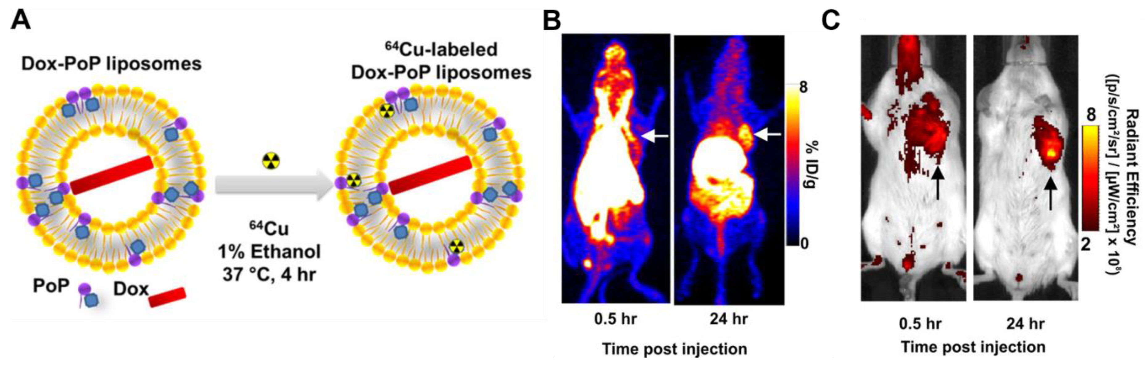
  134. Luo, D.; Goel, S.; Liu, H.-J.; Carter, K.A.; Jiang, D.; Geng, J.; Kutyreff, C.J.; Engle, J.W.; Huang, W.-C.; Shao, S.; et al. Intrabilayer 64Cu Labeling of Photoactivatable, Doxorubicin-Loaded Stealth Liposomes. ACS Nano 2017, 11, 12482–12491. [Google Scholar] [CrossRef]
  135. Record, M.; Subra, C.; Silvente-Poirot, S.; Poirot, M. Exosomes as intercellular signalosomes and pharmacological effectors. Biochem. Pharmacol. 2011, 81, 1171–1182. [Google Scholar] [CrossRef] [Green Version]
  136. van den Boorn, J.G.; Schlee, M.; Coch, C.; Hartmann, G. SiRNA delivery with exosome nanoparticles. Nat. Biotechnol. 2011, 29, 325–326. [Google Scholar] [CrossRef] [PubMed]
  137. Pant, K.; Neuber, C.; Zarschler, K.; Wodtke, J.; Meister, S.; Haag, R.; Pietzsch, J.; Stephan, H. Active Targeting of Dendritic Polyglycerols for Diagnostic Cancer Imaging. Small 2020, 16, 12. [Google Scholar] [CrossRef] [PubMed]
  138. van Onzen, A.; Rossin, R.; Schenning, A.; Nicolay, K.; Milroy, L.G.; Robillard, M.S.; Brunsveld, L. Tetrazine-trans-Cyclooctene Chemistry Applied to Fabricate Self-Assembled Fluorescent and Radioactive Nanoparticles for in Vivo Dual Mode Imaging. Bioconjug. Chem. 2019, 30, 547–551. [Google Scholar] [CrossRef]
  139. Harmsen, S.; Medine, E.I.; Moroz, M.; Nurili, F.; Lobo, J.; Dong, Y.Y.; Turkekul, M.; Pillarsetty, N.V.K.; Ting, R.; Ponomarev, V.; et al. A dual-modal PET/near infrared fluorescent nanotag for long-term immune cell tracking. Biomaterials 2021, 269, 8. [Google Scholar] [CrossRef]
  140. Rasekholghol, A.; Fazaeli, Y.; Moradi Dehaghi, S.; Ashtari, P. Grafting of CdTe quantum dots on thiol functionalized MCM-41 mesoporous silica for 68Ga radiolabeling: Introducing a novel PET agent. J. Radioanal. Nucl. Chem. 2020, 324, 599–608. [Google Scholar] [CrossRef]
  141. Shi, S.X.; Chen, F.; Goel, S.Y.; Graves, S.A.; Luo, H.M.; Theuer, C.P.; Engle, J.W.; Cai, W.B. In Vivo Tumor-Targeted Dual-Modality PET/Optical Imaging with a Yolk/Shell-Structured Silica Nanosystem. Nanomicro Lett. 2018, 10, 11. [Google Scholar] [CrossRef] [Green Version]
  142. Licciardello, N.; Hunoldt, S.; Bergmann, R.; Singh, G.; Mamat, C.; Faramus, A.; Ddungu, J.L.Z.; Silvestrini, S.; Maggini, M.; De Cola, L.; et al. Biodistribution studies of ultrasmall silicon nanoparticles and carbon dots in experimental rats and tumor mice. Nanoscale 2018, 10, 9880–9891. [Google Scholar] [CrossRef] [PubMed] [Green Version]
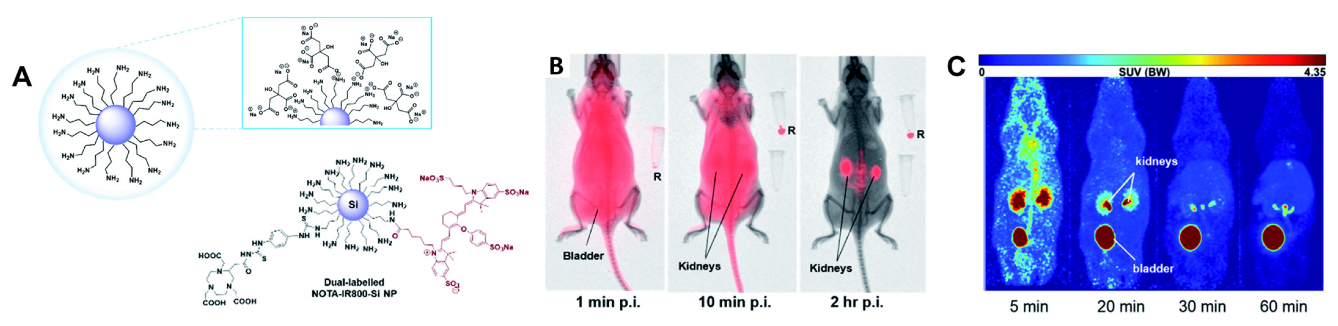
  143. Singh, G.; Ddungu, J.L.Z.; Licciardello, N.; Bergmann, R.; De Cola, L.; Stephan, H. Ultrasmall silicon nanoparticles as a promising platform for multimodal imaging. Faraday Discuss. 2020, 222, 362–383. [Google Scholar] [CrossRef] [PubMed]
  144. Pretze, M.; van der Meulen, N.P.; Wangler, C.; Schibli, R.; Wangler, B. Targeted Cu-64-labeled gold nanoparticles for dual imaging with positron emission tomography and optical imaging. J. Label. Compd. Radiopharm. 2019, 62, 471–482. [Google Scholar] [CrossRef]
  145. Cui, X.J.; Mathe, D.; Kovacs, N.; Horvath, I.; Jauregui-Osoro, M.; de Rosales, R.T.M.; Mullen, G.E.D.; Wong, W.; Yan, Y.; Kruger, D.; et al. Synthesis, Characterization, and Application of Core Shell Co0.16Fe2.84O4@NaYF4(Yb, Er) and Fe3O4@NaYF4(Yb, Tm) Nanoparticle as Trimodal (MRI, PET/SPECT, and Optical) Imaging Agents. Bioconjug. Chem. 2016, 27, 319–328. [Google Scholar] [CrossRef] [PubMed] [Green Version]
  146. Nsubuga, A.; Zarschler, K.; Sgarzi, M.; Graham, B.; Stephan, H.; Joshi, T. Towards Utilising Photocrosslinking of Polydiacetylenes for the Preparation of “Stealth” Upconverting Nanoparticles. Angew. Chem. Int. Ed. 2018, 57, 16036–16040. [Google Scholar] [CrossRef] [PubMed]
  147. Fazaeli, Y.; Zare, H.; Karimi, S.; Feizi, S. 68Ga CdTe/CdS fluorescent quantum dots for detection of tumors: Investigation on the effect of nanoparticle size on stability and in vivo pharmacokinetics. Radiochim. Acta 2020, 108, 565–572. [Google Scholar] [CrossRef]
  148. Resch-Genger, U.; Grabolle, M.; Cavaliere-Jaricot, S.; Nitschke, R.; Nann, T. Quantum dots versus organic dyes as fluorescent labels. Nat. Methods 2008, 5, 763–775. [Google Scholar] [CrossRef]
  149. Thakare, V.; Tran, V.-L.; Natuzzi, M.; Thomas, E.; Moreau, M.; Romieu, A.; Collin, B.; Courteau, A.; Vrigneaud, J.-M.; Louis, C.; et al. Functionalization of theranostic AGuIX® nanoparticles for PET/MRI/optical imaging. RSC Adv. 2019, 9, 24811–24815. [Google Scholar] [CrossRef] [Green Version]
  150. Cusin, F.; Fernandes Azevedo, L.; Bonnaventure, P.; Desmeules, J.; Daali, Y.; Pastor, C.M. Hepatocyte Concentrations of Indocyanine Green Reflect Transfer Rates Across Membrane Transporters. Basic Clin. Pharmacol. Toxicol. 2017, 120, 171–178. [Google Scholar] [CrossRef]
  151. Jing, B.; Qian, R.; Jiang, D.; Gai, Y.; Liu, Z.; Guo, F.; Ren, S.; Gao, Y.; Lan, X.; An, R. Extracellular vesicles-based pre-targeting strategy enables multi-modal imaging of orthotopic colon cancer and image-guided surgery. J. Nanobiotechnol. 2021, 19, 151. [Google Scholar] [CrossRef]
  152. Anselmo, A.C.; Mitragotri, S. Nanoparticles in the clinic: An update. Bioeng. Transl. Med. 2019, 4, e10143. [Google Scholar] [CrossRef] [PubMed] [Green Version]
  153. Johnson, K.K.; Koshy, P.; Yang, J.-L.; Sorrell, C.C. Preclinical Cancer Theranostics—From Nanomaterials to Clinic: The Missing Link. Adv. Funct. Mater. 2021, 31, 2104199. [Google Scholar] [CrossRef]
  154. Palazzolo, S.; Bayda, S.; Hadla, M.; Caligiuri, I.; Corona, G.; Toffoli, G.; Rizzolio, F. The Clinical Translation of Organic Nanomaterials for Cancer Therapy: A Focus on Polymeric Nanoparticles, Micelles, Liposomes and Exosomes. Curr. Med. Chem. 2018, 25, 4224–4268. [Google Scholar] [CrossRef] [PubMed]
  155. Huang, H.; Feng, W.; Chen, Y.; Shi, J. Inorganic nanoparticles in clinical trials and translations. Nano Today 2020, 35, 100972. [Google Scholar] [CrossRef]
  156. Shetty, A.; Chandra, S. Inorganic hybrid nanoparticles in cancer theranostics: Understanding their combinations for better clinical translation. Mater. Today Chem. 2020, 18, 100381. [Google Scholar] [CrossRef]
  157. Nanotechnology Guidance Documents. Available online: https://www.fda.gov/science-research/nanotechnology-programs-fda/nanotechnology-guidance-documents (accessed on 27 March 2022).
  158. van Leeuwen, F.W.B.; Valdés-Olmos, R.; Buckle, T.; Vidal-Sicart, S. Hybrid surgical guidance based on the integration of radionuclear and optical technologies. Br. J. Radiol. 2016, 89, 20150797. [Google Scholar] [CrossRef]
  159. Van Oosterom, M.N.; Rietbergen, D.D.D.; Welling, M.M.; Van Der Poel, H.G.; Maurer, T.; Van Leeuwen, F.W.B. Recent advances in nuclear and hybrid detection modalities for image-guided surgery. Expert Rev. Med. Devices 2019, 16, 711–734. [Google Scholar] [CrossRef] [Green Version]
  160. de Jong, M.; van Leeuwen, F.W.B.; Lahoutte, T.; Evangelista, L.; Barbet, J.; Del Vecchio, S.; Schibli, R. Molecular imaging: The emerging role of optical imaging in nuclear medicine. Eur. J. Nucl. Med. Mol. Imaging 2014, 41, 2150–2153. [Google Scholar] [CrossRef] [Green Version]
  161. Brouwer, O.R.; van den Berg, N.S.; Mathéron, H.M.; van der Poel, H.G.; van Rhijn, B.W.; Bex, A.; van Tinteren, H.; Valdés Olmos, R.A.; van Leeuwen, F.W.B.; Horenblas, S. A Hybrid Radioactive and Fluorescent Tracer for Sentinel Node Biopsy in Penile Carcinoma as a Potential Replacement for Blue Dye. Eur. Urol. 2014, 65, 600–609. [Google Scholar] [CrossRef]
  162. Christensen, A.; Juhl, K.; Charabi, B.; Mortensen, J.; Kiss, K.; Kjær, A.; von Buchwald, C. Feasibility of Real-Time Near-Infrared Fluorescence Tracer Imaging in Sentinel Node Biopsy for Oral Cavity Cancer Patients. Ann. Sur. Oncol. 2016, 23, 565–572. [Google Scholar] [CrossRef] [Green Version]
  163. KleinJan, G.H.; van den Berg, N.S.; Brouwer, O.R.; de Jong, J.; Acar, C.; Wit, E.M.; Vegt, E.; van der Noort, V.; Valdés Olmos, R.A.; van Leeuwen, F.W.B.; et al. Optimisation of Fluorescence Guidance During Robot-assisted Laparoscopic Sentinel Node Biopsy for Prostate Cancer. Eur. Urol. 2014, 66, 991–998. [Google Scholar] [CrossRef] [PubMed]
  164. KleinJan, G.H.; van Werkhoven, E.; van den Berg, N.S.; Karakullukcu, M.B.; Zijlmans, H.J.M.A.A.; van der Hage, J.A.; van de Wiel, B.A.; Buckle, T.; Klop, W.M.C.; Horenblas, S.; et al. The best of both worlds: A hybrid approach for optimal pre- and intraoperative identification of sentinel lymph nodes. Eur. J. Nucl. Med. Mol. Imaging 2018, 45, 1915–1925. [Google Scholar] [CrossRef] [PubMed] [Green Version]
  165. Paredes, P.; Vidal-Sicart, S.; Campos, F.; Tapias, A.; Sánchez, N.; Martínez, S.; Carballo, L.; Pahisa, J.; Torné, A.; Ordi, J.; et al. Role of ICG-99mTc-nanocolloid for sentinel lymph node detection in cervical cancer: A pilot study. Eur. J. Nucl. Med. Mol. Imaging 2017, 44, 1853–1861. [Google Scholar] [CrossRef] [PubMed]
  166. Schaafsma, B.E.; Verbeek, F.P.R.; Rietbergen, D.D.D.; Hiel, B.; Vorst, J.R.; Liefers, G.J.; Frangioni, J.V.; Velde, C.J.H.; van Leeuwen, F.W.B.; Vahrmeijer, A.L. Clinical trial of combined radio- and fluorescence-guided sentinel lymph node biopsy in breast cancer. Br. J. Surg. 2013, 100, 1037–1044. [Google Scholar] [CrossRef] [Green Version]
  167. Stoffels, I.; Leyh, J.; Pöppel, T.; Schadendorf, D.; Klode, J. Evaluation of a radioactive and fluorescent hybrid tracer for sentinel lymph node biopsy in head and neck malignancies: Prospective randomized clinical trial to compare ICG-99mTc-nanocolloid hybrid tracer versus 99mTc-nanocolloid. Eur. J. Nucl. Med. Mol. Imaging 2015, 42, 1631–1638. [Google Scholar] [CrossRef]
  168. Hensbergen, A.W.; van Willigen, D.M.; van Beurden, F.; van Leeuwen, P.J.; Buckle, T.; Schottelius, M.; Maurer, T.; Wester, H.-J.; van Leeuwen, F.W.B. Image-Guided Surgery: Are We Getting the Most Out of Small-Molecule Prostate-Specific-Membrane-Antigen-Targeted Tracers? Bioconjug. Chem. 2020, 31, 375–395. [Google Scholar] [CrossRef]
  169. Schottelius, M.; Wurzer, A.; Wissmiller, K.; Beck, R.; Koch, M.; Gorpas, D.; Notni, J.; Buckle, T.; van Oosterom, M.N.; Steiger, K.; et al. Synthesis and Preclinical Characterization of the PSMA-Targeted Hybrid Tracer PSMA-I&F for Nuclear and Fluorescence Imaging of Prostate Cancer. J. Nucl. Med. 2019, 60, 71–78. [Google Scholar] [CrossRef] [Green Version]
  170. Santini, C.; Kuil, J.; Bunschoten, A.; Pool, S.; de Blois, E.; Ridwan, Y.; Essers, J.; Bernsen, M.R.; van Leeuwen, F.W.B.; de Jong, M. Evaluation of a Fluorescent and Radiolabeled Hybrid Somatostatin Analog In Vitro and in Mice Bearing H69 Neuroendocrine Xenografts. J. Nucl. Med. 2016, 57, 1289–1295. [Google Scholar] [CrossRef] [Green Version]
  171. Wang, X.; Aldrich, M.B.; Marshall, M.V.; Sevick-Muraca, E.M. Preclinical characterization and validation of a dual-labeled trastuzumab-based imaging agent for diagnosing breast cancer. Chin. J. Cancer Res. 2015, 27, 74–82. [Google Scholar] [CrossRef]
  172. Hekman, M.C.; Rijpkema, M.; Muselaers, C.H.; Oosterwijk, E.; Hulsbergen-Van de Kaa, C.A.; Boerman, O.C.; Oyen, W.J.; Langenhuijsen, J.F.; Mulders, P.F. Tumor-targeted Dual-modality Imaging to Improve Intraoperative Visualization of Clear Cell Renal Cell Carcinoma: A First in Man Study. Theranostics 2018, 8, 2161–2170. [Google Scholar] [CrossRef]
  173. Ciarrocchi, E.; Belcari, N. Cerenkov luminescence imaging: Physics principles and potential applications in biomedical sciences. EJNMMI Phys. 2017, 4, 14. [Google Scholar] [CrossRef] [PubMed] [Green Version]
  174. Das, S.; Thorek, D.L.J.; Grimm, J. Chapter Six—Cerenkov Imaging. In Advances in Cancer Research; Pomper, M.G., Fisher, P.B., Eds.; Academic Press: Cambridge, MA, USA, 2014; Volume 124, pp. 213–234. [Google Scholar]
  175. Spinelli, A.E.; Boschi, F. Novel biomedical applications of Cerenkov radiation and radioluminescence imaging. Phys. Med. 2015, 31, 120–129. [Google Scholar] [CrossRef] [PubMed]
  176. Chin, P.T.K.; Welling, M.M.; Meskers, S.C.J.; Valdes Olmos, R.A.; Tanke, H.; van Leeuwen, F.W.B. Optical imaging as an expansion of nuclear medicine: Cerenkov-based luminescence vs fluorescence-based luminescence. Eur. J. Nucl. Med. Mol. Imaging 2013, 40, 1283–1291. [Google Scholar] [CrossRef] [PubMed]
  177. Hustinx, R. Physician centred imaging interpretation is dying out—Why should I be a nuclear medicine physician? Eur. J. Nucl. Med. Mol. Imaging 2019, 46, 2708–2714. [Google Scholar] [CrossRef]
Figure 1. Depiction of PET and SPECT imaging.
Pharmaceuticals 15 00432 g001
Figure 2. (A) The molecular sensitivity and spatial resolution of imaging processes of relevance to this review, * sensitivity not well characterised. Data obtained from [17,18,19]. (B) Schematic of penetration depth at varying wavelengths.
Pharmaceuticals 15 00432 g002
Figure 3. Representative examples of (a) Common NIR-I dyes, (b) NIR-II dyes that are based on the donor–acceptor–donor design, Q8PNap [25] and BPST [26], (c) Alternate NIR-II dyes: Hydro-1080 [27], BTC1070 [28] and NJ1060 [29] and (d) Additional fluorophores, not previously noted, that are relevant to Section 3 and Section 4.
Pharmaceuticals 15 00432 g003
Figure 4. (A) Chemical structure of the -coordinate cationic [YbL]+ complex; (B) Structures of porphyrinates complexes Yb-4, Yb-2, Yb-cis/trans-3 and Ln-L.
Pharmaceuticals 15 00432 g004
Figure 5. (A) Chemical structures of lanthanide-based probes containing pyclen ligands. (B) Left: 2P imaging of paraformaldehyde-fixed T24-cells: [EuL4a], [SmL4a] (λex = 750 nm), [YbL4b] (λex = 800 nm), [TbL4c] and [DyL4c] (λex = 720 nm). Middle: transmitted light DIC images. Right: merged images. Reprinted (adapted) with permission from Ref. [52]. Copyright 2020 American Chemical Society.
Pharmaceuticals 15 00432 g005
Figure 6. (A) Chemical structure of cycLN-ss-CPT. (B) NIR imaging of YbNPs in HeLa cells; CPT emission λex 370 nm and λem 400–450 nm; Yb(III) emission λex 380 nm and λem 900–1700 nm. Reprinted (adapted) with permission from Ref. [54]. Copyright 2021 American Chemical Society.
Pharmaceuticals 15 00432 g006
Figure 7. Chemical structures of [Eu(DO2Aphen)-DUPA]+ and [Tb(DO2Apic)-DUPA].
Pharmaceuticals 15 00432 g007
Figure 8. Chemical structures of the iridium(III) complexes IRCY, IRDAD and PolyIRLA.
Pharmaceuticals 15 00432 g008
Figure 9. Chemical structure of the Ru(II) polypyridyl complex, HL-PEG2K.
Pharmaceuticals 15 00432 g009
Figure 10. Representative chelators discussed in this review.
Pharmaceuticals 15 00432 g010
Figure 11. The ligands visualise PSMA-positive tumours using µSPECT/CT and fluorescence imaging. The panels display (A) µSPECT/CT scans, (B) Fluorescence images of mice with LS174T-PSMA (right) and wild-type LS174T (left) tumours after intravenous injection of 111In (10 MBq/mouse) or 99mTc-labelled (15 MBq/mouse) ligands. Reprinted with permission from Ref. [71]. Copyright 2022 American Chemical Society.
Pharmaceuticals 15 00432 g011
Figure 12. Dual-modal imaging for glioblastoma resection using 68Ga-NOTA-IRDye800CW-bombesin. The glioblastoma tumour is visible by MRI after gadolinium injection. (A) Uptake in the tumour area was observed by PET/MRI, (F) The NIRF assessment displayed the fluorescent tumour ((B): scale bar 5 mm). (C) Displays the fluorescent residual tumour tissue, and (H) the histological staining (scale bar 50 µm). Several antigen-expressing cells were found in the tissue after gastrin-releasing peptide staining ((G): scale bar 20 µm). After resection, no residual fluorescence was detected in the tumour’s cavity. (D) Moreover, the tissue along the cavity was confirmed to be normal (I) and few antigen-expressing cells were found on it (J). The post-operative analysis shows total removal of the malignancy (E). Reprinted with permission from Ref. [79]. Copyright 2018 Ivyspring International Publisher.
Pharmaceuticals 15 00432 g012
Figure 13. (A) Doxorubicin-loaded Cu-porphyrin-phospholipid liposomes. (B) PET scan of mice after administration of DOX-CuPoP. (C) NIR Fluorescence imaging of mice after administration of DOX-CuPoP. Reprinted with permission from [134]. Copyright 2017 American Chemical Society.
Pharmaceuticals 15 00432 g013
Figure 14. (A) Dual-labelled NOTA-IRDye800CW-Si nanoparticle coated with citric acid. (B) In vivo optical imaging scans of healthy nu/nu mice intravenously injected with IRDye800CW-Si NPs after 1 min, 10 min and 2 h p.i. (R: injected IR800-Si NPs as reference). (C) In vivo PET imaging of [64Cu]Cu-NOTA-IRDye800CW-Si NPs 5, 20, 30, 60 min p.i. Reprinted with permission from [143]. Copyright 2020 The Royal Society of Chemistry.
Pharmaceuticals 15 00432 g014
Publisher’s Note: MDPI stays neutral with regard to jurisdictional claims in published maps and institutional affiliations.

